Müller AO Classifi cation of Fractures—Long Bones
10
Müller AO Classification of Fractures—Long Bones This leaflet is designed to provide an introduction to the classification of long-bone fractures.
Transcript of Müller AO Classifi cation of Fractures—Long Bones
Müller AO Classifi cation of Fractures—Long BonesThis leafl et is designed to provide an introduction to the classifi cation of long-bone fractures.

1 Humerus
11 proximal (types according to topography and extent of bone lesion)
13-A1 13-A2 13-A3 13-C1 13-C2 13-C313-B1 13-B2 13-B3
12 diaphyseal
13 distal
11-A1 11-A2 11-A3 11-B1 11-B2 11-B3 11-C1 11-C2 11-C3
12-A1 12-A2 12-A3 12-B1 12-B2 12-B3 12-C1 12-C2 12-C3
13-A extraarticular fracture13-A1 apophyseal avulsion13-A2 metaphyseal simple13-A3 metaphyseal multifragmentary
13-B partial articular fracture13-B1 sagittal lateral condyle13-B2 sagittal medial condyle13-B3 coronal
13-C complete articular fracture13-C1 articular simple, metaphyseal simple13-C2 articular simple, metaphyseal multifragmentary13-C3 articular multifragmentary
11-A extraarticular unifocal fracture11-A1 tuberosity11-A2 impacted metaphyseal11-A3 nonimpacted metaphyseal
11-B extraarticular bifocal fracture11-B1 with metaphyseal impaction11-B2 without metaphyseal impaction11-B3 with glenohumeral dislocation
11-C articular fracture11-C1 with slight displacement11-C2 impacted with marked displacement11-C3 dislocated
12-A simple fracture12-A1 spiral12-A2 oblique (>_ 30°)12-A3 transverse (< 30°)
12-B wedge fracture12-B1 spiral wedge12-B2 bending wedge12-B3 fragmented wedge
12-C complex fracture12-C1 spiral12-C2 segmental12-C3 irregular
21 proximal21-A1 21-A2 21-A3 21-B1 21-B2 21-B3 21-C1 21-C2 21-C3
22 diaphyseal22-A1 22-A2 22-A3 22-B1 22-B2 22-B3 22-C1 22-C2 22-C3
23 distal23-A1 23-A2 23-A3 23-B1 23-B2 23-B3 23-C1 23-C2 23-C3
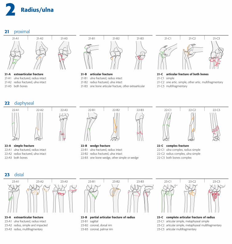
2 Radius/ulna
21-A extraarticular fracture21-A1 ulna fractured, radius intact21-A2 radius fractured, ulna intact21-A3 both bones
21-B articular fracture21-B1 ulna fractured, radius intact21-B2 radius fractured, ulna intact21-B3 one bone articular fracture, other extraarticular
21-C articular fracture of both bones21-C1 simple21-C2 one artic. simple, other artic. multifragmentary21-C3 multifragmentary
22-A simple fracture22-A1 ulna fractured, radius intact22-A2 radius fractured, ulna intact22-A3 both bones
22-B wedge fracture22-B1 ulna fractured, radius intact22-B2 radius fractured, ulna intact22-B3 one bone wedge, other simple or wedge
22-C complex fracture22-C1 ulna complex, radius simple22-C2 radius complex, ulna simple22-C3 both bones complex
23-A extraarticular fracture23-A1 ulna fractured, radius intact23-A2 radius, simple and impacted23-A3 radius, multifragmentary
23-B partial articular fracture of radius23-B1 sagittal23-B2 coronal, dorsal rim23-B3 coronal, palmar rim
23-C complete articular fracture of radius23-C1 articular simple, metaphyseal simple23-C2 articular simple, metaphyseal multifragmentary23-C3 articular multifragmentary
31-A1 31-A2 31-A3 31-B1 31-B2 31-B3 31-C1 31-C2 31-C3
32-A1 32-A2 32-A3 32-B1 32-B2 32-B3 32-C1 32-C2 32-C3
33 distal33-A1 33-A2 33-A3 33-B1 33-B2 33-B3 33-C1 33-C2 33-C3
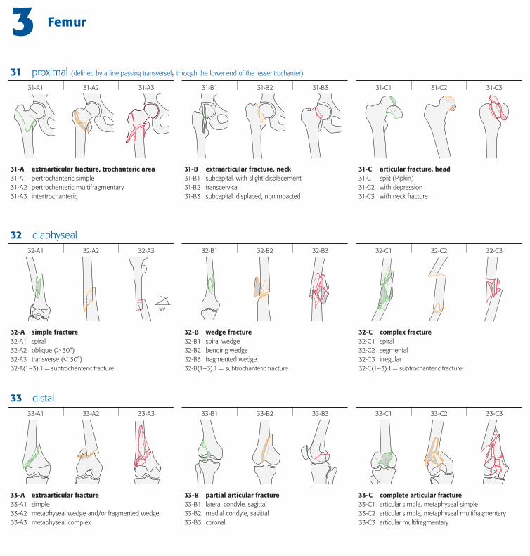
3 Femur
30°
32 diaphyseal
31 proximal (defi ned by a line passing transversely through the lower end of the lesser trochanter)
31-A extraarticular fracture, trochanteric area31-A1 pertrochanteric simple31-A2 pertrochanteric multifragmentary31-A3 intertrochanteric
31-B extraarticular fracture, neck31-B1 subcapital, with slight displacement31-B2 transcervical31-B3 subcapital, displaced, nonimpacted
31-C articular fracture, head31-C1 split (Pipkin)31-C2 with depression31-C3 with neck fracture
32-A simple fracture32-A1 spiral32-A2 oblique (>_ 30°)32-A3 transverse (< 30°)32-A(1–3).1 = subtrochanteric fracture
32-B wedge fracture32-B1 spiral wedge32-B2 bending wedge32-B3 fragmented wedge32-B(1–3).1 = subtrochanteric fracture
32-C complex fracture32-C1 spiral32-C2 segmental32-C3 irregular32-C(1–3).1 = subtrochanteric fracture
33-A extraarticular fracture33-A1 simple33-A2 metaphyseal wedge and/or fragmented wedge 33-A3 metaphyseal complex
33-B partial articular fracture33-B1 lateral condyle, sagittal33-B2 medial condyle, sagittal33-B3 coronal
33-C complete articular fracture33-C1 articular simple, metaphyseal simple33-C2 articular simple, metaphyseal multifragmentary33-C3 articular multifragmentary
41-A1 41-A2 41-A3 41-B1 41-B2 41-B3 41-C1 41-C2 41-C3
42-A1 42-A2 42-A3 42-B1 42-B2 42-B3 42-C1 42-C2 42-C3
43-A1 43-A2 43-A3 43-B1 43-B2 43-B3 43-C1 43-C2 43-C3
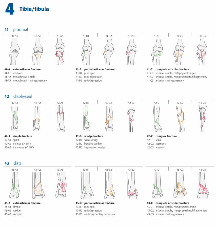
4 Tibia/fi bula
43 distal
42 diaphyseal
41 proximal
41-A extraarticular fracture41-A1 avulsion41-A2 metaphyseal simple41-A3 metaphyseal multifragmentary
41-B partial articular fracture41-B1 pure split41-B2 pure depression41-B3 split-depression
41-C complete articular fracture41-C1 articular simple, metaphyseal simple41-C2 articular simple, metaphyseal multifragmentary41-C3 articular multifragmentary
42-A simple fracture42-A1 spiral42-A2 oblique (>_ 30°)42-A3 transverse (< 30°)
42-B wedge fracture42-B1 spiral wedge42-B2 bending wedge42-B3 fragmented wedge
42-C complex fracture42-C1 spiral42-C2 segmental42-C3 irregular
43-A extraarticular fracture43-A1 simple43-A2 wedge43-A3 complex
43-B partial articular fracture43-B1 pure split43-B2 split-depression43-B3 multifragmentary depression
43-C complete articular fracture43-C1 articular simple, metaphyseal simple43-C2 articular simple, metaphyseal multifragmentary43-C3 articular multifragmentary
44-A1 44-A2 44-A3
44-B1 44-B2 44-B3
44-C1 44-C2 44-C3
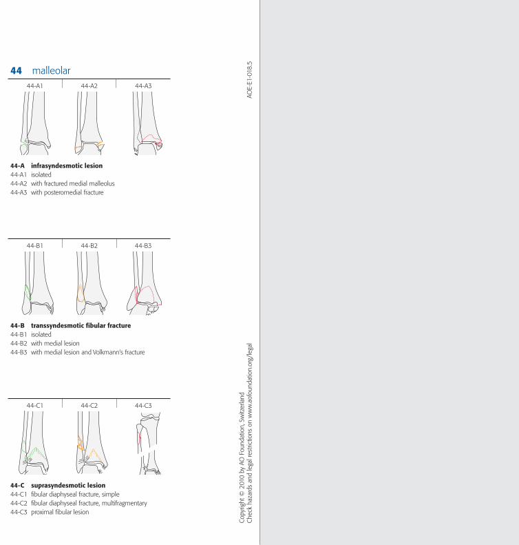
44 malleolar
Cop
yrig
ht ©
201
0 by
AO
Fou
ndat
ion,
Sw
itzer
land
Che
ck h
azar
ds a
nd le
gal r
estri
ctio
ns o
n w
ww
.aof
ound
atio
n.or
g/le
gal
AOE-
E1-0
18.5
44-A infrasyndesmotic lesion44-A1 isolated44-A2 with fractured medial malleolus44-A3 with posteromedial fracture
44-B transsyndesmotic fi bular fracture44-B1 isolated44-B2 with medial lesion44-B3 with medial lesion and Volkmann‘s fracture
44-C suprasyndesmotic lesion44-C1 fi bular diaphyseal fracture, simple44-C2 fi bular diaphyseal fracture, multifragmentary44-C3 proximal fi bular lesion
8- Foot
4- Tibia/fi bula
3- Femur/patella7- Hand
9- Craniomaxillofacial bones
1- Humerus
2- Radius/ ulna
5- Spine
15- Clavicula
91-
92-
44-
43-
42-
41-
33-
32-
31- 23-
22-
21-
13-
12-
11-
51-
53-
52-
6- Pelvis/acetabulum
61-
62-
15-
14- 14- Scapula
34-
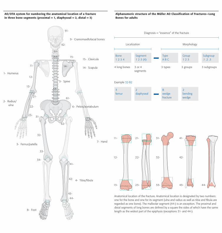
AO/OTA system for numbering the anatomical location of a fracture in three bone segments (proximal = 1, diaphyseal = 2, distal = 3)
Bone1 2 3 4
Segment1 2 3 (4)
TypeA B C
Group1 2 3
Subgroup.1 .2 .3
4 long bones 3 or 4 segments
3 types 3 groups 3 subgroups
Diagnosis = “essence” of the fracture
MorphologyLocalization
-
Anatomical location of the fracture. Anatomical location is designated by two numbers: one for the bone and one for its segment (ulna and radius as well as tibia and fibula are regarded as one bone). The malleolar segment (44-) is an exception. The proximal and distal segments of long bones are defined by a square the sides of which have the same length as the widest part of the epiphysis (exceptions 31- and 44-).
Alphanumeric structure of the Müller AO Classification of Fractures—Long Bones for adults
11-
13-
12-
21-
23-
22-
31-
33-
32-
41-
43-
42-
44-
Example 32-B2
3femur
2diaphyseal
Bwedge fracture
2bendingwedge-
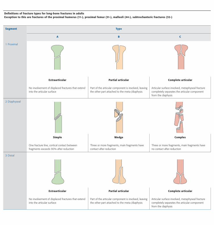
Definitions of fracture types for long-bone fractures in adults Exception to this are fractures of the proximal humerus (11-), proximal femur (31-), malleoli (44-), subtrochanteric fractures (32-)
Segment Type
A B C
1 Proximal
Extraarticular Partial articular Complete articular
No involvement of displaced fractures that extend into the articular surface
Part of the articular component is involved, leaving the other part attached to the meta-/diaphysis
Articular surface involved, metaphyseal fracture completely separates the articular component from the diaphysis
2 Diaphyseal
Simple Wedge Complex
One fracture line, cortical contact between fragments exceeds 90% after reduction
Three or more fragments, main fragments have contact after reduction
Three or more fragments, main fragments have no contact after reduction
3 Distal
Extraarticular Partial articular Complete articular
No involvement of displaced fractures that extend into the articular surface
Part of the articular component is involved, leaving the other part attached to the meta-/diaphysis
Articular surface involved, metaphyseal fracture completely separates the articular component from the diaphysis
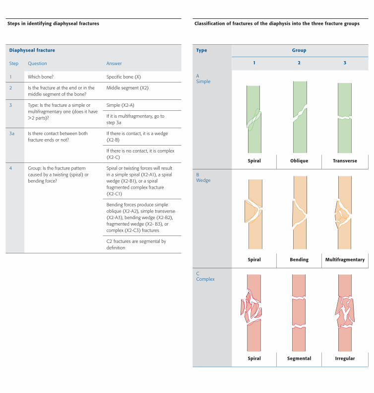
Diaphyseal fracture
Step Question Answer
1 Which bone? Specific bone (X)
2 Is the fracture at the end or in the middle segment of the bone?
Middle segment (X2)
3 Type: Is the fracture a simple or multifragmentary one (does it have > 2 parts)?
Simple (X2-A)
If it is multifragmentary, go to step 3a
3a Is there contact between both fracture ends or not?
If there is contact, it is a wedge (X2-B)
If there is no contact, it is complex (X2-C)
4 Group: Is the fracture pattern caused by a twisting (spiral) or bending force?
Spiral or twisting forces will result in a simple spiral (X2-A1), a spiral wedge (X2-B1), or a spiral fragmented complex fracture (X2-C1)
Bending forces produce simple oblique (X2-A2), simple transverse (X2-A3), bending wedge (X2-B2), fragmented wedge (X2- B3), or complex (X2-C3) fractures
C2 fractures are segmental by definition
Steps in identifying diaphyseal fractures
Type Group
1 2 3
A Simple
Spiral Oblique Transverse
B Wedge
Spiral Bending Multifragmentary
CComplex
Spiral Segmental Irregular
Classification of fractures of the diaphysis into the three fracture groups
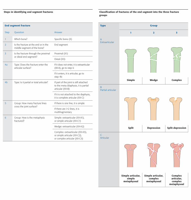
End segment fracture
Step Question Answer
1 Which bone? Specific bone (X)
2 Is the fracture at the end or in the middle segment of the bone?
End segment
3 Is the fracture through the proximal or distal end segment?
Proximal (X1)
Distal (X3)
4a Type: Does the fracture enter the articular surface?
If it does not enter, it is extraarticular (XX-A), go to step 6
If it enters, it is articular, go to step 4b
4b Type: Is it partial or total articular? If part of the joint is still attached to the meta-/diaphysis, it is partial articular (XX-B)
If it is not attached to the diaphysis, it is complete articular (XX-C)
5 Group: How many fracture lines cross the joint surface?
If there is one line, it is simple
If there are > 2 lines, it is multifragmentary
6 Group: How is the metaphysis fractured?
Simple: extraarticular (XX-A1), or simple articular (XX-C1)
Wedge: extraarticular (XX-A2)
Complex: extraarticular (XX-A3), or simple articular (XX-C2), or complex articular (XX-C3)
Steps in identifying end segment fractures
Type Group
1 2 3
A Extraarticular
Simple Wedge Complex
B Partial articular
Split Depression Split-depression
CArticular
Simple articular,simple
metaphyseal
Simple articular,complex
metaphyseal
Complex articular,complex
metaphyseal
Classification of fractures of the end segment into the three fracture groups