Mini-implant assisted rapid palatal expansion: New ...
18
This is an Open Access article distributed under the terms of the Creative Commons Attribution License (http://creativecommons.org/licenses/by/4.0), which permits unrestricted use, distribution, and reproduction in any medium, provided the original work is properly cited. 1 Article received: 19-04-2017. Accepted for publication: 18-05-2017. Address for correspondence: Margaux Montigny University of Bordeaux – CHU Pellegrin Place Amélie Rabat Léon – 33000 Bordeaux E-mail: [email protected] J Dentofacial Anom Orthod 2017;20:405 © The author DOI: 10.1051/odfen/2017021 Transversal anomalies are very common in clinical practice. In 80% of cases, their in- terceptive treatment is essential and must be administered as soon as possible to avoid periodontal, joint, or functional prob- lems. It is based on an orthopedic tech- nique aimed at separating the two maxillae above the level of the intermaxillary palatal suture. In 1860 2 , Angell who first described rap- id maxillary expansion, which was very quickly discredited; however, since the 1960s it has been most widely used treat- ment in our arsenal. Conventionally, rapid maxillary expansion is performed in children who are still grow- ing (no suture synostosis; at approximately 12 years), using a disjunctor (described by Biederman) supported by the teeth (two or four rings) and using a median activa- tor twice a day, or 0.5 mm per day. Many studies, including meta-analyses, have INTRODUCTION Mini-implant assisted rapid palatal expansion: New perspectives M. Montigny Dentofacial Orthopedic Intern, University of Bordeaux, CHU Pellegrin, Place Amélie Rabat Léon, 33000 Bordeaux SUMMARY Maxillary endognathy is a fundamental problem of the transverse dimension, which is very common during childhood. The conventional treatment for this dysmorphia is rapid maxillary expansion using a mid-palatal jack screw. Widely described in literature, it provides many functional and morphological benefits. Unfortunately, there are side effects caused by these devices, especially in patients with thin periodontium. For improving the management of these patients, we use mini-implant assisted rapid pal- atal expansion. The anchorage is directly on the palate, closer to the hemimaxillary resistance center, and it allows for a better skeletal maxillary expansion. It decreases the size of the vestibular cortical is very thin. Benefits, side effects, and implementation of this device will be discussed in this article. KEY WORDS Endognathy, rapid palatal expansion, mini-implant, orthopedic treatment, bone anchorage Article available at https://www.jdao-journal.org or https://doi.org/10.1051/odfen/2017021
Transcript of Mini-implant assisted rapid palatal expansion: New ...

This is an Open Access article distributed under the terms of the Creative Commons Attribution License (http://creativecommons.org/licenses/by/4.0),
which permits unrestricted use, distribution, and reproduction in any medium, provided the original work is properly cited. 1
Article received: 19-04-2017.Accepted for publication: 18-05-2017.
Address for correspondence:
Margaux Montigny University of Bordeaux – CHU PellegrinPlace Amélie Rabat Léon – 33000 BordeauxE-mail: [email protected]
J Dentofacial Anom Orthod 2017;20:405© The author
DOI: 10.1051/odfen/2017021
Transversal anomalies are very common in clinical practice. In 80% of cases, their in-terceptive treatment is essential and must be administered as soon as possible to avoid periodontal, joint, or functional prob-lems. It is based on an orthopedic tech-nique aimed at separating the two maxillae above the level of the intermaxillary palatal suture.
In 18602, Angell who first described rap-id maxillary expansion, which was very
quickly discredited; however, since the 1960s it has been most widely used treat-ment in our arsenal.
Conventionally, rapid maxillary expansion is performed in children who are still grow-ing (no suture synostosis; at approximately 12 years), using a disjunctor (described by Biederman) supported by the teeth (two or four rings) and using a median activa-tor twice a day, or 0.5 mm per day. Many studies, including meta-analyses, have
INTRODUCTION
Mini-implant assisted rapid palatal expansion: New perspectives
M. Montigny Dentofacial Orthopedic Intern, University of Bordeaux, CHU Pellegrin, Place Amélie Rabat Léon,
33000 Bordeaux
SUMMARY
Maxillary endognathy is a fundamental problem of the transverse dimension, which is very common during childhood. The conventional treatment for this dysmorphia is rapid maxillary expansion using a mid-palatal jack screw. Widely described in literature, it provides many functional and morphological benefits. Unfortunately, there are side effects caused by these devices, especially in patients with thin periodontium. For improving the management of these patients, we use mini-implant assisted rapid pal-atal expansion. The anchorage is directly on the palate, closer to the hemimaxillary resistance center, and it allows for a better skeletal maxillary expansion. It decreases the size of the vestibular cortical is very thin. Benefits, side effects, and implementation of this device will be discussed in this article.
KEY WORDS
Endognathy, rapid palatal expansion, mini-implant, orthopedic treatment, bone anchorage
Article available at https://www.jdao-journal.org or https://doi.org/10.1051/odfen/2017021

Montigny M. Mini-implant assisted rapid palatal expansion: New perspectives2
M. MONTIGNY
investigated the actual effects of rapid maxillary expansion at the skeletal, al-veolar, and periodontal levels. Despite the beneficial effects of this method, there are still several disadvantages, which has led to new research.
The introduction of mini-implants and mini-implants over the last 20 years offers patients new treatment alterna-tives by allowing dental displacements when conventional approaches reach their limitations of usefulness. The use
of mini-implants and mini-implants be-comes the first treatment choice when correcting the transverse dimension to counter any undesirable effects and exceed the limits of conventional de-vices.
The aim of this article is to focus on the importance of mini-implants and mini-implants in rapid maxillary expan-sion and to highlight their potential use following a brief review of conventional rapid maxillary expansion.
Skeletal and anatomical
The major effect sought during rap-id maxillary expansion is skeletal, with the separation of the two maxillae at the medial palatal suture in the form of V at the posterosuperior apex, because of the resistance offered by the ptery-goid processes of the sphenoid bone8.
According to a literature review pub-lished in 2005 by Lagravère et al., only 25% of this expansion is skeletal; therefore, three-quarters of the expan-sion is alveolar35. On the other hand, Krebs in 1964 attributed 50% to skele-tal expansion in children31.
This evolution of thought encourag-es us to find a way to increase the ratio of skeletal effects to alveolar ef-fects.
The sutureThe maxilla is the centerpiece of the
middle segment of the face; it con-sists of two maxillae joined by an in-termaxillary suture. Ten bones affect its articulation; rapid maxillary expansion, therefore, directly or indirectly affects
the structures in relation to the max-illa, mandible, nasal cavity, pharyngeal structures, and the pterygoid process-es of the sphenoid bone5.
A direct relationship has been estab-lished between increased resistance to skeletal expansion and patients older than 12–13 years.
Soft tissuesNumerous studies have been pub-
lished on the skeletal effects of maxil-lary expansion, but very few have con-sidered the effects on soft tissues.
In 2010, Johnson et al. published a study of 78 patients treated with rapid maxillary expansion compared to a control group of 437 untreated pa-tients.
In the transverse direction, they ob-served an increase in the interalar distance (measured with calipers) of +1.7 mm on average in growing pa-tients, in correlation with the increase in size of the nasal fossae27.
These results are supported those published by Kim et al., in 2012, on cone-beam computed tomography
BENEFICIAL EFFECTS OF RAPID MAXILLARY EXPANSION
J Dentofacial Anom Orthod 2017;20:405 3
MINI-IMpLANT ASSISTED RApID pALATAL ExpANSION: NEW pERSpECTIVES
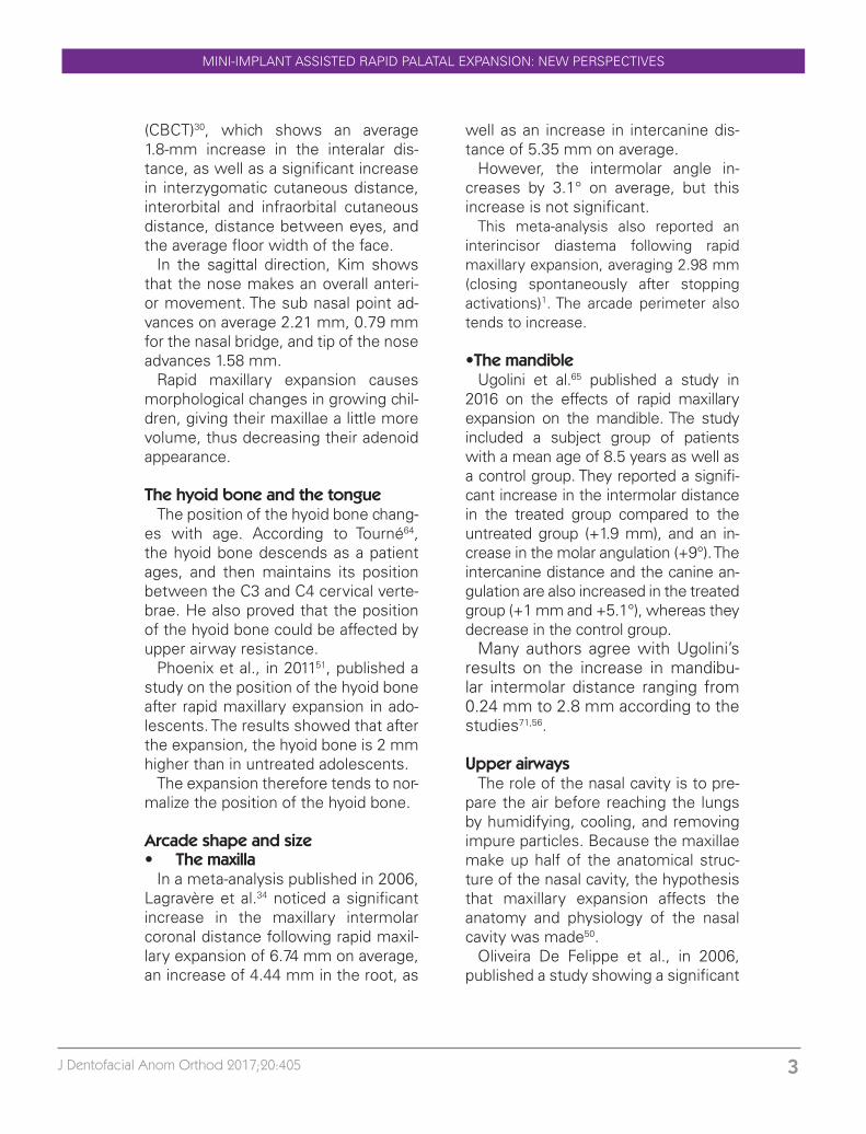
(CBCT)30, which shows an average 1.8-mm increase in the interalar dis-tance, as well as a significant increase in interzygomatic cutaneous distance, interorbital and infraorbital cutaneous distance, distance between eyes, and the average floor width of the face.
In the sagittal direction, Kim shows that the nose makes an overall anteri-or movement. The sub nasal point ad-vances on average 2.21 mm, 0.79 mm for the nasal bridge, and tip of the nose advances 1.58 mm.
Rapid maxillary expansion causes morphological changes in growing chil-dren, giving their maxillae a little more volume, thus decreasing their adenoid appearance.
The hyoid bone and the tongueThe position of the hyoid bone chang-
es with age. According to Tourné64, the hyoid bone descends as a patient ages, and then maintains its position between the C3 and C4 cervical verte-brae. He also proved that the position of the hyoid bone could be affected by upper airway resistance.
phoenix et al., in 201151, published a study on the position of the hyoid bone after rapid maxillary expansion in ado-lescents. The results showed that after the expansion, the hyoid bone is 2 mm higher than in untreated adolescents.
The expansion therefore tends to nor-malize the position of the hyoid bone.
Arcade shape and size• The maxilla
In a meta-analysis published in 2006, Lagravère et al.34 noticed a significant increase in the maxillary intermolar coronal distance following rapid maxil-lary expansion of 6.74 mm on average, an increase of 4.44 mm in the root, as
well as an increase in intercanine dis-tance of 5.35 mm on average.
However, the intermolar angle in-creases by 3.1° on average, but this increase is not significant.
This meta-analysis also reported an interincisor diastema following rapid maxillary expansion, averaging 2.98 mm (closing spontaneously after stopping activations)1. The arcade perimeter also tends to increase.
•The mandibleUgolini et al.65 published a study in
2016 on the effects of rapid maxillary expansion on the mandible. The study included a subject group of patients with a mean age of 8.5 years as well as a control group. They reported a signifi-cant increase in the intermolar distance in the treated group compared to the untreated group (+1.9 mm), and an in-crease in the molar angulation (+9°). The intercanine distance and the canine an-gulation are also increased in the treated group (+1 mm and +5.1°), whereas they decrease in the control group.
Many authors agree with Ugolini’s results on the increase in mandibu-lar intermolar distance ranging from 0.24 mm to 2.8 mm according to the studies71,56.
Upper airwaysThe role of the nasal cavity is to pre-
pare the air before reaching the lungs by humidifying, cooling, and removing impure particles. Because the maxillae make up half of the anatomical struc-ture of the nasal cavity, the hypothesis that maxillary expansion affects the anatomy and physiology of the nasal cavity was made50.
Oliveira De Felippe et al., in 2006, published a study showing a significant

Montigny M. Mini-implant assisted rapid palatal expansion: New perspectives4
M. MONTIGNY
30.12% increase in the volume of na-sal cavities after rapid maxillary expan-sion and a restraint period50.
Kilic et al., in a literature review pub-lished in 2008, which included 11 stud-ies, described an increase of 2–4 mm in the transverse dimension of the na-sal fossae.
A tonsillectomy and, in some cases, continuous positive airway pressure were the treatment of choice for ob-structive sleep apnea (OSA) in children but would not have fully improved the management of this disease.44
Numerous studies have shown that rapid maxillary expansion would have a beneficial effect on OSA. In fact, a re-cent meta-analysis published in 2016 shows a significant decrease in apneas/hypopneas with a drop of −6.86 points on the apnea/hypopnea index after max-illary expansion in patients with OSA43.
Ventilation
The physiological ventilation is nasal. Oral ventilators have traditionally been described as having narrow maxillae, with a deep V-shaped palate and a long face (adenoid profile)38.
Gray studied 310 cases after rap-id maxillary expansion and finds that >80% of cases change their mode of ventilation from oral to nasal. Ap-proximately 50% are more protected against respiratory infections, allergies, and asthma 21.
Nasal resistanceSeveral authors have shown that by
increasing the nasal cavity width, nasal resistance decreases thereby improv-ing nasal ventilation24,69. In fact, the na-sal canals occupy the lowest transverse
section of the nose where nasal resist-ance is greatest. Rapid maxillary ex-pansion separates the maxillary bones in a pyramidal form with maximum ex-pansion at the incisors just above the nasal canals50. Hershel et al. even not-ed that rapid maxillary expansion can decrease nasal resistance by 45%25.
Sleep Apnea syndrome/HypopneaSleep apnea syndrome during child-
hood leads to significant physical and neuro-psychomotor impairments. It is therefore important to detect and treat it as soon as possible to avoid or alle-viate chronic OSA related problems, which can be deleterious to the child’s development43.
Hearing
Several studies have shown benefi-cial effects of rapid maxillary expansion on hearing. Indeed, the middle ear is part of a functional system composed of the nasopharynx, the Eustachian tube, and the mastoid cells. The Eu-stachian tube provides ventilation to the middle ear and protects it from excessive pressure, sounds, and naso-pharyngeal secretions. The Eustachian tube opening is partially controlled by median portion of the tensor veli palat-ini muscle.
Villano et al. in 2006, observed a significant hearing improvement in all their patients with rapid maxillary expansion and concluded that the cor-rection of palatal anatomy by maxillary expansion influenced the muscular function of the Eustachian tube and allows for normal eardrum activity and hearing67.

J Dentofacial Anom Orthod 2017;20:405 5
MINI-IMpLANT ASSISTED RApID pALATAL ExpANSION: NEW pERSpECTIVES
Enuresis/Bedwetting
Timms et al.63 found an association between rapid maxillary expansion and reduction of enuresis. In fact, upper airways obstruction as a contributing factor in persistent enuresis is not a new concept.
Schütz-Fransson conducted a study in 2008 on 23 patients57 and hypoth-esized that maxillary expansion treat-ment, which improves ventilation, pos-itively affects children with enuresis by lowering their alertness thresholds (by decreasing the phases of deep sleep).
The study’s results are in agreement with the pilot study conducted by Kurol in 199832.
Postural Functional Harmonization and Growth Direction
Research in the area of craniofacial growth and development has shown that respiratory function influences fa-cial morphology and cephalic posture.
Ricketts hypothesized that head exten-sion is a functional response to facilitate oral respiration to compensate for nasal obstructions54. According to this theory, one would expect patients with upper airway obstructions to show an increase in craniocervical angulation.
Many authors observe a significant change in cephalic position and crani-ocervical angulation after treatment to improve nasal ventilation (removal of tonsils and adenoids).
Tecco et al. in 200462, performed a ran-dom study to compare the changes of cephalic posture and the craniocervical angle of a female cross section treated with rapid maxillary expansion with a control group. The results show a sig-nificant flexion of the head in the treat-ed group (>4°), whereas an extension of 0.5°–1.6° is observed in the control group. The craniocervical angle decreas-es significantly by about 5° in the treated group, whereas the control group shows a change of only 0°–2.2°.
Rapid maxillary expansion there-fore tends to normalize the posture and improve posture in growing pa-tients.
Linder–Aronson hypothesized that the establishment of nasal ventilation in a patient with a nasopharyngeal ob-struction may be a determining factor in the suppression of mandibular growth. The results of his study published in 1986 show that there is an association between a tonsillectomy with change of ventilation mode and establishment of a more horizontal growth of the mandible39.
Age-dependent sutural resistance
The possibility of carrying out orthope-dic treatment on the intermaxillary su-ture depends essentially on the patient’s age and the force applied, according to
Sander55, Bell7, and Zimring75, it varies from 1.5 to 10.5 kg.
All studies agree, but there is no con-sensus as to the age limit for which it would be necessary to perform an associated surgical procedure. In fact,
IATROGENIC EFFECTS OF CONVENTIONAL METHODS
Montigny M. Mini-implant assisted rapid palatal expansion: New perspectives6
M. MONTIGNY
there are currently few reliable param-eters that allow us to predict the suc-cess or failure of our orthopedic thera-py. The bone age method predicted by a radiograph of the wrist seems to be the most widely used.
Baccetti and Franchi3 have published a method of determining peak growth by the maturation stage of cervical ver-tebrae. They show that up to the cervi-cal stage 3 (CS3), i.e., before the peak, the rapid maxillary expansion produces better skeletal effects.
Melsen investigated the palatal growth and morphology of palatal su-ture from autopsies performed on chil-dren aged 0–18 years. It shows that the transversal growth of medial pal-atal suture continues until age 16 for girls and 18 for boys. She described three stages of endosseous integra-tion, which occur during adolescence and sees the formation of numerous interdigitations between the bone pro-cesses, preventing any separation of the two hemimaxillary muscles with-out causing a fracture of these bone bridges46.
Periodontium
Reduction of alveolar bone thicknessStarnbach et al. described the dental
changes caused by rapid maxillary ex-pansion 50 years ago.
They reported that the forces exerted by the disjunctor on the support teeth produce areas of compression on the alveolodental ligament58.
These data are taken up by several authors like Ballanti4 and Garib et al.19 which, in a study conducted in 2006, show that rapid maxillary expansion induces a decrease in vestibular bone thickness next to the disjunctor teeth.
In fact, rapid maxillary expansion on a ring decreases the thickness of the vestibular bony crest of the supporting teeth from 0.6 to 0.9 mm, whereas that of adjacent teeth is not affected19.
Reduction of the height of the alveolar bone
Numerous studies have linked ortho-dontics and the periodontium, includ-ing the impact of tooth movements on periodontal reduction. Greenbaum and Zachrisson22, Vanarsdall as well as Watson70 hypothesized that rapid maxil-lary expansion could cause dehiscence.
Garib et al.19 validated this hypothesis by studying the height of the vestibu-lar bone crest of the teeth supported by the disjunctor. They report a dehis-cence of 7.1 ± 4.6 mm at the level of the premolars and 3.8 ± 4.4 mm at the level of the molars. These height loss-es vary according to the initial thick-ness of the bone crest.
Dental
TippingIn a study published in 2008 by Garrett
et al.20, the dental version would play an important role on the total expan-sion. In fact, they attribute 39% of the total expansion to the vestibular ver-sion at the level of the first premolars (2.34 mm), and 49% at the level of the first molars (3.27 mm).
A meta-analysis, published in 2012 by Lagravère, also reports a 2–3 mm increase in maxillary interalveolar dis-tance, clearly showing that much of the palatal expansion is dental rather than skeletal34.
As for McNamara et al., they reported an ≤6° maxillary molar vestibuloversion after rapid maxillary expansion45.

J Dentofacial Anom Orthod 2017;20:405 7
MINI-IMpLANT ASSISTED RApID pALATAL ExpANSION: NEW pERSpECTIVES
External resorptionsSince the earliest days of maxillary
rapid expansion, root resorptions of the support teeth have emerged as one of the major adverse effects and have been studied by many authors.
Many report that they appear on the buccal side of the roots. This evaluation was based on the analysis of extracted teeth.
It is with the advent of 3D imag-ing that another study has appeared6, showing that external resorptions were greater at the buccal surface of the root of the first molar (loss of 18.60 mm3 on average) than at the level of the first premolar (13.93-mm loss3).
Posterior skeletal rotation
The vertical dimension is also affect-ed by rapid maxillary expansion and in some cases poses a major problem, which may compromise treatment success, especially in hyperdivergent patients.
Many studies have been conducted describing the effects of rapid maxillary expansion on the vertical dimension and aesthetics14,15.
In fact, after opening the intermaxil-lary suture, the maxilla shows a severe displacement compared to the frontal,
nasal, and ethmoid bones. The maxil-lofacial complex was completely dis-lodged from the pterygoid processes and pushed forward and downward, displacing the latter in the same direc-tion by 2.5 mm.
The mandible has also been rotated downward and backward71.
In a study published in 2016, Conroy-piskai shows that after rap-id maxillary expansion, the Frankfort mandibular plane angle value increases slightly, but that it remains within the standard range14.
This increase may be because of cus-pidal occlusion induced by overcorrec-tion and displacement of the maxilla forward and downward. It would be partly transient, if we consider the sec-ondary linguoversion of maxillary mo-lars recurrence.
However, this statistically significant 1°–2° increase in angulation of the mandibular and palatal plane com-pared to the anterior cranial base (SN) plane is not clinically significant34.
Recurrence
A retention period of three months seems to be sufficient to prevent any relapses. In fact, it is enough time to see a sutural regeneration and allow the stabilization of the separated max-illary segments7.
However, according to Wertz recur-rence would depend on the age at which the rapid maxillary expansion is performed. In a study of 60 patients that examined the changes in inter-molar distance, Wertz shows that in the 12–18-year age group, recurrence is 10% after a retention period of 3 months, and 63% in the >18-year age group71.
Figure 1Hybrid disjunctor with dental and bone joint
supports.

Montigny M. Mini-implant assisted rapid palatal expansion: New perspectives8
M. MONTIGNY
As described above, it is not possible to obtain a purely skeletal expansion and many adverse effects, including gingival recessions and fenestrations, counteract the beneficial effects of this device.
These adverse effects occur in pa-tients who already have an unfavorable periodontal typology (seen in jawbone atresia).
Garib et al. reported that the largest recessions and the largest bone loss were on the first premolars19. Actual-ly, according to a study published in 2011, it is at the level of premolars that the vestibular cortex seems thinnest (0.9 mm)17.
To overcome these disadvantages, the use of mini-implants has been pro-posed as an anchor to the disjunctor replacing the premolar support, offer-ing a new perspective in the use of these devices.
General information
A Hyrax expansion device is support-ed on two molar rings as well as two anterior palatal mini-implants from 1.8 to 2.2 mm in diameter and 7–9 mm in length inserted in paramedian from the premolar region to 2–4 mm from the transverse palatal suture.
This situation would be the most fa-vorable according to the cartographic study of the palate conducted in 2010, respecting 5–6 mm of intraosseous an-chorage9. This situation is also prefer-able because numerous studies have located the resistance center of the nasomaxillary complex in the premo-lar region36,60,40.
The Hyrax expansion device is connect-ed to the two screws and the molar rings by means of a 1.3-mm steel wire, as rigid and short as possible to avoid the torsion-al movements of the device (Fig. 1).
MIXED-SUPPORT APPLIANCES
Figure 1Biomechanical diagram of the forces exerted by the hybrid disjunctor.
J Dentofacial Anom Orthod 2017;20:405 9
MINI-IMpLANT ASSISTED RApID pALATAL ExpANSION: NEW pERSpECTIVES
In some cases, especially when faced with severe nausea and tongue position, the Hyrax® expansion device must be placed in a more anterior po-sition, therefore taking the predefined place of mini-implants. It is therefore possible to place the mini-implants in a more posterior position in relation to the molars18.
The position of the screw can also vary (being more or less anterior) and be adapted to the clinical form of the malocclusion.
Double steel arms and mini-implants at least 1.8 mm in diameter to counter the resistance force since the resist-ance center is at a distance from the expansion force exerted68.
Biomechanics of mixed-support disjunctors
The expansion screw transmits its force via the force transmission sys-tem (steel arms, suprastructures, and mini-implants) to the hemimaxillary muscles (Fig. 2).
The anterior arms deliver the forces via the mini-implants to the anterior palate, whereas the posterior arms de-liver their forces via the molars to the posterior segment of the palate.
The anterior force transmission sys-tem is supported by the bone and transmits force directly to the maxillary bone.
The more central the screw, the more the force will be close to the center of resistance of the hemimaxillary and alve-olar torsion will be less important 11.
A very rigid structure of the force transmission system is therefore nec-essary with, if possible.
Indications
Nowadays, maxillary expansion as-sisted by mini-implants prevents irre-versible damage (periodontal and den-tal). It is proposed to relieve anchoring molars, to decrease the alveolodental effects described above, and to obtain a more stable expansion18.
Toklu23 and Wilmes72, 73 recommend the use of mixed-support disjunctors in the case of weakened vestibular perio-dontium at the premolar level.
Wilmes also proposes to use them in case of weakened anterior dental anchorage because of the absence of temporary or resorbed molars.
In early skeletal class-III cases requir-ing treatment with a Delaire mask in conjunction with a expansion where traction can be skeletal using mini- implant.
The mixed support used for this technique also helps treat OSA syn-drome18.
Implementation
After contact or infiltration anesthe-sia, the thickness of the palatal mu-cosa is measured with a probe, to de-termine the thinnest region (≤2 mm). This data is important for acquiring sufficient primary stability and avoiding excessive leverage73.
The placement of palatal mini-implants (range, 1.8–2.2 mm in diameter, and 7–9 mm long) is performed at an axis of 30° to the maxillary teeth, paramedian in the premolar region, where the bone thickness would be most optimal.
To improve the axis and the position of the mini-implants, in view of the an-
Montigny M. Mini-implant assisted rapid palatal expansion: New perspectives10
M. MONTIGNY
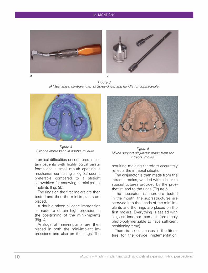
atomical difficulties encountered in cer-tain patients with highly ogival palatal forms and a small mouth opening, a mechanical contra-angle (Fig. 3a) seems preferable compared to a straight screwdriver for screwing in mini-palatal implants (Fig. 3b).
The rings on the first molars are then tested and then the mini-implants are placed.
A double-mixed silicone impression is made to obtain high precision in the positioning of the mini-implants (Fig. 4).
Analogs of mini-implants are then placed in both the mini-implant im-pressions and also on the rings. The
resulting molding therefore accurately reflects the intraoral situation.
The disjunctor is then made from the intraoral molds, welded with a laser to suprastructures provided by the pros-thetist, and to the rings (Figure 5).
The apparatus is therefore tested in the mouth, the suprastructures are screwed into the heads of the mini-im-plants and the rings are placed on the first molars. Everything is sealed with a glass–ionomer cement (preferably photo-polymerizable to have sufficient positioning time).
There is no consensus in the litera-ture for the device implementation.
a b
Figure 3a) Mechanical contra-angle. b) Screwdriver and handle for contra-angle.
Figure 4Silicone impression in double mixture.
Figure 5Mixed support disjunctor made from the
intraoral molds.

J Dentofacial Anom Orthod 2017;20:405 11
MINI-IMpLANT ASSISTED RApID pALATAL ExpANSION: NEW pERSpECTIVES
Some studies recommend activating the device only a few days after the installation of the screws18, whereas others recommend waiting 4–6 weeks after installing the screws allowing for a more organized bone matrix around the mini-implants (lamellar structure)18.
As for the activation of the device, we found different methods in the literature, but the most used seems to be activating the disjunctor twice a day (once in the morning, once in the evening), i.e., an expansion of 0.5 mm/day like a conventional disjunc-tor16,23,47,74. Wilmes recommends acti-vating twice at 90°, i.e., an activation of 0.8 mm/day18.
Benefits
In light of the many disadvantages of the conventional disjunctors described above, the use of mixed anchors seems to be a good alternative to overcome the various problems encountered.
In fact, disjunctors anchored on the posterior side of the first molars and palatal mini-implants on the anterior side provide many advantages:
Compared to the strict bone disjunctor:
- Elimination of the need for invasive surgery. In fact, the strictly bone-sup-ported disjunctors require surgical im-plantation via a palatal flap lift to insert the implant and the osseointegration screw. Whereas the placement of the mini-implant supports for the hybrid disjunctor is flapless because the mini-implants used are self-drilling.
- No need for osseointegration.Unlike implants used in the Dresden
system, osseointegration is not neces-
sary for a miniscrew because the sta-bility is based on mechanical retention.
- Easy to remove. In most case, anes-thesia is not necessary.
Compared to the conventional disjunctor:
- Pushes the boundaries of orthopedic expansion. A recent study published by Choi in 201612 shows that maxillary ex-pansion assisted by mini-implants was effective in almost 87% of patients with an average age of 20 years. In differ-ent studies10,37, patients receiving rapid maxillary expansion with a hybrid dis-junctor appear to be older than those treated with conventional devices. In fact, to minimize the deleterious effects on the teeth and the periodontium in the event of sutural resistance, the age window used is narrower than that for hybrid disjunctors48. In contrast, skele-tal effects are always greater in subjects treated earlier66. It would make it possi-ble to avoid a surgical phase in patients generally presenting with a major sagit-tal problem requiring an additional sagit-tal correction surgery12.
Cases are also described in the liter-ature in young adults, to avoid surgery on the transverse dimension10,37.
- Can be used in patients with weak anterior tooth anchorage (resorption, deciduous teeth). The support being skeletal and not dental, in case of ab-sence of dental organs or weakness of the latter in prior, its implementation is not compromised18,49.
- A repositioning rather than a rota-tion of the two maxillae. The horizontal force generated by the disjunctor an-chored to the mini-implants in an apical position is transmitted to the palate at a
Montigny M. Mini-implant assisted rapid palatal expansion: New perspectives12
M. MONTIGNY
high point located, which is thus closer to the resistance center of the jaw than the dental disjunctors ( horizontal V). This allows more horizontal move-ments and therefore a mass transla-tion of the two hemimaxillae. This de-creases the movement (V frontal) and rotation of the left and right separated jaws (vertical V)61,53.
- Prevents mesialization of the an-chor molars until the premolars erupt. In fact, the rigid structure connected to both mini-implants serves as a space maintainer, and acts as a Nance arch, thus preventing the molars from un-dergoing mesialization and stunting the development of definitive premolars68.
- The versatility of mini-implants palat-ins73. In patients with skeletal class-III dentition who require rapid maxillary expansion, the use of the hybrid dis-junctor seems to be the best option. According to Hourfar et al. who pub-lished a retrospective study in 2015 on 100 patients, maxillary traction would be more effective if supported by two anterior mini-implants connected to the disjunctor rather than a convention-al disjunctor26.
- Increased volume of nasal fossae. It has been proven that rapid maxillary expansion improves nasal ventilation, increases the volume of the upper air-ways, and decreases nasal resistance. A recent study, published in 2014 by Motro et al. reports that the hybrid disjunctor produces exactly the same benefits as a conventional disjunctor on ventilation and the upper airways especially in the nasopharynx and oro-pharynx48. These results have been called into question by the study pub-lished by Kabalan in 2015, which does not report significant differences after rapid maxillary expansion with a hybrid
expansion (none of them with a con-ventional disjunctor) in regard to upper airway volume28.
- Treatment of sleep apnea syndrome.As explained above, the effects of rap-
id maxillary expansion largely exceed the simple correction of the morpho-logical anomaly of endognathy. It helps to standardize functions and is one of the treatments of choice for the correc-tion of sleep apnea syndromes52,13.
Often treated by the removal of the tonsils and adenoids during child-hood without any real skeletal action, it is common to see the appearance or reappearance of sleep apnea syn-drome during adolescence. The mixed- support expansion, pushing the age limits of a conventional expansion, has become the treatment of choice for sleep apnea syndrome in adolescents, also having an indirect action on the repositioning of the tongue16.
- Skeletal stability after removal of the disjunctor. At the end of the ex-pansion, skeletal stability can be main-tained without keeping the disjunctor. Indeed, the Beneplate, the plate to be placed between the two mini-implants, makes it possible to maintain the dis-tance72 (Fig. 6).
- Minimizes loss of vestibular thick-ness of the bony crest in the premolars. Effectively, in a comparative study pub-lished by Toklu et al., show that the loss of thickness of the bone crest at the lev-el of the premolars is 0.80 ± 0.65 mm on average for patients treated with a conventional disjunctor compared to patients treated with a disjunctor with mixed supports, which does not pres-ent a loss of thickness23. These results are in agreement with those found in ChaneFane’s preliminary study, which shows that there would be no loss of

J Dentofacial Anom Orthod 2017;20:405 13
MINI-IMpLANT ASSISTED RApID pALATAL ExpANSION: NEW pERSpECTIVES
vestibular thickness of the bone crest at the level of the premolars following the mixed-support expansion11.
- Fewer alveolar effects and reduc-tion of the dental version at the molar and premolar levels. A comparative study conducted by Lagravère et al. in 2010, reports that a dental disjunctor produces more vestibulo-versions of the premolars than a device with bone supports33. As Toklu shows in his study in 2015, the inclination of the maxillary premolar remains unchanged in the mixed-support expansion compared to conventional expansion.
- Fewer adverse effects on the ver-tical dimension. It goes hand in hand with the reduction of alveolar effects and would increase the therapeutic possibilities, especially for dolichofacial patients42.
Lower cost. In fact, even if the de-vice used seems slightly more expen-sive than a conventional disjunctor (be-cause of the use of mini-implants), this cost is still lower than that of a surgical operation on the transverse dimension requiring general anesthesia and all the equipment associated with the operat-ing theater.
Disadvantages
Anatomical• Bone thickness
To maintain good primary stability of the mini-implant, it is necessary to have a minimum of 6 mm of bone, to pre-vent the perforation of the nasal corti-cal bone. The choice of the positioning of the mini-implants strongly depends on this factor. The preferential location was defined as being at the level of the first maxillary premolar at 2–4 mm of
the intermaxillary suture, representing a low perforation risk29.
• Nasal-palatal canalA mini-implant that is positioned too
anteriorly could damage the nasopala-tal canal and any nerves and vessels passing through it. The risk is low, but this occurs in some cases.
Similarly, at the level of the canine pillars, in the lateral palatal region, this nerve and blood plexus anastomoses with the vessels from the palatal fo-ramen in the anterior direction. An installation position that is too lateral and too anterior will cause problems.
• Salivary glandsThe posterior segment of the hard
palate includes many small salivary glands, a mini-implant placement that is too posterior could affect these structures and cause mucus retention (oral mucocele and/or necrotizing sialo-metaplasia)59.
• Shape of the palatepatients requiring rapid maxillary ex-
pansion most often have narrow pal-ates with deep palatal clefts and lateral palatal bulges. The palatal mucosa is often thick.
This morphology increases the diffi-culty of installation and can compromise the correct position of mini-implants.
• Dental rootsThe insertion of mini-implants at the
palatal level carries the risk of damag-ing the maxillary tooth roots. This risk is especially pronounced at the posteri-or level, at the level of the palatal roots of maxillary molars. This is one of the reasons why the dental anchor is used in later stages18.
But this risk is rare when angulation, length, and position are respected.
Montigny M. Mini-implant assisted rapid palatal expansion: New perspectives14
M. MONTIGNY
Linked to the positionOral opening. The precise positioning
of mini-implants is essential to avoid problems during installation. patients with a small oral opening will have two problems:– poor visibility of the laying position;– interference of the right screwdriver
with the mandible. The use of the mechanical contra-angle solves this problem.Local anesthesia. The placement of
the palatal mini-implants is done under local anesthesia. This step can lead to difficulties, including all complications related to local anesthesia:– local and regional complications:•• equipment complications (needle
breakage, bursting of the cartridge);•• mucosal lesions (ulceration, necro-
sis, hematoma);•• neurological complications (sensi-
tivity, sensory);– general complications:•• toxic accidents;•• allergic reactions.•• Also, it is necessary to have all nec-
essary equipment validated:– syringe;
– needle;– injectable anesthetic solution: artic-
aine, adrenaline 1/200.Loss of the mini-implant and inflam-
mation of the palatal mucosa. One of the problems encountered is the loss of the mini-implant after installation, occurring when primary stability is not achieved16. The loss rate is estimated at 25% in any position, but the palate seems to be the most stable position41.
A new position may be considered after palatal mucosa healing.
Following the positioning, the palatal gingiva may bud around the screw due to compression and create inflamma-tion. To avoid this phenomenon, it is preferable to use mini-implants with high collars and fairly large heads. Good hygiene should also be main-tained (chlorhexidine mouthwash). An-tibiotic therapy may complement the prescription if necessary16.
Pain. The pain recorded on an analog pain scale, appears to be higher after the first activation in patients with a mixed-press disjunctor compared to patients wearing a conventional dis-junctor33.
Rapid maxillary expansion as a treat-ment for maxillary endognathy in chil-dren has been proven time and time again, it is well established. In addition, it has disadvantages that have been discussed previously.
To improve the management of these patients, new techniques have recent-ly emerged.
Through this article, we have tried to put them in perspective, by detailing
their indications, the advantages as well as the disadvantages, and their implementations.
Mixed-feed disjunctors have many advantages but are still seldom used. The habits of the practitioners and the small difficulties that arise from their installation and use mean that they are not always considered as the first choice of treatment.
CONCLUSION

J Dentofacial Anom Orthod 2017;20:405 15
MINI-IMpLANT ASSISTED RApID pALATAL ExpANSION: NEW pERSpECTIVES
They are nevertheless a good alterna-tive in certain clinical situations in chil-dren/adolescents.
Conflict of interest: The author declares that there is no conflict of interest.
1. Akkaya S, Lorenzon S, Uçem TT. Comparison of high-speed and rapid exchange rates. Eur J Orthod 1998;20(3):255-261.
2. Angell E. Treatment of irregularity of the permanent or adult teeth. Dent Cosm 1860;1:540-544,599-600.
3. Baccetti T, crossed L, Mcnamara JA. The Cervical Vertebral Maturation Method for the Assessment of Optimal Treatment in Dentofacial Orthopedics. Semin Orthod 11:119-129.
4. Ballanti F, Lione R, Fanucci E, Franchi L, Baccetti T, Cozza p. Immediate and post-Retention Effects of Rapid Maxillary Expansion Investigated by Computed Tomography in Growing patients. Orthod Angle 2009;79(1):24-29.
5. Baydas¸ B, Yavuz I, Uslu H, Dagsuyu IM. Nonsurgical Rapid Maxillary Expansion Effects on Craniofacial Structures in Young Adult Females, a bone scinti graphy study. Orthod Angle 2006;76(5):3-5.
6. Baysal A, et al. Evaluation of root resorption following rapid maxillary expansion using cone-beam computed tomography. Angle Orthod 2012;82(3):488-494.
7. Bell RA. A review of maxillary expansion in relation to the rate of expansion and patient’s age. Am J Orthod 1982:81(1):32-37.
8. Bishara E, Ortho, Staley RN. Maxillary expansion: Clinical implications. Am J Orthod Dentofac Orthop 1987;91:3-14.
9. Boes M, Darque F. Memory CECSMO: Cartography of the palate - clinical applications in orthodontic mini-implantology 2010.
10. Carlson C, Sung J, McComb RW, Machado AW, Moon W. Microimplant-assisted rapid palatal expansion appliance to orthopedically correct trans verse maxillary deficiency in an adult. Am J Orthod Dentofacial Orthop 2016;149 (5):716-728.
11. Chane-Fane C, Darqué F. Rapid maxillary expansion assisted by palatal mini-implants in adolescents - preliminary study. Int Orthod 2015;13(1):96-111.
12. Choi SH, Shi KK, Cha JY, YC park, Lee KJ. Nonsurgical miniscrew-assisted rapid maxillary expansion in acceptable stability in young adults. Orthod Angle; 2016;86(5):713-720.
13. Cistulli pA, palmisano RG, poole MD. Treatment of obstructive sleep apnea syndrome by rapid maxillary expansion. Sleep 1998;21(8):831-835.
14. Conroy-piskai C, et al. Assessment of vertical change during n. expansion using quad helix gold bonded rapid n. expander. Angle Orthod 2016;86(6):925-933.
15. Cozza p, Giancotti A, petrosino A. Rapid palatal expansion in mixed dentition using a mod-ified expander: a cephalometric investigation. J Orthod 2001;28(2):129-134.
16. Darque F, Ellouze S. Biomechanics of the miniim-anchoring plants: clinical illustrations. International Orthodontics 2007;5(4):357-392.
17. Darque F, Masmoudi K. Memory DU: Mapping the thickness of the buccal alveolar bone 2011.18. Ellouze S, Darque F. Mini implants: The orthodontics of tomorrow. Quintessen 2012.
BIBLIOGRAPHY
˘
Montigny M. Mini-implant assisted rapid palatal expansion: New perspectives16
M. MONTIGNY
19. Garib DG, Henriques JFC, Janson G, Freitas MR, Fernandes AY. periodontal effects of rapid maxillary expansion with tooth-tissue-terminal and tooth-terminal expanders: A com-puted tomography evaluation. Am J Orthod Dentofac Orthop 2006;129(6):749-758.
20. Garrett BJ, JM Caruso, Rungcharassaeng K, JR Boot, Kim JS, Taylor GD. Skeletal effects to the maxilla after rapid maxillary expansion assessed with cone-beam computed tomog-raphy. Am J Orthod Dentofacial Orthop 2008;134(1):8-9.
21. Gray Lp. Results of 310 cases of rapid maxillary expansion selected for medical reasons. J Laryngol Otol 1975;89(6):601-614.
22. Greenbaum KR, Zachrisson BU. The effect of palatal expansion therapy on the periodontal supporting tissues. Am J Orthod 1982;81(1):12-21.
23. Gunyuz Toklu M, Germec-Cakan D, Tozlu M. periodontal, dentoalveolar, and skeletal effects of overthterminal and tooth-bone-terminal expansion appliances. Am J Orthod Dentofacial Orthop 2015;148(1):97-109.
24. Hartgerink D, pS Vig, Abbott DW. The effect of rapid maxillary expansion on nasal airway resistance. Am J Orthod Dentofacial Orthop 1987;92(5):381-389.
25. Hershey HG, BL Stewart, Warren DW. Changes in nasal airway resistance associated with rapid maxillary expansion. Am J Orthod 1976;69(3):274-284.
26. Hourfar J, Kinzinger GSM, Ludwig B, Spindler J, Lisson JA. Differential treatment effects of two anchorage systems for rapid maxillary expansion: a retrospective cephalometric study. Orofac Orthop / Fortschritte der Kieferorthopädie 2016:1-10.
27. Johnson BM, McNamara JA, RL Bandeen, Baccetti T. Changes in soft tissue nasal widths associated with rapid maxillary expansion in prepubertal and postpu-bertal subjects. Angle Orthod 2010;80(6):995-1001.
28. Kabalan O, Gordon J, Heo G, Lagravère MO. Nasal airway changes in bone-terminal and tooth-terminal rapid maxillary expansion treatments. Int Orthod 2015;13(1):1-15.
29. Kawa D, Kunkel M, Heuser L, Jung BA. What is the best position for palatal implants? A CBCT study on bone volume in the growing maxilla. |||UNTRANSLATED_CONTENT_START|||Clin Oral Investig.|||UNTRANSLATED_CONTENT_END||| 2017;21(2):541-549.
30. Kim KB, Adams D, EA Araújo, Behrents RG. Evaluation of immediate soft tissue changes after rapid maxillary expansion. Dental press J Orthod 2012;17(5):157-164.
31. Krebs A. Midpalatal suture expansion studies by the implant method over a seven-year period. Congress Resp Eur Orthod Soc 1964;40:131-142.
32. Kurol J, Modin H, Bjerkhoel A. Orthodontic maxillary expansion and its effect on nocturnal enuresis. Angle Orthod 1998;68(3):225-232.
33. Lagravère MO, Carey J, G, Toogood RW, pW Major Heo. Transverse, vertical, and anter-oposterior changes from bone-anchored maxillary expansion vs traditional rapid maxillary expansion: a randomized clinical trial. Am J Orthod Dentofacial Orthop 2010;137(3):304.e1-12; discussion 304-5.
34. Lagravere MO, Heo G, Major pW, Flores-Mir C. Meta-analysis of immediate changes with rapid maxillary expansion treatment. J Am Dent Assoc 2006;137(1):44-53.
35. Lagravere MO, Major pW, Flores-Mir C. Long-term dental arch changes after rapid maxil-lary expansion treatment: a systematic review. Orthod Angle 2005;75(2):155-161.
36. Lee H, Ting K, Nelson M, Sun N, Sung S-J. N. expansion in customized finite element method models. Am J Orthod Dentofac Orthop 2009;136(3):367-374.

J Dentofacial Anom Orthod 2017;20:405 17
MINI-IMpLANT ASSISTED RApID pALATAL ExpANSION: NEW pERSpECTIVES
37. Lee KJ, YC park, park JY, Hwang WS. Miniscrew-assisted nonsurgical palatal expansion before orthognathic surgery for a patient with severe mandibular prognathism. Am J Or-thod Dentofacial Orthop 2010;137(6):830-839.
38. Lessa FCR, Enoki C, MFN Feres, FCp Valera, Lima WTA, Matsumoto MAN. Influence of pa-drao respiratório na morfologia craniofacial. Rev Bras Otorrinolaringol 2005; 71 (2): 156-160.
39. Linder-Aronson S, Woodside DG, Lundstrom A. Mandibular growth direction following ad-enoidectomy. Am J Orthod 1986;89(4):273-284.
40. Ludwig B, et al. Application of a new viscoelastic finite element method and analysis of miniscrew-supported hybrid hyrax treatment. Am J Orthod Dentofacial Orthop 2013;143(3): 426-435.
41. Ludwig B, Glasl B, SJ, walls B, GSM, Lisson JA Kinzinger Bowman. Anatomical guidelines for miniscrew insertion: palatal sites. J Clin Orthod 2011;45(8):433-41; quiz 467.
42. MacGinnis M, Chu H, Youssef G, Wu KW, Machado W AW, Moon W. The effects of mini-implant assisted rapid palatal expansion (MARpE) on the nasomaxillary complex finite element method analysis. prog Orthod 2014;15:52.
43. Machado-Júnior AJ, Zancanella E, Crespo AN. Rapid maxillary expansion and obstructive apnea sleep: A review and meta-analysis. Med Oral patol Oral Cir Bucal March 2016.
44. Marcus CL, et al. Diagnosis and management of childhood obstructive sleep apnea syn-drome. pediatrics 2012;130(3):576-584.
45. McNamara JA, Baccetti T, Franchi L, Herberger TA. Rapid maxillary expansion followed by fixed appliances: a long-term evaluation of changes in arch dimensions. Orthod Angle 2003;73(4):344-353.
46. Melsen B, Melsen F. The postnatal development of the palatomaxillary region studied on human autopsy material. Am J Orthod 1982;82(4):329-342.
47. Mosleh MI, Kaddah MA, Abd ElSayed FA, ElSayed HS. Comparison of transverse changes during maxillary expansion with 4-point bone-terminal and tooth-terminal maxillary ex-panders. Am J Orthod Dentofacial Orthop 2015;148(4):599-607.
48. Motro M, et al. Rapid-maxillary-expansion induced rhinological effects: a retrospective multicenter study. Eur Arch Otorhinolaryngol 2016;273(3):679-687.
49. Nienkemper M, walls B, pauls, Drescher D. n. protraction using hybrid hyrax-facemask combination. prog Orthod 2013;14(1):5.
50. Oliveira De Felippe NL, Da Silveira AC, Viana G, Kusnoto B, Smith B, Evans CA. Relation-ship between rapid maxillary expansion and nasal cavity size and airway resistance: short- and long-term effects. Am J Orthod Dentofacial Orthop 2008;134(3):370-382.
51. phoenix A, Valiathan M, S Nelson, Strohl Kp, Hans M. Changes in hyoid bone position fol-lowing rapid maxillary expansion in adolescents. Angle Orthod 2011;81(4):632-638.
52. pirelli p, Saponara M, Guilleminault C. Rapid maxillary expansion in children with obstruc-tive sleep apnea syndrome. Sleep 2004;27(4):761-766.
53. Richard O, Nicaud-Léon TM, Facon F. Intermaxillary miniscrew expansion during orthodon-tic treatment in lingual technique. Rev Orthop Dento Faciale 2012;46(4):463-470.
54. Ricketts RM. Respiratory obstruction syndrome. Am J Orthod 1968;54(7):495-507.55. Sander C, Hüffmeier S, Sander FM, Sander FG. Initial results regarding force exertion dur-
ing rapid maxillary expansion in children. Orofac Orthop / Fortschritte der Kieferorthopädie 2006;67(1):19-26.
56. Sandstrom RA, et al. Expansion of the lower arch competitor with rapid maxillary expan-sion. Am J Orthod Dentofac Orthop 1988;94(4):296-302.

Montigny M. Mini-implant assisted rapid palatal expansion: New perspectives18
M. MONTIGNY
57. Schütz-Fransson U, Kurol J. Rapid maxillary expansion effects on nocturnal enuresis in children: a follow-up study. Angle Orthod 2008;78(2):201-208.
58. Starnbach H, Bayne D, Cleall J, Subtelny JD. Facioskeletal and dental changes resulting from rapid maxillary expansion. Angle Orthod 1966;36(2):152-164.
59. Suzuki H, Moon W, prevident LH, SS Suzuki, AS Garcez, Consolaro A. Miniscrew-assisted fast palatal expander (MARpE): the quest for pure orthopedic movement. Dental press J Orthod 21(4):17-23.
60. Tired K, Matsubara S, Sakuda M. Location of the center of resistance for nasomaxillary complex studied in a three-dimensional finite element model. Br J Orthod 1995;22(3): 227-232.
61. Tausche E, Hansen L, Schneider M, Harzer W. [Bone-supported rapid maxillary expansion with an implant-terminal Hyrax screw: the Dresden Distractor]. Orthod en 2008;79(2): 127-135.
62. Tecco S, F, head S, Longhi V Festa, of Attilio M. Changes in head posture after rapid ex-pansion n. in mouth-breathing girls: a controlled study. Orthod Angle 2005; 75 (2): 171-176.
63. DJ Timms. Rapid n. expansion in the treatment of nocturnal enuresis. Angle Orthod 1990; 60(3):229-233.
64. Filmed Lp. Growth of the pharynx and its physiological implications. Am J Orthod Dento-facial Orthop 1991;99 (2):129-139.
65. Ugolini A, Doldo T, Ghislanzoni LTH, Mapelli A, Giorgetti R, Sforza C. Rapid palatal ex-pansion effects on mandibular transverse dimensions in unilateral posterior crossbite pa-tients: a three-dimensional digi-tal imaging study. prog Orthod 2016;17(1):1.
66. JW, Karydis A Vassar, Trojan, Fisher J. Dentoskeletal effects of a temporary skeletal an-chorage device-supported rapid n. expansion appliance (TSADRME): A pilot study. Angle Orthod 2016; 86 (2): 241-249.
67. Villano A, Grampi B, Fiorentini R, Gandini p. Correlations between rapid maxillary expan-sion (RME) and the auditory apparatus. Angle Orthod 2006; 76 (5); 752-758.
68. Walter A., Wendl B, ploder O, Mojal S., puigdollers A. Stability determinants of bone- terminal force-transmitting components in three RME hybrid expanders-in vitro study. Eur J Orthod 2017;39(1):76-84.
69. Warren DW, Hershey HG, TA Turvey, Hinton VA, Hairfield WM. The nasal airway following maxil-lary expansion. Am J Orthod Dentofacial Orthop 1987;91(2):111-116.
70. Watson WG. Expansion and fenestration gold dehis-cence. Am J Orthod 1980;77(3): 330-332.
71. Wertz RA. Skeletal and dental changes midpalatal suture opening. Am J Orthod 1970;58 (1):41-66.
72. Wilmes B, Drescher D, Nienkemper M. A miniplate system for improved stability of skel-etal anchorage. J Clin Orthod 2009;43(8):494-501.
73. Walls B, Nienkemper M, Drescher D. Application and effectiveness of a mini-implant-and tooth-borne rapid palatal expansion device: the hybrid hyrax. World J Orthod 2010;11(4): 323-330.
74. Yılmaz A, Arman-Özçırpıcı A, Erken S, polat-Özsoy Ö. Mini-implant-supported maxillary expansion appliance with standard expansion protocols. Eur J Orthod 2015;37(5):556-564.
75. Zimring JF, Isaacson RJ. Forces produced by rapid maxillary expansion. 3. Forces present during retentioning. Angle Orthod 1965;35:178-186.