Microvascular Free Tissue Transfer - University of Texas ... · Microvascular Free Tissue Transfer...
46
Microvascular Free Tissue Transfer Glen T. Porter, MD Faculty Advisor: Shawn D. Newlands, MD, PhD The University of Texas Medical Branch Department of Otolaryngology Galveston, Texas Grand Rounds Presentation October 2004
Transcript of Microvascular Free Tissue Transfer - University of Texas ... · Microvascular Free Tissue Transfer...

Microvascular Free Tissue
Transfer Glen T. Porter, MD
Faculty Advisor: Shawn D. Newlands, MD, PhD
The University of Texas Medical Branch
Department of Otolaryngology
Galveston, Texas
Grand Rounds Presentation
October 2004

History
Early 1900’s Alexis Carrel
1950’s Jacobsen and Suarez—anastomoses in animals
1959 Seidenberg– free jejunum segments to repair pharyngoesophageal defects (human)
1972 McLean and Buncke – omental flap to cover a cranial defect (first “microvascular” flap)
1973 Daniels and Taylor– “free flap” First free cutaneous flap
1976 Baker and Panje– first free flap in head and neck cancer reconstruction
Groin flap pedicled on the circumflex iliac artery

Advantages of free tissue transfer
Two team approach
Improved vascularity and wound healing
Low rate of resorption
Defect size of little consequence
Potential for sensory and motor innervation
Permits use of osseointegrated implants

Advantages of free tissue transfer
Wide variety of available tissue types
Large amount of composite tissue
Tailored to match defect
Wide range of skin characteristics
More efficient use of harvested tissue
Immediate reconstruction

Recipient vessels
Arteries
Superficial temporal system
–scalp and upper face
Facial artery—midface and
cervical region
(atherosclerosis common)
Superior thyroid or lingual
artery—lower cervical region
Other: thyrocervical trunk,
external carotid, common
carotid

Recipient vessels
Veins
External jugular
Branches of internal jugular
(common facial)
Internal jugular
Retrograde (superficial
temporal, thyroid)
Transverse cervical, occipital
(very small)

Recipient vessels after previous
neck dissection
Gold standard: Angiogram (short-term injury to endothelium reported)
Operative reports
Long-pedicled flaps
Thyrocervical trunk (transverse cervical), Occipital vessels, retrograde drainage (thyroid veins, superficial temporal), external carotid artery
Contralateral vessels (recipient or graft)
End-to-side anastomoses with large vessels
Vein grafts
Arteriovenous loop (poorer results)

Vessel selection
Size
Arterial vs.Venous
Atherosclerosis
XRT-related changes
Vessel geometry (location and
orientation)
Vessel length

Vessel preparation
Arteries need to have strong pulsatile
flow—cut until it flows.
Cut back beyond branches or ligate them if
sufficiently distant from the anastomosis
site.
Atherosclerosis
Intimal inspection
Dilation
Removing the adventitia

Irradiated vessels
Technically more difficult—effects appear specific to arteries
Higher incidence of atherosclerosis
Vessel wall fibrosis, increased wall thickness, more intimal dehiscence
No reported difference in outcome of microvascular anastomoses (Nahabedian MY, et al., 2004, Kroll SS, et al 1998)
Microvascular anastomoses tolerate XRT well long-term (Foote RL., et al., 1994)
Require careful handling, cut off clot (teasing thrombi may denude vessel wall—”sticky” walls), smaller suture, needle introduced from lumen to outside wall (to pin intima to wall)

Prepare vessels Evaluate vessel geometry
Trim, irrigate, dilate
Partial flap insetting (bony cuts and plating done at donor bed, if necessary)
Arterial vs. venous anastomosis first with early or delayed unclamping of first vessel showed no difference. (Braun, et al., 2003)
Anastomosis of remaining vessel
Complete flap insetting
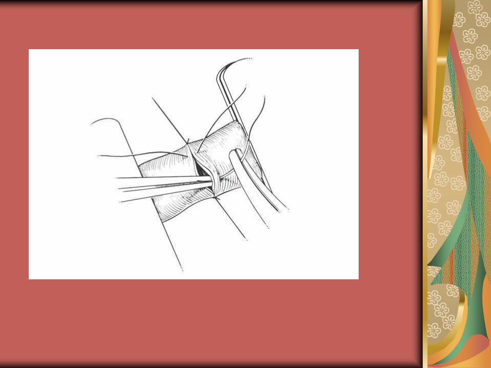
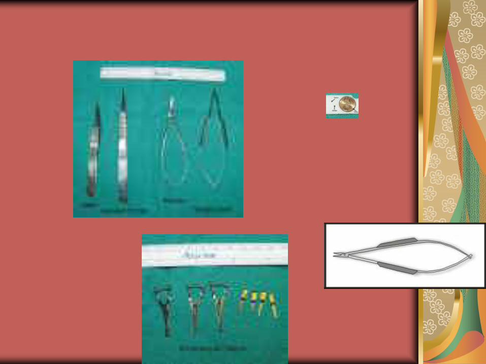
Microvascular Anastomosis

Microvascular surgical technique
Trim adventitia 2-3mm
Gentle handling (no full-thickness)
Trim free edge, if needed
Dissect vessels from surrounding tissues
Irrigate and dilate Heparinized saline
Mechanical dilation (1 ½ times normal –paralyses smooth muscle)
Chemical dilation, if necessary
Suturing

Microvascular suture technique
3 guide sutures (120 degrees apart)
Perpendicular piercing
Entry point 2x thickness of vessel from cut end
Equal bites on either side
Microforceps in lumen vs. retracting adventitia
Pull needle through in circular motion
Surgeon’s knot with guide sutures, simple for others
Avoid backwalling—2 bites/irrigation

3 suture
technique

Vessel size mismatch
Laminar flow vs. turbulent flow
<2:1 – dilation, suture technique
>2:1, <3:1 – beveling or spatulation (no more than 30 degrees to avoid turbulence)
>3:1 – end-to-side

End-to-end vs. End-to-side
Recent reports indicate end-to-side without
increase in flap loss or blood flow rate.
End-to-side overcomes size discrepancy,
avoids vessel retraction, and IJ may act as
venous siphon.
End-to-side felt best when angle is less
than 60 degrees (minimize turbulence)
Vessel incision should be elliptical, not slit
Can use continuous suture technique

End-to-side
Anastomosis
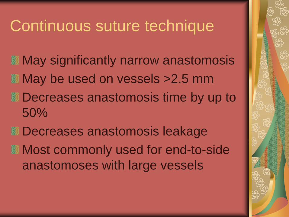
Continuous suture technique
May significantly narrow anastomosis
May be used on vessels >2.5 mm
Decreases anastomosis time by up to
50%
Decreases anastomosis leakage
Most commonly used for end-to-side
anastomoses with large vessels
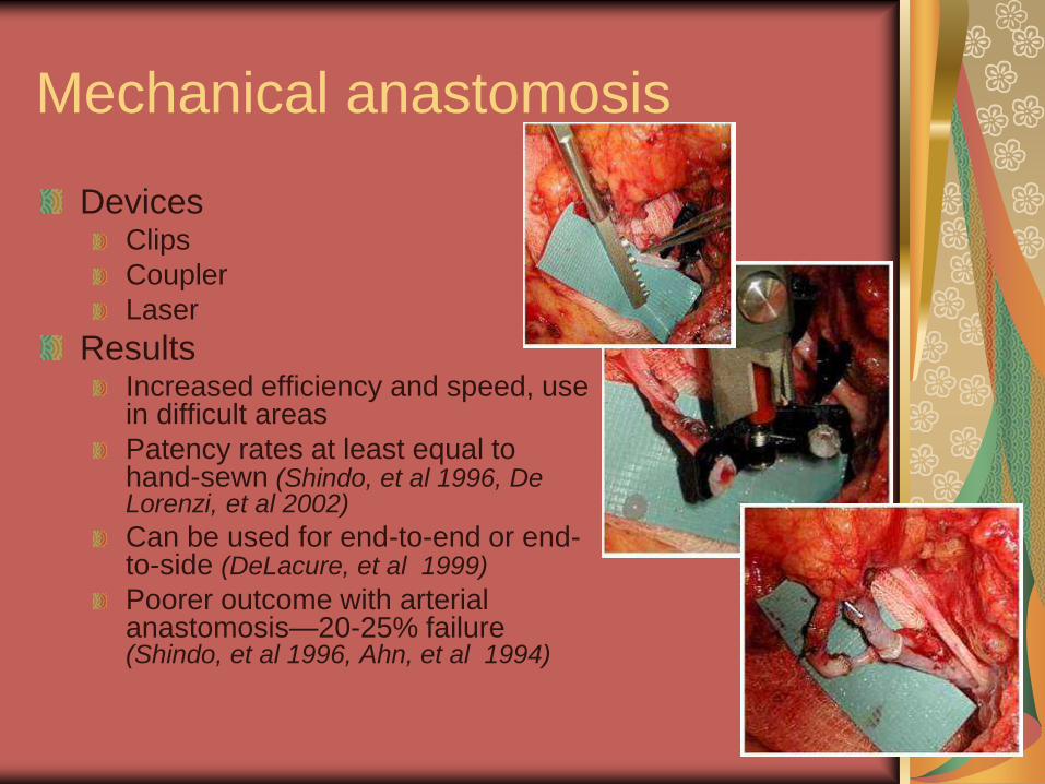
Mechanical anastomosis
Devices Clips
Coupler
Laser
Results Increased efficiency and speed, use in difficult areas
Patency rates at least equal to hand-sewn (Shindo, et al 1996, De Lorenzi, et al 2002)
Can be used for end-to-end or end-to-side (DeLacure, et al 1999)
Poorer outcome with arterial anastomosis—20-25% failure (Shindo, et al 1996, Ahn, et al 1994)

Vein grafts
Used in situation where pedicle is not long enough for tension-free anastomosis
Usually harvested from lower extremity (saphenous system)
Valve orientation is necessary
Avoid anastomosis at level of vein valve
Keep clamps in place until both anastomoses sewn
Prognosis for success controversial (Jones NF, et al., 1996, German, et al. 1996)
Recent literature

Microvascular Hints & Helps
Use background to help visualize suture
Demagnetize instruments, if needed
May reclamp vessels for repair after 15
minutes of flow
Reclamp both arterial and venous vessels
when revising venous anastomosis
Support your hands and hold instruments
like a pencil

Ischemia
Primary and secondary Primary: 2.25-6 hours
Secondary: 1-12 hours
Interrelation
No flow phenomenon
Cold vs. normothermic In vitro studies show benefit to cooling of flaps
In vivo studies show surface cooling (<4hr ischemia time) does not adversely effect flap success (Shaw W. et al 1996)
Tissue specific critical ischemia times Metobolic rate dependent
Perfusates (UW, tissusol, Viaspan, Heparin) Literature unclear

Anastomotic failure
93-95% success rate expected
Venous thrombosis:Arterial thrombosis 4:1, ateriovenous loop, tobacco use significant factors (Nahabedian M., et al, 2004) Other literature indicates 9/10 thromboses secondary to venous thrombus
Tobacco use as contribution controversial (4/5 failures in Nahabedian study - venous thrombosis)
Venous occlusion, Delayed reconstruction, Hematoma significant factors in breast free tissue recon. (Nahabedian M., et al, 2004)
Salvage 50% in breast reconstruction
Age, prior irradiation, DM (well-controlled), method of anastomosis, timing, vein graft, and specific arteries/veins not felt to contribute to failure rate

Anastomotic Failure--timeline
15-20 minutes
<72 hours
5-7 days
>8 days
Thin vs. thick flaps

Thrombus formation
Injury to endothelium and media of vessel Mechanical vs. thermal
Error in suture placement Backwall or loose sutures
Edges not well-aligned (most common in veins—most common site of thrombus)
Intimal discontinuity with exposure of media Oblique sutures, large needles, tight knots
Infection
Hypovolemia and low flow states Nitroprusside at dose to decrease arterial pressure by 30% causes severe reduction in flap blood flow (40%) (Banic, et al. 2003)
Vessel geometry (kinking, tension)

Vessel spasm Causes
Trauma
Contact with blood
Vasoconstrictive drugs Phenylephrine--dose causing 30% increase in arterial pressure shows no effect on flap circulation (Banic A, et al., 1999)
Nicotine
Temperature, drying
Treatment Warmth
Xylocaine
Papavarine, thorazine
Volume repletion

Treatement for anastomotic failure
Revision of anastomoses
Exploration of wound
Streptokinase, urokinase, rt-PA (Atiyeh BS, et al 1999)
Leech therapy
Wound care
Statistics Revisions successful in 50%
Revisions less successful after first 24-48hr
>6 hrs of ischemia leads to poor survival
12 hrs of ischemia leads to “no-flow” phenomenon
After 5 days almost all flaps in rabbit model survived with loss of artery or vein (but not both)—this is rational for other modalities after 48 hours

Post-operative care
Anticoagulation
Attention to wound care
Flap monitoring
Nothing around neck that might compress pedicle
Antibiotics
Hemoglobin/intravascular volume—literature unclear (Velanovich V., et al 1988, Quinlan 2003)
No pressors/nicotine/cooling of flap (literature unclear)

Anticoagulation
Rheology RBC concentration
Plasma viscosity
RBC aggregation
RBC deformability
Other (platelets, thrombogenic mediators)
Agents Aspirin
Heparin
Dextran
Other
Indications Hypercoagulable state (Friedman G, et al, 2001)
Excessive vessel trauma
Complications

Dextran Macromolecule which is a compound of glucose subunit
Thought to improve RBC flexibility, increase electronegativity of vessel wall (which decreases platelet adhesion), act as intravascular volume expander, decrease RBC aggregation
Shown to decrease clotting secondary to exposed collagen in rabbit arteries. Little effect on platelet, rather inhibits fibrin stabilization of thrombi (Weislander, JB, et al., 1986)
No effect on overall flap survival when compared with aspirin. Systemic complications 3.9-7.2 times more common with dextran infusion (Disa J., et al, 2001)
Complications can include renal damage, anaphylactic shock, congestive heart failure, MI, pulmonary edema, pleural effusion, pneumonia

Aspirin
Prevent platelet thrombosis
Inhibits arachidonic acid to prostaglandin synthesis on the platelet—prevents release of platelet granuoles that cause platelet aggregation. Mechanism is biphasic and dose-dependant
High doses of aspirin can have negative effect on endothelial production of prostacyclin which prevents platelet accumulation on exposed collagen and dilates vessels.
ASA PR qd x several weeks (often given at beginning of case)—5 grains (325 mg)
No good studies to confirm benefit of use
Hematoma formation

Heparin
Naturally occuring glycosoaminoglycan which interrupts clotting cascade
Prevents transformation of prothrombin to thrombin, fibrinogen to fibrin
Does not lyse existing thrombi
Strongly adheres to endothelium Concentration on endothelium 100x serum
½ life = 90 minutes
Given at time of first quarter of arterial anastomoses vs. at time of unclamping (bolus only vs. bolus with drip x 3 days)
Literature unconvincing, although it may increase microvascular perfusion after ischemia
Hematoma formation
Used as irrigation solution
Local infusion may possibly be beneficial
Low molecular weight heparin
Appears to decrease vessel thrombosis in renal
transplants (Broyer M, et al.,1991, Alkhunaizi AM,
et al, 1998)
Flap monitoring
Clinical –”flap checks” Most commonly used
Warmth
Color
Pin prick
Wound monitoring (hematoma, fistula)
Frequency
Mechanical Doppler
Implanted vs. external vs. color flow
Other
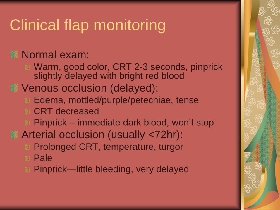
Clinical flap monitoring
Normal exam: Warm, good color, CRT 2-3 seconds, pinprick slightly delayed with bright red blood
Venous occlusion (delayed): Edema, mottled/purple/petechiae, tense
CRT decreased
Pinprick – immediate dark blood, won’t stop
Arterial occlusion (usually <72hr): Prolonged CRT, temperature, turgor
Pale
Pinprick—little bleeding, very delayed

Mechanical flap monitoring
Doppler
External
Implanted
Buried flaps
80-100% salvage (Disa J, et al 1999)
Color flow
Other

Antibiotics
8-20% of patients undergoing free tissue transfer will develop an infection despite intravenous antibiotic coverage.(Cloke DJ., et al, 2004)
1 day vs. 5 day course of Clindamycin showed no significant difference in free flap survival (Carroll WR., et al., 2003)
Topical antibiotics in combination with intervenous antibiotics did not show a significant difference in post-operative complications after free tissue transfer (Simons JP, et al., 2001)

Free flap reconstruction—the costs
Longer ICU stay, more expensive,
longer OR time (McCrory AL, et al.,
2002)


Sources
Broyer M, Gagnadoux MF, Sierro A, Fischer AM, Niaudet P. Preventive treatment of vascular thrombosis after kidney transplantation in children with low molecular weight heparin. Transplant Proc 1991; 23: 1384.
Alkhunaizi AM, Olyaei AJ, Barry JM, et al. Efficacy and safety of low molecular weight heparin in renal transplantation. Transplantation 1998; 66: 533.
Nahabedian MY. Singh N. Deune EG. Silverman R. Tufaro AP. Recipient vessel analysis for microvascular reconstruction of the head and neck. [Journal Article] Annals of Plastic Surgery. 52(2):148-55; discussion 156-7, 2004 Feb.
McCrory AL. Magnuson JS. Free tissue transfer versus pedicled flap in head and neck reconstruction. [Journal Article] Laryngoscope. 112(12):2161-5, 2002 Dec.
Kroll SS. Robb GL. Reece GP. Miller MJ. Evans GR. Baldwin BJ. Wang B. Schusterman MA. Does prior irradiation increase the risk of total or partial free-flap loss?. [Journal Article] Journal of Reconstructive Microsurgery. 14(4):263-8, 1998 May.
Jones NF. Johnson JT. Shestak KC. Myers EN. Swartz WM. Microsurgical reconstruction of the head and neck: interdisciplinary collaboration between head and neck surgeons and plastic surgeons in 305 cases. [Journal Article] Annals of Plastic Surgery. 36(1):37-43, 1996 Jan.
Foote RL. Olsen KD. Meland NB. Schaid DJ. Kunselman SM. Tumor-ablative surgery, microvascular free tissue transfer reconstruction, and postoperative radiation therapy for advanced head and neck cancer. [Journal Article] Mayo Clinic Proceedings. 69(2):122-30, 1994 Feb.
Cloke DJ. Green JE. Khan AL. Hodgkinson PD. McLean NR. Factors influencing the development of wound infection following free-flap reconstruction for intra-oral cancer. [Journal Article] British Journal of Plastic Surgery. 57(6):556-60, 2004 Sep.

Sources
Braun S. Mine R. Syed SA., et al. The optimal sequence of microvascular repair during prolonged clamping in free flap transfer. Plastic and Reconstructive Surgery 111(1):233-241, 2003
Banic A. Krejci V. Erni D., et al. Effects of sodium nitroprusside and phenylephrine on blood flow in free musculocutaneous flaps during general anesthesia. Anesthesiology 90(1):147-155, 1999
German G. Steinau HU. The clinical reliability of vein grafts in free flap transfer. Journal of Reconstructive Surgery 67:194-199, 1996
Nahabedian MY. Bahram M. Manson PN. Factors associated with anastomotic failure after microvascular reconstruction of the breast. Plastic and Reconstructive Surgery 114(1):74-82, 2004
Nahabedian M. Singh N. Deune EG. Recipient vessel analysis for microvascular reconstruction of the head and neck. Annals of Plastic Surgery 52(2):148-155, 2004
Disa JJ. Polvora VP. Pusic AL Singh B. Cordeiro P. Dextran-related complications in head and neck microsurgery: Do the benefits outweigh the risks? A prospective randomized analysis. Head and Neck Microsurgery 112(6):1534-1539, 2001
Carroll WR. Rosenstiel D. Fix JR. de la Torre J. Solomon JS. Brodish B. Rosenthal EL. Heinz T. Niwas S. Peters GE. Three-dose vs extended-course clindamycin prophylaxis for free-flap reconstruction of the head and neck. [Clinical Trial. Journal Article. Randomized Controlled Trial] Archives of Otolaryngology -- Head & Neck Surgery. 129(7):771-4, 2003 Jul.
Simons JP. Johnson JT. Yu VL. Vickers RM. Gooding WE. Myers EN. Pou AM. Wagner RL. Grandis JR. The role of topical antibiotic prophylaxis in patients undergoing contaminated head and neck surgery with flap reconstruction. [Clinical Trial. Journal Article. Randomized Controlled Trial] Laryngoscope. 111(2):329-35, 2001 Feb.

Sources
Shindo M., Costantino P., Nalbone V., et al., Use of a mechanical microvascular anastomotic device in head and neck free tissue transfer. Archives of Otolaryngology—Head & Neck Surgery 122(5):529-532, 1996.
De Lorenzi F., van der Hulst R., Boeckx W., et al., VCS auto suture stapled microvascular anastomoses in lower leg free flaps. Plastic and Reconstructive Surgery 109(6):2023-2030, 2002
DeLacure M., Kuriakose M., Spies A., et al., Clinical experience in end-to-side venous anastomoses with a microvascular anastomotic coupling device in head and neck reconstruction. Archives of Otolaryngology—Head and Neck Surgery 125(8):869-872, 1999
Ahn C., Shaw W., Berns S., et al., Clinical experience with the 3M microvascular coupling anastomotic device in 100 free-tissue transfers. Plastics and Reconstructive Surgery 93:1481-1484, 1994
Berggren A., Ostrup L., Lidman D., et al., Mechanical anastomosis of small arteries and veins with the Unilink apparatus: a histologic and scanning electron microscopic study. Plastic and Reconstructive Surgery, 80:274-283, 1987
Swartz W., Banis J., Head and Neck Microsurgery. Williams & Wilkins, Baltimore, MA, c.1992
Baker S. Microsurgical Reconstruction of the Head and Neck. Churchill Livingstone, New York, NY, c.1989
Panje W., Moran W. Free Flap Reconstruction of the Head and Neck. Thieme Medical Publishers, Inc., New York, NY c.1989
Velanovich V. Smith DJ. Robson MC. Heggers JP. The effect of hemoglobin and hematocrit levels on free flap survival. American Surgeon 54(11):659-663, 1988
Quinlan J. Anaesthesia for reconstructive free flap surgery. Anesthesia and Intensive Care Medicine The Medicine Publishing Company, Ltd., c.2003