MICROVASCULAR FREE TISSUE TRANSFER OF THE RECTUS …
64
I I MICROVASCULAR FREE TISSUE TRANSFER OF THE RECTUS ABDOMINIS MUSCLE IN DOGS by Earl F. Calfee III (Trey) Thesis submitted to the Faculty of the Virginia Polytechnic Institute and State University is partial fulfillment of the requirements for the degree of Master of Science in Veterinary Medical Science Approval Committee: Chair: Otto I. Lanz, D.V.M., Diplomate A.C.V.S. _____________ Robert B. Duncan Jr., D.V.M., Ph.D., Diplomate A.C.V.P. _____________ Richard V. Broadstone, D.V.M., Ph.D., Diplomate A.C.V.A. _____________ Robert A. Martin, D.V.M., Diplomate A.C.V.S. and A.B.V.P. _____________ March 1, 2002 Blacksburg, VA Keywords: Microvascular, Rectus Abdominis, Dogs, Tissue Transfer Copyright 2002, Earl F. Calfee III (Trey)
Transcript of MICROVASCULAR FREE TISSUE TRANSFER OF THE RECTUS …

I
I
MICROVASCULAR FREE TISSUE TRANSFER OF THE RECTUS ABDOMINIS MUSCLE IN DOGS
by
Earl F. Calfee III (Trey)
Thesis submitted to the Faculty of the Virginia Polytechnic Institute and State University is partial fulfillment of the requirements for the degree of
Master of Science
in Veterinary Medical Science
Approval Committee:
Chair: Otto I. Lanz, D.V.M., Diplomate A.C.V.S. _____________ Robert B. Duncan Jr., D.V.M., Ph.D., Diplomate A.C.V.P. _____________ Richard V. Broadstone, D.V.M., Ph.D., Diplomate A.C.V.A. _____________ Robert A. Martin, D.V.M., Diplomate A.C.V.S. and A.B.V.P. _____________
March 1, 2002
Blacksburg, VA Keywords: Microvascular, Rectus Abdominis, Dogs, Tissue Transfer
Copyright 2002, Earl F. Calfee III (Trey)

II
II
MICROVASCULAR FREE TISSUE TRANSFER OF THE RECTUS ABDOMINIS
MUSCLE IN DOGS
by
Earl F. Calfee III
(ABSTRACT)
Objective - To assess donor site morbidity and survival of the rectus abdominis muscle
with an overlying skin graft after free tissue transfer to a medial femorotibial defect in
dogs.
Study Design - Experimental study
Sample Population – Phase one - six canine cadavers / Phase two - seven adult mixed
breed dogs
Methods – Phase one - The rectus abdominis muscle was removed from cadavers,
muscular and vascular dimensions were recorded and angiography was performed. Phase
two - Muscular transfer was performed through anastomosis of the caudal epigastric
vasculature to the saphenous vasculature. Transferred tissues were evaluated on
postoperative days three, six, 10, and 13. Animals were examined daily until euthanasia
between postoperative days 31 and 42. Postmortem angiograms were performed and
tissues collected for histopathologic evaluation.
Results – Phase one - Appropriate vascular dimensions for microvascular anastomosis
were confirmed and surgical technique perfected. Phase two – Muscular excision
produced minimal donor site morbidity. All muscles survived after microvascular
transfer and angiography confirmed vascular patency. All skin grafts survived with one
graft undergoing partial necrosis.
Conclusions - The rectus abdominis muscle can be successfully transferred to a medial
femorotibial defect and serve as a bed for acute skin grafting. No significant donor site
morbidity is associated with its removal.

III
III
Clinical Relevance - Microvascular free tissue transfer of the canine rectus abdominis
muscle has not been previously described. This technique provides an alternative for
repair of appropriate wounds. Additional studies are needed to define its utility in
clinical patients.

IV
IV
TABLE OF CONTENTS
Abstract II
Introduction / Literature Review 1
Indications for free tissue transfer 1
Previously transferred tissues – veterinary 2
Superficial cervical flap 3
Medial saphenous flap 3
Latissimus dorsi flap 4
Carpal pad flap 4
Omental pedicle flap 4
Muscular versus cutaneous coverage 5
Requirements for free tissue transfer candidates 7
Vascular patterns of muscles 7
New vascular classification for canine rectus abdominis muscle 9
Previous studies involving canine rectus abdominis 10
Human rectus abdominis free tissue transfer 10
Study objectives 11
Materials and Methods 12
Phase 1 12
Muscular excision and vascular isolation 12
Angiograms 13
Phase 2 14
Muscular excision and vascular isolation 15
Preparation of recipient site 16
Vascular anastomosis 17
Closure 18

V
V
Bandaging and epidural catheters 18
Post-operative monitoring 19
Euthanasia, angiograms and necropsies 20
Results 21
Phase 1 21
Phase 2 21
Donor and recipient site evaluations 22
Angiograms 24
Histopathology 25
Discussion 26
Rectus abdominis anatomy 26
Advantage/disadvantages of canine rectus abdominis 26
Donor site closures 27
Skin graft morbidity 28
Post-operative monitoring 29
Anticoagulant therapy 31
Tissue cosmesis 33
Conclusions/Future Research 35
References 37
Figures 43
Normal canine rectus abdominis anatomy 43
Femorotibial defect 44
Phase 1 angiogram 45
Day 3 bandage changes 46
Day 6 bandage changes 47
Day 10 bandage changes 48
Skin grafts at study conclusion 49

VI
VI
Distal skin graft necrosis 50
Traumatic distal graft necrosis 51
Incisional bruising 52
Seroma formation 53
Phase 2 angiograms 54
Histopathologic muscle atrophy 55
Distal skin graft necrosis 56
Tables 57
Muscular, vascular and recipient site dimensions 57
Vita 58

1
1
Introduction/Literature Review
Distal extremity and oral defects present some of the most difficult reconstructive
challenges for veterinary surgeons. These anatomic regions lack redundant local tissues
and second intention healing of wounds in these body regions is often not possible
because of the limited ability of peripheral tissues to undergo contraction. If possible,
second intention healing often produces a poor cosmetic outcome and tissues that are
susceptible to repeated trauma. Wound repair of distal extremity and oral cavity defects
therefore often involves transfer of tissues from distant sites where redundant tissues can
be found. Traditionally, this has involved open wound management in preparation for
skin grafting. Microvascular free tissue transfer allows acute one stage repair of
extensive wounds of the distal extremities and oral cavity with excellent cosmesis and a
potentially better functional outcome. Free tissue transfer has the potential to shorten the
length of hospitalization because days to weeks of open wound management are
unnecessary. In people, free tissue transfer techniques have been shown to shorten
unemployment periods and the duration of hospitalization following repair of severe
tibial fractures1. This has in turn led to lower hospitalization costs and increased patient
productivity following these major surgical procedures.
Microvascular free tissue transfer is based on the concept of angiosomes, which
are definable regions of tissue perfused by a single nutrient artery and vein. This concept
was initially applied with reconstructive procedures based on axial pattern flaps. In the
1980’s, several axial pattern flaps were developed for repair of large skin defects in dogs 2-4. Examples include the caudal superficial epigastric, omocervical, thoracodorsal, and
deep circumflex iliac axial pattern flaps 2,5,6. Axial pattern flaps are useful in
reconstruction of certain cutaneous defects in dogs and cats. They are, however, limited
by dependence on preservation of an intact vascular pedicle. This limitation prevents
their use for repair of wounds in the most distal aspects of the limbs and facial region.

2
2
A vascular leash on the other hand does not limit microvascular free tissue
transfers. This in turn allows tissue movement to distant recipient sites. Additionally,
microvascular free tissue transfer reliably produces viable tissues. This has been
documented in both human and veterinary medicine. Fowler, et al retrospectively
evaluated outcomes following free tissue transfers in 57 consecutive cases involving two
veterinary institutions 7. Tissues transferred using microvascular techniques included
muscle, bone, skin, and footpads. Fifty-three of 57 procedures were successful. Only
inexperience of the assistant surgeon and use of the latissimus dorsi muscle flap were
associated with increased flap failure. Khouri, et al prospectively evaluated surgical
outcome following free tissue transfer in people 8. This study described outcomes in
greater than 450 consecutive patients at over 20 institutions. An overall 96% success
rate for viable tissue transfer was reported. Sixty variables were evaluated for their effect
on outcome. Four factors were associated with a higher incidence of post-operative
vascular thrombosis. These included the use of interpositional vein grafts, free tissue
transfer into previously irradiated or chronic wounds and muscle flaps covered by
meshed skin grafts. Both studies concluded that successful free tissue transfer was
primarily dependent on the experience level of the primary and assistant surgeons.
Examples of autogenous tissues transferred using microvascular free tissue
techniques in veterinary medicine include skin, bone, omentum, muscle, footpads, and
various combinations of these tissues 7,9-12. In 1986, Fowler et al performed the first
documented free tissue transfers in veterinary medicine 13. This involved orthotropic
transfer of the superficial cervical skin flap in eight dogs. In this study, the periphery of
the superficial cervical angiosome was sharply incised and undermined to the base of the
vascular pedicle. The superficial cervical artery and vein were then isolated, transected
and microvascular anastomosis performed. The flaps survived in six of eight dogs. In
1987, Miller et al described the successful microvascular transfer of the superficial
cervical free flap in a dog following traumatic injury to the forelimb 14. Later that same

3
3
year Fowler, et al reported successful free tissue transfer of the superficial cervical flap in
six clinical cases 13. The superficial cervical flap was successfully transferred to the rear
limb in four dogs and to the forelimb in two dogs.
Between 1988 and 1996 multiple experimental studies were performed which
involved free tissue transfer of bone, foot pads or muscle 11,15-17. They were performed
almost exclusively at the University of Saskatchewan under the direction of David
Fowler. In these studies, angiosomes of various tissues were initially defined through
angiography and viability was later confirmed in research animals following heterotropic
transfer.
Several studies were performed based on the angiosome of the superficial cervical
vasculature. Initially the vascular supply to the trapezius muscle was defined.
Angiography revealed that the prescapular branch of the superficial cervical artery
supplied the entire cervical portion of the trapezius muscle as well as its overlying skin 18.
This musculocutaneous composite flap was then transferred successfully in three of four
research dogs to a medial femorotibial defect 16. In 1993, the potential viability of an
osteomusculocutaneous free flap using the cervical portion of the trapezius muscle, its
overlying skin and the scapular spine was defined 15. This study revealed that viability of
the scapular spine was maintained with preservation of superficial cervical vascular
pedicle following detachment of the spine from the scapular body and transection of all
other soft tissue attachments.
Between 1996 and 1997, Degner et al published three papers involving the use of
microvascular free tissue transfer. Initially the angiosome of the medial saphenous
vasculature was defined through angiography. The medial saphenous fasciocutaneous
free flap was then successfully transferred to a forelimb defect in six consecutive research
dogs 19. Medial saphenous fasciocutaneous or myocutaneous free flaps were then
successfully transferred in eight clinical cases 20. The myocutaneous flaps incorporated
the distal one half of the caudal head of the sartorius muscle. One-hundred percent tissue

4
4
viability was produced in all eight dogs. Free tissue transfer of the transverses abdominis
muscle was then reported in a two-phase study 10. Phase one defined the perfusion
pattern of the muscle and described the dimensions of the phrenicoabdominal artery and
vein, which supplied the cranial one half of the muscular body. In phase two, the
myoperitoneal flap was successfully used in the repair of a traumatically induced canine
oronasal fistula.
In 1996, Nicoll et al described the transfer of the latissimus dorsi free muscle flap
in a group of research cats 21. This study described the thoracodorsal vascular pedicle
and its distribution to the latissimus dorsi muscle. Heterotropic and orthotropic transfers
of the feline latissimus dorsi muscle were then performed. Muscular viability following
heterotropic transfer was confirmed in two of three cats. Complete viability was
produced in four of four cats following orthotropic transfer. The authors concluded that
the cranial one half of the feline latissimus dorsi muscle is a viable option for free tissue
transfer. The 66 percent survival rate was below what is normally reported for canine
free tissue transfers but the population sample was too small to determine significance.
In 1997, Fowler and Moens published two papers on the use of a carpal footpad
free flap in reconstruction of canine weight bearing surfaces 22,23. The carpal pad blood
supply was described through cadaveric angiograms in order to define the feasibility of
microvascular transfer 23. The carpal pad flap was then successfully transferred in three
clinical cases 22. Surgical revisions were needed in all animals to maintain appropriate
weight bearing forces on the transferred tissues and only partial closure of donor sites
was possible. All donor sites healed uneventfully through second intention.
In 1999, Roa et al described the survival of a free omental pedicle flap to a canine
medial femorotibial defect 12. The benefits of the abundant omental vasculature and long
arterial and venous pedicle length are discussed. Seven omental flaps were transferred in
five dogs with only two flaps surviving. Flaps were evaluated through angiography,
nuclear scintigraphy and histopathology. Failure of two flaps was attributed to self-

5
5
inflicted trauma from the research dogs. The other flap failures were primarily attributed
to a lack of microvascular surgical experience by the investigators.
In 2001, Lanz et al described the effects of epidural lidocaine administration on
blood flow through the medial saphenous fasciocutaneous free flap 24. Lidocaine
epidurals were of theoretical benefit because of vasodilatory effects on peripheral tissues.
The authors found no increase in free flap blood flow but did see a decline in systemic
blood pressures. Administration of lidocaine epidurally was therefore not recommended
during free tissue transfer procedures.
It is evident from this literature review that the depth of experience with
veterinary free tissue transfer is limited. It is however interesting to find that success
rates for human and veterinary free tissue transfers are indistinguishable and that surgical
outcomes in both fields are primarily dependent on surgeon ability and experience. This
is promising for veterinary medicine and makes free tissue transfer a realistic and feasible
option for veterinary patients.
In the past few years attention has been focused on the different capabilities of
muscle versus skin in wound healing following transfer into compromised wound beds.
From the above literature review we find that four muscles have been transferred using
free tissue transfer techniques in the veterinary literature. These include the trapezius,
latissimus dorsi, caudal head of the sartorius and the transverses abdominis 10,16,20,21.
Muscle has an inherently rich blood supply, which has been shown to increase
local tissue oxygen tension as well as facilitate delivery of antibiotics and components of
the immune system to devascularized wounds 25. This quality has made microvascular
transfer of muscle a mainstay in the treatment of chronic osteomyelitis in people 26,27.
Transplanted muscle also promotes healing of concurrent orthopedic injuries through
enhancement of periosteal blood supply 28-30 and increases the strength of early bony
union in devascularized fracture models 30. In addition, transferred muscle provides a

6
6
physical barrier for compromised recipient tissues and can serve as a bed for acute skin
grafting 26,27.
Very little scientific data exists in the veterinary literature comparing the benefits
of muscular and cutaneous transfers into devascularized wounds. Much of the human
literature however involves the use of animal models to compare the effects of muscular
and cutaneous coverage. Porter et al recently compared the effects of devascularized
cortical bone coverage with a free muscle flap versus a cutaneous axial pattern flap in the
dog31. They found no statistical differences between treatment groups when comparing
cortical blood flow, cortical bone porosity, intracortical new bone production and
maximum depth of new periosteal bone formation. There was a trend towards greater
periosteal bone formation beneath muscle than skin but the difference was not significant.
The authors recognized one major fault in their study design. They did not adequately
prevent endosteal revascularization from proximal and distal aspects of the fracture
model. This interfered with their ability to detect differences in periosteal blood flow
contribution between the muscle and skin coverage groups. In light of the fact that
multiple previous studies have documented the superiority of muscular coverage in the
above capacities the results of this study are called into question.
Based on current knowledge levels, it is not clear what qualities make muscle
superior to skin in the promotion of wound healing. Gosain et al compared the effect of
muscular and cutaneous coverage on inhibition of bacterial inoculum growth 25. Their
work revealed that blood flow supplied by muscle coverage as opposed to cutaneous
coverage was significantly greater 24 hours following surgery. The superiority of
muscular blood flow was however temporary with a trend of increasing blood flow in the
skin coverage group over the six days following individual surgical procedures. Blood
flow underlying the cutaneous flaps eventually surpassed that under the muscular flaps.
Throughout the study however bacterial inoculum counts were significantly less in the
muscle coverage group. The authors theorized that the superiority of muscular blood
7
7
flow in the first 24 hours following tissue elevation prevented bacterial tissue
colonization. The authors also observed that muscle was more firmly adherent to the
bacterial inoculum chambers with greater tissue ingrowth than skin. They hypothesized
that this quality of tissue adherence and ingrowth was in part responsible for muscles
ability to resist infection better than skin. This concept is also alluded to in Porter’s
canine study even though differences in tissue adhesion between cutaneous and muscular
coverage groups were not noted31. The authors state the need for overlying tissues to
develop intimate contact with devascularized beds in order to stimulate angiogenesis and
resultant vascular ingrowth. Their study however was not designed to test differences in
adhesion properties between skin and muscle coverage groups. Muscle’s qualities of
tissue ingrowth and malleability may inevitably be responsible for its ability to promote
tissue healing more effectively than skin.
An individual muscle must fulfill certain anatomic requirements prior to
consideration for use in free tissue transfer. Muscles must have an expendable function,
be easily accessible, have a reliable vascular pedicle, and produce minimal donor site
morbidity following removal 32. If a muscle to be used in free tissue transfer does not
have an expendable function then it must have surrounding musculature to perform its
function following removal from the donor site. Additional requirements include a
vascular pedicle length of at least 10.0 mm, arterial and venous diameters of at least 0.5
mm, and an appropriate vascular distribution 21,33,34.
Currently, there are five defined vascular patterns for muscles. Type one muscles
receive blood flow from one major vascular pedicle and the entire muscle can be
transferred based on that pedicle. Type two muscles have one or more dominant pedicles
entering at the muscle’s insertion or origin with additional minor pedicles entering the
muscular body. Transfer of type two muscles can be performed based on the major
vascular pedicle following ligation of the minor pedicles. Type three muscles have two
major vascular pedicles that each supplies approximately 50% of the muscular blood
8
8
flow. Transfer of type three muscles can be based on either of the major pedicles but a
portion of the muscle will routinely die if the entire muscle is transferred. Type four
muscles have no major vascular pedicle but are perfused by several minor pedicles. Type
four muscles therefore have very limited application in free tissue transfer. Type five
muscles have one major vascular pedicle but receive additional blood supply from minor
segmental vessels. The entire muscle will survive if either the major vascular pedicle or
the segmental blood supply is maintained.
The canine rectus abdominis muscle is described as having a type three vascular
pattern 35. It primarily receives blood flow from two major vascular pedicles, the cranial
epigastric artery and vein and the caudal epigastric artery and vein. (See Figure 1)
Terminal branches of the deep circumflex iliac artery and vein coursing with the medial
branches of spinal nerves T13-L3 enter the muscle’s lateral margin and provide
segmental perfusion. Based on the original description of type three vascular patterns a
portion of the rectus abdominis muscle would be expected to die if transfer of the entire
muscle were based on a single vascular pedicle.
Previous studies by Degner et al demonstrated that perfusion of the entire canine
rectus abdominis muscle is maintained following transection of the caudal epigastric
blood supply 36. This study revealed that approximately two-thirds of the abdominal
portion of the rectus abdominis muscle is perfused by the caudal vascular pedicle.
Contrast angiograms revealed anastomotic “choke vessels” between the cranial and
caudal muscular blood supplies. These choke vessels support perfusion of the entire
muscle following ligation of the caudal vascular pedicle. Scintigraphic findings in this
same study found no difference in quantitative blood flow between control rectus
abdominis muscles and contralateral muscles following transection of the caudal vascular
pedicle. Based on these results survival of the entire muscle was predicted following
division of the caudal epigastric pedicle. This was confirmed experimentally in five of
six dogs with a cranially based axial rotation of the entire rectus abdominis muscle and its

9
9
overlying cutaneous paddle 35. Transfer of the overlying skin was unsuccessful in five of
six dogs but the entire rectus abdominis muscle survived in all but one dog. Because
viability of the entire muscle has been documented based on a cranial vascular pedicle
transfer and 2/3 of the muscle is perfused by the caudal epigastric vasculature, I felt the
entire muscle should remain viable following a caudally based transfer.
This predicted viability calls into doubt the categorization of the rectus abdominis
muscle as a type three muscle. Solano et al proposed an additional classification for the
canine semitendinosus muscle, which has a similar vascular pattern to the rectus
abdominis muscle 37. The semitendinosus muscle has two primary vascular pedicles and
the entire muscle survives following ligation of either pedicle. They proposed an
additional category of type three B or type six for muscles that have two primary vascular
pedicles which allow survival of the entire muscle following ligation of either pedicle.
The rectus abdominis muscle appears to fall into this category.
The vessels of both the cranial and caudal vascular pedicles of the rectus
abdominis muscle are of appropriate diameter and length for use in free tissue transfer.
Gregory et al defined the potential use of the canine rectus abdominis, gracilis and
latissimus dorsi myocutaneous tissues in free tissue transfer 33. This study involved
peripheral tissue dissection, vascular isolation and fluorescein staining in 15 canine
cadavers. Based on the results of this study, the authors concluded that the gracilis and
latissimus dorsi myocutaneous flaps were good candidates for free tissue transfer because
of adequate vascular pedicle dimensions, easily performed dissection and tissue viability
following peripheral dissection. The authors however did not recommend use of the
rectus abdominis myocutaneous flap because of unreliable viability of the skin overlying
the caudal aspect of the muscle. The cranial and caudal epigastric vascular pedicle
dimensions were both of adequate dimensions for use in free tissue transfer but the
cranial pedicle was described as being difficult to harvest in dogs with deep thoracic
conformations. Transfer of the rectus abdominis muscle in the present study was based

10
10
on the caudal epigastric blood supply because of the reported harvesting difficulties in
Gregory’s study and the primary role of the caudal epigastric artery and vein in perfusion
of the rectus abdominis muscle.
Successful removal of the canine rectus abdominis muscle from the ventral
abdomen without postoperative morbidity has been previously described by Degner et al.
This was demonstrated during transfer of the cranial rectus abdominis muscle pedicle flap
with its overlying caudal superficial epigastric skin flap 35. The authors did not
specifically intend to describe post-operative morbidity following removal of the rectus
abdominis muscle from the ventral abdomen but noted that none of the dogs developed
abdominal wall hernias following surgery.
Free tissue transfer of the rectus abdominis muscle is reported in the human
literature 38-41. This muscle is consistently and effectively used in reconstruction of lower
extremity defects in people. The reported benefits of its use include minimal donor site
morbidity, a reliable vascular pedicle with dimensions that facilitate microvascular
anastomosis, and an ability to be contoured into distal extremity defects 39. The pedicle
length in people is reported to exceed the mean pedicle length of other free tissue transfer
candidates and therefore often prevents the need for interpositional “bridging” vein
grafts. Vein grafts were recently associated with an increased risk of intra-operative and
post-operative vascular thrombosis in human free tissue transfers 8. A lengthy vascular
pedicle is therefore quite beneficial in minimizing failure of free tissue transfers.
Minimal donor site morbidity is one of the most beneficial qualities of the human rectus
abdominis free flap. In people the muscle is harvested laparoscopically through a limited
inguinal incision 41. This approach allows harvesting of up to 100 percent of the muscle
with a scar that is easily covered by undergarments. Cosmesis undoubtedly plays a larger
role in people than veterinary patients, as donor site morbidity is rarely an issue following
veterinary free tissue transfers.

11
11
Microvascular free tissue transfer of the canine rectus abdominis muscle has not
previously been reported in the veterinary literature. Therefore, the objectives of this
study were to show that the rectus abdominis muscle could be successfully transferred to
a defect created on the medial aspect of the canine femorotibial region using
microvascular surgical techniques. Additionally we hypothesized that the muscle would
be a suitable bed for acute meshed skin grafting, and that removal of the muscle from the
ventral body wall would be associated with minimal morbidity. Previous veterinary
research has shown that the canine rectus abdominis muscle meets the basic requirements
for free tissue transfer candidates including minimal donor site morbidity, an appropriate
vascular angiosome and adequate vascular dimensions. We were therefore confident that
free tissue transfer of the rectus abdominis muscle would produce reliably viable tissue.

12
12
Materials and Methods
The study was divided into two phases. Phase one was performed on canine
cadavers and defined the vascular anatomy and muscular dimensions of the rectus
abdominis muscle. The surgical technique for vascular isolation and muscular excision
was also perfected in phase one. Phase two was performed on live dogs and determined
the feasibility of transferring the rectus abdominis muscle to a defect created on the
medial aspect of the femorotibial region.
Phase 1
Phase one was performed using six canine cadavers weighing between 20 and 40 kg that
were previously used in either student teaching laboratories or continuing education
seminars at the Virginia-Maryland Regional College of Veterinary Medicine. Heparin
sodium (100 units/kg)(Elkins Sinn Inc., Cherry Hill, NJ) was administered intravenously
prior to euthanasia. The skin, subcutaneous tissues and linea alba were incised along
midline from the level of the xiphoid to the cranial border of the pubis. Lateral dissection
was performed along the caudal extent of the incision to identify the lateral margin of the
rectus abdominis muscle and the superficial inguinal ring. The fascia of the internal and
external abdominal oblique muscles was longitudinally incised over the ventral surface of
the rectus abdominis muscle beginning at the craniomedial border of the inguinal canal.
The ventral fascia overlying the rectus abdominis muscle was then dissected from the
ventral muscular surface until it became firmly adherent to the muscle at the first or
second tendinous intersections cranial to the pubis. The fascia was then transected and
dissection continued along the lateral and medial borders of the rectus abdominis muscle.
Laterally, perforating neurovascular bundles were ligated and transected near their entry
into the muscle. The muscle was then transected at the level of the xiphoid. Blunt
dissection of fascial tissues dorsal to the rectus abdominis muscle allowed lateral

13
13
retraction of the muscle. This facilitated visualization of the caudal epigastric artery and
vein entering the caudodorsal muscular surface. The caudal epigastric vasculature was
then followed proximally to its origin from the deep femoral artery and vein as the
pudendoepigastric arterial and venous trunks. The deep femoral artery and vein were
ligated and transected proximal and distal to the origin of their respective
pudendoepigastric trunks. These points of vascular transection allowed removal of the
caudal epigastric vasculature while maintaining the integrity of the entire
pudendoepigastric trunk. This in turn facilitated measurement of the caudal epigastric
vascular pedicle length and arterial diameter. The external pudendal artery and vein were
ligated and transected distal to their branching from the pudendoepigastric trunk. The
muscle was then transected at its insertion along the cranial border of the pubis allowing
its removal from the abdomen with an intact vascular pedicle.
After removal of the muscle, a 20 gauge, 1.88-inch over-the-needle intravenous
catheter (Angiocath, Becton Dickinson, Sandy, UT) was inserted into the arterial
pudendoepigastric trunk. A 3:1 water: barium sulfate suspension (Liquid E-Z-Paque, EZ-
EM Inc., Westbury, NY) was infused using a 12 cc syringe until resistance was felt. The
muscles were then refrigerated for 24 hours at minus nine degrees Celsius to allow
hardening of the barium infusion.
Twenty-four hours following muscular removal from the ventral abdomen,
angiography was performed with a cabinet x-ray system (Faxitron x-ray system-43805N,
Hewlett Packard, McMinnville, OR). After angiography was completed, measurements
of muscular width, length and thickness were recorded. Arterial pedicle length and base
diameter were also determined using calipers. Arterial diameter measurements were
taken from the base of the pudendoepigastric trunk as it branched from the deep femoral
artery. Pedicle length was measured from the origin of the pudendoepigastric trunk at the
deep femoral artery to the entry of the caudal epigastric artery into the rectus abdominis
muscle.

14
14
Phase 2
The Virginia Tech Animal Care Committee approved all animal use in this
project. Four female and three male mixed breed dogs were used for the live animal
portion of the study. A thorough physical examination, a serum chemistry profile, and a
complete blood count were performed on each dog prior to entrance into the study to rule
out any systemic disease processes that might interfere with tissue healing after free
tissue transfer. Dogs were housed in the research facilities for 10 days prior to initiation
of the study to acclimate them to frequent handling and their new environment. Aspirin
(81 mg/dog)(Rugby Laboratories, Norcross, GA) was administered orally at 12-hour
intervals beginning 24 hours prior to surgery and continuing for 72 hours afterwards.
Animals were pre-medicated with intramuscular injections of acepromazine maleate (0.3
mg/kg)(Fort Dodge Animal Health, Fort Dodge, IO), morphine sulfate (0.25
mg/kg)(Elkins-Sinn, Cherry Hill, NJ) and atropine (0.3 mg/kg)(Phoenix Pharmaceuticals,
Inc., St. Joseph, MO). Anesthetic induction was achieved with intravenous sodium
thiopental (10 mg/kg)(Pentothal, Abbott Laboratories, North Chicago, IL). After
orotracheal intubation anesthesia was maintained with isoflurane in 100% oxygen (Isoflo,
Abbott Laboratories, North Chicago, IL). Intravenous cefazolin (22mg/kg) (Apothecan,
Princeton, NJ) was administered after anesthetic induction and at two-hour intervals
during the procedure. Intravenous fluids (Lactated Ringers Solution, Baxter Healthcare
Corporation, Deerfield, IL) were administered at a rate of 22 ml/kg/hr during surgery and
a warm air blanket (Bair Hugger, Augustine Medical, Eden Prarie, MN) was used to
maintain body temperature during surgery and recovery from anesthesia.
After they were anesthetized, all of the dogs had a surgical clip and aseptic skin
preparation of their ventral abdomen and left femorotibial region. The left rear limb was
circumferentially clipped from the mid-metatarsal region to the dorsal midline. Removal

15
15
of the rectus abdominis muscle and creation of the rear limb defect were performed
simultaneously by two separate surgical teams.
The skin was incised on the ventral midline from the xiphoid to the cranial border
of the pubis. In male dogs a parapreputial incision was created. The initial skin incision
was deepened to the level of the linea alba. Subcutaneous tissues were undermined
laterally to identify the left superficial inguinal ring. The ventral fascia overlying the
rectus abdominis muscle was incised along the muscular midline from the inguinal canal
cranially to the level of cranial muscular transection. Adherent fascia was sharply
dissected from the ventral surface of the muscle to expose the lateral and medial muscular
margins. This allowed visualization of the entire ventral muscular surface. Dorsally
adherent fascia was bluntly dissected from the muscle. Laterally perforating, segmental
neurovascular bundles were ligated using hemoclips (Weck Closure Systems, Research
Triangle Park, NC) and transected near their entry into the muscle. The muscle was then
transected cranially. Hemoclips (Weck Closure Systems, Research Triangle Park, NC)
were used to control hemorrhage encountered during cranial transection of the rectus
abdominis muscle. The point of cranial muscular transection was determined by the
proximal to distal measurements of the femorotibial defect. The excised segment of
rectus abdominis muscle was however intentionally shorter than the femorotibial defect
to allow for muscular stretching after transfer into the defect. A 10 mm section of
adherent ventral rectus fascia was maintained on the cranial border of the muscle to
facilitate suture purchase at the recipient site. Prior to cranial and caudal muscular
transection, measurements of the length and width of the rectus abdominis muscle were
recorded.
The muscle was reflected laterally allowing visualization of the caudal epigastric
artery and vein as they entered the caudodorsal surface of the muscle. The muscle was
transected at its insertion along the cranial border of the pubis. Surrounding adventitial
tissues were meticulously dissected from the caudal epigastric vasculature to the level of

16
16
vascular branching from the deep femoral artery and vein. The external pudendal artery
and vein were ligated and transected near their origin from the pudendoepigastric trunk.
Measurements of the caudal epigastric pedicle length, as well as the arterial and venous
pedicle base diameters, as described in phase one, were determined and recorded at this
time. The muscle was then covered with a moistened laparotomy sponge while awaiting
transfer into the cutaneous defect created on the medial aspect of the femorotibial region.
Prior to skin incision at the recipient site, a sterile marking pen was used to
outline the dimensions of the proposed femorotibial cutaneous defect. The defect started
at the level of the mid-femur and ended just proximal to the tarsocrural joint and involved
an estimated 50 percent of the limb’s circumference. (See Figure 2) Defect dimensions
were recorded and the skin was sharply incised. Subcutaneous tissues were dissected
from the underlying dermis and the saphenous artery and medial saphenous vein
identified. A curvilinear skin incision originating at the cranioproximal margin of the
defect and extending proximally was made. The skin edges were undermined to allow
additional visualization of the saphenous vascular pedicle. The previously excised skin
was then prepared for use as a full thickness meshed skin graft by removal of all
subcutaneous tissues and meshing with a No. 15 scalpel blade.
Adventitial tissue was removed from the saphenous artery and medial saphenous
vein in preparation for transection and eventual anastomosis. After the adventitia was
removed the arterial and venous diameters were measured and recorded. The saphenous
artery and medial saphenous vein were then ligated with hemoclips at the level of the mid
femur and microvascular vessel clamps were placed proximal to the hemoclips. The
vessels were transected between the microvascular clamps and the hemoclips.
The caudal epigastric artery and vein were similarly ligated with hemoclips and
divided immediately distal to where the vessels branched from the deep femoral artery
and vein. A microvascular clamp was placed on the vein distal to the hemoclips prior to

17
17
venous transection to prevent passive hemorrhage from the vein during arterial
anastomosis. The rectus abdominis muscle was then removed from the abdomen.
The muscle was placed into the femorotibial defect with the ventral muscular
surface positioned superficially and the caudal muscular border placed proximally. This
positioning allowed approximation of the saphenous and caudal epigastric vascular
pedicles. Stay sutures were placed in the proximal margins of the muscle to inhibit
movement during vessel anastomosis. The caudal epigastric artery and the saphenous
artery were positioned in a microvascular-approximating clamp (Accurate Surgical and
Scientific Instruments, Westbury, NY). The vascular lumens were flushed with
heparinized saline (10 units/ml) and additional adventitial tissue was removed from
vessel ends with microvascular dissection scissors (ASSI, Westbury, NY). An end-to-
end arterial anastomosis was performed with the aid of an operating microscope using
10/0 nylon suture (Braun Surgical, Emmenbrucke, Germany) on a 100 um taper needle in
a simple interrupted pattern. Venous anastomosis was performed with a 2.0 mm
polyethylene coupling ring (Medical Co. Alliances, Inc, Park City, UT). Two percent
lidocaine (Phoenix Pharmaceuticals Inc., St. Joseph, MO) was applied topically to the
arteries and veins to minimize vasospasm.
After arterial and venous anastomoses, the microvascular clamps were removed
and the vessels were evaluated for patency. Patency was confirmed by visualization of a
flash of blood crossing the venous anastomotic site, return of a reddish-pink coloration to
the muscle, and arterial pulse transmission distal to the arterial anastomosis. Total
ischemic time was then recorded.
After the confirmation of patency, the peripheral borders of the rectus abdominis
muscle were sutured to the surrounding subcutaneous tissues with 4/0 glycomer 60
(Monosyn, Braun Surgical, Melsungen, Germany) in a simple continuous pattern. The
proximal curvilinear incision was closed with 4/0 glycomer 60 in an interrupted
subcuticular pattern. The previously prepared full thickness meshed skin graft was
18
18
placed over the transferred rectus abdominis muscle and sutured to the margins of the
rear limb defect with a combination of interrupted and continuous suture patterns using
4/0 glycomer 60.
The abdominal incision was closed in four layers in females and five layers in
males. The lateral fascial margins of the internal and external abdominal oblique
musculature were apposed to the medial fascial margins using 0 polydioxanone (PDS,
Ethicon Inc., Johnson and Johnson, Somerville, NJ) in a simple interrupted pattern. In
male dogs, a two layer subcutaneous closure was performed using 3/0 glycomer 60
(Monosyn, Braun Surgical, Melsungen, Germany) in a simple continuous pattern. In
females, a single subcutaneous layer was placed. Bites were taken intermittently into
deep fascial tissues to obliterate dead space and prevent seroma formation. The skin
margins were apposed using 3/0 glycomer 60 in a subcuticular pattern. Final skin closure
was accomplished with either 3/0 monofilament nylon (Dermalon, Sherwood/Davis and
Geck, St. Louis, MO) in a cruciate pattern or skin staples (Davis and Geck, St. Louis,
MO).
After surgery a non-adherent dressing (Adaptic, Johnson and Johnson, Arlington,
TX) was coated with triple antibiotic ointment (Qualitest Pharmaceuticals, Inc.,
Huntsville, AL) and placed over the skin graft. Several layers of cast padding and
compressive wrap were then applied circumferentially around the limb, followed by a
fiberglass cylindrical cast (Delta Lite”s”, Johnson and Johnson, Raynham, MA). The cast
was cut along its cranial and caudal surfaces producing medial and lateral halves. The
cast was then covered with another wrap (Vetwrap, 3M Animal Care Products, St. Paul,
MN and Elasticon, Johnson and Johnson, Arlington, TX). An epidural catheter (Arrow
International Inc., Reading, PA) was placed in all dogs for delivery of Duramorph to
provide analgesia after surgery.
Epidural morphine (Duramorph) (0.1 mg/kg) injections were administered every
eight hours until the time of catheter removal. Morphine sulfate (0.25 mg/kg) (Elkins-

19
19
Sinn Cherry Hill, NJ) was also available for intramuscular injection if the dogs appeared
persistently painful after epidural morphine administration. The catheters were removed
at the time of the first bandage change three days after surgery. Animals were given
cephalexin (22 mg/kg PO) (Teva Pharmaceuticals, Sellersville, PA) three times daily for
10 days following surgery. Daily evaluations of bandage condition, abdominal incisional
condition, appetite, general attitude, urination, and defecation were noted. Bandages
were changed three, six, and 10 days after surgery. Prior to bandage changes, animals
were sedated with intravenous acepromazine maleate (1.0 mg/dog) (Fort Dodge Animal
Health, Fort Dodge, IO). During bandage changes, photographs of the skin grafts and
donor sites were taken. Viability of the skin grafts was subjectively evaluated using
tissue color, muscular bleeding induced by bandage removal, and superficial temperature
as criteria. Grafts were cleaned with nolvasan scrub on days six and 10 prior to re-
bandaging. On day six, the medial aspect of the bivalve cast was removed and animals
were maintained in a lateral splint. On day 12, the lateral splints were removed and the
limbs were left uncovered for the remainder of the study.
Animals were housed in the clinical research facilities for the remainder of the
study and evaluated twice daily until euthanasia was performed with an intravenous
injection of pentobarbital sodium/phenytoin solution (1.0 ml/10 #) (Beuthanasia-D
Special, Schering-Plough Animal Health, Union, NJ). Animals were euthanatized
between four and one-half and six weeks after surgery.
After the dogs were killed, the skin and subcutaneous tissues overlying the
femoral artery were incised. Subcutaneous tissue dissection was continued to the level of
the femoral artery. An 18 gauge, 1.88-inch over-the-needle intravenous catheter
(Angiocath, Becton Dickinson, Sandy, UT) was introduced into the femoral artery
proximal to the branching of the saphenous artery. The catheter was directed into the
saphenous artery and the distal tip of the catheter advanced to a point proximal to the
arterial anastomosis. The catheter was secured in the vessel with multiple circumferential

20
20
sutures and flushed with heparinized saline. Seven milliliters of intravenous iothalamate
sodium (Conray 400, Mallinckrodt inc., St. Louis, MO) were injected into the catheter
and radiographs (lateral and craniocaudal views) were made of the femorotibial regions.
Determinations of arterial and venous anastomotic patency and muscular perfusion were
assessed from the resultant angiograms.
After angiograms were made, gross necropsies were performed and representative
samples collected from the recipient and donor sites of all dogs. The meshed skin graft
and underlying rectus abdominis muscle were incised peripherally and undermined
beginning distally up to the level of the arterial and venous anastomoses. A single
ligature was placed around the saphenous artery and medial saphenous vein proximal to
the anastomoses and the vessels were transected proximal to the ligature. The entire
rectus abdominis muscle flap was placed on cardboard in a fully expanded state and
immersed in a solution of 10 percent neutral buffered formalin. The cutaneous tissues of
the donor site were then incised and sharp dissection continued to the level of the
abdominal body wall closure. The cranial two-thirds of the body wall closure were
excised in entirety and placed in the formalin solution.
The following day samples were collected from the preserved tissues. A single
full thickness cross-sectional sample was removed from the previously excised donor site
closure at a point just cranial to the umbilicus. Samples were removed from three
separate regions of the recipient site. One sample was obtained from the area of the
venous and arterial anastomoses. Longitudinal and cross-sectional samples were
collected from both the proximal and distal portions of the skin grafts and underlying
rectus abdominis muscles. The samples were then routinely processed, embedded in
paraffin, sectioned and stained with hematoxylin and eosin. Microscopic slides were
evaluated for changes in the muscular and cutaneous tissues. Samples of normal muscle
from the contralateral rectus abdominus muscle were collected from the donor site for
comparison purposes.

21
21
Results
Phase 1
The results of the phase one cadaveric angiographic studies confirmed that the
caudal epigastric artery and vein perfused approximately two-thirds of the length of the
rectus abdominis muscle. The caudal epigastric artery and vein entered the caudodorsal
surface of the muscle and coursed cranially near the midline of the muscular body. Prior
to vessel termination, three to five medial and lateral vascular branches were visualized
exiting the main arterial trunk. Lateral branches were generally more prominent than
medial branches. Anastomotic communications between the caudal and cranial epigastric
angiosomes were visualized at the cranial termination of the caudal epigastric artery. (See
Figure 3) In all six dogs, the arterial and venous pudendoepigastric trunks originated
from the deep femoral vasculature. The pudendoepigastric trunk then divided into the
external pudendal and caudal epigastric vasculature. Mean width, length, and thickness
(+/- SD) of the rectus abdominis muscles (n=6) were 41 +/- 2 mm, 205 +/- 33 mm and 7
+/- 2 mm, respectively. The mean vascular pedicle length was 29 +/- 9 mm. The mean
diameter of the caudal epigastric arteries at the base of the pudendoepigastric trunk was 2
+/- 1 mm.
Muscular dissection consistently revealed three to four irregular transverse
tendinous intersections, which were adherent to the ventral surface of the rectus
abdominis muscle. The ventral rectus fascia became progressively more adherent to the
muscle as cranial dissection was performed. Four to five laterally perforating segmental
neurovascular bundles were consistently encountered during muscular dissection.
Phase 2
Hematology tests and the results of general physical examination, performed prior
to entrance into phase two of the study, were within normal limits in all dogs. The dogs

22
22
weighed between 16.4 and 27.7 kg. The mean length and width of the excised rectus
abdominis muscles (n=7) were 225 +/- 48 mm and 55 +/- 2 mm, respectively. Mean
length of the caudal epigastric vascular pedicle (n=7) was 28 +/- 2 mm. The mean
diameter of the caudal epigastric artery and vein (n=7) were 1.9 +/- 0.2 mm and 2.9 +/-
0.5 mm, respectively. The mean diameters of the saphenous artery and medial saphenous
vein (n=7) at the level of vessel anastomosis were 2.1 +/- 0.4 mm and 3.1 +/- 0.6 mm,
respectively. The mean medial femorotibial wound (n=7) length and width were 258 +/-
10 mm and 66 +/- 6 mm, respectively. (See Table 1) Mean rectus abdominis ischemic
time (n=7) was 40 +/- 6 minutes.
In five of seven dogs, the pudendoepigastric trunk was visualized branching from
the deep femoral artery and vein. Prior to entry of the caudal epigastric artery and vein
into the caudodorsal border of the rectus abdominis muscle, the pudendoepigastric trunk
divided into the external pudendal and caudal epigastric arteries and veins. Two of seven
dogs had deviations from the aforementioned anatomy. These two dogs did not have a
pudendoepigastric trunk. Instead, the external pudendal artery and vein originated
directly from the deep femoral vasculature. This did not interfere with vascular isolation,
transection, or anastomosis but was noted during vascular dissection.
Epidural catheters were removed three days after the surgical procedure. No
significant complications occurred with their use. Two dogs were treated with
intermittent urinary catheterization for urine retention, which resolved within 48 hours.
The meshed skin grafts survived in all seven dogs. One hundred percent
survival, however, only occurred in six dogs. On day three (See Figure 4), all skin grafts
were discolored with multifocal areas of pale pink, red, and dark purple. Mild to
moderate bruising was visualized along the cutaneous margin peripheral to the skin
grafts. Pink to red bleeding muscle was clearly visible through the fenestrations of the
skin grafts in all dogs. All grafts appeared moderately edematous in comparison to
surrounding tissues with the distal aspect of the grafts being most affected.

23
23
On day six, cutaneous bruising peripheral to the skin grafts had resolved in all
dogs (See Figure 5). The skin grafts developed a more homogenous pink to red
coloration. The rectus abdominis muscle was no longer clearly visualized through the
fenestrations of the skin grafts in most dogs. Three of seven skin grafts (numbers 2, 4,
and 5) had one to three areas of superficial epidermal necrosis that were approximately
one centimeter in diameter. The skin grafts continued to appear moderately edematous
and raised in comparison with adjacent skin margins.
At day ten, the skin grafts had similar coloration to surrounding skin but were
slightly erythematous. The edema had decreased and the skin grafts were only slightly
raised in comparison with surrounding skin. Most skin graft fenestrations had closed and
bleeding granulation tissue was visualized protruding from the fenestrations that had not
closed. (See Figure 6)
At four weeks, the skin grafts showed no discoloration as compared to
surrounding normal tissues. Minimal fenestrations were evident and early hair growth
was visible on most skin grafts. At the conclusion of the study, four and one-half to six
weeks after surgery, the grafts had a similar appearance to surrounding normal skin and
moderate hair growth was visualized on all skin grafts. Hair density varied between
individual dogs. Fenestration of the skin grafts was not readily apparent. The skin grafts
were only minimally raised in comparison to surrounding tissues (See Figure 7).
Three days after surgery, one dog (number 6) had an area of clear demarcation
along the distal aspect of its skin graft with uniform pale discoloration distal to the line of
demarcation. (See Figure 8) At day six, the distal portion of the skin graft appeared
somewhat desiccated and dark purple in color. At day 10, a line of black demarcation
indicating skin necrosis was visualized. This area was not debrided and by day 21 the
area of necrosis had sloughed. Four weeks after surgery epithelial margins of the
cutaneous defect had advanced to nearly close the underlying defect. At the conclusion
24
24
of the study, a defect of less than 5 mm in length was present at the distal end of the skin
graft.
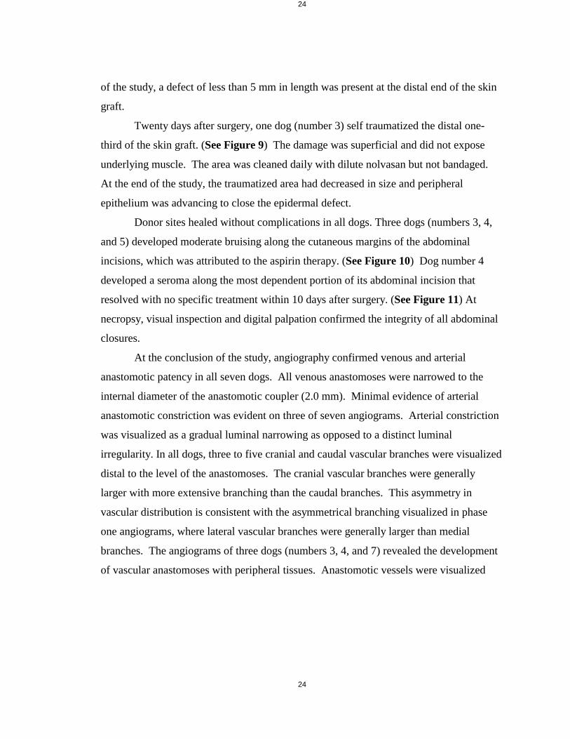
Twenty days after surgery, one dog (number 3) self traumatized the distal one-
third of the skin graft. (See Figure 9) The damage was superficial and did not expose
underlying muscle. The area was cleaned daily with dilute nolvasan but not bandaged.
At the end of the study, the traumatized area had decreased in size and peripheral
epithelium was advancing to close the epidermal defect.
Donor sites healed without complications in all dogs. Three dogs (numbers 3, 4,
and 5) developed moderate bruising along the cutaneous margins of the abdominal
incisions, which was attributed to the aspirin therapy. (See Figure 10) Dog number 4
developed a seroma along the most dependent portion of its abdominal incision that
resolved with no specific treatment within 10 days after surgery. (See Figure 11) At
necropsy, visual inspection and digital palpation confirmed the integrity of all abdominal
closures.
At the conclusion of the study, angiography confirmed venous and arterial
anastomotic patency in all seven dogs. All venous anastomoses were narrowed to the
internal diameter of the anastomotic coupler (2.0 mm). Minimal evidence of arterial
anastomotic constriction was evident on three of seven angiograms. Arterial constriction
was visualized as a gradual luminal narrowing as opposed to a distinct luminal
irregularity. In all dogs, three to five cranial and caudal vascular branches were visualized
distal to the level of the anastomoses. The cranial vascular branches were generally
larger with more extensive branching than the caudal branches. This asymmetry in
vascular distribution is consistent with the asymmetrical branching visualized in phase
one angiograms, where lateral vascular branches were generally larger than medial
branches. The angiograms of three dogs (numbers 3, 4, and 7) revealed the development
of vascular anastomoses with peripheral tissues. Anastomotic vessels were visualized

25
25
between the vascular beds of the transferred rectus abdominis muscles and venous
structures of the lateral saphenous vasculature (See Figure 12).
Histological evaluation confirmed muscular viability after microvascular free
tissue transfer in all seven dogs. All dogs had evidence of mild to moderate muscle fiber
atrophy. Hypertrophy and rounding of nuclei were visible both peripherally and
occasionally centrally in individual muscle fibers of all dogs. Occasional centrally
located nuclear rows were present in longitudinal sections of the transferred muscles (See
Figure 13). All samples collected at necropsy from the contralateral rectus abdominis
muscles revealed normal skeletal muscle with no evidence of atrophy.
In the two dogs that developed epidermal ulceration viable muscle was present
beneath the ulcerative beds. Marked acanthosis and hyperkeratosis were visualized at the
margins of the ulcers in both dogs and there was maintenance of viable dermis and
adnexa overlying the supporting muscular beds. (See Figure 14) Regions of fibroplasia
and fibrovascular connective tissue joined the superficial and deep muscular margins to
the pre-existing connective tissue of the femorotibial defects and overlying meshed skin
grafts in all dogs.

26
26
Discussion
The rectus abdominis is a long, flat-paired muscle located on the ventral midline
that originates from the first costal cartilages and inserts on the cranial border of the
pubis. It is encased within the rectus sheath that is composed of the insertional tendons
of the internal abdominal oblique, external abdominal oblique, and transversus abdominis
muscles. The rectus sheath does not surround the most caudodorsal aspect of the rectus
abdominis muscle. Instead, a thin layer of the transversalis fascia and peritoneum covers
this surface. The abdominal portion of the rectus abdominis muscle receives innervation
from the medial branches of spinal nerves T13-L3. (See Figure 1) 36
The rectus abdominis muscle has several beneficial anatomic qualities when used
in free tissue transfer. First, its long rectangular shape is ideal for placement in wounds of
the distal extremities. This is in comparison to the triangular shape of the trapezius
muscle and the broad rectangular shape of the latissimus dorsi muscle. Second, complete
enclosure of the rectus abdominis muscle within the rectus sheath allows its precise
removal from the abdomen without associated subcutaneous tissues. This, in turn,
produces less bulk after transfer and facilitates its use as a bed for acute skin grafting.
Third, the consistent thickness of the rectus abdominis muscle throughout its length
produces excellent cosmetic results. Finally, the peritoneal covering of the muscle’s
caudodorsal border makes the rectus abdominis a candidate for use as a specialized
myoperitoneal flap as is reported in people 38.
One potential disadvantage of the rectus abdominis muscle is its relatively narrow
dimensions. This may prevent its use in covering defects involving greater than 50
percent of the circumference of the distal extremities. The trapezius muscle with its
triangular shape may be more appropriate for repair of wider defects. Another potential
disadvantage of the rectus abdominis muscle for free tissue transfer is its relatively short
vascular pedicle, as compared to reported pedicle lengths of other muscles used in

27
27
veterinary surgery. The prescapular branch of the superficial cervical artery and vein
supplying the trapezius muscle and the phrenicoabdominal artery and vein supplying the
transversus abdominis muscle are 44 +/- 10 mm and 46.7 +/- 5.0 mm in length,
respectively 10,18. The mean length of the caudal epigastric vascular pedicle, 28 +/- 2
mm, as measured in phase two of this study easily exceeds the minimal length
requirements for microvascular transfer but may be limiting in situations where a longer
vascular pedicle would be needed.
The overall length of the rectus abdominis muscle (mean – 22.5 cm) may however
negate the limitations of the relatively short vascular pedicle. This length should allow
anastomosis of the rectus vascular pedicle with donor vessels that are outside the area of
recipient site injury. This in turn should allow use of the rectus abdominis muscle in free
tissue transfer where extensive damage of recipient site vascular beds has occurred and
anastomosis must be performed away from the primary defect.
All donor site closures healed without major complications. Muscle excision did
require entrance into the abdominal cavity but in an otherwise stable animal this would
not be expected to produce increased morbidity as compared to muscle excisions from
other body regions. Bruising, which was attributed to aspirin therapy, was noted around
the abdominal incisions in three dogs and a dependent seroma formed in one dog.
Aspirin predisposed to incisional bruising through inhibition of the cyclooxygenase
pathway.
Seroma formation is commonly reported, if not expected, with excision of
muscular or myocutaneous donor tissues from the scapular region in dogs and cats 15,21.
Nicoll et al reported seroma formation in seven of seven cats following orthotropic and
heterotropic transfer of the latissimus dorsi muscle flap 21. Philibert et al, reported
seroma formation in four of four dogs following free tissue transfer of the trapezius
musculocutaneous flap 15. Finally, Degner reported seroma formation and partial donor
site dehiscence in two of six medial saphenous fasciocutaneous free flaps 19. In

28
28
comparison to the morbidity associated with tissue harvest from other canine free tissue
transfer donor sites, harvesting of the rectus abdominis is associated with minimal
morbidity.
The full thickness, distal skin graft necrosis seen on one dog is not easily
explained. The most common etiologies for skin graft failure include excessive graft
movement, sub-graft hematoma or seroma formation and infection. These causes
however seem unlikely in the dog of our study because the pelvic limbs were bandaged in
all dogs for approximately two weeks following individual surgical dates and no evidence
of infection, seroma or hematoma was seen in this dog prior to skin necrosis
visualization.
Excessive bandage pressure or inadequate subcutaneous tissue removal are other
possible etiologies for distal necrosis in this dog. Attempts were made to produce even
pressure throughout the bandaged limbs in all dogs. The most distal aspects of the grafts
were however located just proximal to the tarsus. This is known to be a natural region of
bandage constriction because of relatively reduced limb circumference 42. Bandaging of
the distal tibia produces a focal “tourniquet effect” if additional padding is not provided.
This in turn predisposes to distal extremity ischemic injuries. It is possible in our study
that the skin grafts under these areas were predisposed to excessive bandage pressure and
this could be responsible for the distal graft necrosis in this dog. We are however unable
to explain viability in the rest of the distal skin grafts with the above theory.
Inadequate removal of subcutaneous tissues could also have been responsible for
the aforementioned graft necrosis. Meshed skin graft viability is dependent on nutrition
from underlying tissues through plasmatic imbibition. This process involves bathing of
the deep graft surface with serum from the recipient tissue bed. Excessive subcutaneous
graft tissues interfere with this process. Graft revascularization through inosculation and
vascular ingrowth is also inhibited by inadequate subcutaneous tissue removal from the
deep surface of skin grafts. Revascularization begins as early as 48 hours following skin

29
29
graft placement. Capillary rich granulation tissue produces vascular sprouts, which enter
the deep surface of skin grafts. If sprouting capillaries enter pre-existing vascular
channels vessel growth is facilitated because of minimal tissue resistance. Inadequate
removal of graft subcutaneous tissues interferes with revascularization and therefore is a
plausible explanation for distal graft failure in the one dog of our study.
Intensive post-operative monitoring was not performed following free tissue
transfer in this study because of the logistical difficulties. In people, flap monitoring is a
priority and has been the subject of extensive research attempting to determine the ideal
monitoring modality 43-48. Intensive monitoring has proven essential in people because of
a reported post-operative thrombosis rate of approximately 10% 8. In addition, salvage
rates as high as 70% have been reported following re-operation for thromboses flaps.
The success of a second exploratory surgery is however largely dependent on rapid
recognition of vascular compromise. Early re-operation minimizes the deleterious effects
of temporary tissue ischemia and the so-called “no reflow” phenomenon.
Monitoring methods can be divided into two primary categories; direct vascular
pedicle blood flow measurements and determinations of tissue perfusion. Ultrasonic
doppler blood flow probes have been one of the most commonly used instruments to
determine blood flow across vascular anastamoses 43,47,48 These instruments determine
blood flow through detection of changes in sound wave frequencies reflected from
moving blood cells. Within the last 10 years implantable Doppler probes have been
developed that allow measurement of blood flow in buried muscle flaps 48. Probes are
easily placed and removed through simple retraction preventing the need for an additional
surgery for probe removal. Probes are however expensive, have the potential to
traumatize vasculature anastomoses, can provide inaccurate information if displaced and
will not accurately read when subjects are moving 43. Their use has not been evaluated in
veterinary free tissue transfer patients.

30
30
Duplex Doppler transcutaneous ultrasonography has also been used in people to
monitor vascular pedicle patency. This technique uses a combination of gray scale
ultrasound imaging and Doppler flow measurements. Vessels as small as 1.0 mm in
diameter can be visualized and arterial and venous flow can be differentiated. This
method however does not allow continuous monitoring and is highly dependent on the
skills of the ultrasonographer. Instrumentation for its usage is also very expensive. Other
methods of determining vascular patency include electromagnetic flowmetry and arterial
thermometry 47.
Two common methods of determining tissue perfusion include dermofluorometry
and radioisotope scanning. Fluoroscein staining has historically been considered the best
means of identifying inadequate flap perfusion 47. This originally involved the use of a
Wood’s lamp to visualize areas of tissue fluorescence. Dermofluorometry differs from
traditional fluorescein staining through the use of a dermofluorometer to quantify surface
fluorescence. The dermofluorometer requires small volumes of fluorescein and allows
sequential measurements of increased fluorescein following repeated injections. This
method however underestimates tissue viability and cannot be used for continuous
monitoring because of a delay in tissue drug removal when perfusion decreases.
Fluoroscein injection using the traditional Wood’s lamp is associated with an
unacceptably high rate of allergic reactions in people 44. Dermofluorometry virtually
eliminates these reactions but still underestimates tissue viability by as much as 37% 46.
Radioisotope scanning has frequently been used to determine tissue viability and
fluid flow in both human and veterinary medicine 12,17,49-51. It is reliable in defining
perfusion and tissue viability but cannot be used for continuous monitoring. It also
involves radioisotopes, which require special licensure for use and isolation facilities.
This methodology is most useful in determining viability of vascularized free bone
transfers 17,48,51. Other methods of monitoring tissue perfusion and viability including
pulse oximetry, skin surface thermometry, impedence, tissue pH and hematocrit

31
31
measurements, magnetic resonance imaging, radioisotopic washout, thermal clearance,
reflection spectrophotometry, photoplethysmography, transcutaneous oxygen monitoring
and interstitial fluid pressure monitoring have been described in people but are beyond
the scope of this discussion.
Even with the development of numerous sophisticated and expensive techniques
to monitor tissue viability the most reliably accepted method continues to be subjective
flap evaluation by a member of the surgical team. The aforementioned monitoring
techniques have been developed to allow continuous monitoring by less experienced
personnel. This in turn allows notification of the surgical team when potential flap
compromise occurs. In veterinary medicine flap monitoring is made difficult by the fact
that surgical sites must be protected from animal mutilation and movement is often
uncontrollable. This frequently necessitates covering transferred tissues with protective
bandages or casts. In our study, we did not attempt to monitor the rectus flaps because of
concern for potential tissue compromise from repeated bandage changes, exposed tissues,
or adjacent monitoring instrumentation.
In the current study both topical and systemic anticoagulants were used. Topical
heparinized saline was used intraoperatively because of its inhibitory effects on thrombin
and coagulation factor Xa and its promotion of antithrombin 3 activity 52. Aspirin was
also used in the perioperative period in order to block the production of thromboxane A2
that is known to stimulate platelet aggregation. Other anticoagulants that have been
routinely used in human reimplantations and elective microsurgical procedures include
Dextran, dipyridamole, pentoxifylline, chlorpromazine, streptokinase, urokinase and
tissue plasminogen activator 53. Their use is more aggressively administered with re-
implantation procedures for traumatic extremity amputations than for elective free tissue
transfers because of extensive vascular damage and subendothelial collagen exposure
associated with re-implantation procedures.

32
32
Extensive human research has been performed attempting to define the ideal
topical or systemic anticoagulants 52. There is no agreement about which agents are ideal
but it has been shown that some form of anticoagulant administration is beneficial to the
patency rates of experimental microvascular anastomoses 52-54. Clinical studies are often
based on exaggerated subendothelial collagen exposure through extensive endothelium
removal, vascular lumen inversion or vascular crushing methods. In these studies
antithrombotic agents are clearly beneficial with dramatic differences in patency rates
between treatment and control groups 52-54.
In clinical studies the benefits of anti-thrombotic agents are not so clear. A 1998
prospective clinical study by Khouri found that anti-thrombotic administration had no
effect on the overall success rate of free tissue transfers 8. The authors concluded that use
of antithrombotic agents is relatively unimportant in comparison to precise surgical
technique. The benefits of peri-operative anticoagulant therapy for re-implantation
procedures have on the other hand been documented in the human literature 55.
Salemark et al demonstrated the benefits of anticoagulant therapy in re-implantation
procedures but were unable to demonstrate significant benefits of antithrombotic
administration for elective free tissue transfers. This most likely reflects differences in
the degree of endothelial injury with traumatic reimplantations more closely mimicking
experimental models where endothelial damage is significant.
Controversy still exists about the role of antithrombotic agents in successful
microvascular free tissue transfers. Antithrombotic therapy appears most beneficial when
vascular damage is extensive. Because traumatic extremity reimplantations have not
been reported in the veterinary literature antithrombotic therapy most likely plays a minor
role at best in veterinary patients. With atraumatic free tissue transfers endothelial
damage should be minimal and successful transfer is likely therefore almost entirely
dependent on appropriate microvascular technique. General agreement although not
universal exists that anticoagulants play a supporting role in the success of free tissue

33
33
transfers. Agreement is however universal that meticulous technique and adequate
surgical experience are prerequisites for successful free tissue transfers.
The transferred rectus abdominis muscle with its overlying meshed skin graft
produced excellent cosmesis in this study. The muscle conformed well to the
femorotibial defect and was not noticeably bulky. Subjectively, a moderate amount of
tissue edema was evident immediately after transfer. This was most apparent in the distal
aspect of the transferred tissues. Ten to fourteen days later, the thickness of the
transferred tissues had decreased and skin grafts were only slightly elevated in
comparison to the peripheral skin margin. The time frame required for resolution of
edema coincided with the predictable return of lymphatic drainage in tissues transferred
by microvascular techniques. This time frame of lymphatic return has been demonstrated
in experimental animal models and clinical human patients following limb re-
implantations and free tissue transfers 49,50.
Muscle atrophy seen histologically at the studies conclusion was expected.
Denervation atrophy is reported in both the human and veterinary literature after free
muscle transfer 16,21,35,56,57. Atrophy following free tissue transfer is reported to be fiber
type specific with preferential atrophy of type-1 muscle fibers 56. Increased muscle
atrophy is associated with prolonged immobilization and post-operative infection. This
quantitative decrease in muscle fiber mass does however not correlate with a decrease in
overall thickness of transferred tissues. In people, no statistically significant decrease in
overall tissue thickness is reported up to four years after microvascular free tissue transfer
of muscles with overlying meshed skin grafts 58. Muscle transfers have actually been
reported to increase in thickness a mean of 2.4 times preoperative measurements by two
weeks postoperatively and maintain 1.7 times their original thickness at six weeks 58.
Replacement of muscle fibers with adipose and fibrous tissues may account for the lack
of an overall decrease in tissue volume after microvascular free tissue transfer.

34
34
Often with veterinary cutaneous or myocutaneous free tissue transfer the direction
of hair growth and hair density are less than ideal because skin orientation is dictated by
the position of the vascular pedicle. Acute skin grafting over muscle flaps allows
orientation of hair growth to approximate surrounding tissues. Additionally, the skin
graft donor site can be selected to minimize visible scar formation and to more closely
approximate the qualities of the skin surrounding the recipient site defect. Meshed skin
grafting over the transferred rectus abdominis muscle was performed in this study
because of the previously mentioned cosmetic benefits and because of documented poor
perfusion to the caudal aspects of the cutaneous angiosome overlying the canine rectus
abdominis muscle33.
35
35
Conclusion
In conclusion, this study confirmed that the canine rectus abdominis muscle could
be successfully transferred to a femorotibial defect using microvascular techniques.
Phase one verified that the caudal epigastric vascular pedicle is of appropriate diameter
and length for use in free tissue transfer. In phase two, muscular dissection and pedicle
isolation were relatively straightforward. Minimal donor site morbidity was associated
with removal of the muscle from the ventral abdomen and the transferred rectus
abdominis muscle served as a viable bed for acute skin grafting. Further work is needed
to demonstrate the reproducible utility of the rectus abdominis muscle for free tissue
transfer in clinical cases.
One potential area of future research is in defining the utility of the canine rectus
abdominis muscle in oral cavity reconstruction. The canine transverses abdominis muscle
has been successfully used in reconstruction of a traumatic canine maxillary defect 10.
Degner et al described the benefits of myoperitoneal reconstruction for oral cavity defects
with primary healing of oral mucosa to the transferred peritoneal surface 10. The human
rectus abdominis muscle with its associated peritoneal lining is successfully used in
intraoral reconstructive surgery. Mixter et al described five cases of successful intra-oral
rectus abdominis myoperitoneal free flap transfer in people 38. This article briefly
describes the benefits of oral myoperitoneal transfers, which again include primary
healing of the muscles peritoneal lining with adjacent oral squamous epithelium and
resistance of the peritoneal covering to oral and gastric secretions.
The canine rectus abdominis muscles partial peritoneal lining makes it a candidate
for use as a myoperitoneal flap in oral reconstructive surgery. The muscle has been
successfully used in this fashion in one clinical case at the Virginia-Maryland Regional
College of Veterinary Medicine following a traumatic maxillary wound. Prior to free
tissue transfer multiple unsuccessful attempts were made using local tissue advancement

36
36
techniques. Final repair was achieved with free tissue transfer of the rectus into the oral
cavity. Thus far the use of specialized free tissue transfers in veterinary medicine have
been rarely described. With time however such research will inevitably begin to appear
in the veterinary literature.

37
37
References
1. Francel TJ. Improving reemployment rates after limb salvage of acute severe
tibial fractures by microvascular soft-tissue reconstruction. Plast Reconstr Surg
1994;93:1028-34.
2. Pavletic MM. Canine axial pattern flaps, using the omocervical, thoracodorsal and
deep circumflex iliac direct cutaneous arteries. Am J Vet Res 1981;42:391-406.
3. Kostolich M, Pavletic MM. Axial pattern flap based on the genicular branch of
the saphenous artery in the dog. Vet Surg 1987;16:217-22.
4. Henney LH, Pavletic MM. Axial pattern flap based on the superficial brachial
artery in the dog. Vet Surg 1988;17:311-7.
5. Pavletic MM. Caudal superficial epigastric arterial pedicle grafts in the dog. Vet
Surg 1980;9:103-107.
6. Pavletic MM, Kostolich M, Koblik P, et al. A comparison of the cutaneous trunci
myocutaneous flap and latissimus dorsi myocutaneous flap in the dog. Vet Surg
1987;16:283-93.
7. Fowler JD, Degner DA, Walshaw R, et al. Microvascular free tissue transfer:
results in 57 consecutive cases. Vet Surg 1998;27:406-12.
8. Khouri RK, Cooley BC, Kunselman AR, et al. A prospective study of
microvascular free-f lap surgery and outcome. Plast Reconstr Surg
1998;102:711-21.
9. Basher AW, Fowler JD, Bowen CV, et al. Microneurovascular free digital pad
transfer in the dog. Vet Surg 1990;19:226-31.
10. Degner DA, Lanz OI, Walshaw R. Myoperitoneal microvascular free flaps in
dogs: an anatomical study and a clinical case report. Vet Surg 1996;25:463-70.
11. Fowler JD, Levitt L, Bowen CV. Microsurgical free bone transfer in the dog.
Microsurgery 1991;12:145-50.

38
38
12. Roa DM, Bright RM, Daniel GB, et al. Microvascular transplantation of a free
omental graft to the distal extremity in dogs. Vet Surg 1999;28:456-65.
13. Fowler JD, Miller CW, Bowen V, et al. Transfer of free vascular cutaneous flaps
by microvascular anastomosis. Results in six dogs. Vet Surg 1987;16:446-50.
14. Miller C. Microvascular distant transfer of a axial-pattern flap in a dog. JAVMA
1987;190:203-204.
15. Philibert D, Fowler JD. The trapezius osteomusculocutaneous flap in dogs. Vet
Surg 1993;22:444-50.
16. Philibert D, Fowler JD, Clapson JB. Free microvascular transplantation of the
trapezius musculocutaneous flap in dogs. Vet Surg 1992;21:435-40.
17. Szentimrey D, Fowler D, Johnston G, et al. Transplantation of the canine distal
ulna as a free vascularized bone graft. Vet Surg 1995;24:215-25.
18. Philibert D, Fowler JD, Clapson JB. The anatomic basis for a trapezius muscle
flap in dogs. Vet Surg 1992;21:429-34.
19. Degner DA, Walshaw R, Lanz O, et al. The medial saphenous fasciocutaneous
free flap in dogs. Vet Surg 1996;25:105-13.
20. Degner DA, Walshaw R. Medial saphenous fasciocutaneous and myocutaneous
free flap transfer in eight dogs. Vet Surg 1997;26:20-5.
21. Nicoll SA, Fowler JD, Remedios AM, et al. Development of a free latissimus
dorsi muscle flap in cats. Vet Surg 1996;25:40-8.
22. Fowler J. The microvascular carpal foot pad flap: results in three clinical cases.
VCOT 1997;10:187-191.
23. Moens N. The microvascular carpal foot pad flap: vascular anatomy and surgical
technique. VCOT 1997;10:183-186.
24. Lanz OI, Broadstone RV, Martin RA, et al. Effects of epidural anesthesia on
microcirculatory blood flow in free medial saphenous fasciocutaneous flaps in
dogs. Vet Surg 2001;30:374-9.

39
39
25. Gosain A, Chang N, Mathes S, et al. A study of the relationship between blood
flow and bacterial inoculation in musculocutaneous and fasciocutaneous flaps.
Plast Reconstr Surg 1990;86:1152-62; discussion 1163.
26. Musharafieh R, Osmani O, Musharafieh U, et al. Efficacy of microsurgical free-
tissue transfer in chronic osteomyelitis of the leg and foot: review of 22 cases. J
Reconstr Microsurg 1999;15:239-44.
27. Anthony JP, Mathes SJ, Alpert BS. The muscle flap in the treatment of chronic
lower extremity osteomyelitis: results in patients over 5 years after treatment.
Plast Reconstr Surg 1991;88:311-8.
28. Fisher J, Wood MB. Experimental comparison of bone revascularization by
musculocutaneous and cutaneous flaps. Plast Reconstr Surg 1987;79:81-90.
29. Richards RR, Schemitsch EH. Effect of muscle flap coverage on bone blood flow
following devascularization of a segment of tibia: an experimental investigation in
the dog. J Orthop Res 1989;7:550-8.
30. Richards RR, McKee MD, Paitich CB, et al. A comparison of the effects of skin
coverage and muscle flap coverage on the early strength of union at the site of
osteotomy after devascularization of a segment of canine tibia. J Bone Joint Surg
Am 1991;73:1323-30.
31. Porter CD, Fowler JD, Matte GG. A comparison of skin and muscle for cortical
revascularization. V.C.O.T. 2000;13:128-134.
32. Fowler J. Reconstructive Microsurgical Applications In: W. a. Wilkins, ed.
Current Techniques in Small Animal Surgery. Fourth ed. Baltimore, MD,
1998;607-621.
33. Gregory CR, Gourley IM, Koblik PD, et al. Experimental definition of latissimus
dorsi, gracilis, and rectus abdominus musculocutaneous flaps in the dog. Am J Vet
Res 1988;49:878-84.

40
40
34. Fujimaki A, O'Brien BM, Kurata T, et al. Experimental micro-anastomosis of 0.4-
0.5 mm vessels. Br J Plast Surg 1977;30:269-72.
35. Degner DA, Walshaw R, Arnoczky SP, et al. Evaluation of the cranial rectus
abdominus muscle pedicle flap as a blood supply for the caudal superficial
epigastric skin flap in dogs. Vet Surg 1996;25:292-9.
36. Degner D. The cranial rectus abdominus muscle pedicle flap in the dog. VCOT
1994;7:21-24.
37. Solano M, Purinton PT, Chambers JN, et al. Effects of vascular pedicle ligation
on blood flow in canine semitendinosus muscle. Am J Vet Res 1995;56:731-5.
38. Mixter RC, Mayfield K, Dibbell DG, et al. Intraoral reconstruction with a
microvascular peritoneal flap [see comments]. Plast Reconstr Surg 1991;88:452-
7.
39. Reath DB, Taylor JW. Free rectus abdominis muscle flap: advantages in lower
extremity reconstruction. South Med J 1989;82:1143-6.
40. Reath DB, Taylor JW. The segmental rectus abdominis free flap for ankle and
foot reconstruction. Plast Reconstr Surg 1991;88:824-8; discussion 829-30.
41. Salgado CJ, Orlando GS, Herceg S, et al. Pfannenstiel incision as an alternative
approach for harvesting the rectus abdominis muscle for free-tissue transfer. Plast
Reconstr Surg 2000;105:1330-3.
42. Anderson DM, White RA. Ischemic bandage injuries: a case series and review of
the literature. Vet Surg 2000;29:488-98.
43. Furnas H, Rosen JM. Monitoring in microvascular surgery. Ann Plast Surg
1991;26:265-72.
44. Graham BH, Walton RL, Elings VB, et al. Surface quantification of injected
fluorescein as a predictor of flap viability. Plast Reconstr Surg 1983;71:826-33.
45. Khouri RK, Shaw WW. Monitoring of free flaps with surface-temperature
recordings: is it reliable? Plast Reconstr Surg 1992;89:495-9; discussion 500-2.

41
41
46. Myers B, Donovan W. An evaluation of eight methods of using fluorescein to
predict the viability of skin flaps in the pig. Plast Reconstr Surg 1985;75:245-50.
47. Stepnick DW, Hayden RE. Postoperative monitoring and salvage of
microvascular free flaps. Otolaryngol Clin North Am 1994;27:1201-17.
48. Machens HG, Mailaender P, Rieck B, et al. Techniques of postoperative blood
flow monitoring after free tissue transfer: an overview. Microsurgery
1994;15:778-86.
49. Anthony JP, Foster RD, Price DC, et al. Lymphatic regeneration following
microvascular limb replantation: a qualitative and quantitative animal study. J
Reconstr Microsurg 1997;13:327-30.
50. Khazanchi RK, Rakshit K, Bal CS, et al. Evaluation of lymphatic drainage in free
flaps by lymphoscintigraphy: a preliminary study. Br J Plast Surg 1996;49:123-8.
51. Levitt L, Fowler JD, Longley M, et al. A developmental model for free
vascularized bone transfers in the dog. Vet Surg 1988;17:194-202.
52. Das SK, Miller JH. Current status of topical antithrombotic agents in
microvascular surgery. Microsurgery 1994;15:630-2.
53. Siegel DB. Use of anticoagulants in replantation and elective microsurgery.
Microsurgery 1991;12:277-80.
54. Rothkopf DM, Chu B, Bern S, et al. The effect of dextran on microvascular
thrombosis in an experimental rabbit model [see comments]. Plast Reconstr Surg
1993;92:511-5.
55. Salemark L. International survey of current microvascular practices in free tissue
transfer and replantation surgery. Microsurgery 1991;12:308-11.
56. Kauhanen MS, Salmi AM, von Boguslawsky EK, et al. Muscle fiber diameter and
muscle type distribution following free microvascular muscle transfers: a
prospective study. Microsurgery 1998;18:137-44.

42
42
57. Schmalbruch H, al-Amood WS, Lewis DM. Morphology of long-term denervated
rat soleus muscle and the effect of chronic electrical stimulation. J Physiol
1991;441:233-41.
58. Salmi A, Tukiainen E, Harma M, et al. A prospective study of changes in muscle
dimensions following free- muscle transfer measured by ultrasound and CT
scanning. Plast Reconstr Surg 1996;97:1443-50.

43
43
Figure 1 - Normal Canine Rectus Abdominis Anatomy – The muscle receives blood
flow from two primary sources, the cranial and caudal epigastric arteries and veins.
Additional perfusion is supplied by lateral perforating vessels, which are terminal
branches of the deep circumflex iliac artery and vein. The muscle has four to five
tendinous intersections, which are points of firm attachment by the rectus abdominis
sheath. The rectus sheath completely surrounds the muscle and is composed of the
insertional tendons of the internal abdominal oblique, external abdominal oblique and
transversus abdominis muscles.

44
44
Figure 2 - Femorotibial Defect – Cutaneous tissues were removed from the distal aspect
of the femur to the level of the distal tibia and included approximately ½ the
circumference of the limb. An additional proximal incision was made to allow
visualization of the recipient site vasculature.

45
45
Figure 3 - Phase One Angiograms - Angiograms confirmed that the caudal epigastric
artery and vein perfused approximately two-thirds of the length of the rectus abdominis
muscle. Prior to vessel termination, three to five medial and lateral vascular branches
were visualized exiting the main arterial trunk. Lateral branches were generally more
prominent than medial branches. Anastomotic communications between the caudal and
cranial epigastric angiosomes were visualized at the cranial termination of the caudal
epigastric artery.

46
46
Figure 4 - Day Three Bandage Change – Note skin graft discoloration with multifocal
areas of pale pink, red, and dark purple. Mild to moderate bruising was visualized along
the cutaneous margin peripheral to the skin grafts. Pink to red bleeding muscle was
clearly visible through the fenestrations of the skin grafts in all dogs. All grafts appeared
moderately edematous.

47
47
Figure 5 - Day Six Bandage Change – Note cutaneous bruising peripheral to the skin
grafts had resolved. The skin grafts developed a more homogenous pink to red
coloration. The rectus abdominis muscle was no longer clearly visualized through the
fenestrations of the skin grafts in most dogs.

48
48
Figure 6 - Day Ten Bandage Change - The skin grafts had similar coloration to
surrounding skin but were slightly erythematous. The edema had decreased and the skin
grafts were only minimally raised in comparison with surrounding skin. Most skin graft
fenestrations had closed and bleeding granulation tissue was visualized protruding from
the fenestrations that had not closed.

49
49
Figure 7 - Grafts at Study Conclusion - The grafts had a similar appearance to
surrounding normal skin and moderate hair growth was visualized in all dogs. Hair
density varied between individual dogs. Fenestrations of the skin grafts were not readily
apparent. The skin grafts were only minimally raised in comparison to surrounding
tissues.

50
50
Figure 8 - Distal Graft Necrosis - At post-operative day six in this dog skin graft
necrosis was visualized as a line of clear color demarcation. The area of distal
discoloration eventually sloughed and was in the process of healing by second intention
at the studies conclusion.
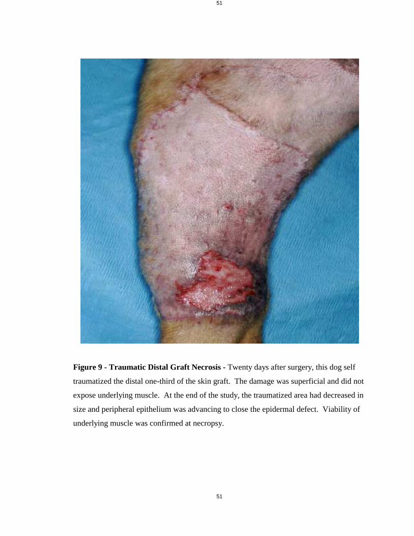
51
51
Figure 9 - Traumatic Distal Graft Necrosis - Twenty days after surgery, this dog self
traumatized the distal one-third of the skin graft. The damage was superficial and did not
expose underlying muscle. At the end of the study, the traumatized area had decreased in
size and peripheral epithelium was advancing to close the epidermal defect. Viability of
underlying muscle was confirmed at necropsy.

52
52
Figure 10 - Incisional Bruising - Donor sites healed without complications in all dogs.
Three dogs developed moderate bruising along the cutaneous margins of the abdominal
incisions. At necropsy, visual inspection and digital palpation confirmed the integrity of
all abdominal closures

53
53
Figure 11 - Seroma Formation - Donor sites healed without complications in all dogs.
This dog developed a dependent seroma along its abdominal incision that resolved with
no specific treatment within 10 days after surgery. At necropsy, visual inspection and
digital palpation confirmed the integrity of all abdominal closures.

54
54
Figure 12 - Phase Two Angiograms – (Craniocaudal and lateral views) Three to five
cranial and caudal vascular branches were visualized distal to the level of the
anastomoses. The cranial vascular branches were generally larger with more extensive
branching than the caudal branches (left image). The angiograms of three dogs revealed
the development of vascular anastomoses with peripheral tissues. Anastomotic vessels
were visualized between the vascular beds of the transferred rectus abdominis muscles
and venous structures of the lateral saphenous vasculature. Contrast is visible within the
lateral saphenous vasculature of this dog on both the lateral and craniocaudal views.

55
55
Figure 13 - Histopathologic Muscle Atrophy - Histological evaluation confirmed
muscular viability after free tissue transfer in all dogs. All dogs had evidence of mild to
moderate muscle atrophy. Enlarged and rounded nuclei were visible both peripherally
and occasionally centrally in individual muscle fibers as seen in the bottom image
(longitudinal view). Occasional centrally located nuclear rows were present in
longitudinal sections of the transferred muscles as well. Increased prominence of spacing
around individual muscle fibers and enlarged peripherally located nuclei are seen on the
top image (cross-sectional view). These changes are all consistent with neurogenic
muscle atrophy.

56
56
Figure 14 - Distal Skin Graft Necrosis – In the two dogs that developed epidermal
ulceration viable muscle was present beneath the ulcerative beds. Marked acanthosis and
hyperkeratosis were visualized at the margins of the ulcers in both dogs but there was
maintenance of viable dermis and adnexal structures overlying the supporting muscular
beds.

57
57
Table 1 – Physical Characteristics of rectus abdominis muscle, donor and recipient
vascular pedicles and recipient site defect
Rectus
abdominis
muscle
(mean in
mm)
Caudal epigastric
vasculature (mean
in mm)
Medial saphenous
vasculature
(mean in mm)
Recipient
site defect
(mean in
mm)
Length 225 +/- 48 28 +/- 2 NA 258 +/- 10
Diameter NA 1.9 +/- 0.2
(arterial)
2.9 +/- 0.5
(venous)
2.1 +/- 0.4
(arterial)
3.1 +/- 0.6
(venous)
NA
Width 55 +/- 2 NA NA 66 +/- 6

58
58
Earl F. Calfee III (Trey)
VITA
Earl F. Calfee III (Trey) was born on August 7,1971 in Cleveland, TN. He attended high
school in his hometown at Cleveland High School then spent two years at Cleveland
State Community College. He earned a Bachelor of Science Degree from the University
of Tennessee in Knoxville in 1993. He attained his Doctorate of Veterinary Medicine
from the University of Tennessee College of Veterinary Medicine in 1998. He then spent
one year in a rotating small animal internship in Boston, MA at the Angell Memorial
Animal Hospital. In 1998, he began a small animal surgical residency and master’s
program at the VA-MD Regional College of Veterinary Medicine in Blacksburg, VA.
He is currently in the third and final year of his residency training, which will be
completed in July of 2002.
![Bone Transport versus Acute Shortening for the Management ...676639/UQ676639_OA.pdf · combined approaches with both microvascular tissue transfers and bone grafts [3] . Masquelet](https://static.fdocuments.in/doc/165x107/5fa3f7aa1a49a1064954e5b0/bone-transport-versus-acute-shortening-for-the-management-676639uq676639oapdf.jpg)