MICROSPORIDIA Biology and Evolution of Highly Reduced ... · The focal point of the microsporidian...
24
Annu. Rev. Microbiol. 2002. 56:93–116 doi: 10.1146/annurev.micro.56.012302.160854 Copyright c 2002 by Annual Reviews. All rights reserved First published online as a Review in Advance on April 16, 2002 MICROSPORIDIA: Biology and Evolution of Highly Reduced Intracellular Parasites Patrick J. Keeling and Naomi M. Fast Department of Botany, Canadian Institute for Advanced Research, University of British Columbia, Vancouver BC, V6T 1Z4, Canada; e-mail: [email protected] Key Words metabolism, genomics, infection, phylogeny, fungi ■ Abstract Microsporidia are a large group of microbial eukaryotes composed exclusively of obligate intracellular parasites of other eukaryotes. Almost 150 years of microsporidian research has led to a basic understanding of many aspects of mi- crosporidian biology, especially their unique and highly specialized mode of infection, where the parasite enters its host through a projectile tube that is expelled at high velocity. Molecular biology and genomic studies on microsporidia have also drawn attention to many other unusual features, including a unique core carbon metabolism and genomes in the size range of bacteria. These seemingly simple parasites were once thought to be the most primitive eukaryotes; however, we now know from molecular phylogeny that they are highly specialized fungi. The fungal nature of microsporidia indicates that microsporidia have undergone severe selective reduction permeating ev- ery level of their biology: From cell structures to metabolism, and from genomics to gene structure, microsporidia are reduced. CONTENTS INTRODUCTION ..................................................... 93 THE MICROSPORIDIAN SPORE ........................................ 95 MICROSPORIDIAN INFECTION AND LIFE CYCLE ....................... 96 MICROSPORIDIAN GENOMICS ........................................ 99 ORIGIN AND EVOLUTION OF MICROSPORIDIA ......................... 100 CORE METABOLISM ................................................. 107 MICROSPORIDIA IN THE NEXT 150 YEARS ............................. 110 INTRODUCTION The microsporidia (or Microspora) are an unusual group of eukaryotic, obligate intracellular parasites that have attracted the curiosity of biologists for more than 100 years. Like many other intracellular parasites, microsporidia are highly special- ized and have evolved an extremely sophisticated and unique infection mechanism along with other adaptations to life inside other cells. These adaptations are pri- marily characterized by reduction. Compared with other eukaryotes, microsporidia 0066-4227/02/1013-0093$14.00 93
Transcript of MICROSPORIDIA Biology and Evolution of Highly Reduced ... · The focal point of the microsporidian...

14 Aug 2002 10:49 AR AR168-MI56-05.tex AR168-MI56-05.sgm LaTeX2e(2002/01/18)P1: IKH10.1146/annurev.micro.56.012302.160854
Annu. Rev. Microbiol. 2002. 56:93–116doi: 10.1146/annurev.micro.56.012302.160854
Copyright c© 2002 by Annual Reviews. All rights reservedFirst published online as a Review in Advance on April 16, 2002
MICROSPORIDIA: Biology and Evolution of HighlyReduced Intracellular Parasites
Patrick J. Keeling and Naomi M. FastDepartment of Botany, Canadian Institute for Advanced Research, University of BritishColumbia, Vancouver BC, V6T 1Z4, Canada; e-mail: [email protected]
Key Words metabolism, genomics, infection, phylogeny, fungi
■ Abstract Microsporidia are a large group of microbial eukaryotes composedexclusively of obligate intracellular parasites of other eukaryotes. Almost 150 yearsof microsporidian research has led to a basic understanding of many aspects of mi-crosporidian biology, especially their unique and highly specialized mode of infection,where the parasite enters its host through a projectile tube that is expelled at highvelocity. Molecular biology and genomic studies on microsporidia have also drawnattention to many other unusual features, including a unique core carbon metabolismand genomes in the size range of bacteria. These seemingly simple parasites were oncethought to be the most primitive eukaryotes; however, we now know from molecularphylogeny that they are highly specialized fungi. The fungal nature of microsporidiaindicates that microsporidia have undergone severe selective reduction permeating ev-ery level of their biology: From cell structures to metabolism, and from genomics togene structure, microsporidia are reduced.
CONTENTS
INTRODUCTION . . . . . . . . . . . . . . . . . . . . . . . . . . . . . . . . . . . . . . . . . . . . . . . . . . . . . 93THE MICROSPORIDIAN SPORE. . . . . . . . . . . . . . . . . . . . . . . . . . . . . . . . . . . . . . . . 95MICROSPORIDIAN INFECTION AND LIFE CYCLE . . . . . . . . . . . . . . . . . . . . . . . 96MICROSPORIDIAN GENOMICS. . . . . . . . . . . . . . . . . . . . . . . . . . . . . . . . . . . . . . . . 99ORIGIN AND EVOLUTION OF MICROSPORIDIA . . . . . . . . . . . . . . . . . . . . . . . . . 100CORE METABOLISM . . . . . . . . . . . . . . . . . . . . . . . . . . . . . . . . . . . . . . . . . . . . . . . . . 107MICROSPORIDIA IN THE NEXT 150 YEARS. . . . . . . . . . . . . . . . . . . . . . . . . . . . . 110
INTRODUCTION
The microsporidia (or Microspora) are an unusual group of eukaryotic, obligateintracellular parasites that have attracted the curiosity of biologists for more than100 years. Like many other intracellular parasites, microsporidia are highly special-ized and have evolved an extremely sophisticated and unique infection mechanismalong with other adaptations to life inside other cells. These adaptations are pri-marily characterized by reduction. Compared with other eukaryotes, microsporidia
0066-4227/02/1013-0093$14.00 93

14 Aug 2002 10:49 AR AR168-MI56-05.tex AR168-MI56-05.sgm LaTeX2e(2002/01/18)P1: IKH
94 KEELING ¥ FAST
are highly reduced at every level: from morphology and ultrastructure, to biochem-istry and metabolism, and even at the level of their molecular biology, genes andgenomes.
The first microsporidian was described in the middle of the nineteenth centurywhen pebrine, or “pepper disease,” was ravaging silkworms in southern Europeand threatening to destroy the European silk industry. The p´ebrine agent wasobserved to be a microscopic parasite that was namedNosema bombycisby Nageliin 1857 (66). Nageli consideredNosemato be a member of the schizomycete fungi,although classification at that time did not reflect the true diversity of microbiallife, and schizomycetes were a grab bag of yeasts and bacteria. After furtherstudy, Balbiani accordingly created a new group forNosemain 1882, calling itMicrosporidia (2), the name still in use.
Today, the microsporidia are known to be an extremely diverse group of par-asites. There are currently approximately 150 described genera of microsporidiawith over 1200 individual species (78, 79). By far, most microsporidia infect ani-mals, where they have been characterized in all vertebrate orders as well as mostinvertebrates, including the parasitic myxosporidia (95). While these animal par-asites account for the vast majority of microsporidia, a few species have beenshown to infect certain protists, such as ciliates and gregarine apicomplexa (95). Itis interesting that these gregarines and some of the ciliates are themselves animalparasites, which suggests that the microsporidian probably once infected the sameanimal hosts as these protists and later adapted to parasitize its neighbor. Giventhe diversity and abundance of microsporidia known in animals today, it seemslikely that the actual number of microsporidia far exceeds those which have beendescribed and that the number of microsporidian species could perhaps approachthe number of species of animals.
Although widespread among animals, microsporidia are apparently most preva-lent in arthropods and fish. They are used as biological control agents against insectpests and, under natural circumstances, are found to be destructive to apiculture,fish, and some crustacea important to aquaculture (4, 75). The first microsporidianinfection described in mammals wasEncephalitozoon cuniculi, originally found inrabbits in 1922 (106). This species is now known to frequently infect a broad rangeof mammalian hosts. In 1959 the first clear case of a human microsporidian in-fection was recorded (60), but such cases were relatively rare until the mid-1970s,when a dramatic increase in recorded infections accompanied the increased preva-lence of immunosuppressed individuals, either resulting from infection with HIVor due to the use of immunosuppressing drugs (98). The most common of theseopportunistic human microsporidia isEnterocytozoon bieneusi, which was firstdescribed as a gastrointestinal parasite causing “wasting syndrome” (a potentiallylethal diarrhea) in 1985 (20).E. bieneusiis now known to infect a wide range of hu-man tissues in AIDS patients and occasionally infects healthy immunocompetenthumans where it results in an acute but self-limiting intestinal disorder (74).Presently, 13 species of microsporidia have been found to infect humans (30, 92, 99)leading to a long list of human diseases, including chronic diarrhea and wasting

14 Aug 2002 10:49 AR AR168-MI56-05.tex AR168-MI56-05.sgm LaTeX2e(2002/01/18)P1: IKH
MICROSPORIDIA: REDUCED PARASITES 95
syndrome, keratoconjunctivitis, pneumonia, bronchitis, nephritis, urethritis, pro-statitis, hepatitis, encephalitis, myostitis, and peritonitis (30, 98, 99, 105).
THE MICROSPORIDIAN SPORE
The focal point of the microsporidian infection strategy, life history, and diagnosisis the spore, a single, highly organized cell (Figure 1). Spores are the only easilyrecognizable stage of microsporidia, they are the stage where species can be dif-ferentiated, and they are the only stage of microsporidia that is viable outside ofa host cell. Spores range in size from as little as 1µm in E. bieneusito 40µmin Bacillidium filiferum(92) and can be spherical, ovoid, rod-shaped, or crescent-shaped, although most are ovoid. Within a species, spore morphology tends to befairly regular, although some species do possess different spore types in differentstages of their life cycles (92). The spore is bound by a normal unit membraneand two rigid extracellular walls. The exospore wall is composed of a dense, gran-ulofibrous, proteinaceous matrix (7, 93) and is generally uniform at the surface,although it can be highly ornamented in aquatic microsporidia (92). The endosporewall is composed of alpha-chitin (92) and proteins and is of uniform thickness,except at the apex of the spore where the endospore wall is considerably thinnerthan elsewhere. Within the spore membrane is the sporoplasm, or the cytoplasmof the spore, which is the infectious material of microsporidia. The sporoplasmcontains a single nucleus or two nuclei arranged as a diplokaryon (two closelyappressed nuclei), cytoplasm enriched with ribosomes, and is otherwise dominatedby structures relating to infection. There are three principal structures related toinfection: the polaroplast, the polar filament or polar tube, and the posterior vac-uole. The polaroplast is a large organization of membranes occupying the anterior
Figure 1 Diagram of a microsporidian spore show-ing the major structures discussed in the text.

14 Aug 2002 10:49 AR AR168-MI56-05.tex AR168-MI56-05.sgm LaTeX2e(2002/01/18)P1: IKH
96 KEELING ¥ FAST
part of the spore. The anterior portion of the polaroplast exists as highly organized,closely stacked membranes called the lammellar polaroplast, whereas the posteriorportion is more loosely organized and is called the vesicular polaroplast. The mostobvious organelle associated with infection is the polar filament or polar tube. Inthe sporoplasm this filament is composed of membrane and glycoprotein layersand ranges from 0.1 to 0.2µm in diameter and 50 to 500µm in length (51, 92). Thepolar filament is attached at the apex of the spore via an umbrella-shaped structurecalled the anchoring disk, from which it extends to the posterior of the spore. Forapproximately one third to one half the length of the spore the polar filament isstraight, and the remainder is helically coiled about the contents of the sporoplasm.The number of coils, their arrangement relative to one another, and even the angleof helical tilt are conserved and diagnostic for a particular species (79, 92). Thepolar filament terminates at the third major organelle associated with infection,the posterior vacuole. There is apparently some physical association between theend of the polar filament and the posterior vacuole (59, 100), but whether the polarfilament actually enters the vacuole or simply contacts it is not known, as the pointof contact has never been observed (51, 92, 94).
MICROSPORIDIAN INFECTION AND LIFE CYCLE
Germination of the microsporidian spore is one of the most interesting and dra-matic series of subcellular events in biology, involving a build-up and controlledrelease of tremendous force, a cascade of extremely rapid events in close succes-sion, and a complete alteration of the sporoplasm, which includes some uniquerestructuring of membrane topology.
Spore germination begins with an environmental trigger that varies for differentspecies depending on their habitat (85) but is largely poorly understood (51). Invitro, spores may be germinated by a number of physical and chemical stimuli in-cluding, but not limited to, alterations in pH, dehydration followed by rehydration,hyperosmotic conditions, the presence of anions or cations, or exposure to ultra-violet light or peroxides [for an extensive review of these conditions for variousspecies see (51)]. When a spore is induced to germinate, the first sign is a generalswelling of the spore and a specific swelling of the polaroplast and posterior vacuole(59). This is the result of an increase in osmotic pressure in the spore (52, 67, 86),but how this pressure builds up is the subject of some debate. Spores are equippedwith aquaporins that specifically transport water across the sporoplasm membrane(31), which explains how the influx of water can take place, but not necessarilywhy. One intriguing possibility was raised after it was observed that levels oftrehalose drop significantly during the germination ofNosema algerae(84, 88).Trehalose is a glucose-glucose disaccharide found widely in nature and is the majorcarbohydrate storage material of microsporidian spores (84, 90) and fungal spores(1, 29). It was suggested that during spore activation in microsporidia, trehalose isdegraded to constituent glucose monomers, effectively increasing the number ofsoluble molecules within the spore and leading to the import of large quantities of
14 Aug 2002 10:49 AR AR168-MI56-05.tex AR168-MI56-05.sgm LaTeX2e(2002/01/18)P1: IKH
MICROSPORIDIA: REDUCED PARASITES 97
water and the concomitant increase in osmotic pressure (84, 86, 88). This modelis a fascinating possibility and would represent an ingenious system to generateforce in the cell. However, the germination of the spore involves a great numberof changing conditions, so the levels of trehalose cannot be tied unequivocally tothe rise in internal osmotic pressure. In fungal spores trehalose does not appear toprimarily act as an energy reserve but rather as an anti-stress metabolite (1). Thisalso may be the role of trehalose in microsporidia, and trehalose degradation mayonly be a step in the germination process. Moreoever, the trehalose levels in a re-lated species ofNosemado not change during germination (19), suggesting some,or potentially many, other causes for water influx. Indeed, alternative models pointto the intracellular concentration of calcium ions as a cause for the influx of water(50) and a possible role for calmodulin in the process (102). Here it is suggestedthat membrane breakdown during spore activation could release calcium ions fromthe endomembrane system into the sporoplasm. These ions could induce the influxof water and could also induce the activation of enzymes such as trehalase whoseactivity could further enhance the hypertonic shift in the sporoplasm (50).
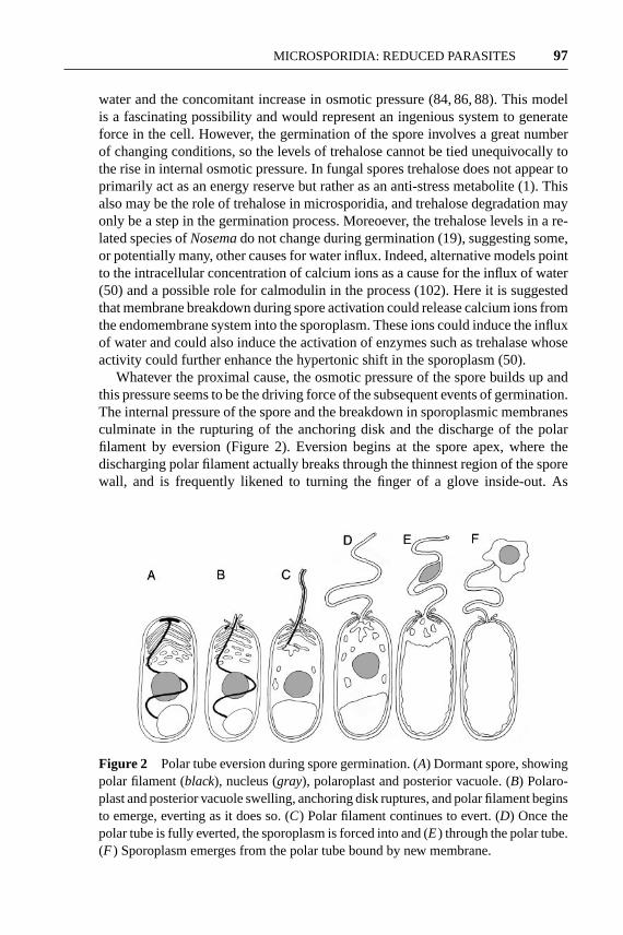
Whatever the proximal cause, the osmotic pressure of the spore builds up andthis pressure seems to be the driving force of the subsequent events of germination.The internal pressure of the spore and the breakdown in sporoplasmic membranesculminate in the rupturing of the anchoring disk and the discharge of the polarfilament by eversion (Figure 2). Eversion begins at the spore apex, where thedischarging polar filament actually breaks through the thinnest region of the sporewall, and is frequently likened to turning the finger of a glove inside-out. As
Figure 2 Polar tube eversion during spore germination. (A) Dormant spore, showingpolar filament (black), nucleus (gray), polaroplast and posterior vacuole. (B) Polaro-plast and posterior vacuole swelling, anchoring disk ruptures, and polar filament beginsto emerge, everting as it does so. (C) Polar filament continues to evert. (D) Once thepolar tube is fully everted, the sporoplasm is forced into and (E) through the polar tube.(F ) Sporoplasm emerges from the polar tube bound by new membrane.

14 Aug 2002 10:49 AR AR168-MI56-05.tex AR168-MI56-05.sgm LaTeX2e(2002/01/18)P1: IKH
98 KEELING ¥ FAST
the filament everts, it becomes a tube and the dense granular, glycoproteinaceousmaterial that filled the filament when inside the sporoplasm is deposited on theoutside of the tube (51). The remarkable nature and violent speed of this eventcannot be overstated. The discharged polar tube can range in length from 50–500µm in length (potentially 100 times the length of the spore) and is generatedby turning inside out a filament that is coiled around the sporoplasm. Yet, theentire event of germination takes place in fewer than two seconds and the tipof the discharging tube can move through the medium at velocities exceeding100µm/s (32).
Clearly, the polar tube is a significant projectile, and if a potential host cell liesnearby, the discharging tube can strike this cell and pierce its membrane. Once thepolar tube is fully discharged, the continued pressure within the spore (most likelyfrom the swelling of the posterior vacuole) forces the sporoplasm through the polartube. Although somewhat elastic, the polar tube is a narrow conduit ranging fromjust 0.1 to 0.25µm in diameter, but the sporoplasm is nevertheless forced throughthe polar tube rapidly, emerging at the tip of the tube in only 15–500 ms (32). Ifthe polar tube has penetrated a host cell, the sporoplasm emerges from the tubedirectly into the host cytoplasm, thus infecting without the host recognizing theparasite as a foreign invader. This invasion is a remarkable event, since the sporo-plasm membrane has been left behind in the now empty spore (87, 101), raisingsome intriguing questions about membrane topology surrounding spore germi-nation. First, an intracellular membrane-based organelle has broken through thesporoplasm membrane and spore wall while being turned inside out. Yet, the sporo-plasm passes out of its bounding membrane and through the polar tube to emerge atthe other side with a brand-new membrane. This is thought to be possible becausepolaroplast membrane is forced into the polar tube at the onset of germination, andit is this membrane that forms the bounding plasma membrane of the intracellularstage of the parasite (101), a remarkable feat of membrane manipulation.
Once inside the host cell, the parasite is referred to as a meront and it begins astage of growth and division characterized by a high degree of interspecies varia-tion. In general, however, the parasite is found directly within the host cytoplasmand not within a host phagocytotic vacuole, as is the case for many other intracel-lular parasites. Occasionally the parasite induces the formation of a surroundingmembrane known as a parasitophorous vacuole at an early stage of infection (55),but most often they do not (although they commonly do during sporogenesis). Atthis stage, the parasite is intimately associated with its host and often induces sig-nificant changes to the host that are not obviously deleterious. Often the host cellreorganizes around the parasite, so that the parasite can be found surrounded byhost organelles such as endoplasmic reticulum (ER), nuclei, or mitochondria (92).In some instances the microsporidian also physically interacts with the host nu-cleus, resulting in the enlargement of host nuclear pores (57) or even the invasionand infection of the nucleus itself (35). Host cells may also transform in shapeand size, often enlarging (103). The archetype of microsporidian-induced host celltransformation is the xenoma, a host cell harnessed and transformed by the parasite

14 Aug 2002 10:49 AR AR168-MI56-05.tex AR168-MI56-05.sgm LaTeX2e(2002/01/18)P1: IKH
MICROSPORIDIA: REDUCED PARASITES 99
to promote its own growth and development. In such cases, the host cell is inducedto enlarge and undergo many rounds of nuclear division resulting in an enormousplasmodium filled with parasites in highly ordered strata depending on their stageof development: mature spores at the center with the earlier stages radiating towardthe periphery of the xenoma (12, 54, 103).
The onset of sporogony is marked in some species by the separation of diplokary-otic nuclei (11) and in other specific cases by meiosis (34), although synaptonemalcomplexes have been observed in all life stages of other microsporidia. Morpholog-ical features are more consistent indicators of sporogony and include an apparentthickening of the plasma membrane (due to the accumulation of electron-densematerial on the membrane) and the increased presence of ER and ribosomes. Boththe ER and ribosomes change organization throughout sporogony, with the ERbecoming highly ordered, and the ribosomes increasingly forming arrays attachedto the ER, known as polyribosomes (92). Although sporogony can occur in directcontact with the host cytoplasm, some species produce a sporophorous vesicle inwhich the sporonts develop. In most species, this stage of the life cycle is alsoaccompanied by some degree of division, although the number of sporoblasts(presporal cells) produced varies among species from two (bisporous) to many(polysporous). Following division, the extrusion apparatus (including the polarfilament, polaroplast, and posterior vacuole) begins to develop. Early observationsimplicated the Golgi as giving rise to the polar filament (91), and this has beenconfirmed by histochemical labeling for activity of the Golgi marker, thiaminepyrophosphatase (80). Further, histochemical studies assaying activity of nucleo-side diphosphatase, a marker of the ER and sometimes of the outercis-Golgi, alsoimplicate the ER in the formation of the polar filament and associated polaroplastmembranes (81). These results suggest that the Golgi and ER (which themselvesare homologous membrane systems) give rise to the sophisticated extrusion ap-paratus of microsporidia. As the extrusion apparatus nears complete formationand the sporoblasts approach maturity, the cells decrease in size and the chitinousendospore layer develops. Once complete, the mature spores are released. Somespecies produce autoinfective spores that germinate immediately in an attempt toinfect the same host, thus spreading the infection quickly though an individual(76). Alternatively, spores are released into the environment (i.e., via host urine,feces, or decomposition) where they can infect other individuals, most often byway of the digestive system.
MICROSPORIDIAN GENOMICS
The genomes of microsporidia have aroused interest since they were first kary-otyped and found to be much smaller than expected for eukaryotes. Presently, thesizes of 13 microsporidian genomes are known and they fall between 19.5 Mbp andonly 2.3 Mbp [(6, 70); for a summary table see (63)]. Apart from their small size,microsporidian genomes are in all characteristics eukaryotic: They have multiple

14 Aug 2002 10:49 AR AR168-MI56-05.tex AR168-MI56-05.sgm LaTeX2e(2002/01/18)P1: IKH
100 KEELING ¥ FAST
linear chromosomes, telomeres, and segregate by closed mitosis. However, thereduced size of microsporidian genomes has led to two genome sequence surveys(25, 36), the sequencing of an entire chromosome (69) and now the completelysequenced genome ofE. cuniculi(44). The general characteristics of the 2.9-MbpEncephalitozoongenome mirror what has been observed in other highly reducedeukaryotic genomes (23): Genes are typically flanked by short intergenic regions(although there are no overlapping genes), there are few repeat sequences, littleevidence of selfish elements, and few introns. Introns were predicted to be presentin microsporidia based on studies that characterized elements of the spliceosome(21, 27) and one putative intron in a ribosomal protein-coding gene (5). In the 1997predicted open reading frames in the completeE. cuniculigenome, only 11 intronswere identified, givingEncephalitozoonone of the lowest intron densities amongeukaryotes. One of the surprising characteristics of theEncephalitozoongenomethat demonstrates the extreme degree of reduction is the finding thatEncephalito-zoongenes themselves are actually shorter on average than their homologs fromother organisms (44). This suggests that the selection for reduction is very strongindeed, perhaps even overwhelming selection against marginally disadvantageousdeletions in protein- and RNA-encoding gene sequences.
ORIGIN AND EVOLUTION OF MICROSPORIDIA
Our conception of the evolutionary history of microsporidia has been radicallyrewritten on a number of occasions since their original description in the middleof the nineteenth century. When N¨ageli consideredNosemato be a yeast-likefungus, the concept of a “protist” or a “protozoan” was in its infancy, so it wascommon to pigeonhole microbial organisms into animals, plants, or sometimesfungi depending on their characteristics. The unique mode of infection seen inNosemaeventually led to their separation from fungi, but with no obvious similarityto any other group of eukaryotes, the evolutionary origins of microsporidia werenot much clearer. As the real diversity of microbial eukaryotes began to dawnon biologists, more complex hierarchical classification schemes were developedbased on certain common features of morphology. One of the cell types to beidentified and classified together were spore-forming parasites, collectively calledSporozoa. This group contains what are now known as apicomplexa, myxosporidia,actinomyxidia, haplosporidia, microsporidia, and a handful of individual genera.Within Sporozoa, microsporidia were considered to be most closely related to avariety of other parasites at different times but were most often believed to be akinto myxosporidia and actinomyxidia, which, with microsporidia, were collectivelycalled the Cnidosporidia (53).
For some time, microsporidia were considered to be either Cnidosporidia [al-though the vast differences between microsporidia on one hand and myxosporidiaand actinomyxidia on the other were pointed out (58)] or an independent protist
14 Aug 2002 10:49 AR AR168-MI56-05.tex AR168-MI56-05.sgm LaTeX2e(2002/01/18)P1: IKH
MICROSPORIDIA: REDUCED PARASITES 101
lineage of uncertain affinity (56). However, in 1983, attention was drawn to thepossible evolutionary significance of microsporidia in a new way. Cavalier-Smith(13) proposed that the origin of eukaryotes might have preceded the endosymbioticorigin of the mitochondrion by some considerable span of time, implying that theremay be protists that evolved before the mitochondrial origin. In other words, theremay be primitively amitochondriate eukaryotes, and focusing attention on theseprotists could unlock some of the secrets surrounding the origin of eukaryotes.Four lineages of amitochondriate protists that could hold this pivotal position wereidentified, and these were collectively named Archezoa: Archamoebae (e.g.,En-tamoeba), Metamonada (e.g.,Giardia), Parabasalia (e.g.,Trichomonas), and Mi-crosporidia (13). Archezoa were also known to have other characteristics that couldbe considered primitive, for instance the microsporidia and some other Archezoacontain 70S ribosomes with bacterial-sized rRNAs rather than the 80S ribosomestypical of eukaryotes (18, 39). In some articulations of the Archezoa hypothesis,microsporidia were actually singled out as perhaps the most primitive and ancientlineage of all eukaryotes, since in addition to lacking mitochondria they also lackflagella and other 9+ 2 structures and were sometimes thought to lack Golgias well (68). Shortly after the Archezoa hypothesis was formulated, the tools ofmolecular phylogenetics began to be applied vigorously to microbial eukaryotes,and the first molecular data from microsporidia lent extraordinary support to theArchezoa hypothesis. The small subunit ribosomal RNA (SSU rRNA) from themicrosporidianVairimorphawas shown to be the earliest branch on the eukary-otic tree (96), and of greater interest, the microsporidia were found to be the onlyeukaryotes to retain the prokaryotic trait of having their 5.8S rRNA fused to thelarge subunit (LSU) rRNA (97). These two pieces of evidence bolstered the no-tion that microsporidia were indeed an ancient and primitive lineage, and furtherevidence seemed to accumulate with the sequencing of every new microsporidiangene: Phylogenies based on elongation factor 1α (43), elongation factor 2 (42),as well as isoleucyl tRNA synthetase (9), all showed the microsporidia branchingdeeply.
The same apparent early phylogenetic position was also seen with other Arche-zoa (with the possible exception ofEntamoeba), and altogether the case of anancient origin and primitive lack of mitochondria for microsporidia and otherArchezoa seemed neatly sewn up. Despite the accumulated evidence in favor ofthe Archezoa, the highly adapted parasitic lifestyle of the microsporidia was alwaysa source of doubt: Such specialized parasites may appear primitive when in factthey reflect a process of reduction from a more complex ancestor. It was also notedthat microsporidian gene sequences are highly divergent and possibly misleadingdue to an artifact in phylogenetic reconstruction known as “long branch attraction.”It was even noted that the fused 5.8S-LSU rRNA in microsporidia could easilybe a secondarily derived state since microsporidian rRNAs are extremely strangecompared with other eukaryotes, or even when compared with prokaryotes. Micro-sporidian rRNAs are exceptionally small because they have sustained a number

14 Aug 2002 10:49 AR AR168-MI56-05.tex AR168-MI56-05.sgm LaTeX2e(2002/01/18)P1: IKH
102 KEELING ¥ FAST
of deletions, sometimes in regions of the sequence that are highly conserved evenbetween prokaryotes and eukaryotes. It was reasoned that a deletion that affectedone of the rRNA operon processing sites could generate the fused microsporidianrRNA from an ancestral eukaryotic rRNA operon (14).
These concerns eventually proved to be well founded, as further sampling ofmicrosporidian genes soon revealed that not all evidence supported an ancientorigin for the group. The first phylogenies to contradict the evidence for an earlyorigin of microsporidia were those of alpha-tubulin and beta-tubulin (24, 46). Herethe microsporidia formed a surprising but extremely well-supported group withfungi. This alternative, fungal origin for microsporidia is in complete disagreementwith earlier evidence that microsporidia are ancient or primitive organisms becausefungi are not considered ancient or primitive and are now known to be close rela-tives of animals. How could different genes from the same organisms provide suchdifferent phylogenetic trees? One possibility is that the accelerated rates of substi-tution common to many microsporidian genes led to artifacts in the phylogeneticreconstruction, and it was proposed that the ancient origin of microsporidia waserroneously suggested by the highly derived genes of rRNA and elongation factors.However, tubulin phylogenies are not immune to this problem, and it was notedthat fungal and microsporidian tubulins are both highly divergent [perhaps becauseneither group contains 9+ 2 structures, releasing evolutionary constraints on theirtubulin gene sequences (46)], suggesting that these phylogenies may be in error. Ithas now been demonstrated, however, that both alpha- and beta-tubulins from theflagellated chytrid fungi are highly conserved, and in phylogenies where chytridsare the only fungal representatives, microsporidia still branch with fungi with highstatistical support. This shows that the relationship between microsporidia andfungi in tubulin phylogenies is not a long branch artifact (48).
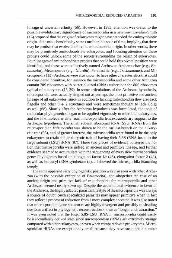
The evidence from tubulin phylogenies led to an immediate re-evaluation ofcertain characteristics of microsporidia such as an insertion in the EF-1α protein,(43) as well as unusual features of their meiosis that had previously been recog-nized as resembling homologous processes in fungi (28). In addition, genes encod-ing TATA-box-binding protein (26), mitochondrial HSP70 (33, 37, 71), glutamyl-tRNA synthetase (10), and the largest subunit of RNA polymerase II (RPB1) (38)were also sequenced from microsporidia, and the phylogenies of each of thesegenes further supported the fungal origin of the group—strongly in the case ofRPB1. The genome ofE. cuniculialso yielded a number of proteins with strongfungal affinities (44), and a recent analysis of combined molecular data from fourgenes further supports the fungal origin of microsporidia (3). Also of critical im-portance is that much of the evidence for the ancient origin of microsporidia hasrecently been undermined by the re-analysis of genes that had previously supportedthe early-branching position. Re-analyzing EF-2 and LSU rRNA data using meth-ods that take into account variations in substitution rates at different sites in asequence showed that these genes not only fail to support the ancient position ofmicrosporidia but actually weakly support the fungal relationship (38, 89). Someexamples of these phylogenies are shown in Figure 3, and a summary of molecular

14 Aug 2002 10:49 AR AR168-MI56-05.tex AR168-MI56-05.sgm LaTeX2e(2002/01/18)P1: IKH
MICROSPORIDIA: REDUCED PARASITES 103
Fig
ure
3(C
onti
nued
on
next
pag
e)

14 Aug 2002 10:49 AR AR168-MI56-05.tex AR168-MI56-05.sgm LaTeX2e(2002/01/18)P1: IKH
104 KEELING ¥ FAST
14 Aug 2002 10:49 AR AR168-MI56-05.tex AR168-MI56-05.sgm LaTeX2e(2002/01/18)P1: IKH
MICROSPORIDIA: REDUCED PARASITES 105
phylogenies including microsporidia is given in Table 1. Furthermore, the notionthat microsporidia are primitively amitochondriate has been disproved by twomeans. First, if they are fungi then we know that microsporidia must be derivedfrom mitochondrial ancestors since fungi and all their close relatives contain mito-chondria. Second, genes for proteins derived from mitochondria have been foundin microsporidia. Most mitochondria contain a small genome, but the majority ofgenes for mitochondrial proteins are encoded in the nucleus and their products arepost-translationally targeted to the organelle. Therefore, it has been reasoned thatan organism that has drastically altered its mitochondrion, or even lost it altogether,could retain relic mitochondrial genes in the nuclear genome (17). One such gene,encoding mitochondrial HSP70, has now been characterized in three species ofmicrosporidia:N. locustae, Vairimorpha necatrix, andE. cuniculi. The presenceof this gene in these organisms gives solid confirmation that microsporidia hada mitochondriate ancestry, and in some analyses even supports a fungal origin(33, 37, 71). Now, several more mitochondrion-derived genes have been found inmicrosporidia (25, 44), the evolutionary implications of which will be consideredwhen the current presence or absence of mitochondria is discussed below.
Altogether, there are now a number of gene phylogenies that provide robust sup-port for some relationship between microsporidia and other fungi, but what exactlyis this relationship? Most genes that have been used to test this have only includedascomycetes and occasionally basidiomycetes. With such poor sampling of fungi,it is not clear from these studies if microsporidia actually are fungi, or if they aremerely a closely related sister group of fungi. Unfortunately, only two genes havecurrently been sampled from diverse fungi to better define this relationship, andthese are alpha- and beta-tubulins. In the case of beta-tubulin there is strong supportfor microsporidia actually evolving from within the fungi, but phylogenies fail todistinguish whether microsporidia are specifically related to ascomycetes or zy-gomycetes (48). Alpha-tubulin also strongly supports microsporidia evolving fromwithin the fungi, but in this case, and in analyses combining both genes, the mi-crosporidia show a specific relationship to zygomycetes (P.J. Keeling, unpublished
←−−−−−−−−−−−−−−−−−−−−−−−−−−−−−−−−−−−−−−−−−−−−−−−−−−−−−−−−−Figure 3 Phylogenies of four microsporidian proteins showing several types of mi-crosporidian phylogeny. (A) Beta-tubulin and (B) RPB1 phylogenies show strong sup-port for microsporidia being related to, or derived from, fungi. (C) Elongation factor-2was originally reported to show microsporidia branching early among eukaryotes,but re-analysis taking into account site-to-site rate variation actually shows the mi-crosporidia branching weakly with fungi. (D) Elongation factor-1α is an example of aphylogeny that consistently places microsporidia early among eukaryotes; however, itis an extremely divergent gene, calling into question its use as a phylogenetic markerfor microsporidia. This is particularly interesting in the case of EF-1α since the mi-crosporidian gene actually contains an insertion otherwise found only in animals andfungi (inset). All trees were inferred using gamma-corrected distances and weightedneighbor joining as described in (25).

14 Aug 2002 10:49 AR AR168-MI56-05.tex AR168-MI56-05.sgm LaTeX2e(2002/01/18)P1: IKH
106 KEELING ¥ FAST
TABLE 1 Summary of published phylogenetic trees showing position of microsporidia
Unresolved/Gene “Deep” branching intermediate Fungal
Alpha-tubulin (24, 46, 48)
Beta-tubulin (24, 46, 48)
RPB1 (38)
TBP (26)
Glu-tRNA synthetase (10)
Ser-tRNA synthetase (44)
V-ATPase-A (44)
TF IIB (44)
GTPase (44)
mt HSP70 (37, 71) (71) (33, 37)
LSU rRNA (70) (70) (89)
EF-2 (42) (38)
Ile-tRNA synthtase (9)
mt PDH-alpha (25)
mt PDH-beta (25)
EF-1α (42, 43) (38)
SSU rRNA (96) (89)
eIF-2γ (47)
Gln-tRNA synthetase (10)
Proteosome alpha (8)
data). Interestingly, a zygomycete origin for microsporidia was proposed basedon the superficial similarities between the polar tube and the apical spore body ofharpellalean zygomycetes (15). However, molecular evidence to date does not sug-gest a specific relationship between microsporidia and harpellalean zygomycetesor any other group of zygomycetes, except in certain analyses where microsporidiaare related to entomophthorales (48).
While the exact relationship between microsporidia and fungi remains to be clar-ified, nearly all current evidence does support one major conclusion: Microsporidiaare not ancient eukaryotes, but are instead highly evolved fungi. This conclusioncolors nearly all other aspects of microsporidia in a new light: No longer arethey primitive in lacking mitochondria, flagella, or peroxisomes—these featuresresult from reductive evolution, probably in response to their growing adaptationto intracellular parasitism (the mitochondrion is a special case discussed below).Similarly, microsporidian biochemistry is not primitive, it is reduced. Even at themolecular level, the tiny genomes of microsporidia evolved from larger genomes

14 Aug 2002 10:49 AR AR168-MI56-05.tex AR168-MI56-05.sgm LaTeX2e(2002/01/18)P1: IKH
MICROSPORIDIA: REDUCED PARASITES 107
by gene loss and compaction, and the unusual genes and gene sequences we findtoday are highly derived, not ancient (49).
CORE METABOLISM
Much of the metabolism of eukaryotes centers around the mitochondrion, the so-called powerhouse of the cell. However, ultrastructural studies on microsporidiain the 1960s revealed no mitochondrion (91), and no study since then has actuallyvisualized an organelle answering to the description of a typical mitochondrionin any microsporidian (63, 92). We now know that microsporidia evolved frommitochondriate fungi, but two interesting questions remain: What was the fate ofthe microsporidian mitochondrion, and how has their core metabolism adapted?
Metabolic pathways for energy generation from carbohydrates have more orless been worked out from several amitochondriate protists, in particular theparabasalianTrichomonas vaginalis, the diplomonadGiardia lamblia, and theentamoebidEntamoeba histolytica(data are also beginning to accumulate fromthe apicomplexan,Cryptosporidium) (65, 73). In addition, a number of genes en-coding enzymes involved in core metabolic pathways have also been characterizedfrom these organisms. The metabolic picture that has emerged from these stud-ies shows some commonality between these disparate organisms, but also a greatdeal of variation. In general all these organisms lack electron transfer chains, ox-idative phosphorylation, and the tricarboxylic acid (TCA) cycle. All break downglucose using the glycolytic pathway, which is like that of other eukaryotes exceptthat phosphofructokinase is pyrophosphate-dependent rather than ATP-dependent(65). From phosphoenol pyruvate, pyruvate is formed directly or can be formedusing a malate bypass not found in typical eukaryotes. Pyruvate metabolism isperhaps the defining difference between these amitochondriates and other eukary-otes. Typically, pyruvate enters the mitochondrion and is oxidatively decarboxy-lated by the pyruvate dehydrogenase complex (PDHC), but in the amitochondriateparabasalia, diplomonads, and entamoebids, it is decarboxylated by a single en-zyme, pyruvate:ferredoxin oxidoreductase (PFOR), an iron-sulphur protein alsoused by anaerobic bacteria. Electrons are transferred from pyruvate to ferredoxinby PFOR, then to NADH, and ultimately to an organic terminal electron acceptor.In the parabasalia, the terminal electron acceptor is ionic hydrogen, resulting inthe production of hydrogen gas, and the reactions following pyruvate productiontake place within a membrane-bound organelle called the hydrogenosome (64).Conversely, inGiardiaandEntamoebaall the reactions of core carbon metabolismare cytosolic. Altogether, sugar metabolism in these organisms is substantially lessefficient than that of mitochondriate eukaryotes, relying as it does on what has beentermed “extended glycolysis” and substrate-level phosphorylation (65).
Until recently, microsporidia could not be compared to these other “amito-chondriates” since next to nothing was known about their metabolic capacities,largely because their obligate intracellular growth and division presents seriouschallenges to biochemical assays (83). Nevertheless, some biochemical assays

14 Aug 2002 10:49 AR AR168-MI56-05.tex AR168-MI56-05.sgm LaTeX2e(2002/01/18)P1: IKH
108 KEELING ¥ FAST
were carried out using purified spores and in vitro–germinated spores maintainedfor a short time in a cell culture medium (102). Altogether, these studies con-firmed the suspected lack of TCA cycle, showed a requirement for ATP in thesustaining media (buttressing suspicions that these parasites probably import ATPfrom their hosts), determined that the parasites produce lactic acid and pyruvicacid, and demonstrated the presence of several enzymes involved in glycolysis,the pentose-phosphate pathway, as well as trehalose synthesis and degradation(22, 84, 102).
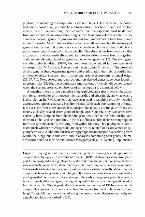
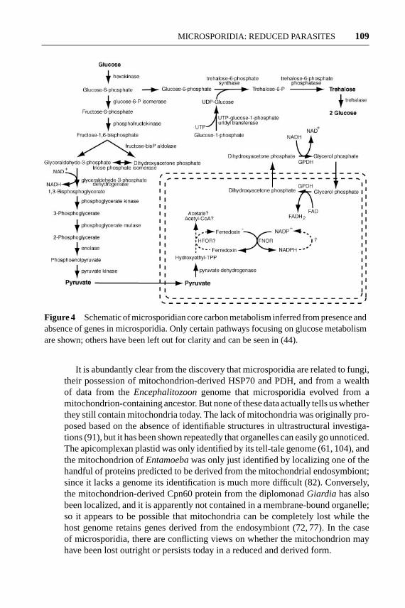
These studies indicate that microsporidia have retained the glycolytic pathwayand suggest that they probably use extended glycolysis. However, the first mi-crosporidian gene encoding an enzyme for core carbon metabolism proved thatthey are very different indeed. TheN. locustaegenome was found to encode boththe alpha and beta subunits of pyruvate dehydrogenase (PDH, or PDHC E1), thefirst enzyme of the pyruvate dehydrogenase complex (25). In mitochondriate eu-karyotes, PDH is responsible for the decarboxylation of pyruvate and gives riseto the “active aldehyde” intermediate, 2-alpha-hydroxyethyl-thiamine pyrophos-phate (HETPP), which is then converted by PDHC E2 (dihydrolipoamide acetyltransferase) and E3 (dihydrolipoamide dehydrogenase) into acetyl-CoA. Genesencoding PDH subunits have also been found in the genome ofE. cuniculi, butPDHC E2 and E3 are absent, which suggests that the role of PDH in microsporidiais unique. One possible explanation stems from the observation that PDH andPFOR actually share certain similarities in both structure (16) and biochemical ac-tivity; in particular, both use HETPP as an active intermediate (62). Accordingly,microsporidian PDH might be synthesizing HETPP to reduce the iron-sulphurcenter of a second protein, which then transfers electrons to ferredoxin, in ef-fect mimicking the activity of PFOR but without actually using the enzyme (25).The catalog of genes present in theEncephalitozoongenome does indeed con-firm that microsporidia use a completely novel form of core metabolism. Mostimportantly, theEncephalitozoongenome encodes genes for PDHC E1 (but notE2 or E3) as well as ferredoxin, ferredoxin:NADPH oxidoreductase, and a numberof proteins involved in synthesizing iron-sulphur centers (44). Microsporidia alsodiffer from other amitochondriates in that phosphofructokinase is related to theATP-dependent enzyme of other fungi rather than being pyrophosphate dependent.A schematic representation of certain pathways inferred to be important in coreenergy metabolism in microsporidia is shown in Figure 4.
The presence of PDH-based pyruvate metabolism inNosemaandEncephal-itozoonbreaks one of the main “rules” of amitochondriate metabolism, namelythe use of PFOR or some derivative of PFOR rather than PDH (65). Yet, thisdifference also epitomizes “amitochondriate” metabolism in that the core carbonmetabolic machinery of these protists appears to have been cobbled together pieceby piece, probably directed more by what enzymes happened to be available thanby which ones might be brought together to make the most efficient system (65).This metabolism provides a fine example of evolution working as a “tinkerer” asproposed by Jacob (40, 41, 45).
14 Aug 2002 10:49 AR AR168-MI56-05.tex AR168-MI56-05.sgm LaTeX2e(2002/01/18)P1: IKH
MICROSPORIDIA: REDUCED PARASITES 109
Figure 4 Schematic of microsporidian core carbon metabolism inferred from presence andabsence of genes in microsporidia. Only certain pathways focusing on glucose metabolismare shown; others have been left out for clarity and can be seen in (44).
It is abundantly clear from the discovery that microsporidia are related to fungi,their possession of mitochondrion-derived HSP70 and PDH, and from a wealthof data from theEncephalitozoongenome that microsporidia evolved from amitochondrion-containing ancestor. But none of these data actually tells us whetherthey still contain mitochondria today. The lack of mitochondria was originally pro-posed based on the absence of identifiable structures in ultrastructural investiga-tions (91), but it has been shown repeatedly that organelles can easily go unnoticed.The apicomplexan plastid was only identified by its tell-tale genome (61, 104), andthe mitochondrion ofEntamoebawas only just identified by localizing one of thehandful of proteins predicted to be derived from the mitochondrial endosymbiont;since it lacks a genome its identification is much more difficult (82). Conversely,the mitochondrion-derived Cpn60 protein from the diplomonadGiardia has alsobeen localized, and it is apparently not contained in a membrane-bound organelle;so it appears to be possible that mitochondria can be completely lost while thehost genome retains genes derived from the endosymbiont (72, 77). In the caseof microsporidia, there are conflicting views on whether the mitochondrion mayhave been lost outright or persists today in a reduced and derived form.

14 Aug 2002 10:49 AR AR168-MI56-05.tex AR168-MI56-05.sgm LaTeX2e(2002/01/18)P1: IKH
110 KEELING ¥ FAST
The best evidence to date focuses on the potential amino-terminal leadersof mitochondrion-derived proteins to see if they are potentially mitochondrion-targeting transit peptides. The three initial reports of mitochondrial HSP70 genesall came to different conclusions regarding the amino termini of these genes: Itwas proposed that they were targeted to a mitochondrion, that they likely were not,or that they could be targeted to peroxisomes instead. A re-analysis of the HSP70leaders, as well as those of PDH alpha and beta, came to the conclusion thatthey did not appear to have amino-terminal mitochondrion-targeting sequences(25). However, there is now credible evidence from a variety of proteins encodedin theEncephalitozoongenome that several proteins may contain mitochondrial-targeting transit peptides, leading to the proposition that a cryptic mitochondrion, amitosome, would likely be found in microsporidia (44). If the metabolic profile ofmicrosporidia inferred from the presence and absence of genes in core metabolicpathways is examined, one can also see that some reactions may necessitate thepresence of a membrane-bound organelle. One example from theEncephalitozoongenome is the glycerol-3-phosphate shuttle, where dihydroxyacetone phosphate(DHAP) is reduced to glycerol-3-phosphate (oxidizing NADH in the process),which is transported to the mitochondrion where it is oxidized to re-form DHAP(reducing FAD in the process).Encephalitozooncontains both cytosolic and mi-tochondrial homologs of the key enzyme in this shuttle, glycerol phosphate de-hydrogenase (GPDH) (44). The significance of this pathway in microsporidia isthat the shuttle is present in order to move electrons across the mitochondrialenvelope, so the presence of this pathway implies that microsporidia do indeedcontain a membrane-bound relic mitochondrion and not just a few leftover en-zymes. Conclusive evidence for this hypothesis will only come from localizingmitochondrion-derived enzymes to an organelle in microsporidia.
MICROSPORIDIA IN THE NEXT 150 YEARS
In 2001, microsporidian research was transformed by the completion of theE. cuniculigenome sequence (44). These data immediately provided insight andcompelling answers to a number of questions about microsporidian biology thathad previously been matters of hypothesis. Yet, we are really only beginning toscratch the surface of understanding just how these parasites have become sowell adapted to their parasitic lifestyle. In terms of microsporidian genome size,Encephalitozoonis more of an exception, rather than the rule, having one of themost reduced genomes. Different lineages of microsporidia have reduced theirgenome sizes at different rates, doubtless losing and retaining pathways differen-tially. One question that arises from theEncephalitozoongenome is, “What makesup the difference between small and large microsporidian genomes?” Are largergenomes less compact, have they undergone less-severe reduction, or have theirenvironments simply allowed for the loss of fewer genes? In addition to questionsof biology, our current interpretations of microsporidian origins are still clouded byour lack of specific information about their ancestors. Although we are confident

14 Aug 2002 10:49 AR AR168-MI56-05.tex AR168-MI56-05.sgm LaTeX2e(2002/01/18)P1: IKH
MICROSPORIDIA: REDUCED PARASITES 111
that microsporidia and fungi are related, the specifics of their relationship await fur-ther analysis. Once the origin of microsporidia has been unambiguously resolved,it may be possible to reconstruct how they might have evolved to be the highlyspecialized parasites we see today. This reconstruction process could provide im-portant insights into microsporidian biology, and may also give us a glimpse ofthe process of adapting to parasitism.
ACKNOWLEDGMENTS
We thank Miklos Muller and William Martin for continuing advice and discussionson anaerobic metabolism, and Jir´ı Vavra for correspondence on microsporidianbiology. We also thank members of the Keeling lab and Jir´ı Vavra for critical read-ing of the manuscript. Microsporidian research in the Keeling lab is supported bya New Investigator award in Pathogenic Mycology from the Burroughs-WellcomeFund and a grant from the Canadian Institutes of Health Research (CIHR). P. J.Keeling is a Scholar of the Canadian Institute for Advanced Research (CIAR)and the Michael Smith Foundation for Health Research (MSFHR). N. M. Fast issupported by postdoctoral fellowships from MSFHR and CIHR.
The Annual Review of Microbiologyis online at http://micro.annualreviews.org
LITERATURE CITED
1. Arguelles JC. 2000. Physiological roles oftrehalose in bacteria and yeasts: a compar-ative analysis.Arch. Microbiol.174:217–24
2. Balbiani G. 1882. Sur les microsporidiesou sporospermies des articules.C. R.Acad. Sci.95:1168–71
3. Baldauf SL, Roger AJ, Wenk-Siefert I,Doolittle WF. 2000. A kingdom-levelphylogeny of eukaryotes based on com-bined protein data.Science 290:972–77
4. Becnel JJ, Andreadis TG. 1999. Micro-sporidia of insects. See Ref. 105, pp. 447–501
5. Biderre C, Metenier G, Vivares CP. 1998.A small spliceosomal-type intron occursin a ribosomal protein gene of the micro-sporidianEncephalitozoon cuniculi. Mol.Biochem. Parasitol.94:283–86
6. Biderre C, Pag`es M, Metenier G, David D,Bata J, et al. 1994. On small genomes ineukaryotic organisms: molecular karyo-
types of two microsporidian species (Pro-tozoa) parasites of vertebrates.C. R. Acad.Sci. III 317:399–404
7. Bigliardi E, Selmi MG, Lupetti P, CoronaS, Gatti S, et al. 1996. Microsporidianspore wall: ultrastructural findings onEn-cephalitozoon hellemexospore.J. Eu-karyot. Microbiol.43:181–86
8. Bouzat JL, McNeil LK, Robertson HM,Solter LF, Nixon JE, et al. 2000. Phyloge-nomic analysis of the alpha proteasomegene family from early-diverging eukary-otes.J. Mol. Evol.51:532–43
9. Brown JR, Doolittle WF. 1995. Root ofthe universal tree of life based on ancientaminoacyl-tRNA synthetase gene dupli-cations.Proc. Natl. Acad. Sci. USA92:2441–45
10. Brown JR, Doolittle WF. 1999. Gene de-scent, duplication, and horizontal transferin the evolution of glutamyl- and gluta-minyl-tRNA synthetases.J. Mol. Evol.49:485–95

14 Aug 2002 10:49 AR AR168-MI56-05.tex AR168-MI56-05.sgm LaTeX2e(2002/01/18)P1: IKH
112 KEELING ¥ FAST
11. Cali A, Takvorian PM. 1999. Develop-mental morphology and life cycles ofthe microsporidia. See Ref. 105, pp. 85–128
12. Canning EU, Lom J, Nicholas JP. 1982.GenusGlugeaThelohane, 1891 (PhylumMicrospora): redescription of the typespeciesGlugea anomala(Moniez, 1887)and recognition of its sporogonic develop-ment within sporophorous vesicles (pans-poroblastic membranes).Protistologica18:193–210
13. Cavalier-Smith T. 1983. A six-kingdomclassification and a unified phylogeny. InEndocytobiology II: Intracellular Spaceas Oligogenetic, ed. HEA Schenk, WSSchwemmler, pp. 1027–34. Berlin: deGruyter
14. Cavalier-Smith T. 1993. Kingdom proto-zoa and its 18 phyla.Microbiol. Rev.57:953–94
15. Cavalier-Smith T. 1998. A revised six-kingdom system of life.Biol. Rev.73:203–66
16. Chabriere E, Charon MH, Volbeda A, Pie-ulle L, Hatchikian EC, Fontecilla-CampsJC. 1999. Crystal structures of the keyanaerobic enzyme pyruvate:ferredoxinoxidoreductase, free and in complex withpyruvate.Nat. Struct. Biol.6:182–90
17. Clark CG, Roger AJ. 1995. Direct evi-dence for secondary loss of mitochondriain Entamoeba histolytica. Proc. Natl.Acad. Sci. USA92:6518–21
18. Curgy JJ, V´avra J, Vivares CP. 1980. Pres-ence of ribosomal RNAs with prokary-otic properties in Microsporidia, eukary-otic organisms.Biol. Cell.38:49–51
19. de Graaf DC, Masschelein G, Vander-geynst F, De Brabander HF, Jacobs FJ.1993. In vitro germination ofNosema apis(Microspora: Nosematidae) spores and itseffect on their a-trehalose/d-glucose ratio.J. Invert. Pathol.62:220–25
20. Desportes I, Le Charpentier Y, Galian A,Bernard F, Cochand-Priollet B, et al.1985. Occurrence of a new microspori-dan:Enterocytozoon bieneusin. g., n. sp.,
in the enterocytes of a human patient withAIDS. J. Protozool.32:250–54
21. DiMaria P, Palic B, Debrunner-Voss-brinck BA, Lapp J, Vossbrinck CR. 1996.Characterization of the highly divergentU2 RNA homolog in the microsporidianVairimorpha necatrix. Nucleic Acids Res.24:515–22
22. Dolgikh VV, Sokolova JJ, Issi IV. 1997.Activities of enzymes of carbohydrate andenergy metabolism of the spores of the mi-crosporidian,Nosema grylli. J. Eukaryot.Microbiol. 44:246–49
23. Douglas S, Zauner S, Fraunholz M, Bea-ton M, Penny S, et al. 2001. The highlyreduced genome of an enslaved algal nu-cleus.Nature410:1091–96
24. Edlind TD, Li J, Visvesvara GS, VodkinMH, McLaughlin GL, Katiyar SK. 1996.Phylogenetic analysis ofβ-tubulin se-quences from amitochondrial protozoa.Mol. Phylogenet. Evol.5:359–67
25. Fast NM, Keeling PJ. 2001. Alpha andbeta subunits of pyruvate dehydroge-nase E1 from the microsporidianNosemalocustae: mitochondrion-derived carbonmetabolism in microsporidia.Mol. Bio-chem. Parasitol.117:201–9
26. Fast NM, Logsdon JM Jr, Doolittle WF.1999. Phylogenetic analysis of the TATAbox binding protein (TBP) gene fromNosema locustae: evidence for a micro-sporidia-fungi relationship and spliceoso-mal intron loss.Mol. Biol. Evol.16:1415–19
27. Fast NM, Roger AJ, Richardson CA, Doo-little WF. 1998. U2 and U6 snRNA genesin the microsporidianNosema locustae:evidence for a functional spliceosome.Nucleic Acids Res.26:3202–7
28. Flegel TW, Pasharawipas T. 1995. A pro-posal for typical eukaryotic meiosis in mi-crosporidians.Can. J. Microbiol.41:1–11
29. Francois J, Parrou JL. 2001. Reserve car-bohydrates metabolism in the yeastSac-charomyces cerevisiae. FEMS Microbiol.Rev.25:125–45

14 Aug 2002 10:49 AR AR168-MI56-05.tex AR168-MI56-05.sgm LaTeX2e(2002/01/18)P1: IKH
MICROSPORIDIA: REDUCED PARASITES 113
30. Franzen C, Muller A. 2001. Microspori-diosis: human diseases and diagnosis.Mi-crobes Infect.3:389–400
31. Frixione E, Ruiz L, Cerbon J, Undeen AH.1997. Germination ofNosema algerae(Microspora) spores: Conditional inhibi-tion by D2O, ethanol and Hg2+ suggestsdependence of water influx upon mem-brane hydration and specific transmem-brane pathways.J. Eukaryot. Microbiol.44:109–16
32. Frixione E, Ruiz L, Santillan M, de Var-gas LV, Tejero JM, Undeen AH. 1992.Dynamics of polar filament discharge andsporoplasm expulsion by microsporidianspores.Cell Motil. Cytoskelet.22:38–50
33. Germot A, Philippe H, Le Guyader H.1997. Evidence for loss of mitochondriain Microsporidia from a mitochondrial-type HSP70 inNosema locustae. Mol.Biochem. Parasitol.87:159–68
34. Hazard EI, Brookbank JW. 1984. Karyo-gamy and meiosis in anAmbylosporasp.(Microspora) in the mosquitoCulex sali-narius. J. Invert. Pathol.44:3–11
35. Hendrick RP, Groff JM, Baxa DV. 1991.Experimental infections withNucleo-spora salmonisn. g., n. s.: an intranuclearmicrosporidium from chinook salmon(Oncorhynchus tshawitscha). Am. Fish.Soc. Newsl.19:5
36. Hinkle G, Morrison HG, Sogin ML.1997. Genes coding for reverse transcrip-tase, DNA-directed RNA polymerase, andchitin synthase from the microsporid-ianSpraguea lophii. Biol. Bull. 193:250–51
37. Hirt RP, Healy B, Vossbrinck CR, Can-ning EU, Embley TM. 1997. A mito-chondrial Hsp70 orthologue inVairimor-pha necatrix: molecular evidence thatmicrosporidia once contained mitochon-dria.Curr. Biol. 7:995–98
38. Hirt RP, Logsdon JM Jr, Healy B, DoreyMW, Doolittle WF, Embley TM. 1999.Microsporidia are related to Fungi: evi-dence from the largest subunit of RNA
polymerase II and other proteins.Proc.Natl. Acad. Sci. USA96:580–85
39. Ishihara R, Hayashi Y. 1968. Some prop-erties of ribosomes from the sporoplasmof Nosema bombycis. J. Invert. Pathol.11:377–85
40. Jacob F. 1977. Evolution and tinkering.Science196:1161–66
41. Jacob F. 2001. Complexity and tinkering.Ann. NY Acad. Sci.929:71–73
42. Kamaishi T, Hashimoto T, Nakamura Y,Masuda Y, Nakamura F, et al. 1996. Com-plete nucleotide sequences of the genesencoding translation elongation factors 1alpha and 2 from a microsporidian para-site,Glugea plecoglossi: implications forthe deepest branching of eukaryotes.J.Biochem.120:1095–103
43. Kamaishi T, Hashimoto T, Nakamura Y,Nakamura F, Murata S, et al. 1996. Proteinphylogeny of translation elongation factorEF-1α suggests Microsporidians are ex-tremely ancient eukaryotes.J. Mol. Evol.42:257–63
44. Katinka MD, Duprat S, Cornillot E, M´ete-nier G, Thomarat F, et al. 2001. Genomesequence and gene compaction of the eu-karyote parasiteEncephalitozoon cuni-culi. Nature414:450–53
45. Keeling PJ. 2001. Parasites go the FullMonty. Nature414:401–2
46. Keeling PJ, Doolittle WF. 1996. Alpha-tubulin from early-diverging eukaryoticlineages and the evolution of the tubulinfamily. Mol. Biol. Evol.13:1297–305
47. Keeling PJ, Fast NM, McFadden GI.1998. Evolutionary relationship betweentranslation initiation factor eIF-2gammaand selenocysteine-specific elongationfactor SELB: change of function in trans-lation factors.J. Mol. Evol.47:649–55
48. Keeling PJ, Luker MA, Palmer JD. 2000.Evidence from beta-tubulin phylogenythat microsporidia evolved from withinthe fungi.Mol. Biol. Evol.17:23–31
49. Keeling PJ, McFadden GI. 1998. Originsof microsporidia.Trends Microbiol.6:19–23

14 Aug 2002 10:49 AR AR168-MI56-05.tex AR168-MI56-05.sgm LaTeX2e(2002/01/18)P1: IKH
114 KEELING ¥ FAST
50. Keohane EM, Weiss LM. 1998. Charac-terization and function of the microsporid-ian polar tube: a review.Folia Parasitol.45:117–27
51. Keohane EM, Weiss LM. 1999. The struc-ture, function, and composition of the mi-crosporidian polar tube. See Ref. 105, pp.196–224
52. Kudo RR. 1918. Experiments on the ex-trusion of polar filaments of cnidosporid-ian spores.J. Parasitol.4:141–47
53. Kudo RR. 1947.Protozoology. Spring-field, IL: Thomas. 778 pp.
54. Larsson JIR. 1983. A revisionary studyof the taxonTuzetiaMaurand, Fize, Fen-qick and Michel, 1971, and related forms(Microspora, Tuzetiidae).Protistologica19:323–55
55. Larsson JIR, Ebert D, V´avra J, VoroninVN. 1996. Redescription ofPleistophoraintestinalisChatton, 1907, a microsporid-ian parasite ofDaphnia magnaandDaph-nia pulex, with establishment of a newgenus Glugoides (Microspora, Glugei-gae).Eur. J. Protistol.32:251–61
56. Levine ND, Corliss JO, Cox FE, DerouxG, Grain J, et al. 1980. A newly revisedclassification of the protozoa.J. Proto-zool.27:37–58
57. Liu TP. 1972. Ultrastructural changesin the nuclear envelope of larval fatbody cells ofSimulium vittatum(Diptera)induced by microsporidian infection ofThelohaniabracteata. Tissue Cell4:493–502
58. Lom J, Vavra J. 1962. A proposal to theclassification within the subphylumCnidospora.Syst. Zool.11:172–75
59. Lom J, Vavra J. 1963. The mode of sporo-plasm extrusion in microsporidian spores.Acta Protozool.1:81–92
60. Matsubayashi H, Koike T, Mikata I, TakeiH, Hagiwara S. 1959. A case ofEncepha-litozoon-like body infection in man.Arch.Pathol.67:181–87
61. McFadden GI, Reith ME, Munholland J,Lang-Unnasch N. 1996. Plastid in humanparasites.Nature381:482
62. Menon S, Ragsdale SW. 1997. Mecha-nism of theClostridium thermoaceticumpyruvate:ferredoxin oxidoreductase: evi-dence for the common catalytic intermedi-acy of the hydroxyethylthiamine pyropy-rosphate radical.Biochemistry36:8484–94
63. Metenier G, Vivares CP. 2001. Molecu-lar characteristics and physiology of mi-crosporidia.Microbes Infect.3:407–15
64. Muller M. 1993. The hydrogenosome.J.Gen. Microbiol.139:2879–89
65. Muller M. 1998. Enzymes and compart-mentataion of core energy metabolismof anaerobic eukaryotes—a special casein eukaryotic evolution? InEvolutionaryRelationships Among Protozoa, ed. GHCoombs, K Vickerman, MA Sleigh, AWarren, pp. 109–32. London: Chapman& Hall
66. Nageli K. 1857.Uber die neue Krankheitder Seidenraupe und verwandte Organis-men.Bot. Ztg.15:760–61
67. Oshima K. 1937. On the function of thepolar filament inNosema bombycis. Par-asitology29:220–24
68. Patterson DJ. 1994. Protozoa: evolutionand systematics. InProgress in Protozo-ology, ed. K Hausmann, N H¨ulsmann, pp.1–14. Stuttgart: Fischer-Verlag
69. Peyret P, Katinka MD, Duprat S, DuffieuxF, Barbe V, et al. 2001. Sequence and anal-ysis of chromosome I of the amitochon-driate intracellular parasiteEncephalito-zoon cuniculi(Microspora).Genome Res.11:198–207
70. Peyretaillade E, Biderre C, Peyret P,Duffieux F, Metenier G, et al. 1998. Mi-crosporidianEncephalitozoon cuniculi, aunicellular eukaryote with an unusualchromosomal dispersion of ribosomalgenes and a LSU rRNA reduced to the uni-versal core.Nucleic Acids Res.26:3513–20
71. Peyretaillade E, Broussolle V, Peyret P,Metenier G, Gouy M, Vivar`es CP. 1998.Microsporidia, amitochondrial protists,possess a 70-kDa heat shock protein gene

14 Aug 2002 10:49 AR AR168-MI56-05.tex AR168-MI56-05.sgm LaTeX2e(2002/01/18)P1: IKH
MICROSPORIDIA: REDUCED PARASITES 115
of mitochondrial evolutionary origin.Mol. Biol. Evol.15:683–89
72. Roger AJ, Svard SG, Tovar J, Clark CG,Smith MW, et al. 1998. A mitochondrial-like chaperonin 60 gene inGiardia lam-blia: evidence that diplomonads once har-bored an endosymbiont related to the pro-genitor of mitochondria.Proc. Natl. Acad.Sci. USA95:229–34
73. Rotte C, Stejskal F, Zhu G, Keithly JS,Martin W. 2001. Pyruvate: NADP+ oxi-doreductase from the mitochondrion ofEuglena gracilisand from the apicomple-xan Cryptosporidium parvum: a bioche-mical relic linking pyruvate metabolismin mitochondriate and amitochondri-ate protists.Mol. Biol. Evol. 18:710–20
74. Sandfort J, Hannemann A, Gelderblom H,Stark K, Owen RL, Ruf B. 1994.Ente-rocytozoon bieneusiinfection in an im-munocompetent patient who had acute di-arrhea and who was not infected with thehuman immunodeficiency virus.Clin. In-fect. Dis.19:514–16
75. Shaw RW, Kent ML. 1999. Fish micro-sporidia. See Ref. 105, pp. 418–46
76. Solter LF, Maddox JV. 1998. Timing of anearly sporulation sequence of microspo-ridia in the genusVairimorpha (Micro-sporidia: Burenellidae).J. Invertebr. Pa-thol. 72:323–29
77. Soltys BJ, Gupta RS. 1994. Presence andcellular distribution of a 60-kDa proteinrelated to mitochondrial Hsp60 inGiar-dia lamblia. J. Protistol.80:580–88
78. Sprague V, Becnel JJ. 1999. Checklist ofavailable generic names for microsporidiawith type species and type hosts. See Ref.105, pp. 517–30
79. Sprague V, Becnel JJ, Hazard EI. 1992.Taxonomy of phylum microspora.Crit.Rev. Microbiol.18:285–395
80. Takvorian PM, Cali A. 1994. Enzymehistochemical identification of the Golgiapparatus in the microsporidian,Glugeastephani. J. Eukaryot. Microbiol.41:63S–64S
81. Takvorian PM, Cali A. 1996. Polar tubeformation and nucleoside diphospha-tase activity in the microsporidian,Glugeastephani. J. Eukaryot. Microbiol. 43:102S–3S
82. Tovar J, Fischer A, Clark CG. 1999. Themitosome, a novel organelle related to mi-tochondria in the amitochondrial parasiteEntamoeba histolytica. Mol. Microbiol.32:1013–21
83. Trager W. 1974. Some aspects of intracel-lular parasitism.Science183:269–73
84. Undeen AH, Elgazzar LM, Vander MeerRK, Narang S. 1987. Trehalose levelsand trehalase activity in germinated andungerminated spores ofNosema algerae(Microspora: Nosematidae).J. Invertebr.Pathol.50:230–37
85. Undeen AH, Epsky ND. 1990.In vitro andin vivo germination ofNosema locustae(Microsporidia: Nosematidae) spores.J.Invertebr. Pathol.56:371–79
86. Undeen AH, Frixione E. 1990. The role ofosmotic pressure in the germination ofNosema algeraespores.J. Protozool.37:561–67
87. Undeen AH, Frixione E. 1991. Structu-ral alteration of the plasma membranein spores of the microsporidiumNosemaalgerae on germination.J. Protozool.38:511–18
88. Undeen AH, Vander Meer RK. 1994. Con-version of intrasporal trehalose into reduc-ing sugars during germination ofNosemaalgerae (Protista: Microspora) spores: aquantitative study.J. Eukaryot. Microbiol.41:129–32
89. Van de Peer Y, Ben Ali A, Meyer A. 2000.Microsporidia: accumulating molecularevidence that a group of amitochondriateand suspectedly primitive eukaryotes arejust curious fungi.Gene246:1–8
90. Van der Meer JW, Gochnauer TA. 1971.Trehalose activity associated with sporesof Nosema apis. J. Invertebr. Pathol.17:38–41
91. Vavra J. 1965.Etude au microscopeelectronique de la morphologie et du

14 Aug 2002 10:49 AR AR168-MI56-05.tex AR168-MI56-05.sgm LaTeX2e(2002/01/18)P1: IKH
116 KEELING ¥ FAST
developpement de quelques microspori-dies.C. R. Acad. Sci.261:3467–70
92. Vavra J, Larsson JIR. 1999. Structure ofthe microsporidia. See Ref. 105, pp. 7–84
93. Vavra J, Vinckier D, Torpier G, Porchet E,Vivier E. 1986. A freeze-fracture studyof the microsporidia (Protozoa: Micro-sporidia). I. The sporophorous vesicle, thespore wall, the spore plasma membrane.Protistologica22:143–54
94. Vinckier D, Porchet E, Vivier E, V´avra J,Torpier G. 1993. A freeze-fracture studyof the microsporidia (Protozoa: Micro-spora). II. The extrusion apparatus: polarfilament polaroplast, posterior vacuole.Eur. J. Biochem.29:370–80
95. Vivier E. 1975. The microsporidia of theprotozoa.Protistology11:345–61
96. Vossbrinck CR, Maddox JV, FriedmanS, Debrunner-Vossbrinck BA, Woese CR.1987. Ribosomal RNA sequence suggestsmicrosporidia are extremely ancient eu-karyotes.Nature326:411–14
97. Vossbrinck CR, Woese CR. 1986. Eukary-otic ribosomes that lack a 5.8S RNA.Na-ture320:287–88
98. Weber R, Bryan RT, Schwartz DA, OwenRL. 1994. Human microsporidial infec-tions.Clin. Microbiol. Rev.7:426–61
99. Weber R, Schwartz DA, Deplazes P. 1999.
Laboratory diagnosis of microsporidiosis.See Ref. 105, pp. 315–62
100. Weidner E. 1972. Ultrastructural study ofmicrosporidian invasion into cells.Z. Par-asitenkd.40:227–42
101. Weidner E, Byrd W, Scarbourough A,Pleshinger J, Sibley D. 1984. Microspo-ridian spore discharge and the transfer ofpolaroplast organelle into plasma mem-brane.J. Protozool.31:195–98
102. Weidner E, Findley AM, Dolgikh V, Soko-lova J. 1999. Microsporidian biochem-istry and physiology. See Ref. 105, pp.172–95
103. Weissenberg R. 1976. Microsporidian in-teractions with host cells. InComparativePathobiology, Vol. 1. Biology of the Mi-crosporidia, ed. LA Bulla, TC Cheng, pp.203–38. New York: Plenum
104. Wilson RJ, Denny PW, Preiser PR, Ranga-chari K, Roberts K, et al. 1996. Completegene map of the plastid-like DNA of themalaria parasitePlasmodium falciparum.J. Mol. Biol.261:155–72
105. Wittner M, Weiss LM. 1999.The Micro-sporidia and Microsporidiosis. Washing-ton, DC: ASM. 553 pp.
106. Wright JH, Craighead EM. 1922. Infec-tious motor paralysis in young rabbits.J.Exp. Med.36:135–49