CHAPTER 3 STUDIES ON THE IMPACT OF ISOLATED MICROSPORIDIA...
54
Chapter 3 148 CHAPTER 3 STUDIES ON THE IMPACT OF ISOLATED MICROSPORIDIA ON THE ECONOMIC CHARACTERS OF THE SILKWORM, BOMBYX MORI L. AND THEIR MODE OF TRANSMISSION icrosporidia are a diverse group of spore-forming obligatory parasitic amitochondrial protozoans that currently include approximately 150 described genera with over 1200 individual species (Patrick and Naomi, 2002). They are eukaryotes with distinct nucleus and nuclear envelope but they do not have centrioles or mitochondria and are considered unique among the eukaryotes because their small sub-unit ribosomal RNA (SSU-rRNA) genes are smaller than those of typical eukaryotes (Vossbrinck et al., 1993). Recent analysis using tabulin, rRNA and Hsp 70 data, support the placement of microsporidia within Fungi (Edlind et al., 1996; Keeling and Doolittle, 1996; Fast et al., 1999; Hirt et al, 1997, 1999; Peer et al., 2000; Keeling and Fast, 2002). These organisms have been shown to be pathogens of a wide range of animal hosts, including insects, fishes and mammals (Cali and Takvorian, 2003; Lom and Nilsen, 2003). Microsporidiosis of silkworm is caused by a highly virulent parasitic microsporidian, Nosema bombycis Naegeli. Different strains and species have since been isolated from the infected silkworms and the disease epizootic has become increasingly complex as more number of strains and species are being identified to be infecting silkworm (Ananthalakshmi et al., 1994; Kishore et al., 1994; Samson et al., 1999a, b; Sharma et al., 2003; Singh and Saratchandra, 2003; Nageswara Rao et al., 2004; Shabir Ahmad Bhat and Nataraju, 2004; Selvakumar et al., 2005). All microsporidia are intracellular parasites of great reproductive capacity and are characterized by the possession of thick walled spores containing a polar filament and a sporoplasm. Microsporidian infection has been reported to have an impact on the economic characters of silkworm. Pebrine infection leads to slower larval growth, thereby prolonging the larval duration and reducing the larval weight (Baig et al., 1988; Baig, 1994). Kudo (1931) reported that heavily infected larvae do not spin cocoons and die, whereas mild infection allows the larvae to spin cocoons. In pebrine infected multivoltine and bivoltine races, inferior cocoon characters have been observed (Noamani et al., 1971; Patil and Geethabai, 1989). Pebrine infected silkworm larvae `
-
Upload
truongdang -
Category
Documents
-
view
223 -
download
0
Transcript of CHAPTER 3 STUDIES ON THE IMPACT OF ISOLATED MICROSPORIDIA...

Chapter 3
148
CHAPTER 3
STUDIES ON THE IMPACT OF ISOLATED MICROSPORIDIA ON THE ECONOMIC CHARACTERS OF THE SILKWORM, BOMBYX MORI L. AND
THEIR MODE OF TRANSMISSION
icrosporidia are a diverse group of spore-forming obligatory parasitic
amitochondrial protozoans that currently include approximately 150
described genera with over 1200 individual species (Patrick and Naomi, 2002). They
are eukaryotes with distinct nucleus and nuclear envelope but they do not have
centrioles or mitochondria and are considered unique among the eukaryotes because
their small sub-unit ribosomal RNA (SSU-rRNA) genes are smaller than those of
typical eukaryotes (Vossbrinck et al., 1993). Recent analysis using tabulin, rRNA and
Hsp 70 data, support the placement of microsporidia within Fungi (Edlind et al., 1996;
Keeling and Doolittle, 1996; Fast et al., 1999; Hirt et al, 1997, 1999; Peer et al., 2000;
Keeling and Fast, 2002). These organisms have been shown to be pathogens of a wide
range of animal hosts, including insects, fishes and mammals (Cali and Takvorian,
2003; Lom and Nilsen, 2003). Microsporidiosis of silkworm is caused by a highly
virulent parasitic microsporidian, Nosema bombycis Naegeli. Different strains and
species have since been isolated from the infected silkworms and the disease epizootic
has become increasingly complex as more number of strains and species are being
identified to be infecting silkworm (Ananthalakshmi et al., 1994; Kishore et al., 1994;
Samson et al., 1999a, b; Sharma et al., 2003; Singh and Saratchandra, 2003;
Nageswara Rao et al., 2004; Shabir Ahmad Bhat and Nataraju, 2004; Selvakumar et
al., 2005). All microsporidia are intracellular parasites of great reproductive capacity
and are characterized by the possession of thick walled spores containing a polar
filament and a sporoplasm.
Microsporidian infection has been reported to have an impact on the economic
characters of silkworm. Pebrine infection leads to slower larval growth, thereby
prolonging the larval duration and reducing the larval weight (Baig et al., 1988; Baig,
1994). Kudo (1931) reported that heavily infected larvae do not spin cocoons and die,
whereas mild infection allows the larvae to spin cocoons. In pebrine infected
multivoltine and bivoltine races, inferior cocoon characters have been observed
(Noamani et al., 1971; Patil and Geethabai, 1989). Pebrine infected silkworm larvae
`

Chapter 3
149
spin flimsy and poor quality cocoons (Jameson, 1922; Ghosh, 1944). Silk from the
cocoons of pebrine infected larvae is inferior in strength and uniformity of thickness
compared to that of healthy larvae (Steinhaus, 1949). Pathogen load also plays a
significant role in microsporidian disease incidence. If the host is infected with a very
less pathogen load, many of the individuals may survive to adulthood and only few of
these adults may be infected. According to Choi et al. (2002), a microsporidian isolated
from cabbage white butterfly resulted in death of all the host larvae prior to adult
eclosion at a dosage of 1×10⁸ spores/ml whereas, at a lower dosage of 1×10⁴ spores/ml,
many of the individuals survived to adulthood and only few of these adults were
infected.
The growth of silkworm larvae has been reported to be reduced due to infection
with N. bombycis (Baig, 1994). In honey bee, Nosema apis has been reported to cause
severe cytopathology, such as the disintegration and vacuolation of the cytoplasm of
the glands (Wang and Moeller, 1971) and decreased RNA synthesis in the midgut cells
(Hartwig and Przelecka, 1971) leading to retarded growth.
Transmission of pathogen is a key factor in pathogen-host interactions that can
influence the population dynamics of the host (Anderson and May, 1981; Mc Callum et
al., 2001). There are several potential pathways by which pathogens are transmitted
within a host population – the most common are vertical transmission, i.e. the direct
transfer of infection from parent to progeny (Fine, 1975; Becnel and Andreadis, 1999),
and horizontal transmission, i.e. the transmission of the pathogens from one individual
to another of the same generation (Steinhaus and Martignoni, 1970). Microsporidia- the
obligate pathogens that are commonly found infecting insects may be transmitted
vertically, horizontally or by both means, depending on species-specific
microsporidium-host interactions. Vertical transmission may be by one or more of
several mechanisms including trans-ovum, trans-ovarial and venereal transmission.
Trans-ovarial transmission is known for a wide range of microsporidian species (Becnel
and Andreadis, 1999) and is defined as transmission within the egg yolk or embryo.
Trans-ovum transmission, a broader term that encompasses transovarial transmission,
also includes infection that occurs when hatching neonate larvae feed on egg chorion
contaminated with spores (Becnel and Andreadis, 1999). In trans-ovum transmission,
the pathogen is transmitted via the egg, either in the embryo or yolk, or adhered to the
surface of the egg chorion. The pathogen may actively reproduce and mature in the
Chapter 3
150
embryo or may be occult, continue to develop and reproduce only after embryonation or
hatching (Brooks, 1968; Nordin, 1975; Kellen and Lindegren, 1973). Only a few cases
of venereal transmission via the male host have been documented and even fewer
unequivocally (Kellen and Lindegren, 1971; Toguebaye and Marchand, 1984; Solter,
2006).
Transovarial transmission is common among the microsporidia in both terrestrial
and aquatic insect hosts. Little is known about how the pathogens achieve entry into
developing eggs but there is evidence for injection of sporoplasms by internally infective
(primary) spores (Fries, 1989; Fries et al., 1992; Iwano and Ishihara, 1991 a), movement
to the ovaries of vegetative forms or primary spores via oenocytes in the haemolymph
(Becnel et al., 1989) or possibly movement of vegetative forms across cells (Dunn et al.,
2001).
The microsporidian, Nosema bombycis has been known for many years as a
pathogen causing pebrine disease of the silkworm, Bombyx mori L. The transovarial
transmission of N. bombycis to the silkworm progeny and the destructive effect on the
sericulture industry were first clarified by Louis Pasteur (1870). N. bombycis is
transmitted transovarially in the mulberry pests viz., Spilosoma obliqua Walker
(Lepidoptera: Arctiidae) (Chandra, 1987) and leaf roller, Diaphania pulverulentalis
(Ramegowda and Geethabai, 2005). The percent transovarial transmission differs with
different microsporidia. It is highest with N. bombycis. The transovarial transmission of
Nosema sp. NIS-M11 has been demonstrated in silkworm by Han and Watanabe (1988).
However, the rate of transmission is lower than that of N. bombycis (Iwashita et al., 1990;
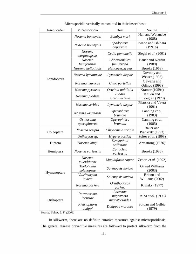
Fujiwara, 1980). Transmission cycles that include vertical transmission have been
observed for Lepidoptera infected with Nosema type microsporidia as well as for other
microsporidian genera in a variety of insect hosts as mentioned in the following Table.
Chapter 3
151
Microsporidia vertically transmitted in their insect hosts
Insect order Microsporidia Host Source
Nosema bombycis Bombyx mori Han and Watanabe
(1988)
Nosema bombycis Spodoptera deparvata
Iwano and Ishihara (1991b)
Nosema carpocapsae
Cydia pomonella Siegel et al. (2001)
Nosema fumiferanae
Choristoneura fumiferana
Bauer and Nordin (1989)
Nosema heliothidis Helicoverpa zea Brooks (1968)
Nosema lymantriae Lymantria dispar Novotny and
Weiser (1993)
Nosema marucae Chilo partellus Ogwang and
Odindo (1993) Nosema pyrausta Ostrinia nubilalis Kramer (1959a)
Nosema plodiae Plodia
interpunctella Kellen and
Lindegren (1973)
Nosema serbica Lymantria dispar Pilarska and Vavra
(1991)
Nosema wistmansi Operophtera
brumata Canning et al.
(1983)
Lepidoptera
Orthosoma operophterae
Operophtera brumata
Canning et al. (1985)
Nosema scripta Chrysomela scripta Bauer and
Prankratz (1993) Coleoptera Unikaryon sp. Hypera postica Solter et al. (1993)
Diptera Nosema kingi Drosophila willistoni
Armstrong (1976)
Hemiptera Nosema varivestis Epilachna varivestis
Brooks (1986)
Nosema mucidifurax
Mucidifurax raptor Zchori et al. (1992)
Thelohania solenopsae
Solenopsis invicta Oi and Williams
(2003) Vairimorpha
invicta Solenopsis invicta
Briano and Williams (2002)
Hymenoptera
Nosema parkeri Ornithodoros
parkeri Krinsky (1977)
Paranosema locustae
Locustae migratoria
migratorioides Raina et al. (1995)
Orthoptera Pleistophora
dixippi Dixippus morosus
Soldan and Gelbic (1979)
Source: Solter, L. F. (2006)
In silkworm, there are no definite curative measures against microsporidiosis.
The general disease preventive measures are followed to protect silkworm from the
Chapter 3
152
microsporidiosis. The preventive measures are more of general type involving
disinfection of silkworm rearing environment, practice of hygiene and application of
prophylactory measures. Maximum precaution is taken to avoid transovarially
transmitted infection. Only the disease free layings are used for silkworm rearing and
seed production, thus eliminating the chances of disease occurrence by transovarial
transmission. Transovum transmission is avoided by surface disinfection of eggs. The
other preventive measures emphasize on elimination of secondary sources of
infection. The major sources of secondary infection are the diseased and dead larvae,
faeces and the gut juices vomited by the diseased larvae. Alternate hosts of different
microsporidia especially the lepidopteran pests of mulberry and nearby agricultural
crops also pose a serious problem by contaminating the mulberry leaf with the spores
carried by them and thus cross infect the silkworm through contaminated mulberry
leaf.
Use of certain chemicals fed to silkworm either through artificial diet or through
mulberry leaf spray method has been reported to be an effective way to control the
microsporidiosis of silkworm (Brooks et al., 1978; Iwano and Ishihara, 1981;
Hyasaka, 1991; Frankenhuyzen et al., 2004). As prevention is better than cure, the
disinfection of silkworm rearing house, silkworm rearing appliances and the silkworm
seed production centers is generally followed to eliminate the microsporidian spores
which otherwise may lead to secondary infection. It is coupled with meticulous
hygiene practices in silkworm rearing and egg production centers. Various
disinfectants viz., Chlorinated lime and hydrochloric acid (Miyajima, 1979b),
Formalin (Kagawa, 1980), Asiphore (Venkata Reddy et al., 1990), Chlorine dioxide
(Nataraju, 1995; Balavenkatasubbaiah et al., 1999), Kao haiter (Balavenkatasubbaiah
et al., 2003) and Serichlor (Balavenkatasubbaiah et al., 2006) have been reported to
be effective against silkworm pathogens including N. bombycis. In addition to
disinfection of silkworm rearing house and appliances, disinfection of mulberry
leaves also has been reported to be an efficient method for the prevention and control
of microsporidiosis of silkworm (Singh et al., 2007a).
It is observed from the results of Chapter 1 and 2 that five different
microsporidia isolated from insect pests of mulberry and some other agricultural crops
differ from N. bombycis and also from each other with respect to their infectivity,
morphology, ultrastructure, serological affinity, germination response, rate of
Chapter 3
153
sporulation at different temperatures, mode of infection, site of infection,
pathogenicity, rate of spread and in terms of their impact on the rearing performance
of different productive breeds of silkworm. In the present chapter, the results of
investigations on the impact of infection by the isolated microsporidia on the
economic characters of silkworm, impact of different pathogen loads on the health
status and rearing performance of silkworm, impact of microsporidian infection on the
morphology of silkworm larvae and the mode of transmission of the isolated
microsporidia are presented and discussed. The study on the effect of disinfection of
microsporidian contaminated mulberry leaf on microsporidian disease incidence and
rearing performance of silkworm also constitutes the subject matter of the present
chapter.
MATERIALS AND METHODS
Impact of infection by isolated microsporidian spores on the economic characters
of silkworm: One popular bivoltine and one multivoltine breed were selected for the
study on the impact of microsporidial infection on the economic characters of
silkworm. The eggs of the selected bivoltine and multivoltine breed viz., CSR2 and
Pure Mysore respectively were received from the germplasm bank of CSR&TI,
Mysore for the study. The larvae of the selected breeds were reared under hygienic
conditions till the beginning of 3rd instar. The microsporidian spores were inoculated
at a concentration of 1×10⁷ spores/ml/100 larvae on day zero of 3rd instar. The
inoculum was prepared from purified spores of the microsporidia and quantified by
standard method using Neubar haemocytometer as described in earlier chapters. The
silkworm larvae were inoculated with different microsporidia by feeding mulberry
leaf smeared with one ml inoculum of 1×10⁷ spores/ml to 100 larvae. The larvae
were allowed to feed on the treated leaves for 24 h to ensure complete consumption of
the treated leaves. After 24 h, the larvae were reared as per standard conditions till
cocooning. Two controls for each of the breed were maintained for comparison
purpose. The first control larvae were treated with mulberry leaves contaminated with
1 ml of 1×10⁷ spores/ml of N. bombycis to 100 larvae and the second control larvae
were treated with mulberry leaves smeared with sterilized distilled water and reared
on uncontaminated mulberry leaves. The controls and treatments had three
replications of 100 larvae each. The larvae were observed for growth, larval duration
and survival. Data with regard to other economic characters such as larval weight,
Chapter 3
154
single cocoon weight, single shell weight and shell ratio was recorded. Data with
respect to average filament length was also recorded and the data was analyzed
statistically.
Impact of lower pathogen loads on the health status and rearing performance of
silkworm: To study the impact of lower pathogen loads on the health status and
rearing performance of silkworm, a popular bivoltine silkworm breed (CSR2) was
selected. The eggs of the said breed were received from the germplasm bank of C.S.R.
& T.I., Mysore, surface disinfected and then incubated at 25±1ºC and 80±5% RH. The
hatched larvae were reared till the beginning of third instar following standard
silkworm rearing practices under hygienic conditions. The third instar silkworm larvae
were inoculated separately with different concentrations of the isolated microsporidian
spores. Also, one set of larvae inoculated with different concentrations of Nosema
bombycis spores was maintained separately for comparison purpose. Four different
concentrations of the pathogen inoculum viz., 1×10², 1×10³, 1×10⁴ and 1×10⁵
spores/ml were tested to determine their impact on the health status and rearing
performance of silkworm. These different concentrations were prepared from purified
spores of each isolated microsporidia by serial dilution of the quantified stock
inoculum. The quantification was done following the standard method using Neubar
haemocytometer (Cantwell, 1970). Each inoculum concentration formed a treatment.
For each treatment five replications of 100 larvae were maintained. Different sets of
larvae were inoculated separately with different concentrations of each of the
microsporidian spores isolated from insect pests of mulberry and other agricultural
crops. One ml of specific concentration of specific microsporidian spores was smeared
on the ventral surface of mulberry leaf and fed to the silkworm larvae just out of 2nd
moult. Also, one normal control batch without any inoculation was maintained for
comparison purpose. Observations with regard to larval mortality, pupal mortality,
percentage moths infected and total infection percent were recorded. Data with regard
to larval weight, larval duration, pupation rate, cocoon weight, shell weight and shell
percentage also was recorded. The data was statistically analyzed to arrive at
conclusion.
Impact of infection by the isolated microsporidia on the morphology of the
silkworm, Bombyx mori L.: To study the impact of infection by the isolated
microsporidia on the larval morphology, CSR2 breed of the silkworm, Bombyx mori
Chapter 3
155
L. was selected. The eggs of the said breed were received from the germplasm bank
of CSR&TI, Mysore. The larvae were reared under hygienic conditions till the
beginning of third instar. Immediately after second moult, the spores of the isolated
microsporidia were fed separately to the silkworm larvae (100 larvae/treatment) at a
concentration of 1×10⁷ spores/ml by smearing it on the mulberry leaf disc of 100 cm2
surface area. One set of larvae was inoculated with Nosema bombycis spores (1×10⁷
spores/ml) which served as control for comparison purpose. The second feeding was
provided with normal leaves to each inoculated batch after 24 hours of microsporidian
inoculation and the rearing was continued as per standard methods till the onset of the
spinning. From the day of inoculation till the onset of spinning, daily 10 larvae were
taken randomly and observations with regard to changes in average larval length,
width and weight were recorded. The same observations were recorded daily in
healthy control batches also.
Mode of transmission: To determine the mode of transmission of the isolated
microsporidia, a popular bivoltine silkworm breed (CSR2) was selected and on day
zero of fourth instar, larvae of the said breed were per orally inoculated separately
with the spores of the isolated microsporidia at a dosage of 1×10⁷ spores/ml. To
compare the results, one set of larvae was inoculated with spores of Nosema
bombycis. The inoculum containing 1×10⁷ spores/ml of the isolated microsporidia or
N. bombycis was prepared from the stock inoculums by proper quantification using
Neubar haemocytometer (Cantwell, 1970). One ml of inoculum (1×10⁷ spores/ml) of
each microsporidia was smeared separately on 100 sq. cms surface area of mulberry
leaf disc and fed to 100 larvae immediately after 3rd moult. The larvae were allowed
to feed on the contaminated leaves for 24 hours. The second normal feeding was
given after 24 hours and the rearing on uncontaminated mulberry leaves was
continued till cocooning. After cocoon formation, the cocoons from each treated batch
were cut open for sex separation of the pupae. The male and female pupae were kept
in separate trays for moth emergence. Yet another set of larvae was reared without
inoculation till spinning and moth emergence.
The moths obtained after inoculation of the larvae of CSR2 breed with an
inoculum dosage of 1×10⁷ spores/ml of the isolated microsporidia or Nosema
bombycis were provisionally regarded as infected. The moths obtained from the
batches without inoculation were provisionally regarded as healthy. To find out the

Chapter 3
156
mode of transmission of the microsporidia, the male and female moths were allowed
to mate in three different combinations viz., Infected female×Infected male (IF×IM);
Infected female×Healthy male (IF×HM) and Healthy female×Infected male (HF×IM).
After 3 hours of pairing, the male and female moths were depaired and the gravid
female moths were allowed to lay eggs for 24 hours on egg sheets. After egg laying,
gonads of each male and female moth were dissected out and tested individually for
the presence of microsporidian spores and also, the whole body (excluding the
gonads) of these moths was subjected to microscopic examination (Phase contrast
microscope, Nikon-Type 104) for the presence of microsporidian spores. The egg
layings obtained from different treatments were surface sterilized by immersing in 2%
formalin solution for 5 minutes at room temperature. The layings were then treated
with hydrochloric acid of specific gravity 1.076 at 46.1oC for 5 minutes in a hot water
bath to terminate the egg diapause and washed in running tap water to remove the
traces of HCl. The layings were incubated for normal embryonic development at
25±1oC temperature and 80±5% RH. During the pinhead stage, the layings were
black-boxed to ensure uniform development of embryo and hatching. After two days
of black boxing, the layings were exposed to light to stimulate hatching. Fecundity
and hatching percent of each laying were recorded. The dead eggs in each laying were
also examined for infection by examination of whole wet mount of egg content
following its removal from chorion to confirm the infection passing to progeny
through infection in gonads. The progeny larvae were reared as per standard methods
(Datta, 1992). After I moult, 100 larvae/batch were collected randomly and
homogenized individually and the smear was observed under phase contrast
microscope for the presence of microsporidian spores and thus, the transmission rate
was calculated by the standard formula (Han and Watanabe, 1988) which is as
follows;
(A × B) + (C × D) Transmission rate =
A + C
where A- Number of dead eggs.
B- % of dead eggs infected.
C- Number of larvae hatched.
D- % of larvae infected.
Chapter 3
157
Also, the rearing of the remaining progeny larvae was continued to record the
mortality, if any, during different larval instars and the pupal stage in the
transovarially infected batches. The observations were recorded and analyzed.
Transmission through surface contaminated layings: To study the transmission of
the microsporidia through surface contaminated layings, disease free layings of CSR2
breed at the blue egg stage were smeared with microsporidian spores (dosage of
1×10⁷ spores/ml) of each microsporidia separately on the egg surface with the help of
a camelin brush. The egg layings were air dried, wrapped in tissue paper and
subjected to black boxing for uniform hatching. After two days, the layings were
exposed to indirect light for hatching. The newly hatched larvae were reared as per
standard methods. Mortality due to microsporidian infection in the progeny, if any,
was recorded in different larval instars and pupal stage.
Effect of disinfectants against the spores of the isolated microsporidia and N.
bombycis through disinfection of mulberry leaves: To determine the effect of
mulberry leaf disinfection on the spores of the isolated microsporidia and N.
bombycis, two popular disinfectants viz., Decol and Sanitech were selected and the
selected disinfectants were used at a concentration of 1.0% and 400 ppm respectively.
The said concentrations of the selected disinfectants were fixed on the basis of their
palatability to silkworm and for the preparation of Sanitech solution (400 ppm),
addition of lime was avoided as it is a feed-repellent. To conduct the test, a popular
bivoltine silkworm breed CSR2 was selected. The larvae of the said breed were reared
as per standard methods (Datta, 1992) up to the second moult. Mulberry leaf discs
were smeared with the spores of the isolated microsporidia and N. bombycis
separately at an infective concentration of 1×10⁷ spores/ml. The mulberry leaf discs
were then air dried and dipped separately in Decol (1.0%) and Sanitech (400 ppm)
solutions for 30 minutes and then again air dried and fed to silkworm larvae (100
larvae/replication/treatment) of CSR2 breed on day zero of third instar. The larvae
were allowed to feed on the treated leaf discs for 24 hours to ensure the complete
consumption of the treated leaf. After 24 hours, the fresh mulberry leaf without any
contamination was provided to each treated batch. In one set of experiment, the dead
larvae if any, from all the treatments were collected and subjected to microscopic
examination to reveal the cause of mortality. Also, on the 13th day of post inoculation,
all the survived larvae were homogenized individually in distilled water and the
Chapter 3
158
smears were observed under phase contrast microscope (Nikon, Type-104) to record
the total percentage of microsporidian infection. In yet another set of experiment, the
treated larvae were reared as per standard methods up to cocooning to determine the
effect of mulberry leaf disinfection on the rearing performance of microsporidian
inoculated silkworm. The data with regard to larval weight, larval duration, pupation
rate, cocoon weight, shell weight and shell percentage was recorded and analysed. For
each larval batch fed with the mulberry leaf discs smeared separately with the
different microsporidia followed by dipping in Decol and Sanitech solutions, one
control batch fed with the mulberry leaf disc smeared with the spores of the concerned
microsporidia (1×10⁷ spores/ml) without any treatment was also maintained for
comparison purpose. Also one normal control batch fed with the mulberry leaf disc
dipped in distilled water was maintained for comparison purpose.
RESULTS
Impact of infection by isolated microsporidian spores on the economic characters
of silkworm: The results of the studies on the impact of microsporidian infection on
economic characters of two silkworm breeds viz., CSR2 and Pure Mysore are
presented in Table 3.1 and Figures 3.1 and 3.2. It is observed that the infection by all
the five isolated microsporidia had a significant impact on the economic parameters of
both the silkworm breeds tested. A comparison with respective healthy control batch
indicates that the infection due to the five microsporidia viz., NIK-1Pr, NIK-1Cc,
NIK-1Cpy, NIK-1So and NIK-1Dp lowered the pupation rate (ERR%), larval weight,
single cocoon weight, single shell weight, percent silk content and filament length.
The infection also prolonged the larval duration of both the silkworm breeds tested.
Larval weight: The results on the impact of the isolated microsporidia on the weight
of 10 larvae on sixth day of final instar of the two silkworm breeds tested viz., CSR2
and Pure Mysore as presented in Table 3.1 show that the microsporidian infection
significantly reduced the larval weight of both the breeds tested. The highest
reduction in larval weight of CSR2 batches was recorded in the larvae inoculated with
the standard microsporidian strain N. bombycis (26.6 g) followed by NIK-1So (30.0
g), NIK-1Cpy (30.3 g), NIK-1Pr (31.1 g), NIK-1Dp (31.8 g) and NIK-1Cc (33.0 g)
compared to the normal control batches where a larval weight of 35.7 g was recorded.
A similar trend with respect to the impact of microsporidian infection on larval weight
Chapter 3
159
was recorded in Pure Mysore batches also and the highest reduction in larval weight
was recorded in silkworm larvae inoculated with N. bombycis (18.8 g) followed by
NIK-1So (21.1 g), NIK-1Cpy (21.4 g), NIK-1Pr (22.5 g), NIK-1Dp (22.9 g) and NIK-
1Cc (23.9 g) compared to the normal control batches where a larval weight of 25.8 g
was recorded.
Larval duration: In CSR2 batches inoculated separately with the isolated
microsporidia and Nosema bombycis, the larval duration got increased from 23 to 25
days when compared with the normal control batch. Similarly, the larval duration was
prolonged in Pure Mysore batches inoculated separately with the isolated
microsporidia and was recorded as 29 days compared to the 28 days recorded in
normal control batches.
Pupation rate (ERR%): The data as presented in Table 3.1 shows that the pupation
rate (ERR%) in CSR2 breed inoculated separately with five different microsporidia
was lowered significantly and was recorded to be lowest in NIK-1So inoculated
batches (73.0%) followed by NIK-1Cpy (75.0%), NIK-1Pr (79.0%), NIK-1Dp
(81.0%) and NIK-1Cc (86.3%) inoculated batches as against 95.7% pupation rate
recorded in healthy host population. Also, in Pure Mysore breed inoculated separately
with five different microsporidia, the lowest pupation rate was recorded in NIK-1So
inoculated batches (79.0%) followed by NIK-1Cpy (80.3%), NIK-1Pr ( 82.0%), NIK-
1Dp (86.0% ) and NIK-1Cc (88.3%) which in turn was significantly lower than the
normal control batches (95.3%).
When the pupation rate of the batches inoculated with the standard
microsporidian strain, Nosema bombycis is compared with that of the batches
inoculated with the isolated microsporidia, it is observed that in N. bombycis
inoculated CSR2 batches, the pupation rate was significantly lower (64.7%) compared
to the five microsporidia tested. Similarly, in Pure Mysore batches inoculated with
Nosema bombycis also, pupation rate of only 73.3% was recorded clearly suggesting
the five microsporidia isolated from insect pests of mulberry and other agricultural
crops are less pathogenic to silkworm than the standard strain, Nosema bombycis.
Single cocoon weight: In both the silkworm breeds viz., CSR2 and Pure Mysore
inoculated separately with different microsporidia, cocoon weight was reduced
significantly compared to the healthy control batches. The highest reduction in cocoon

Chapter 3
160
weight of CSR2 batches was recorded in the batches inoculated with N. bombycis
(1.428 g) followed by NIK-1So (1.470 g), NIK-1Cpy (1.478 g), NIK-1Pr (1.480 g),
NIK-1Dp (1.490 g) and NIK-1Cc (1.495 g) compared to the healthy control batches
(1.600 g). Similarly, in Pure Mysore batches also, the highest reduction in cocoon
weight was recorded in the batches inoculated with N. bombycis (0.964 g) followed
by NIK-1So (0.996 g), NIK-1Cpy (1.005 g), NIK-1Pr (1.044 g), NIK- 1Dp (1.082 g)
and NIK-1Cc (1.113 g) compared to the healthy control batches (1.127 g) (Table 3.1).
Single shell weight: In the CSR2 batches inoculated separately with the isolated
microsporidia and N. bombycis, there was a significant reduction in shell weight. The
highest reduction in shell weight was recorded in the batches inoculated with N.
bombycis (0.276 g) followed by NIK-1So (0.292 g), NIK-1Cpy (0.296 g), NIK-1Pr
(0.320 g), NIK-1Dp (0.325 g) and NIK-1Cc (0.328 g) compared to the healthy control
batches wherein a shell weight of 0.366 g was recorded (Table 3.1). A similar trend
with respect to the reduction in shell weight due to microsporidian infection was
recorded in Pure Mysore batches also and the highest reduction in shell weight was
recorded in the batches inoculated with N. bombycis (0.107 g) followed by NIK-1So
(0.117 g), NIK-1Cpy (0.122 g), NIK-1Pr (0.130 g), NIK-1Dp (0.137 g) and NIK-1Cc
(0.141 g) inoculated batches. In the control Pure Mysore batches, shell weight of
0.154 g was recorded.
Shell percentage: The infection due to the isolated microsporidia significantly
reduced the shell percentage of both the breeds tested. The highest reduction in shell
percentage of CSR2 batches inoculated with the different microsporidia was recorded
in the batches inoculated with N. bombycis (19.3%) followed by NIK-1So (19.8%),
NIK-1Cpy (20.0%), NIK-1Pr (21.6%), NIK-1Dp (21.8%) and NIK-1Cc (21.9%)
compared to the healthy control batches (22.8%). A similar trend was observed in
Pure Mysore batches also and the highest reduction in shell percentage was recorded
in N. bombycis inoculated batches (11.1%) followed by NIK-1So (11.7%), NIK-1Cpy
(12.1%), NIK-1Pr (12.4%), NIK-1Dp (12.6%) and NIK-1Cc (12.7%) compared to the
healthy control batches (13.6%) (Table 3.1). The percentage reduction in the shell
percentage of the CSR2 and Pure Mysore batches inoculated with the isolated
microsporidia and N. bombycis compared to that of the healthy control batches is
graphically represented in Figures 3.3 and 3.4 respectively.

Chapter 3
161
Filament length: The infection with the isolated microsporidia had a significant
impact on the Filament length of both the breeds tested (Table 3.1). The highest
decrease in filament length of CSR2 breed was recorded in N. bombycis inoculated
batches (617.0 m) followed by NIK-1So (635.0 m), NIK-1Cpy (644.0 m), NIK-1Pr
(721.0 m), NIK-1Dp (782.0 m) and NIK-1Cc (807.0 m) compared to the filament
length of 980.0 m in healthy control batches. Similarly, the highest decrease in
filament length of Pure Mysore breed was recorded in the N. bombycis inoculated
batches (284.0 m) followed by NIK-1So (291.3 m), NIK-1Cpy (325.0 m), NIK-1Pr
(369.0 m), NIK-1Dp (371.0 m) and NIK-1Cc (400.7 m) compared to the filament
length of 443.0 m in the healthy Pure Mysore batches. The percentage decrease in the
filament length of the CSR2 and Pure Mysore batches inoculated with the isolated
microsporidia and N. bombycis compared to that of the healthy control batches is
graphically represented in Figures 3.5 and 3.6 respectively.
Impact of lower pathogen loads on the health status and rearing performance of
silkworm: The results on the impact of lower pathogen loads on the health status and
rearing performance of the CSR2 breed of the silkworm, Bombyx mori L. are
presented in Table 3.2. It is clear from the data as presented in the said table that
inoculation of the isolated microsporidia to silkworm at the two lowest concentrations
viz., 1×10² and 1×10³ spores/ml did not cause any larval and pupal mortality whereas
at the same two concentrations, larval mortality of 1.0 and 3.0% and pupal mortality
of 3.7 and 7.3% respectively was recorded in the batches inoculated with the standard
strain N. bombycis. Also, in the batches inoculated with the spores of the
microsporidia viz., NIK-1Pr, NIK-1Cc and NIK-1Dp at a concentration of 1×10²
spores/ml, all the emerged moths were found to be free from microsporidian infection,
thereby the total infection percentage was nil. However, in the batches inoculated with
the microsporidia viz., NIK-1Cpy, NIK-1So and N. bombycis at the same
concentration, 8.0, 13.0 and 63.3% of the emerged moths were infected, thereby
resulting in a total infection of 8.0, 13.0 and 68.0% respectively. At a concentration of
1×10³ spores/ml, the highest total infection percent was recorded in the batches
inoculated with the standard microsporidian strain N. bombycis (86.0%) followed by
NIK-1So (35.0%), NIK-1Cpy (30.0%), NIK-1Pr (28.0%), NIK-1Dp (17.0%) and
NIK-1Cc (14.0%). Inoculation of the isolated microsporidia and N. bombycis to
silkworm at a concentration of 1×10⁴ spores/ml caused a larval mortality of 1.0, 6.7,

Chapter 3
162
9.7 and 10.7% in NIK-1Pr, NIK-1Cpy, NIK-1So and N. bombycis inoculated batches
respectively whereas at the same concentration, the larval mortality in NIK-1Cc and
NIK-1Dp inoculated batches was nil. A similar trend with regard to pupal mortality
also was recorded in the batches inoculated with NIK-1Cc and NIK-1Dp at the same
concentration whereas in NIK-1Pr, NIK-1Cpy, NIK-1So and N. bombycis inoculated
batches, pupal mortality of 1.7, 3.7, 4.7 and 10.0% respectively was recorded at a
concentration of 1×10⁴ spores/ml. The highest total infection % at the concentration of
1×10⁴ spores/ml was recorded in N. bombycis inoculated batches (89.3%) followed by
NIK-1So (50.7%), NIK-1Cpy (45.7%), NIK-1Pr (35.0%), NIK-1Dp (21.7%) and
NIK-1Cc (17.3%) inoculated batches. At the highest spore concentration of 1×10⁵
spores/ml, the larval and pupal mortality in case of NIK-1Cc inoculated batches was
nil whereas the same in case of the batches inoculated with NIK-1Pr, NIK-1Cpy,
NIK-1So, NIK-1Dp and N. bombycis was recorded as 6.7, 16.3, 20.3, 1.0 and 21.7%
(larval mortality) and 3.7, 4.3, 6.3, 1.0 and 14.7% (pupal mortality) respectively. The
highest total infection percent at the concentration of 1×10⁵ spores/ml was recorded in
the batches inoculated with N. bombycis (92.0%), followed by NIK-1So (87.0%),
NIK-1Cpy (80.3%), NIK-1Pr (44.0%), NIK-1Dp (27.0%) which in turn was followed
by NIK-1Cc inoculated batches (21.3%).
The impact of the different pathogen loads on the rearing performance of
silkworm as presented in Table 3.2 is mentioned in detail as follows:
Larval weight: At a concentration of 1×10² spores/ml, there was not any significant
impact on the larval weight in case of the batches inoculated with the isolated
microsporidia and it ranged from 38.70 to 39.17 g whereas at the same concentration,
there was a significant reduction in larval weight of N. bombycis inoculated batches
(37.77 g) as against a larval weight of 39.18 g in case of the normal control batches.
Similarly, at a concentration of 1×10³ spores/ml, the larval weight in case of the
batches inoculated with the isolated microsporidia ranged from 38.10 to 38.87 g
whereas, the same in case of N. bombycis inoculated batches was recorded as 37.17 g.
The larval weight ranged from 38.02 to 38.74 g in the batches inoculated with the
isolated microsporidia at a concentration of 1×10⁴ spores/ml. The same in N.
bombycis inoculated batches was recorded as 36.95 g only. At the highest spore
concentration (1×10⁵ spores/ml), there was a significant reduction in the larval weight
of the inoculated batches and the lowest larval weight was recorded in the batches

Chapter 3
163
inoculated with N. bombycis (36.64 g) followed by NIK-1So (37.74 g), NIK-1Cpy
(37.90 g), NIK-1Pr (38.23 g), NIK-1Dp (38.61 g) which in turn was followed by the
batches inoculated with NIK-1Cc (38.68 g). In the normal control batches, the same
was recorded as 39.18 g.
Larval duration: Inoculation of the silkworm larvae with the spores of the isolated
microsporidia at the concentrations ranging from 1×10² to 1×10⁵ spores/ml did not
cause any effect on the larval duration and the same was recorded as 24 days which
was similar to that of the normal control batches. Similarly, in the batches inoculated
with the spores of N. bombycis at the concentrations of 1×10² and 1×10³ spores/ml,
there was no effect on the larval duration and the same was recorded as 24 days.
However, in case of the batches inoculated with N. bombycis spores at the
concentrations of 1×10⁴ and 1×10⁵ spores/ml, the larval duration was prolonged by
one day and was recorded as 25 days.
Pupation rate: Inoculation of silkworm larvae with the spores of the isolated
microsporidia at the two lowest concentrations viz., 1×10² and 1×10³ spores/ml did
not lead to any significant impact on the pupation rate and the same ranged from 92.0
to 95.0% whereas, in case of the batches inoculated with the spores of N. bombycis,
the pupation rate of 91.0 and 85.3% respectively was recorded at the same two
concentrations compared to the normal control batches wherein a pupation rate of
95.7% was recorded. Also, at the inoculum concentration of 1×10⁴ spores/ml, there
was no significant impact on the pupation rate of the batches inoculated with NIK-
1Pr, NIK-1Cc and NIK-1Dp microsporidia and the pupation rate of 93.0, 94.0 and
94.0% respectively was recorded. On the other hand, at the same concentration, there
was a significant reduction in the pupation rate of the batches inoculated with the
microsporidia viz., NIK-1Cpy, NIK-1So and N. bombycis and the pupation rate of
85.3, 81.3 and 75.0% respectively was recorded. Inoculation of the microsporidia viz.,
NIK-1Cc and NIK-1Dp to silkworm at the highest concentration (1×10⁵ spores/ml)
also did not lead to any significant impact on the pupation rate and the same was
recorded as 94.0 and 93.7% respectively whereas, at the same concentration, the
pupation rate was significantly reduced in the batches inoculated with NIK-1Pr, NIK-
1Cpy, NIK-1So and N. bombycis and was recorded as 85.3, 75.0, 69.0 and 59.3%
respectively.

Chapter 3
164
Single cocoon weight: The isolated microsporidia did not cause any significant
impact on the cocoon weight of the batches inoculated at the two lowest
concentrations viz., 1×10² and 1×10³ spores/ml and the same ranged from 1.580 to
1.604 g whereas, in the batches inoculated with the spores of the standard strain N.
bombycis at the same concentrations, the cocoon weight was slightly less and ranged
from 1.542 to 1.558 g. In the batches inoculated with the spores of the microsporidia
viz., NIK-1Pr, NIK-1Cc, NIK-1Cpy, NIK-1So and NIK-1Dp at the concentration of
1×10⁴ spores/ml, cocoon weight of 1.560, 1.572, 1.551, 1.541and 1.570 g respectively
was recorded whereas at the same concentration, the cocoon weight in the N.
bombycis inoculated batches was recorded as 1.507 g. Inoculation of the spores of the
isolated microsporidia and N. bombycis to silkworm at a concentration of 1×10⁵
spores/ml significantly reduced the cocoon weight and the lowest cocoon weight was
recorded in the batches inoculated with N. bombycis (1.487 g) followed by NIK-1So
(1.519 g), NIK-1Cpy (1.529 g), NIK-1Pr (1.537 g), NIK-1Dp (1.560 g) which in turn
was followed by the batches inoculated with NIK-1Cc (1.565 g) compared to the
normal control batches wherein the cocoon weight of 1.607 g was recorded.
Single shell weight: At the two lowest concentrations viz., 1×10² and 1×10³
spores/ml, there was no significant impact on the shell weight of the batches
inoculated with the isolated microsporidia and the same ranged from 0.349 to 0.374 g
whereas in the batches inoculated with N. bombycis, the shell weight was slightly less
and ranged from 0.320 to 0.335 g. In the batches inoculated with the spores of the
microsporidia viz., NIK-1Pr, NIK-1Cc, NIK-1Cpy, NIK-1So and NIK-1Dp at the
concentration of 1×10⁴ spores/ml, shell weight of 0.347, 0.356, 0.342, 0.335 and 0.335
g respectively was recorded whereas at the same concentration, the shell weight in the
N. bombycis inoculated batches was recorded as 0.302 g only. Inoculation of the
spores of the isolated microsporidia and N. bombycis to silkworm at a concentration
of 1×10⁵ spores/ml significantly reduced the shell weight and the lowest shell weight
was recorded in the batches inoculated with N. bombycis (0.295 g) followed by NIK-
1So (0.326 g), NIK-1Cpy (0.332 g), NIK-1Pr (0.337 g), NIK-1Dp (0.348 g) which in
turn was followed by the batches inoculated with NIK-1Cc (0.352 g) compared to the
normal control batches wherein the shell weight was recorded as 0.375 g.
Shell percentage: In the batches inoculated with the spores of the isolated
microsporidia at a concentration of 1×10² spores/ml, there was no impact of
Chapter 3
165
microsporidian infection on the shell percentage and the same was recorded as 23.29,
23.31, 22.91, 22.78 and 23.30% respectively in the batches inoculated with NIK-1Pr,
NIK-1Cc, NIK-1Cpy, NIK-1So and NIK-1Dp whereas in the batches inoculated with
N. bombycis, there was a significant reduction in shell percentage even at the lowest
concentration and the same was recorded as 21.49% as against the shell percentage of
23.32% in the normal control batches. In the batches inoculated with the spores of the
isolated microsporidia at a concentration of 1×10³ spores/ml, the shell percentage
ranged from 22.09 to 22.86% whereas, at the same concentration, the shell percentage
in N. bombycis inoculated batches was recorded as 20.78% only. Similarly, in the
batches inoculated with the spores of the isolated microsporidia at a concentration of
1×10⁴ spores/ml, the shell percentage ranged from 21.74 to 22.65% whereas, at the
same concentration, the shell percentage in N. bombycis inoculated batches was
recorded as 20.03 % only. At the highest concentration of 1×10⁵ spores/ml, there was
a significant reduction in the shell percentage and the lowest shell percentage was
recorded in the batches inoculated with N. bombycis (19.82%) followed by NIK-1So
(21.47%), NIK-1Cpy (21.74%), NIK-1Pr (21.93%), NIK-1Dp (22.32%) and NIK-1Cc
(22.52%).
Impact of infection by the isolated microsporidia on the morphology of the
silkworm, Bombyx mori L.:
Larval length: The results with regard to the impact of the infection caused by
different isolated microsporidia on the daily increase in larval length of silkworm
(CSR2 breed) are presented in Table 3.3. The data indicates that there was no
significant impact of infection caused by the isolated microsporidia and Nosema
bombycis on the progressive increase in the larval length upto 6th day of post
inoculation. However, from 7th day of PI onwards, the progressive increase in larval
length was significantly reduced in the batches inoculated with NIK-1Cpy (34.50
mm), NIK-1So (34.33 mm) and Nosema bombycis (33.33 mm) compared to the
healthy control batches wherein the larval length was recorded as 36.00 mm on 7th
day PI. In the batches inoculated with the microsporidia NIK-1Pr, NIK-1Cc and NIK-
1Dp, there was not any significant reduction in the increase of larval length on 7th day
post inoculation which was recorded as 35.33, 35.67 and 35.50 mm respectively. On
8th day PI, the infection with different microsporidia caused significant reduction in
the increase in larval length in all the inoculated batches compared to healthy control

Chapter 3
166
batches and the larval length was recorded as 35.67. 36.00, 35.17, 35.50, 36.00 and
34.33 mm in the batches inoculated with NIK-1Pr, NIK-1Cc, NIK-1Cpy, NIK-1So,
NIK-1Dp and Nosema bombycis respectively when compared to the 37.00 mm larval
length in healthy control batch. On 9th and 10th day PI, the larval length ranged from
36.00 to 37.50 mm in the inoculated batches whereas the same ranged from 38.50 to
39.50 mm in healthy control batches. The same trend with regard to impact of
infection by the isolated microsporidia and N. bombycis on the daily increase in larval
length was recorded on succeeding days which ranged from 36.00 to 37.67 mm, 45.50
to 50.50 mm and 49.00 to 55.00 mm on 11th, 12th and 13th day of post inoculation
respectively compared to the healthy control batches wherein the same was recorded
as 40.00, 53.33 and 57.67 mm respectively. With progressive infection, the impact of
microsporidiosis on larval length was more pronounced and on 14th day PI, the lowest
larval length was recorded in the batches inoculated with NIK-1So (56.67 mm)
followed by NIK-1Cpy (57.33 mm), NIK-1Pr (58.33 mm) and NIK-1Dp (60.33 mm).
The highest larval length was recorded in the batches inoculated with NIK-1Cc (60.50
mm). In N. bombycis inoculated batches, the same was recorded as 55.50 mm only
compared to the healthy control batches (62.00 mm). One day prior to the spinning
i.e. on 15th day PI, the larval length was recorded as 62.67, 64.50, 60.50, 58.50 and
63.50 mm in the batches inoculated with NIK-1Pr, NIK-1Cc, NIK-1Cpy, NIK-1So
and NIK-1Dp respectively compared to healthy control batches wherein a larval
length of 66.17 mm was recorded. In N. bombycis inoculated batches, the same was
recorded as 56.00 mm. The percentage difference in the larval length of the batches
inoculated with different microsporidia compared to that of the healthy control
batches one day prior to the onset of spinning is graphically represented in Figure 3.7.
Larval width: The data with respect to the impact of infection by the isolated
microsporidia on the daily increase in larval width is presented in Table 3.4. The data
indicates that there was no significant impact of infection by the isolated
microsporidia on the progressive increase in larval width up to 12th day of post
inoculation. However, in the batches inoculated with N. bombycis, the increase in
larval width got significantly reduced from 7th day of PI onwards and was recorded as
5.50, 6.00, 6.00, 6.17, 6.37 and 7.50 mm against a larval width of 6.67, 7.00, 7.33,
7.33, 7.50 and 8.50 mm in healthy control batches on 7th, 8th, 9th, 10th, 11th and 12th
day PI respectively. On 13th day PI, a significant impact of infection by the isolated

Chapter 3
167
microsporidia on the larval width was observed (9.00, 9.00, 8.00, 8.00 and 9.00 mm in
the batches inoculated with NIK-1Pr, NIK-1Cc, NIK-1Cpy, NIK-1So and NIK-1Dp
respectively) as against a larval width of 10.00 mm in healthy control batches. In case
of N. bombycis inoculated batches, only 7.67 mm larval width was recorded. On 14th
day PI, larvae inoculated with the isolated microsporidia attained a larval width of
10.00 mm in all the batches whereas in N. bombycis inoculated batches, only 8.33 mm
larval width was attained as against a larval width of 11.00 mm in healthy control
batches. Before the onset of spinning (15th day PI), the lowest larval width was
recorded in NIK-1Cpy and NIK-1So batches (10.00 mm) followed by NIK-1Pr (10.33
mm), NIK-1Dp (10.50 mm) and NIK-1Cc (11.00 mm). In the healthy control batches,
the same was recorded as 12.00 mm. In the batches inoculated with the standard strain
N. bombycis, larval width of 8.50 mm was recorded on 15th day of PI. The percentage
difference in the larval width of the microsporidia inoculated batches compared to that
of the healthy control batches one day prior to the onset of spinning is graphically
represented in Figure 3.8.
Larval weight: The results with regard to the impact of infection by the isolated
microsporidia on daily increase in larval weight are presented in Table 3.5. From the
said table, it is clear that the infection by the isolated microsporidia did not result in
any significant impact on the larval weight from the day of inoculation up to 11th day
of PI and the larval weight ranged from 0.44 to 9.46 g/10 larvae from the day of
inoculation up to 11th day PI. In the healthy control batches, the same ranged from
0.45 to 9.56 g/10 larvae. On the other hand, in case of N. bombycis inoculated
batches, though there was no significant impact of infection on larval weight upto 6th
day of PI, however, from 7th day of PI onwards, the progressive increase in larval
weight was slightly reduced and the larval weight was recorded as 7.50, 8.62, 9.00,
9.06 and 9.10 g on 7th, 8th, 9th, 10th and 11th day PI respectively. On 12th day of PI,
larval weight of 21.14, 22.04, 20.16, 20.08 and 22.00 g was recorded in the batches
inoculated with NIK-1Pr, NIK-1Cc, NIK-1Cpy, NIK-1So and NIK-1Dp respectively
whereas the same in case of N. bombycis inoculated batches was recorded as 18.47 g
as against the larval weight (23.94 g) of healthy control batches showing significant
reduction in the progressive increase in larval weight in the inoculated batches. The
trend continued on 13th and 14th day of PI also where the larval weight ranged from
29.57 to 31.19 g and 33.01 to 34.05 g respectively in the batches inoculated with the

Chapter 3
168
isolated microsporidia. This reduction in larval weight was comparatively more
pronounced in N. bombycis inoculated batches, where larval weight of 25.17 and
27.02 g on 13th and 14th day of PI respectively was recorded. In healthy control
batches, 32.53 and 35.32 g larval weight respectively was recorded which was
significantly higher than observed in inoculated batches. One day prior to the onset of
spinning (15th day PI), the adverse impact of microsporidian infection on larval
weight was further-more pronounced. The lowest larval weight was recorded in the
batches inoculated with NIK-1So (34.32 g) followed by NIK-1Cpy (34.54 g), NIK-
1Pr (34.94 g), NIK-1Dp (35.31 g) and NIK-1Cc (36.45 g). In case of the batches
inoculated with N. bombycis, 28.00 g larval weight was recorded as against a larval
weight of 38.52 g in the healthy control batches. The percentage reduction in the
larval weight of the inoculated batches compared to that of the healthy control batches
one day prior to the onset of spinning is graphically represented in Figure 3.9.
Mode of Transmission: Microscopic examination of the gonads of the moths from
the batches inoculated with the isolated microsporidia revealed infection in NIK-1Pr,
NIK-1Cpy, NIK-1So and NIK-1Dp inoculated batches. However, the gonads of the
moths from the batches inoculated with the microsporidian NIK-1Cc were devoid of
microsporidian infection. Data on transmission of the five isolated microsporidia in
silkworm is presented in Table 3.6. The table indicates that dead eggs laid by NIK-
1Cc infected female mated with infected male (IF×IM) and also infected female
mated with healthy male (IF×HM) were not found infected with the said
microsporidian whereas the dead eggs laid by NIK-1Cpy and NIK-1So infected
females mated with infected males (IF×IM) and also infected females mated with
healthy males (IF×HM) show 100% infection. Infection percentage in the dead eggs
laid by NIK-1Pr and NIK-1Dp infected females mated with infected males (IF×IM)
and infected females mated with healthy males (IF×HM) ranged from 80.3 to 81.0%.
In the standard strain, N. bombycis, the infection in dead eggs was 100%. The dead
eggs laid by the healthy females mated with infected males (HF×IM) were devoid of
infection in all the microsporidian inoculated batches tested. The fecundity and
hatching percentage in the combinations viz., IF×IM, IF×HM and HF×IM (in the
batches inoculated with the isolated microsporidia and N. bombycis) were
significantly less and ranged between 367 to 468 and 66.1 to 92.5% respectively
compared to the healthy control batches wherein the same was recorded as 512 and
Chapter 3
169
97.0% respectively. The microsporidian NIK-1Pr showed 94.6 and 90.6%
transmission and NIK-1Dp showed 82.6 and 80.0% transmission to progeny larvae
hatched from the layings obtained after mating infected females with infected males
(IF×IM) and infected females with healthy males (IF×HM) respectively. NIK-1Cpy
and NIK-1So showed 100 % transmission to progeny in the combinations viz., IF× IM
and IF×HM which was similar to Nosema bombycis (100%). Significantly, the
microsporidian NIK-1Cc did not show any transmission to progeny larvae. No
infection was observed in the progeny larvae hatched from eggs laid by healthy
females mated with infected males (HF×IM) in all the batches indicating that there is
no venereal transmission of the isolated microsporidia through male.
Mortality of transovarially infected progeny populations:
IF×IM combinations: The results as presented in Table 3.7 show that there was no
mortality in the progeny batches of NIK-1Cc infected female mated with infected
male upto the end of observation period confirming the non-transovarial transmission
of the said microsporidian whereas in the progeny batches obtained from the parents
infected with NIK-1Cpy, NIK-1So and N. bombycis, mortality of 9.3, 14.0 and 20.0%
respectively was recorded during I instar, however, the same in case of NIK-1Pr and
NIK-1Dp was nil. During II instar, mortality of 9.3, 17.3, 23.3, 9.0 and 24.0% was
recorded due to NIK-1Pr, NIK-1Cpy, NIK-1So, NIK-1Dp and N. bombycis
respectively. During III instar, the mortality in the transovarially infected progeny
batches ranged from 34.0 to 59.3%. During IV instar, mortality of 31.3, 15.3, 3.3 and
31.0% was recorded in NIK-1Pr, NIK-1Cpy, NIK-1So and NIK-1Dp batches
respectively. In case of N. bombycis infected progeny, all the larvae died before
reaching IV instar whereas in case of NIK-1Cpy and NIK-1So infected progenies, all
the larvae died before reaching V instar. In case of NIK-1Pr and NIK-1Dp infected
progenies, mortality of 20.0 and 26.0% respectively was recorded during V instar.
There was no pupation in all the batches under study. Thus, the total mortality in the
batches transovarially infected with the isolated microsporidia and N. bombycis was
100%.
IF×HM combinations: The data as presented in Table 3.8 shows that in the progeny
batches of NIK-1Cc infected female mated with healthy male, there was no mortality
upto the end of observation period whereas in the progeny batches obtained from the
parents infected with NIK-1Cpy, NIK-1So and N. bombycis, mortality of 9.3, 12.3
Chapter 3
170
and 20.3% respectively was recorded during I instar, however, the same in case of
NIK-1Pr and NIK-1Dp was nil. During II instar, mortality of 8.0, 12.3, 21.3, 6.7 and
23.7% was recorded due to NIK-1Pr, NIK-1Cpy, NIK-1So, NIK-1Dp and N.
bombycis respectively. During III instar, the mortality in the transovarially infected
progeny batches ranged from 24.7 to 57.7%. During IV instar, mortality of 30.3, 21.0,
8.7 and 28.3% was recorded in NIK-1Pr, NIK-1Cpy, NIK-1So and NIK-1Dp batches
respectively. In case of N. bombycis infected progeny, all the larvae died before
reaching IV instar whereas in case of NIK-1Cpy and NIK-1So infected progenies, all
the larvae died before reaching V instar. In case of NIK-1Pr and NIK-1Dp infected
progenies, mortality of 33.3 and 40.3% respectively was recorded during V instar. In
this moth combination also, there was no pupation in all the batches under study, thus
showing a total mortality of 100% within the larval stage.
Transmission through surface contaminated layings: The results on the percent
mortality observed in the progeny batches obtained from layings externally
contaminated with different microsporidia are presented in Table 3.9. No mortality
was observed during I instar in any of the microsporidia contaminated batches,
whereas in II instar, NIK-1Cpy and NIK-1So contaminated batches showed 9.0 and
9.7% mortality respectively. The same in case of Nosema bombycis contaminated
batches was recorded as 18.7%. During III instar, mortality of 9.7, 17.0, 19.0, 4.3 and
23.7% was recorded in the batches hatched from the layings externally contaminated
with NIK-1Pr, NIK-1Cpy, NIK-1So NIK-1Dp and N. bombycis respectively whereas
in the batches contaminated with NIK-1Cc, mortality during III instar also was nil.
During IV instar, mortality of 22.0, 15.3, 21.0, 21.7, 17.0 and 39.3% was recorded in
NIK-1Pr, NIK-1Cc, NIK-1Cpy, NIK-1So, NIK-1Dp and N. bombycis contaminated
batches respectively whereas, the same during V instar was recorded as 23.7, 23.0,
27.3, 29.0, 24.3 and 18.3% respectively. Data also shows that in case of the batches
obtained from the layings externally contaminated with N. bombycis, all the larvae
died before pupation whereas in the batches contaminated with NIK-1Pr, NIK-1Cc,
NIK-1Cpy, NIK-1So and NIK-1Dp, the metamorphosis of survived larvae into pupae
was observed but a mortality of 20.3, 17.7, 13.0, 13.3 and 18.7% respectively was
recorded at pupal stage. It is clear from the results as presented in Table 3.9 that the
highest total mortality percent was recorded in the batches obtained from the layings
Chapter 3
171
externally contaminated with N. bombycis (100%) followed by NIK-1So (92.7%),
NIK-1Cpy (87.3%), NIK-1Pr (75.7%), NIK-1Dp (64.3%) and NIK-1Cc (56.0%).
Effect of disinfectants against the spores of the isolated microsporidia and N.
bombycis through disinfection of mulberry leaves: The results with regard to the
effect of two popular disinfectants against the isolated microsporidia and N. bombycis
through disinfection of mulberry leaves are presented in Table 3.10. No mortality was
observed in the larval batches fed with the mulberry leaves smeared separately with
the isolated microsporidia and N. bombycis followed by dipping in Decol (1.0%) and
Sanitech (400 ppm) solutions separately. However, in the inoculated control batches,
a mortality of 40.0, 28.0, 70.0, 85.0, 31.7 and 100.0% was recorded due to NIK-1Pr,
NIK-1Cc, NIK-1Cpy, NIK-1So, NIK-1Dp and N. bombycis respectively. In Decol
treated NIK-1Pr, NIK-1Cc, NIK-1Cpy, NIK-1So, NIK-1Dp and N. bombycis batches,
total microsporidian infection of 35.3, 18.0, 44.7, 52.7, 28.0 and 62.0% respectively
was recorded whereas the same in case of Sanitech treated batches was recorded as
14.7, 7.0, 18.3, 22.3, 10.3 and 33.3% respectively as against the inoculated control
batches wherein a total microsporidian infection of 72.7, 53.7, 100.0, 100.0, 65.0 and
100.0% was recorded due to NIK-1Pr, NIK-1Cc, NIK-1Cpy, NIK-1So, NIK-1Dp and
N. bombycis microsporidia respectively. The data as presented in Table 3.10 clearly
indicates that among the two disinfectants tested, Sanitech (400 ppm) was
comparatively more effective in suppression of the microsporidian disease and the
treating of NIK-1Pr, NIK-1Cc, NIK-1Cpy, NIK-1So, NIK-1Dp and N. bombycis
microsporidia smeared mulberry leaf with Sanitech solution resulted in a disease
suppression of 79.7, 86.9, 81.7, 77.7, 84.0 and 66.7% respectively when compared to
inoculated control batches. Similarly, treating of the mulberry leaf smeared with the
microsporidia viz., NIK-1Pr, NIK-1Cc, NIK-1Cpy, NIK-1So, NIK-1Dp and N.
bombycis, with Decol (1.0%) solution resulted in a disease suppression of 51.3, 66.4,
55.3, 47.3, 56.8 and 38.0% respectively when compared to the inoculated control
batches.
The results on the effect of feeding microsporidian smeared leaf dipped in Decol
(1.0%) and Sanitech (400 ppm) solutions on the rearing performance of silkworm are
presented in Table 3.11.
Larval weight: With respect to the two disinfectants tested, the larval weight was
comparatively more in the batches fed with the mulberry leaf smeared separately with

Chapter 3
172
the isolated microsporidia followed by dipping in Sanitech (400 ppm) solution and
was recorded to be highest in NIK-1Cc treated batches (42.16 g) followed by NIK-
1Dp (42.01 g), NIK-1Pr (41.54 g), NIK-1Cpy (40.60 g), NIK-1So (40.42 g) and N.
bombycis (40.27 g) treated batches whereas, in the batches fed with the mulberry leaf
smeared with the isolated microsporidia followed by dipping in Decol (1.0%)
solution, the highest larval weight was recorded in NIK-1Cc treated batches (40.84 g)
followed by NIK-1Dp (40.66 g), NIK-1Pr (40.35 g), NIK-1Cpy (39.85), NIK-1So
(39.78 g) which in turn was followed by the batches treated with N. bombycis (39.58
g). However, in the inoculated control batches, the larval weight was significantly less
and was recorded as 39.32, 40.11, 38.31, 38.11, 40.00 and 35.24 g in NIK-1Pr, NIK-
1Cc, NIK-1Cpy, NIK-1So, NIK-1Dp and N. bombycis inoculated batches
respectively. On the other hand, the larval weight was significantly higher (43.08 g) in
the normal control batches compared to that of the disinfectant treated as well as
inoculated control batches.
Larval duration: In the batches fed with mulberry leaf smeared with the isolated
microsporidia and N. bombycis followed by dipping in Decol and Sanitech solutions
separately, there was no effect on the larval duration and it was recorded to be same
as that of the normal control batches (24 days). On the other hand, in the inoculated
control batches of all the isolated microsporidia as well as N. bombycis, the larval
duration was recorded to be prolonged by one day and was recorded as 25 days.
Pupation rate: In the batches fed with the mulberry leaf smeared with the
microsporidian spores followed by dipping separately in the two selected disinfectants
(Decol and Sanitech), the pupation rate ranged from 94.3 to 95.7% which was similar
to that of the normal control batches whereas, in the microsporidia inoculated control
batches, there was a significant reduction in pupation rate and it ranged from 10.7 to
67.7% only. Significantly, in the N. bombycis inoculated control batches, 100% larval
mortality was recorded and as a result, there was no pupation.
Single cocoon weight: The cocoon weight was comparatively more in the batches fed
with mulberry leaf smeared with the different microsporidia followed by dipping in
400 ppm Sanitech solution and ranged from 1.605 to 1.680 g whereas the same in
case of the batches fed with the microsporidian smeared leaf dipped in 1.0% Decol
ranged from 1.554 to 1.647 g. In the inoculated control batches, the cocoon weight
was significantly less and ranged from 1.532 to 1.595 g. Compared to the disinfectant
Chapter 3
173
treated batches and inoculated control batches, the cocoon weight was higher in the
normal control batches (1.735 g).
Single shell weight: In the batches fed with mulberry leaf smeared with the different
microsporidia followed by dipping in 400 ppm Sanitech solution, the shell weight was
comparatively more than that of the batches treated with 1.0% Decol and ranged from
0.337 to 0.373 g. The same in case of the Decol treated batches ranged from 0.319 to
0.358 g. In case of the inoculated control batches, there was a significant reduction in
the shell weight and it ranged from 0.305 to 0.331 g only compared to the normal
control batches wherein the shell weight of 0.402 g was recorded.
Shell percentage: The infection due to the isolated microsporidia though led to the
reduction in the shell percentage of the treated batches but this reduction was more
pronounced in the inoculated control batches compared to that of the batches treated
separately with Decol and Sanitech solutions. Among the two disinfectants tested, the
shell percentage was more in the Sanitech treated batches compared to that of the
Decol treated batches. In the batches fed with the mulberry leaf smeared separately
with the spores of the isolated microsporidia and N. bombycis followed by dipping of
the leaf in 400 ppm Sanitech solution, the highest shell percentage was recorded in
NIK-1Cc inoculated batches (22.23%) followed by NIK-1Dp (22.04%), NIK-1Pr
(21.78%), NIK-1Cpy (21.42%), NIK-1So (21.23%) and N. bombycis inoculated
batches (21.03%). The same trend was recorded in Decol treated batches also and the
highest shell percentage was recorded in NIK-1Cc inoculated batches (21.73%)
followed by NIK-1Dp (21.59%), NIK-1Pr (21.25%), NIK-1Cpy (21.03%), NIK-1So
(20.83%) which in turn was followed by N. bombycis inoculated batches (20.56%). In
the inoculated control batches, the shell percentage was significantly less and was
recorded to be the lowest in the batches inoculated with NIK-1So (19.94 %) followed
by NIK-1Cpy (20.06%), NIK-1Pr (20.23%), NIK-1Dp (20.52%) and NIK-1Cc
(20.74%) compared to the normal control batches wherein a shell percentage of
23.20% was recorded.
DISCUSSION
Mulberry, which is a sole food plant for the silkworm, Bombyx mori L. is
frequented by a number of insects either for food or a casual visit from nearby
agricultural crops. A number of workers have reported that these pests may harbour
Chapter 3
174
different types of microsporidia (Kishore et al., 1994; Sharma et al., 1989, 2003;
Singh et al., 2008). Accordingly, in the present study, insect pests collected from
mulberry gardens and agricultural crop fields were screened for the presence of
microsporidia. The microsporidian infection was recorded in samples of Pieris rapae,
Catopsilia crocale, Catopsilia pyranthe, Spilosoma obliqua and Diaphania
pulverulentalis specimens.
A comparison of the infectivity of the isolated microsporidia with that of N.
bombycis indicates that the isolated microsporidia cause significantly less mortality in
larval and pupal stages compared to that caused by N. bombycis. The isolated
microsporidia differ from N. bombycis with respect to infectivity, spore morphology,
spore ultra structure, serological affinity, germination response, rate of sporulation at
different temperatures, mode of infection, site of infection and pathogenicity. Also,
the ability of the isolated microsporidia to spread infection within a healthy colony of
silkworm is limited compared to Nosema bombycis. The isolated microsporidia are
capable of infecting different breeds of silkworm but the susceptibility of different
breeds to infection by the isolated microsporidia is less when compared to their
susceptibility to infection by N. bombycis.
The isolated microsporidia were found to adversely affect the economic
characters of silkworm. The survival percentage of the larvae was significanly
lowered and the larval duration prolonged. The larval weight, cocoon weight, shell
weight and percent silk content in the resultant cocoons was also reduced compared to
the healthy control. Kudo (1931) reported that heavily infected larvae of Bombyx mori
do not spin cocoons and die whereas mild infection allows the larvae to spin cocoons.
Noamani et al. (1971) and Patil and Geethabai (1989) also reported inferior cocoon
characters in pebrine treated multivoltine and bivoltine races. According to Baig
(1994), as the larval weight decreases due to progressive pebrine infection, it results
in inferior cocoon characters. Also due to increase in the time duration from the day
of inoculation to spinning, the silk glands get infected and due to their impaired
function, the cocoons obtained are significantly inferior compared to the cocoons
obtained from healthy silkworm larvae. The cocoon characters viz., single cocoon
weight, single shell weight and silk ratio are adversely affected. Silk from the cocoons
of pebrine infected larvae is inferior in strength and uniformity of thickness to that of
healthy larvae (Steinhaus, 1949). Jameson (1922) and Ghosh (1944) have also
Chapter 3
175
reported that pebrine infected silkworms spin flimsy and poor quality cocoons.
Similar findings have also been reported in a recent study wherein the impact of a
microsporidian (Lbms) isolated from a silkworm breed of North East Indian origin on
economic characters of different bivoltine and multivoltine breeds of silkworm has
been studied. According to the study, the microsporidian infection significantly
lowered the percent survival (ERR%), larval weight, single cocoon weight, shell
weight and percent silk content of all the silkworm breeds studied (Shabir Ahmad
Bhat and Nataraju, 2005a).
Inoculation of the spores of the isolated microsporidia to third instar silkworm
larvae at two lowest concentrations viz., 1×10² and 1×10³ spores/ml did not cause any
significant impact on the health status and rearing performance of silkworm. At the
said spore concentrations, there was no larval or pupal mortality in the batches
inoculated with the microsporidia isolated from insect pests of mulberry and
agricultural crops. The results are in conformity with an earlier study by Choi et al.
(2002) wherein it has been reported that at lower spore dosages, many of the
individuals survive to adulthood and only few of these adults are infected. Also, at
these two concentrations, there was no significant impact on the larval weight, larval
duration, pupation rate, cocoon weight, shell weight and percent silk content of the
inoculated batches. However, at the higher concentrations, there was a significant
impact on the health status and rearing performance of silkworm. Compared to the
isolated microsporidia, the inoculation of the spores of N. bombycis to silkworm at the
lowest concentration (1×10² spores/ml) resulted in a significant impact on the health
status of silkworm leading to a larval and pupal mortality of 1.0 and 3.7%
respectively. Also, 63.3% of the emerged moths were infected and a total infection of
68.0% was recorded. Also, there was a reduction in larval weight, cocoon weight,
shell weight and percent silk content. However, at this concentration, there was no
impact on the larval duration. The results of the present study, therefore reveal that as
the infection of silkworm larvae by the isolated microsporidia at the lower spore
dosages does not cause any significant impact on the health status and rearing
performance of the host and if this level of infection is detected in a silkworm crop,
such cocoons though considered unfit for seed preparation (as some degree of
infection is recorded at the moth stage) can be used for commercial purpose to avoid
the economic loss.

Chapter 3
176
Infection due to the isolated microsporidia caused significant changes in the
larval morphology. The progressive increase in larval length, width and weight were
found to be significantly affected in the inoculated batches compared to the healthy
control batches. In a few earlier studies also, the growth of silkworm larvae has been
reported to be reduced due to infection with N. bombycis and also other parasites
(Baig, 1994; Nath et al., 1990; Rath et al., 2000; Geortz et al., 2004). In insects, the
initial phase of microsporidian infection is a stimulation of the cell to accumulate
endoplasmic reticula around the pathogen, an increase in the number of mitochondria
and stimulation of nucleic acid synthesis (Issi, 1986). However, the destruction of the
cell begins when the microsporidia go into sporogony leading to the reduction in the
number of endoplasmic reticula and an increased vacuolation in the cell (Tanabe,
1971; Liu and Davies, 1972; Issi, 1986). At the terminal stage, only the nucleus and
mitochondria remain, with the result, the growth of the host is inhibited. In the present
study also, the growth of silkworm larvae (host) was found to be markedly inhibited
by microsporidian infection. However, a recent study carried out by Shabir Ahmad
Bhat and Nataraju (2007c) on the effect of infection caused by a microsporidian
isolated from Lamerin breed of silkworm collected from north eastern state of India
shows that it does not cause any perceptible effect on the morphology of silkworm
larvae of the infected batches. In yet another study, microsporidian infection has been
reported to cause marked ultrastructural changes in the cells of certain glands leading
to depletion of ribosomes, disarrangement of endoplasmic reticula, decrease in size of
mitochondria, formation of vacuoles in the cytoplasm and thereby, retarded growth
(Jurand et al., 1967). Brooks (1971) reported that due to microsporidian infection,
inflammatory responses develop in some insects and such responses, in severe cases,
produce nodules that are infiltrated with haemocytes and melanized leading to
derangement of the infected organ. In honey bee also, Nosema apis has been reported
to cause severe cytopathology, such as the disintegration and vacuolation of the
cytoplasm of the glands (Wang and Moeller, 1971) and decreased RNA synthesis in
the midgut cells (Hartwig and Przelecka, 1971). Kucera and Weiser (1975) reported
that the infection by Vairimorpha plodiae in Barathra brassicae and Galleria
mellonella causes enzymatic disturbances. The activity of lactate dehydrogenase
isoenzyme increases substantially by the fifth day of infection leading to marked
inhibition in the host growth.
Chapter 3
177
Transmission of microsporidian infection is a key factor in pathogen-host
interactions that can influence the population dynamics of the host (Anderson and
May, 1981; Mc Callum et al., 2001). There are several potential pathways by which
pathogens are transmitted within a host population - the most common pathway is the
vertical transmission i.e. the direct transfer of infection from parent to progeny (Fine,
1975; Becnel and Andreadis, 1999). Vertical transmission of pathogens may include
one or more of several mechanisms including transovum, transovarial (or
transovarian) and venereal transfer, and may involve intermediate hosts, sex ratio
distorting (eg., host-feminizing and male sterilization/killing), farming of
microorganisms and vectoring of pathogens (Solter, 2006). Vertical transmission via
the gametes among microsporidian parasites has been reported by a number of
workers (Ishihara and Fujiwara, 1965; Andreadis, 1983; Canning et al., 1985; Becnel
et al., 1989; Dunn et al., 1998). However, in certain cases, even if the microsporidian
is within the female gamete, vertical transmission does not occur. In a recorded case
in the winter moth, Operophthera brumata (Lepidoptera), the embryo within the
infected egg was not infected but the larvae subsequently became infected when the
microsporidian spores were ingested together with the remains of the yolk as the
larvae eat their way through the egg shell (Canning et al., 1985). In the present study
also, the larvae hatched from the eggs externally contaminated with the isolated
microsporidia and the standard strain, N. bombycis were found to be infected.
According to Dunn et al. (1993) and Terry et al. (1997), vertical transmission from
generation to generation might maintain the parasite within the host population. In
addition to N. bombycis, several other strains and species of microsporidia have been
reported to infect silkworm (Ananthalakshmi et al., 1994; Kishore et al., 1994;
Sharma et al., 1989, 2003; Shabir Ahmad Bhat and Nataraju, 2004, Singh et al.,
2008). It has been observed that some of the microsporidia are not transmitted by
transovarial means and others have low rate of transovarial transmission (Fujiwara,
1980, 1985; Han and Watanabe, 1988; Iwashita et al., 1990; Ananthalakshmi et al.,
1994; Nageswara Rao et al., 2004; Shabir Ahmad Bhat and Nataraju, 2005b, Singh et
al., 2008). The microsporidian strains viz., Nosema bombycis and Nosema sp., NIK-2r
transmit infection to progeny to an extent of 100% (Han and Watanabe, 1988;
Ananthalakshmi et al., 1994), whereas the microsporidia viz., Vairimorpha sp. NIS-
M12, NIK-4m, Microsporidium sp. NIS-M25, Pleistophora sp. NIS-M27 and

Chapter 3
178
Thelohania sp. NIS-M32 do not transmit infection to the progeny (Kawarabata, 2003;
Singh and Saratchandra, 2003). In the present study also the microsporidian NIK-1Cc
isolated from Catopsilia crocale did not exhibit transovarial transmission in
silkworm. However, four microsporidia viz., NIK-1Pr, NIK-1Cpy, NIK-1So and NIK-
1Dp isolated from lepidopteran pests showed varying degrees of transovarial
transmission to F1 progeny. This finding is significant as it adds one more dimension
to the already existing problem of Nosema bombycis, a prominent microsporidian
causing pebrine disease in silkworm showing 100 % transovarial transmission. Siegel
et al. (1986) reported that a high percentage of transovarially infected larvae die. In
the present study also, there was significantly higher level of mortality in the
transovarially infected larvae. According to Solter (2006), many of the microsporidian
species are transmitted from infected females to nearly 100% of offsprings resulting
in decreased fecundity and significantly higher mortality in transovarially infected
larvae.
The microsporidian infected adults of European corn borer, P. nubilalis
(Zimmack and Brindley, 1957) and Spruce budworm, Choristoneura fumiferana
(Thomson, 1958) have been reported to lay fewer eggs than the normal females.
Talukdar (1960, 1962) working with Philosoma ricini observed that the fecundity of
the infected moths was only 62 as against 324 in healthy females. Verber and Jassic
(1961) observed reduction in fecundity of Hyphantria cunea infected by N. bombycis.
They also reported reduced fecundity of females of Bombyx mori infected with N.
bombycis. Jolly and Sen (1972) observed reduction in egg laying capacity of the
females of Antheraea mylitta infected with a Nosema sp. The reasons for the
decreased fecundity have been given by different workers as follows- Fecundity is
dependent on the reserves of nutritious substances accumulated in the larvae before
pupation, as the silk moths do not consume any food (Verber and Jassic, 1961). The
microsporidia mainly develop in the fat bodies of host wherein many metabolic
processes occur (Wyatt, 1980) and the destruction/depletion of the nutritive reserves
due to the infection and multiplication of microsporidia may result in less fecund
females (Armstrong and Bass, 1986).Continuous multiplication and deriving of
nutrients by the parasite from the host results in retarded growth which is evidenced
by depletion of host cytoplasmic components. This has a direct effect on the
fecundity, development of embryo and subsequently on hatching (Jyothi and Patil,
Chapter 3
179
2008). In the present study also, a significant decrease in fecundity of microsporidian
inoculated batches (IF×IM and IF×HM combinations) was recorded which is in
confirmation with a recent report by Jyothi and Patil (2008) wherein fecundity has
been found to be significantly reduced in the batches where both male and female
moths were pebrine infected and crossed (IF×IM) and also where pebrine infected
female moths were crossed with uninfected male moths (IF×UM).
Transovarial transmission of Nosema bombycis has been reported in several
insects in addition to Bombyx mori viz., European cabbage worm, Pieris brassicae L.
(Blunk, 1952), imported cabbage worm, Pieris rapae L. (Tanada, 1955), European
corn borer, P.nubilalis (Zimmack et al., 1954; Kramer, 1959b), spruce bud worm,
Choristoneura fumiferana Clem. (Thomson, 1958), fall web worm, Hyphantria cunea
(Weiser, 1957), alfa alfa snout beetle, Brachyrhinus lingustici L. (Weiser, 1958) and
mulberry leaf roller, Diaphania pulverulentalis (Ramegowda and Geethabai, 2005).
Venereal transmission also has been observed in ceratin microsporidia such as
Nosema kingi in drosophilids (Armstrong, 1976) and Thelohania species in
mosquitoes (Kellen et al., 1965). Venereal transmission of a microsporidian in the
Indian meal moth, Plodia interpunctella has been reported by Kellen and Lindegren
(1971). Also, a few cases of venereal transmission have been documented by Solter
(2006). However, in the present study, the isolated microsporidia did not exhibit any
venereal transmission showing that though the male moth carries infection, it does not
have any role in vertical transmission of the pathogen via sperm into the progeny
eggs. Patil et al. (2002) also reported that male moth does not play any significant role
in disease transmission to progeny but the infected male moth may transfer the spores
along with the seminal fluid to the bursa copulatrix of the female moth during mating
and the same is not transferred to the progeny.
The disinfection of silkworm rearing house, silkworm rearing appliances and the
silkworm seed production centers is generally followed to eliminate the
microsporidian spores which otherwise may lead to secondary infection. Chemical
disinfectants such as Hilite, Sodium hypochlorite, Bleaching powder and Formalin
have been reported to be effective against the spores of N. bombycis (Baig et al.,
1989). Iwashita and Zhou (1988) and Patil (1991) also have reported the efficacy of
Calcium chloride against N. bombycis. Nataraju (1995) and Balavenktasubbaiah et al.
(1999) have reported that Chlorine dioxide is an effective disinfectant against the

Chapter 3
180
silkworm pathogens including N. bombycis. In a recent study by Singh et al. (2007a),
disinfection of mulberry leaf in addition to the disinfection of rearing house and
appliances also has been reported to be effective against the spores of N. bombycis.
Hence, in the present study also, disinfection of the microsporidian spore smeared
mulberry leaf with two popular disinfectants viz., Decol (1.0%) and Sanitech (400
ppm) was carried out to test their efficacy against the spores of the isolated
microsporidia as well as N. bombycis. Both the disinfectants were found to be
effective against the spores of the isolated microsporidia and N. bombycis. In the
batches treated with the two disinfectants, there was no mortality due to
microsporidiosis whereas in the inoculated control batches, mortality due to
microsporidiosis was recorded which ranged from 28.0 to 100.0%. Among the two
disinfectants tested, Sanitech (400 ppm) was comparatively more effective in
microsporidian disease suppression and led to the disease suppression percentage
ranging from 66.7 to 86.9% whereas Decol (1.0%) led to a disease suppression
ranging from 38.0 to 66.4%. The two disinfectants also ensured better rearing
performance of the microsporidia treated batches compared to the inoculated control
batches. The rearing parameters viz., larval weight, pupation rate, cocoon weight,
shell weight and shell percentage were recorded to be better in the batches fed with
the mulberry leaf smeared with the spores of the different microsporidia followed by
dipping separately in Sanitech (400 ppm) and Decol (1.0%) solutions compared to the
inoculated control batches. However, the disease suppression due to the treatment of
microsporidian contaminated mulberry leaf with the said disinfectants was not
100.0%, therefore, the only available option is that the insect pests of mulberry and
other agricultural crops should be strictly kept under check as they carry
microsporidian spores cross infective to silkworm which may not be eliminated to
cent percent level by the use of the disinfectants which are commonly used in
Sericulture industry.
Present study, therefore, establishes that a number of microsporidian strains are
harboured by wild lepidopterans and can cause cross infection in silkworm. This also
explains sudden and sporadic outbreak of pebrine disease from time to time in
sericultural areas in India. The study concludes that out of five different microsporidia
isolated from insect pests collected from mulberry gardens and other agricultural
fields, four microsporidia showed considerable infectivity and transovarial
Chapter 3
181
transmission in silkworm. These microsporidia, therefore, constitute a potential threat
of gaining entry into silkworm rearing and perpetuate the infection despite routine
care taken in mother moth examination and sanitations. To keep this disease under
check, sericulture industry has to keep in place a robust system of mother moth
examination in seed production centers. Present study also underlines the need for
detailed studies on the population dynamics of various insect pests of mulberry and
other agricultural crops and outbreak of microsporidian disease in silkworm.
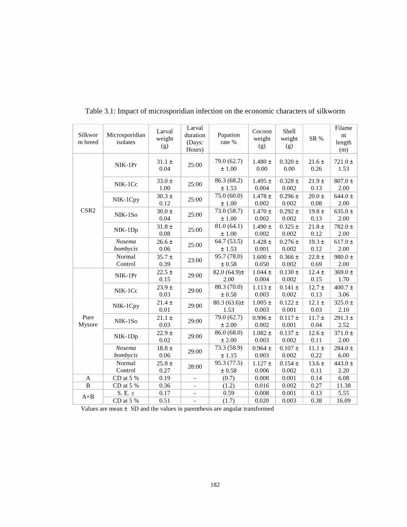
182
Table 3.1: Impact of microsporidian infection on the economic characters of silkworm
Silkworm breed
Microsporidian isolates
Larval weight
(g)
Larval duration (Days: Hours)
Pupation rate %
Cocoon weight
(g)
Shell weight
(g) SR %
Filament
length (m)
NIK-1Pr 31.1 ± 0.04
25:00 79.0 (62.7)
± 1.00 1.480 ±
0.00 0.320 ±
0.00 21.6 ± 0.26
721.0 ± 1.53
NIK-1Cc 33.0 ± 1.00
25:00 86.3 (68.2)
± 1.53 1.495 ± 0.004
0.328 ± 0.002
21.9 ± 0.13
807.0 ± 2.00
NIK-1Cpy 30.3 ± 0.12
25:00 75.0 (60.0)
± 1.00 1.478 ± 0.002
0.296 ± 0.002
20.0 ± 0.08
644.0 ± 2.00
NIK-1So 30.0 ± 0.04
25:00 73.0 (58.7)
± 1.00 1.470 ± 0.002
0.292 ± 0.002
19.8 ± 0.13
635.0 ± 2.00
NIK-1Dp 31.8 ± 0.08
25:00 81.0 (64.1)
± 1.00 1.490 ± 0.002
0.325 ± 0.002
21.8 ± 0.12
782.0 ± 2.00
Nosema bombycis
26.6 ± 0.06
25:00 64.7 (53.5)
± 1.53 1.428 ± 0.001
0.276 ± 0.002
19.3 ± 0.12
617.0 ± 2.00
CSR2
Normal Control
35.7 ± 0.39
23:00 95.7 (78.0)
± 0.58 1.600 ± 0.050
0.366 ± 0.002
22.8 ± 0.69
980.0 ± 2.00
NIK-1Pr 22.5 ± 0.15
29:00 82.0 (64.9)± 2.00
1.044 ± 0.004
0.130 ± 0.002
12.4 ± 0.15
369.0 ± 1.70
NIK-1Cc 23.9 ± 0.03
29:00 88.3 (70.0)
± 0.58 1.113 ± 0.003
0.141 ± 0.002
12.7 ± 0.13
400.7 ± 3.06
NIK-1Cpy 21.4 ± 0.01
29:00 80.3 (63.6)± 1.53
1.005 ± 0.003
0.122 ± 0.001
12.1 ± 0.03
325.0 ± 2.10
NIK-1So 21.1 ± 0.03
29:00 79.0 (62.7)
± 2.00 0.996 ± 0.002
0.117 ± 0.001
11.7 ± 0.04
291.3 ± 2.52
NIK-1Dp 22.9 ± 0.02
29:00 86.0 (68.0)
± 2.00 1.082 ± 0.003
0.137 ± 0.002
12.6 ± 0.11
371.0 ± 2.00
Nosema bombycis
18.8 ± 0.06
29:00 73.3 (58.9)
± 1.15 0.964 ± 0.003
0.107 ± 0.002
11.1 ± 0.22
284.0 ± 6.00
Pure Mysore
Normal Control
25.8 ± 0.27
28:00 95.3 (77.5)
± 0.58 1.127 ± 0.006
0.154 ± 0.002
13.6 ± 0.11
443.0 ± 2.20
A CD at 5 % 0.19 - (0.7) 0.008 0.001 0.14 6.08 B CD at 5 % 0.36 - (1.2) 0.016 0.002 0.27 11.38
S. E. ± 0.17 - 0.59 0.008 0.001 0.13 5.55 A×B
CD at 5 % 0.51 - (1.7) 0.020 0.003 0.38 16.09 Values are mean ± SD and the values in parenthesis are angular transformed

183
Table 3.2: Impact of lower pathogen loads on the health status and rearing performance of the silkworm, Bombyx mori L.
Mortality due to infection (%) Microsporidian
isolates
Spore concentration (spores/ml) Larva Pupa
Moths infected
(%)
Total infection
(%)
Larval weight
(g)
Larval duration (D:H)
Pupation rate (%)
Single cocoon weight
(g)
Single shell
weight (g)
SR %
1×10² 0.0(0.0) 0.0(0.0) 0.0(0.0) 0.0(0.0) 39.17 24.00 95.0(77.1) 1.602 0.373 23.29 1×10³ 0.0(0.0) 0.0(0.0) 28.0(31.9) 28.0(31.9) 38.66 24.00 93.0(74.6) 1.587 0.357 22.48 1×10⁴ 1.0(5.7) 1.7(7.5) 32.3(34.6) 35.0(36.3) 38.54 24.00 93.0(74.6) 1.560 0.347 22.23
NIK-1Pr
1×10⁵ 6.7(15.0) 3.7(11.1) 33.7(35.5) 44.0(41.5) 38.23 24.00 85.3(67.4) 1.537 0.337 21.93 1×10² 0.0(0.0) 0.0(0.0) 0.0(0.0) 0.0(0.0) 39.17 24.00 95.0(77.1) 1.604 0.374 23.31 1×10³ 0.0(0.0) 0.0(0.0) 14.0(21.9) 14.0(21.9) 38.87 24.00 94.0(75.8) 1.591 0.364 22.86 1×10⁴ 0.0(0.0) 0.0(0.0) 17.3(24.6) 17.3(24.6) 38.74 24.00 94.0(75.8) 1.572 0.356 22.65
NIK-1Cc
1×10⁵ 0.0(0.0) 0.0(0.0) 21.3(27.5) 21.3(27.5) 38.68 24.00 94.0(75.8) 1.565 0.352 22.52 1×10² 0.0(0.0) 0.0(0.0) 8.0(16.4) 8.0(16.4) 38.80 24.00 94.3(76.2) 1.593 0.365 22.91 1×10³ 0.0(0.0) 0.0(0.0) 30.0(33.2) 30.0(33.2) 38.30 24.00 92.0(73.6) 1.581 0.350 22.16 1×10⁴ 6.7(15.0) 3.7(11.1) 35.3(36.4) 45.7(42.5) 38.21 24.00 85.3(67.4) 1.551 0.342 22.06
NIK-1Cpy
1×10⁵ 16.3(23.8) 4.3(11.9) 59.7(50.6) 80.3(63.6) 37.90 24.00 75.0(60.0) 1.529 0.332 21.74 1×10² 0.0(0.0) 0.0(0.0) 13.0(21.1) 13.0(21.1) 38.70 24.00 93.0(74.6) 1.590 0.362 22.78 1×10³ 0.0(0.0) 0.0(0.0) 35.0(36.3) 35.0(36.3) 38.10 24.00 92.0(73.6) 1.580 0.349 22.09 1×10⁴ 9.7(18.1) 4.7(12.5) 36.3(37.0) 50.7(45.4) 38.02 24.00 81.3(64.4) 1.541 0.335 21.74
NIK-1So
1×10⁵ 20.3(26.8) 6.3(14.5) 60.3(50.9) 87.0(68.8) 37.74 24.00 69.0(56.2) 1.519 0.326 21.47
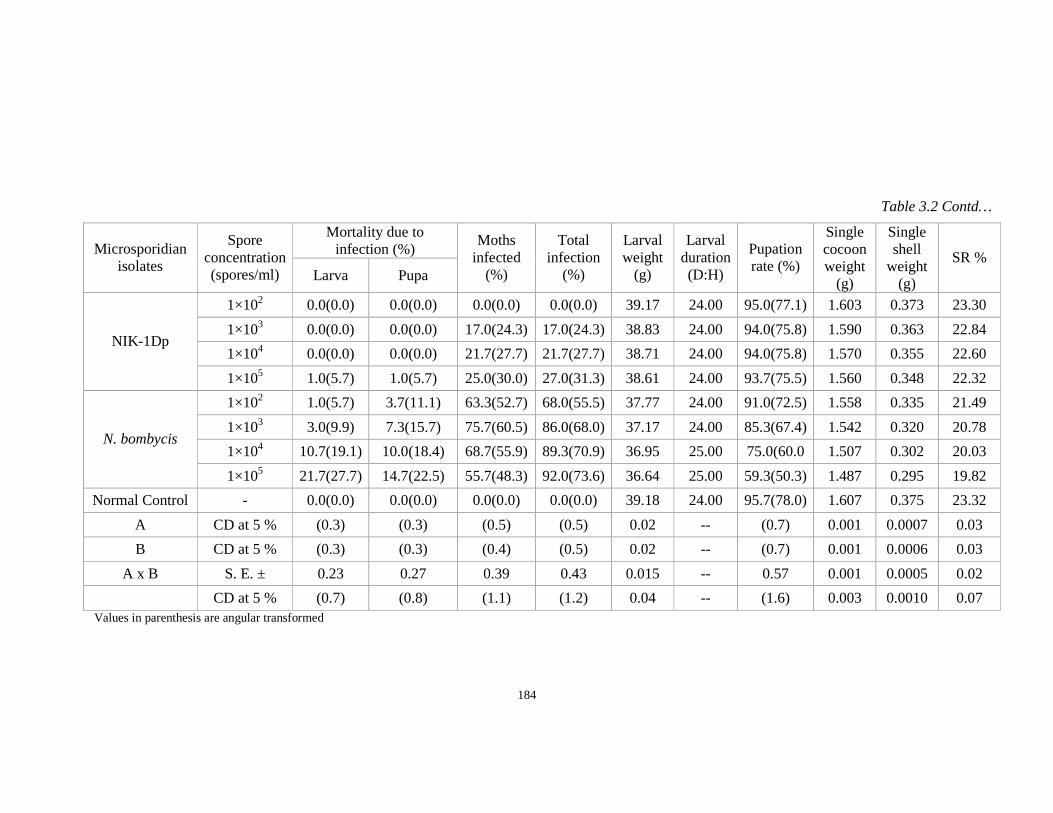
184
Table 3.2 Contd…
Mortality due to infection (%) Microsporidian
isolates
Spore concentration (spores/ml) Larva Pupa
Moths infected
(%)
Total infection
(%)
Larval weight
(g)
Larval duration (D:H)
Pupation rate (%)
Single cocoon weight
(g)
Single shell
weight (g)
SR %
1×10² 0.0(0.0) 0.0(0.0) 0.0(0.0) 0.0(0.0) 39.17 24.00 95.0(77.1) 1.603 0.373 23.30
1×10³ 0.0(0.0) 0.0(0.0) 17.0(24.3) 17.0(24.3) 38.83 24.00 94.0(75.8) 1.590 0.363 22.84
1×10⁴ 0.0(0.0) 0.0(0.0) 21.7(27.7) 21.7(27.7) 38.71 24.00 94.0(75.8) 1.570 0.355 22.60 NIK-1Dp
1×10⁵ 1.0(5.7) 1.0(5.7) 25.0(30.0) 27.0(31.3) 38.61 24.00 93.7(75.5) 1.560 0.348 22.32
1×10² 1.0(5.7) 3.7(11.1) 63.3(52.7) 68.0(55.5) 37.77 24.00 91.0(72.5) 1.558 0.335 21.49
1×10³ 3.0(9.9) 7.3(15.7) 75.7(60.5) 86.0(68.0) 37.17 24.00 85.3(67.4) 1.542 0.320 20.78
1×10⁴ 10.7(19.1) 10.0(18.4) 68.7(55.9) 89.3(70.9) 36.95 25.00 75.0(60.0 1.507 0.302 20.03 N. bombycis
1×10⁵ 21.7(27.7) 14.7(22.5) 55.7(48.3) 92.0(73.6) 36.64 25.00 59.3(50.3) 1.487 0.295 19.82
Normal Control - 0.0(0.0) 0.0(0.0) 0.0(0.0) 0.0(0.0) 39.18 24.00 95.7(78.0) 1.607 0.375 23.32
A CD at 5 % (0.3) (0.3) (0.5) (0.5) 0.02 -- (0.7) 0.001 0.0007 0.03
B CD at 5 % (0.3) (0.3) (0.4) (0.5) 0.02 -- (0.7) 0.001 0.0006 0.03
A x B S. E. ± 0.23 0.27 0.39 0.43 0.015 -- 0.57 0.001 0.0005 0.02
CD at 5 % (0.7) (0.8) (1.1) (1.2) 0.04 -- (1.6) 0.003 0.0010 0.07 Values in parenthesis are angular transformed
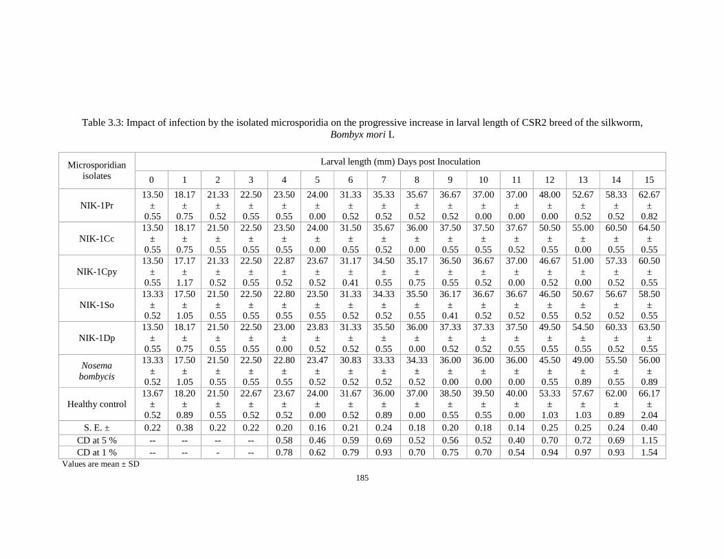
185
Table 3.3: Impact of infection by the isolated microsporidia on the progressive increase in larval length of CSR2 breed of the silkworm, Bombyx mori L
Larval length (mm) Days post Inoculation Microsporidian isolates 0 1 2 3 4 5 6 7 8 9 10 11 12 13 14 15
NIK-1Pr 13.50
± 0.55
18.17 ±
0.75
21.33 ±
0.52
22.50 ±
0.55
23.50 ±
0.55
24.00 ±
0.00
31.33 ±
0.52
35.33 ±
0.52
35.67 ±
0.52
36.67 ±
0.52
37.00 ±
0.00
37.00 ±
0.00
48.00 ±
0.00
52.67 ±
0.52
58.33 ±
0.52
62.67 ±
0.82
NIK-1Cc 13.50
± 0.55
18.17 ±
0.75
21.50 ±
0.55
22.50 ±
0.55
23.50 ±
0.55
24.00 ±
0.00
31.50 ±
0.55
35.67 ±
0.52
36.00 ±
0.00
37.50 ±
0.55
37.50 ±
0.55
37.67 ±
0.52
50.50 ±
0.55
55.00 ±
0.00
60.50 ±
0.55
64.50 ±
0.55
NIK-1Cpy 13.50
± 0.55
17.17 ±
1.17
21.33 ±
0.52
22.50 ±
0.55
22.87 ±
0.52
23.67 ±
0.52
31.17 ±
0.41
34.50 ±
0.55
35.17 ±
0.75
36.50 ±
0.55
36.67 ±
0.52
37.00 ±
0.00
46.67 ±
0.52
51.00 ±
0.00
57.33 ±
0.52
60.50 ±
0.55
NIK-1So 13.33
± 0.52
17.50 ±
1.05
21.50 ±
0.55
22.50 ±
0.55
22.80 ±
0.55
23.50 ±
0.55
31.33 ±
0.52
34.33 ±
0.52
35.50 ±
0.55
36.17 ±
0.41
36.67 ±
0.52
36.67 ±
0.52
46.50 ±
0.55
50.67 ±
0.52
56.67 ±
0.52
58.50 ±
0.55
NIK-1Dp 13.50
± 0.55
18.17 ±
0.75
21.50 ±
0.55
22.50 ±
0.55
23.00 ±
0.00
23.83 ±
0.52
31.33 ±
0.52
35.50 ±
0.55
36.00 ±
0.00
37.33 ±
0.52
37.33 ±
0.52
37.50 ±
0.55
49.50 ±
0.55
54.50 ±
0.55
60.33 ±
0.52
63.50 ±
0.55
Nosema bombycis
13.33 ±
0.52
17.50 ±
1.05
21.50 ±
0.55
22.50 ±
0.55
22.80 ±
0.55
23.47 ±
0.52
30.83 ±
0.52
33.33 ±
0.52
34.33 ±
0.52
36.00 ±
0.00
36.00 ±
0.00
36.00 ±
0.00
45.50 ±
0.55
49.00 ±
0.89
55.50 ±
0.55
56.00 ±
0.89
Healthy control 13.67
± 0.52
18.20 ±
0.89
21.50 ±
0.55
22.67 ±
0.52
23.67 ±
0.52
24.00 ±
0.00
31.67 ±
0.52
36.00 ±
0.89
37.00 ±
0.00
38.50 ±
0.55
39.50 ±
0.55
40.00 ±
0.00
53.33 ±
1.03
57.67 ±
1.03
62.00 ±
0.89
66.17 ±
2.04 S. E. ± 0.22 0.38 0.22 0.22 0.20 0.16 0.21 0.24 0.18 0.20 0.18 0.14 0.25 0.25 0.24 0.40
CD at 5 % -- -- -- -- 0.58 0.46 0.59 0.69 0.52 0.56 0.52 0.40 0.70 0.72 0.69 1.15 CD at 1 % -- -- - -- 0.78 0.62 0.79 0.93 0.70 0.75 0.70 0.54 0.94 0.97 0.93 1.54
Values are mean ± SD
186
Table 3.4: Impact of infection by the isolated microsporidia on the progressive increase in larval width of CSR2 breed of the silkworm, Bombyx mori L.
Larval width (mm) Days post inoculation Microsporidian isolates 0 1 2 3 4 5 6 7 8 9 10 11 12 13 14 15
NIK-1Pr 3.00
± 0.00 3.33
± 0.52 4.00
± 0.00 4.00
± 0.00 4.00
± 0.00 4.00
± 0.00 5.00
± 0.00 6.33
± 0.52 6.50
± 0.55 6.50
± 0.55 6.67
± 0.52 6.67
± 0.52 8.00
± 0.00 9.00
± 0.00 10.00 ± 0.00
10.33 ± 0.52
NIK-1Cc 3.00
± 0.00 3.50
± 0.55 4.00
± 0.00 4.00
± 0.00 4.00
± 0.00 4.00
± 0.00 5.00
± 0.00 6.50
± 0.55 7.00
± 0.00 7.00
± 0.00 7.00
± 0.00 7.00
± 0.00 8.50
± 0.55 9.00
± 0.00 10.00 ± 0.00
11.00 ± 0.55
NIK-1Cpy 3.00
± 0.00 3.33
± 0.52 4.00
± 0.00 4.00
± 0.00 4.00
± 0.00 4.00
± 0.00 5.00
± 0.00 6.33
± 0.52 6.50
± 0.55 7.00
± 0.00 7.00
± 0.00 7.00
± 0.00 8.00
± 0.00 8.00
± 0.00 10.00 ± 0.00
10.00 ± 0.00
NIK-1So 3.00
± 0.00 3.33
± 0.52 4.00
± 0.00 4.00
± 0.00 4.00
± 0.00 4.00
± 0.00 5.00
± 0.00 6.33
± 0.52 6.50
± 0.55 7.00
± 0.00 7.00
± 0.00 7.00
± 0.00 8.00
± 0.00 8.00
± 0.00 10.00 ± 0.00
10.00 ± 0.00
NIK-1Dp 3.00
± 0.00 3.50
± 0.55 4.00
± 0.00 4.00
± 0.00 4.00
± 0.00 4.00
± 0.00 5.00
± 0.00 6.50
± 0.55 6.67
± 0.52 7.00
± 0.00 7.00
± 0.00 7.00
± 0.00 8.33
± 0.52 9.00
± 0.00 10.00 ± 0.00
10.50 ± 0.55
Nosema bombycis 3.00
± 0.00 3.33
± 0.52 4.00
± 0.00 4.00
± 0.00 4.00
± 0.00 4.00
± 0.00 5.00
± 0.00 5.50
± 0.55 6.00
± 0.00 6.00
± 0.00 6.17
± 0.52 6.37
± 0.52 7.50
± 0.55 7.67
± 0.52 8.33
± 0.52 8.50
± 0.55
Healthy control 3.00
±0.00 4.00
± 0.00 4.00
± 0.00 4.00
± 0.00 4.00
± 0.00 4.00
± 0.00 5.00
± 0.00 6.67
± 0.52 7.00
± 0.00 7.33
± 0.52 7.33
± 0.52 7.50
± 0.55 8.50
± 0.55 10.00 ± 0.00
11.00 ± 0.52
12.00 ± 0.00
S. E. ± -- 0.20 -- -- -- -- -- 0.22 0.17 0.12 0.14 0.14 0.17 0.08 0.11 0.18
CD at 5% -- -- -- -- -- -- -- 0.62 0.48 0.33 0.39 0.40 0.48 0.23 0.32 0.53
CD at 1% -- -- -- -- -- -- -- 0.83 0.64 0.44 0.53 0.54 0.64 0.30 0.43 0.71
Values are mean ± SD
187
Table 3.5: Impact of infection by the isolated microsporidia on the progressive increase in larval weight of CSR2 breed of the silkworm, Bombyx mori L.
Weight (g/10 larvae) Days post inoculation Microsporidian isolates 0 1 2 3 4 5 6 7 8 9 10 11 12 13 14 15
NIK-1Pr 0.44
± 0.01
1.07 ±
0.01
1.66 ±
0.01
2.06 ±
0.01
2.08 ±
0.01
2.09 ±
0.01
5.12 ±
0.01
7.62 ±
0.01
8.96 ±
0.01
9.13 ±
0.01
9.15 ±
0.03
9.19 ±
0.03
21.14 ±
0.02
30.08 ±
0.05
33.51 ±
0.14
34.94 ±
0.07
NIK-1Cc 0.45
± 0.01
1.07 ±
0.01
1.66 ±
0.01
2.06 ±
0.01
2.08 ±
0.01
2.09 ±
0.01
5.12 ±
0.01
7.64 ±
0.01
9.02 ±
0.02
9.44 ±
0.01
9.44 ±
0.01
9.46 ±
0.01
22.04 ±
0.02
31.19 ±
0.01
34.05 ±
0.03
36.45 ±
0.08
NIK-1Cpy 0.44
± 0.01
1.07 ±
0.01
1.66 ±
0.01
2.06 ±
0.01
2.07 ±
0.01
2.08 ±
0.01
5.11 ±
0.01
7.62 ±
0.01
8.9 ±
0.01
9.13 ±
0.02
9.13 ±
0.02
9.14 ±
0.02
20.16 ±
0.04
29.81 ±
0.18
33.02 ±
0.02
34.54 ±
0.03
NIK- 1So 0.45
± 0.01
1.07 ±
0.01
1.66 ±
0.01
2.06 ±
0.01
2.07 ±
0.01
2.07 ±
0.01
5.11 ±
0.01
7.62 ±
0.01
8.90 ±
0.01
9.12 ±
0.01
9.12 ±
0.02
9.13 ±
0.02
20.08 ±
0.02
29.5 ±
0.01
33.01 ±
0.01
34.32 ±
0.04
NIK- 1Dp 0.45
± 0.01
1.07 ±
0.01
1.67 ±
0.01
2.06 ±
0.01
2.07 ±
0.01
2.08 ±
0.01
5.12 ±
0.01
7.63 ±
0.01
9.00 ±
0.02
9.43 ±
0.01
9.44 ±
0.01
9.45 ±
0.01
22.00 ±
0.00
31.05 ±
0.04
33.95 ±
0.02
35.31 ±
0.04
Nosema bombycis
0.44 ±
0.01
1.07 ±
0.01
1.66 ±
0.01
2.06 ±
0.00
2.06 ±
0.01
2.07 ±
0.01
5.09 ±
0.01
7.50 ±
0.02
8.62 ±
0.03
9.00 ±
0.02
9.06 ±
0.01
9.10 ±
0.01
18.47 ±
0.26
25.17 ±
0.04
27.02 ±
0.22
28.00 ±
0.19
Healthy control 0.45
± 0.01
1.08 ±
0.01
1.67 ±
0.01
2.08 ±
0.01
2.09 ±
0.01
2.12 ±
0.01
5.13 ±
0.01
7.69 ±
0.01
9.04 ±
0.01
9.48 ±
0.01
9.54 ±
0.01
9.56 ±
0.01
23.94 ±
0.01
32.53 ±
0.04
35.32 ±
0.06
38.52 ±
0.03
S.E. ± 0.002 0.003 0.003 0.002 0.002 0.002 0.003 0.004 0.007 0.005 0.007 0.006 0.041 0.030 0.042 0.036
CD at 5% -- -- -- 0.01 0.01 0.01 0.01 0.01 0.02 0.02 0.02 0.02 0.12 0.09 0.12 0.10 CD at 1% -- -- -- 0.01 0.01 0.01 0.01 0.02 0.03 0.02 0.03 0.02 0.16 0.12 0.16 0.14
Values are mean ± SD
188
Table 3.6: Assay on transovarial and venereal transmission of the isolated microsporidia in silkworm
Microsporidian isolates
Moths paired
Fecundity (No.)
No. of dead eggs
Infected dead eggs (%)
No. of larvae hatched
Hatching % Larvae infected
(%) Transmission (%)
IF×IM 435.0± 6.5 18.0 ± 1.5 81.0(64.1) ± 1.7 370.0 ± 5.0 85.1(67.2) ± 0.1 95.3(77.4) ± 0.2 94.6(76.5) ± 0.1 IF×HM 436.0 ± 7.0 18.0 ± 0.6 80.3(63.6) ± 1.5 372.0 ± 5.6 85.3(67.4) ± 0.2 91.2(72.7) ± 0.1 90.6(72.1) ± 0.0 NIK-1Pr HF×IM 468.0 ± 5.6 11.0 ± 1.2 0.0(0.0)± 0.0 432.0 ± 6.0 92.3(73.8) ± 0.3 0.0(0.0)± 0.0 0.0(0.0)± 0.0 IF×IM 455.0 ± 3.0 11.0 ± 1.2 0.0(0.0)± 0.0 407.0 ± 1.5 89.4(71.0) ± 0.3 0.0(0.0)± 0.0 0.0(0.0)± 0.0 IF×HM 458.0 ± 3.5 11.0 ± 1.0 0.0(0.0)± 0.0 410.0 ± 2.5 89.5(71.1) ± 0.1 0.0(0.0)± 0.0 0.0(0.0)± 0.0 NIK-1Cc HF×IM 468.0 ± 3.1 10.0 ± 0.6 0.0(0.0)± 0.0 433.0 ± 1.5 92.5(74.1) ± 0.3 0.0(0.0)± 0.0 0.0(0.0)± 0.0 IF×IM 427.0 ± 2.5 26.0 ± 2.5 100.0(90.0) ± 0.0 357.0 ± 2.6 83.5(66.0) ± 0.2 100.0(90.0) ± 0.0 100.0(90.0) ± 0.0 IF×HM 430.0 ± 2.1 22.0 ± 2.0 100.0(90.0) ± 0.0 360.0 ± 2.1 83.7(66.2) ± 0.1 100.0(90.0) ± 0.0 100.0(90.0) ± 0.0 NIK-1Cpy HF×IM 463.0 ± 2.5 12.0 ± 1.0 0.0(0.0)± 0.0 424.0 ± 3.1 91.7(73.2) ± 0.2 0.0(0.0)± 0.0 0.0(0.0)± 0.0 IF×IM 423.0 ± 1.5 29.0 ± 2.1 100.0(90.0) ± 0.0 350.0 ± 1.5 82.6(65.3) ± 0.1 100.0(90.0) ± 0.0 100.0(90.0) ± 0.0 IF×HM 428.0 ± 0.6 23.0 ± 1.5 100.0(90.0) ± 0.0 355.0 ± 0.6 82.9(65.5) ± 0.0 100.0(90.0) ± 0.0 100.0(90.0) ± 0.0 NIK-1So HF×IM 462.0 ± 0.6 13.0 ± 0.6 0.0(0.0)± 0.0 422.0 ± 0.6 91.2(72.7) ± 0.1 0.0(0.0)± 0.0 0.0(0.0)± 0.0 IF×IM 442.0 ± 2.5 18.0 ± 0.6 80.3(63.6) ± 1.5 385.0 ± 2.5 87.1(68.9) ± 0.1 82.7(65.4) ± 3.1 82.6(65.3) ± 0.1 IF×HM 445.0 ± 2.0 17.0 ± 0.6 80.3(63.6) ± 0.6 389.0 ± 1.5 87.3(69.1) ± 0.1 80.0(63.4) ± 4.0 80.0(63.4) ± 0.1 NIK-1Dp HF×IM 468.0 ± 1.5 10.0 ± 0.6 0.0(0.0)± 0.0 433.0 ± 1.0 92.5(74.1) ± 0.1 0.0(0.0)± 0.0 0.0(0.0)± 0.0 IF×IM 367.0 ± 2.1 43.0 ± 1.2 100.0(90.0) ± 0.0 243.0 ± 2.0 66.1(54.3) ± 0.2 100.0(90.0) ± 0.0 100.0(90.0)± 0.0 IF×HM 370.0 ± 1.5 40.0 ± 1.5 100.0(90.0) ± 0.0 249.0 ± 1.0 67.4(55.2) ± 0.3 100.0(90.0) ± 0.0 100.0(90.0) ± 0.0
Nosema bombycis
HF×IM 418.0 ± 3.5 18.0 ± 2.0 0.0(0.0)± 0.0 377.0 ± 3.0 90.1(71.6) ± 0.1 0.0(0.0)± 0.0 0.0(0.0)± 0.0 CD at 5% 3.2 1.3 (0.5) 2.8 (0.2) (0.8) (0.03) CD at 5% 2.3 0.9 (0.3) 2.0 (0.1) (0.6) (0.02)
S.E. 2.0 0.8 0.3 1.6 0.1 0.5 0.0 CD at 5 % 5.6 2.3 (0.8) 4.8 (0.3) (1.5) (0.05)
Values are mean ± SD and the values in parenthesis are angular transformed.
189
Table 3.7: Assay on mortality of transovarially infected progeny populations (IF×IM) combinations
Larval mortality % Microsporidian
isolates I instar II instar III instar IV instar V instar
Total mortality
%
NIK-1Pr 0.0(0.0) ± 0.0
9.3(17.7) ± 1.2
39.3(38.8) ± 0.6
31.3(34.0) ± 0.6
20.0(26.5) ± 0.0
100.0(90.0) ± 0.0
NIK-1Cc 0.0(0.0) ± 0.0
0.0(0.0) ± 0.0
0.0(0.0) ± 0.0
0.0(0.0) ± 0.0
0.0(0.0) ± 0.0
0.0(0.0) ± 0.0
NIK-1Cpy 9.3(17.7)
± 2.5 17.3(24.5)
± 1.2 58.0(49.6)
± 1.0 15.3(23.0)
± 2.5 --
100.0(90.0) ± 0.0
NIK-1So 14.0(21.9)
± 2.0 23.3(28.8)
± 4.2 59.3(50.3)
± 3.1 3.3(10.4)
± 1.2 --
100.0(90.0) ± 0.0
NIK-1Dp 0.0(0.0) ± 0.0
9.0(17.4) ± 1.0
34.0(35.6) ± 1.0
31.0(33.8) ± 1.0
26.0(30.6) ± 1.0
100.0(90.0) ± 0.0
Nosema bombycis
20.0(26.5) ± 0.0
24.0(29.3) ±1.0
56.0(48.4) ± 1.0
-- -- 100.0(90.0)
± 0.0
CD at 5 % (2.2) (2.4) (1.5) (0.5) (2.2) --
Values are mean ± SD and the values in parenthesis are angular transformed.
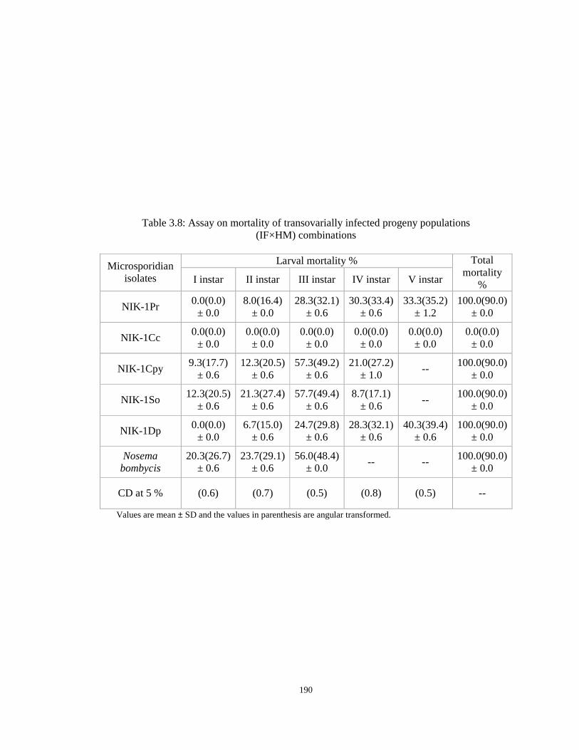
190
Table 3.8: Assay on mortality of transovarially infected progeny populations (IF×HM) combinations
Larval mortality % Microsporidian
isolates I instar II instar III instar IV instar V instar
Total mortality
%
NIK-1Pr 0.0(0.0) ± 0.0
8.0(16.4) ± 0.0
28.3(32.1) ± 0.6
30.3(33.4) ± 0.6
33.3(35.2) ± 1.2
100.0(90.0) ± 0.0
NIK-1Cc 0.0(0.0) ± 0.0
0.0(0.0) ± 0.0
0.0(0.0) ± 0.0
0.0(0.0) ± 0.0
0.0(0.0) ± 0.0
0.0(0.0) ± 0.0
NIK-1Cpy 9.3(17.7)
± 0.6 12.3(20.5)
± 0.6 57.3(49.2)
± 0.6 21.0(27.2)
± 1.0 --
100.0(90.0) ± 0.0
NIK-1So 12.3(20.5)
± 0.6 21.3(27.4)
± 0.6 57.7(49.4)
± 0.6 8.7(17.1)
± 0.6 --
100.0(90.0) ± 0.0
NIK-1Dp 0.0(0.0) ± 0.0
6.7(15.0) ± 0.6
24.7(29.8) ± 0.6
28.3(32.1) ± 0.6
40.3(39.4) ± 0.6
100.0(90.0) ± 0.0
Nosema bombycis
20.3(26.7) ± 0.6
23.7(29.1) ± 0.6
56.0(48.4) ± 0.0
-- -- 100.0(90.0)
± 0.0
CD at 5 % (0.6) (0.7) (0.5) (0.8) (0.5) --
Values are mean ± SD and the values in parenthesis are angular transformed.

191
Table 3.9: Percent mortality in the progeny populations hatched from eggs externally contaminated with the isolated microsporidia
Larval mortality % Microsporidian
isolates I instar II instar III instar IV instar V instar
Pupal mortality%
Total mortality
%
NIK-1Pr 0.0 (0.0) ± 0.0
0.0 (0.0) ± 0.0
9.7 (18.1) ± 0.6
22.0 (27.9) ± 0.0
23.7 (29.1) ± 0.6
20.3 (26.7) ± 0.6
75.7 (60.4) ± 1.5
NIK-1Cc 0.0 (0.0)
± 0.0 0.0 (0.0)
± 0.0 0.0 (0.0)
± 0.0 15.3 (23.0)
± 0.6 23.0 (28.6)
± 0.0 17.7 (24.8)
± 0.6 56.0 (48.4)
± 0.0
NIK-1Cpy 0.0 (0.0)
± 0.0 9.0 (17.4)
± 0.0 17.0 (24.3)
± 0.0 21.0 (27.2)
± 0.0 27.3 (31.5)
± 0.6 13.0 (21.1)
± 0.0 87.3 (69.1)
± 0.6
NIK-1So 0.0 (0.0)
± 0.0 9.7 (18.1)
± 0.6 19.0 (25.8)
± 1.0 21.7 (27.7)
± 0.6 29.0 (32.5)
± 1.0 13.3 (21.4)
± 0.6 92.7 (74.3)
± 2.1
NIK-1Dp 0.0 (0.0)
± 0.0 0.0 (0.0)
± 0.0 4.3 (11.9)
± 0.6 17.0 (24.3)
± 0.0 24.3 (29.5)
± 0.6 18.7 (25.6)
± 0.6 64.3 (53.3)
± 1.2
Nosema bombycis
0.0 (0.0) ± 0.0
18.7 (25.6) ± 1.2
23.7 (29.1) ± 0.6
39.3 (38.8) ± 0.6
18.3 (25.3) ± 1.2
-- 100.0
(90.0) ± 0.0
CD at 5 % -- (0.7) (0.9) (0.5) (0.9) (0.6) (1.9)
Values are mean ± SD and the values in parenthesis are angular transformed.

192
Table 3.10: Effect of mulberry leaf disinfection on microsporidian disease incidence
Microspo-ridian isolates
Treatment (Leaf dipping)
Concentration
Mortality due to
infection (%)
Total infection
(%)
Disease suppression
% over inoculated
control Decol 1.0 % 0.0 (0.0) 35.3 (36.4) 51.3 (45.7)
Sanitech 400 ppm 0.0 (0.0) 14.7 (22.5) 79.7 (63.2) NIK-1Pr Inoculated control - 40.0 (39.2) 72.7 (58.5) --
Decol 1.0 % 0.0 (0.0) 18.0 (25.1) 66.4 (54.5) Sanitech 400 ppm 0.0 (0.0) 7.0 (15.3) 86.9 (68.8) NIK-1Cc
Inoculated control - 28.0 (31.9) 53.7 (47.1) -- Decol 1.0 % 0.0 (0.0) 44.7 (41.9) 55.3 (48.0)
Sanitech 400 ppm 0.0 (0.0) 18.3 (25.3) 81.7 (64.6) NIK-1Cpy Inoculated control - 70.0 (56.8)
100.0 (90.0)
--
Decol 1.0 % 0.0 (0.0) 52.7 (46.5) 47.3 (43.4) Sanitech 400 ppm 0.0(0.0) 22.3 (28.2) 77.7 (61.8) NIK-1So
Inoculated control - 85.0 (67.2) 100.0 (90.0)
--
Decol 1.0 % 0.0 (0.0) 28.0 (31.9) 56.8 (48.9) Sanitech 400 ppm 0.0 (0.0) 10.3 (18.7) 84.0 (66.4) NIK-1Dp
Inoculated control - 31.7 (34.3) 65.0 (53.7) -- Decol 1.0 % 0.0 (0.0) 62.0 (51.9) 38.0 (38.0)
Sanitech 400 ppm 0.0 (0.0) 33.3 (35.2) 66.7 (54.7) N. bombycis Inoculated control - 100.0 (90.0)
100.0 (90.0)
--
Normal control
Distilled water
- 0.0 (0.0) 0.0 (0.0) --
A CD at 5 % - (0.5) (0.9) (0.9) B CD at 5 % - (0.4) (0.7) (0.8)
S.E ± - 0.33 0.61 0.66 A x B
CD at 5 % - (0.9) (1.7) (1.9) Values in parenthesis are angular transformed
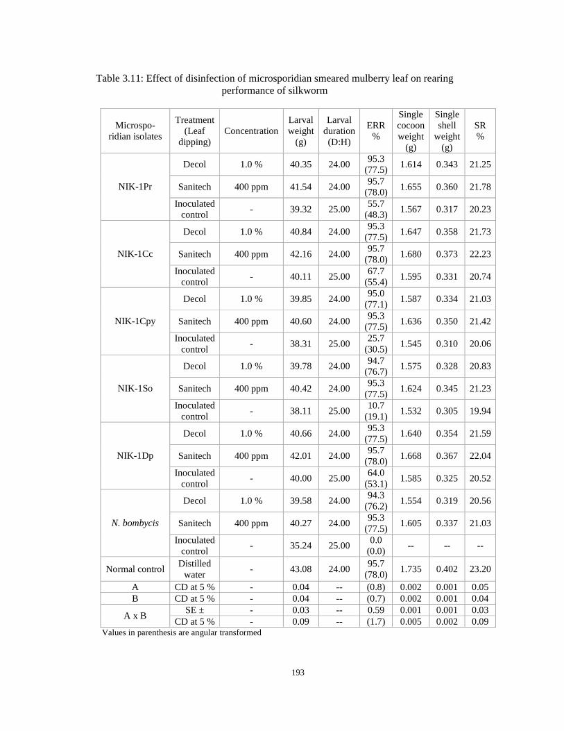
193
Table 3.11: Effect of disinfection of microsporidian smeared mulberry leaf on rearing performance of silkworm
Microspo-ridian isolates
Treatment (Leaf
dipping) Concentration
Larval weight
(g)
Larval duration (D:H)
ERR %
Single cocoon weight
(g)
Single shell
weight (g)
SR %
Decol 1.0 % 40.35 24.00 95.3
(77.5) 1.614 0.343 21.25
Sanitech 400 ppm 41.54 24.00 95.7
(78.0) 1.655 0.360 21.78 NIK-1Pr
Inoculated control
- 39.32 25.00 55.7
(48.3) 1.567 0.317 20.23
Decol 1.0 % 40.84 24.00 95.3
(77.5) 1.647 0.358 21.73
Sanitech 400 ppm 42.16 24.00 95.7
(78.0) 1.680 0.373 22.23 NIK-1Cc
Inoculated control
- 40.11 25.00 67.7
(55.4) 1.595 0.331 20.74
Decol 1.0 % 39.85 24.00 95.0
(77.1) 1.587 0.334 21.03
Sanitech 400 ppm 40.60 24.00 95.3
(77.5) 1.636 0.350 21.42 NIK-1Cpy
Inoculated control
- 38.31 25.00 25.7
(30.5) 1.545 0.310 20.06
Decol 1.0 % 39.78 24.00 94.7
(76.7) 1.575 0.328 20.83
Sanitech 400 ppm 40.42 24.00 95.3
(77.5) 1.624 0.345 21.23 NIK-1So
Inoculated control
- 38.11 25.00 10.7
(19.1) 1.532 0.305 19.94
Decol 1.0 % 40.66 24.00 95.3
(77.5) 1.640 0.354 21.59
Sanitech 400 ppm 42.01 24.00 95.7
(78.0) 1.668 0.367 22.04 NIK-1Dp
Inoculated control
- 40.00 25.00 64.0
(53.1) 1.585 0.325 20.52
Decol 1.0 % 39.58 24.00 94.3
(76.2) 1.554 0.319 20.56
Sanitech 400 ppm 40.27 24.00 95.3
(77.5) 1.605 0.337 21.03 N. bombycis
Inoculated control
- 35.24 25.00 0.0
(0.0) -- -- --
Normal control Distilled
water - 43.08 24.00
95.7 (78.0)
1.735 0.402 23.20
A CD at 5 % - 0.04 -- (0.8) 0.002 0.001 0.05 B CD at 5 % - 0.04 -- (0.7) 0.002 0.001 0.04
SE ± - 0.03 -- 0.59 0.001 0.001 0.03 A x B
CD at 5 % - 0.09 -- (1.7) 0.005 0.002 0.09 Values in parenthesis are angular transformed
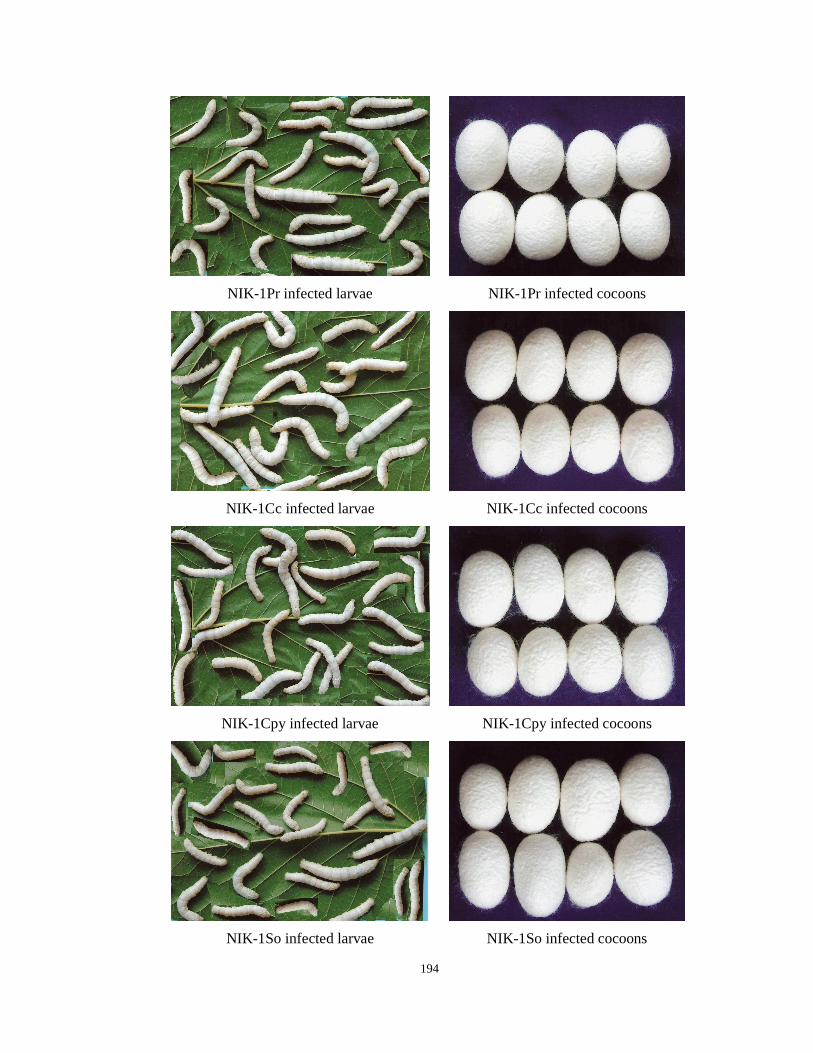
194
NIK-1Pr infected larvae NIK-1Pr infected cocoons
NIK-1Cc infected larvae NIK-1Cc infected cocoons
NIK-1Cpy infected larvae NIK-1Cpy infected cocoons
NIK-1So infected larvae NIK-1So infected cocoons
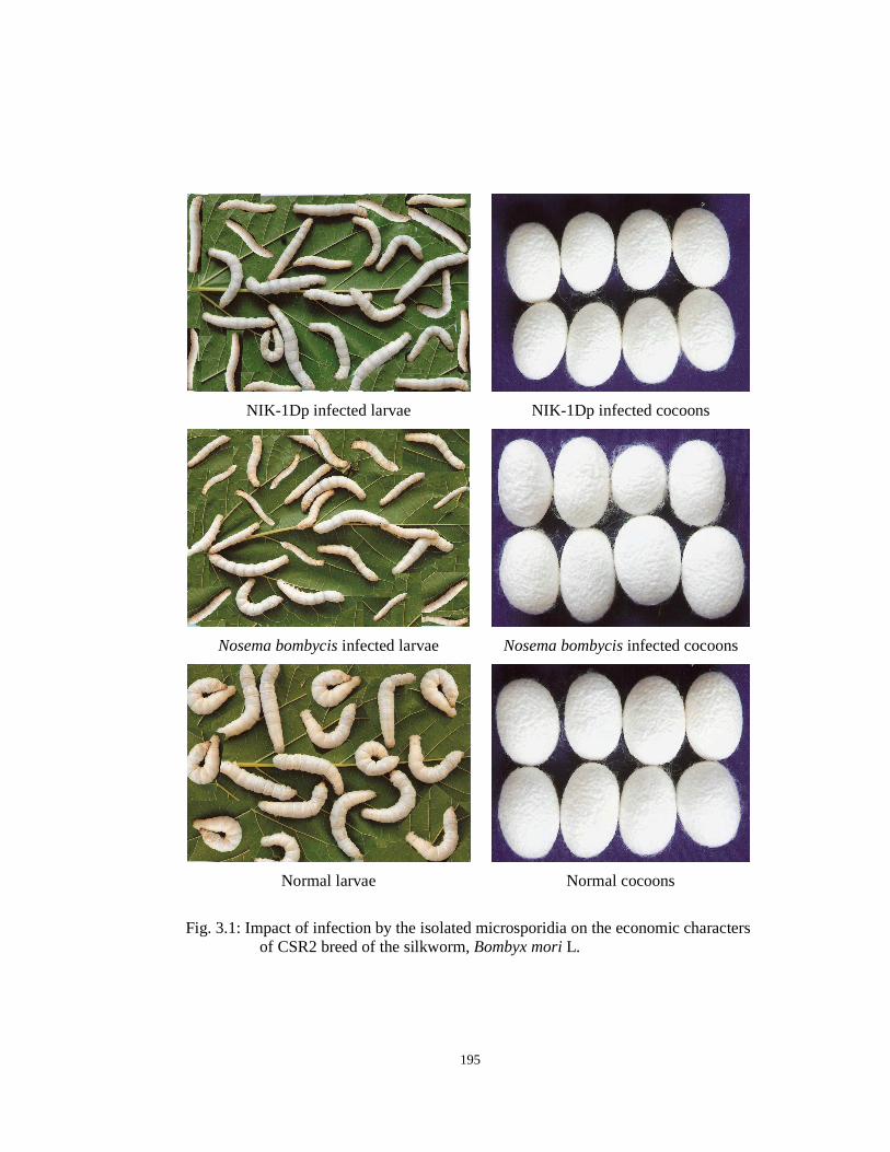
195
NIK-1Dp infected larvae NIK-1Dp infected cocoons
Nosema bombycis infected larvae Nosema bombycis infected cocoons
Normal larvae Normal cocoons
Fig. 3.1: Impact of infection by the isolated microsporidia on the economic characters
of CSR2 breed of the silkworm, Bombyx mori L.
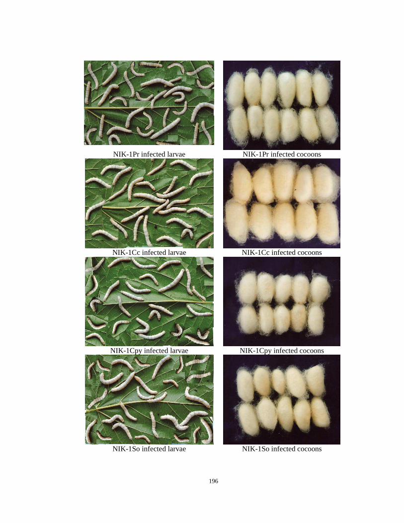
196
NIK-1Pr infected larvae NIK-1Pr infected cocoons
NIK-1Cc infected larvae NIK-1Cc infected cocoons
NIK-1Cpy infected larvae NIK-1Cpy infected cocoons
NIK-1So infected larvae NIK-1So infected cocoons
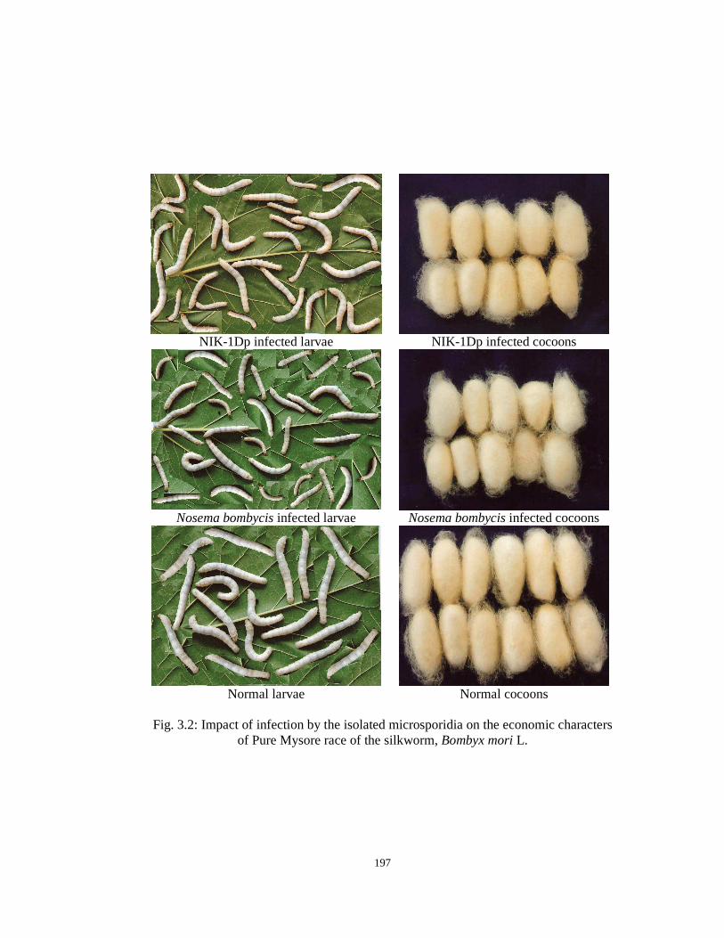
197
NIK-1Dp infected larvae NIK-1Dp infected cocoons
Nosema bombycis infected larvae Nosema bombycis infected cocoons
Normal larvae Normal cocoons
Fig. 3.2: Impact of infection by the isolated microsporidia on the economic characters
of Pure Mysore race of the silkworm, Bombyx mori L.

198
5.263.94
12.2813.15
4.38
15.35
0
5
10
15
20
NIK- 1Pr NIK- 1Cc NIK- 1Cpy NIK- 1So NIK- 1Dp N.bombycis
Microsporidian isolates
% R
ed
uct
ion
Fig. 3.3: Percentage reduction in SR% of CSR2 batches inoculated with different microsporidia compared to that of the healthy control
8.82
6.61
11.02
13.97
7.35
18.38
0
5
10
15
20
NIK- 1Pr NIK- 1Cc NIK- 1Cpy NIK- 1So NIK- 1Dp N.bombycisMicrosporidian isolates
% R
ed
uct
ion
Fig. 3.4: Percentage reduction in SR% of Pure Mysore batches inoculated with different microsporidia compared to that of the healthy control
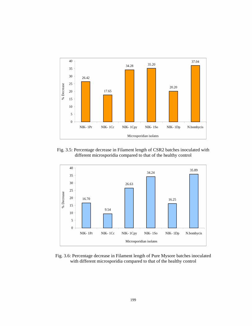
199
26.42
17.65
34.28 35.20
20.20
37.04
0
5
10
15
20
25
30
35
40
NIK- 1Pr NIK- 1Cc NIK- 1Cpy NIK- 1So NIK- 1Dp N.bombycis
Microsporidian isolates
% D
ecre
ase
Fig. 3.5: Percentage decrease in Filament length of CSR2 batches inoculated with different microsporidia compared to that of the healthy control
16.70
9.54
26.63
34.24
16.25
35.89
0
5
10
15
20
25
30
35
40
NIK- 1Pr NIK- 1Cc NIK- 1Cpy NIK- 1So NIK- 1Dp N.bombycis
Microsporidian isolates
% D
ecre
ase
Fig. 3.6: Percentage decrease in Filament length of Pure Mysore batches inoculated with different microsporidia compared to that of the healthy control
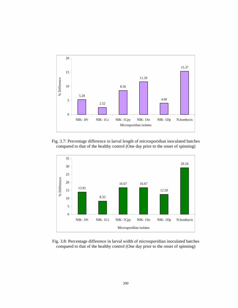
200
5.28
2.52
8.56
11.59
4.00
15.37
0
5
10
15
20
NIK- 1Pr NIK- 1Cc NIK- 1Cpy NIK- 1So NIK- 1Dp N.bombycis
Microsporidian isolates
% D
iffer
ence
Fig. 3.7: Percentage difference in larval length of microsporidian inoculated batches compared to that of the healthy control (One day prior to the onset of spinning)
13.91
8.33
16.67 16.67
12.50
29.16
0
5
10
15
20
25
30
35
NIK- 1Pr NIK- 1Cc NIK- 1Cpy NIK- 1So NIK- 1Dp N.bombycis
Microsporidian isolates
% D
iffer
ence
Fig. 3.8: Percentage difference in larval width of microsporidian inoculated batches compared to that of the healthy control (One day prior to the onset of spinning)
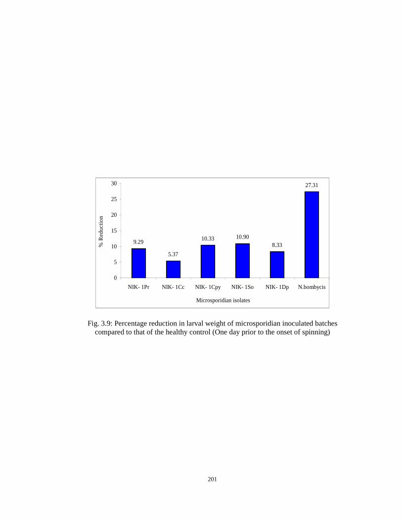
201
9.29
5.37
10.33 10.908.33
27.31
0
5
10
15
20
25
30
NIK- 1Pr NIK- 1Cc NIK- 1Cpy NIK- 1So NIK- 1Dp N.bombycis
Microsporidian isolates
% R
educ
tion
Fig. 3.9: Percentage reduction in larval weight of microsporidian inoculated batches compared to that of the healthy control (One day prior to the onset of spinning)