Microscopy 1
30
Microscopy 1 Biology 101A
-
Upload
cally-lowe -
Category
Documents
-
view
27 -
download
1
description
Microscopy 1. Biology 101A. Announcements. Quiz- Wed, not today- 8:10am-8:25am. Magnification and Resolution. Magnification provides no additional information Resolution often requires magnification. Magnification without resolution. Magnification without resolution. - PowerPoint PPT Presentation
Transcript of Microscopy 1

Microscopy 1
Biology 101A

Announcements
• Quiz- Wed, not today- 8:10am-8:25am

Magnification and Resolution
• Magnification provides no additional information
• Resolution often requires magnification

Magnification without resolution

Magnification without resolution

Magnification without resolution

Magnification without resolution

Magnification without resolution

Magnification without resolution

Contrast


Resolution is a measure of distance• Resolution = d = (.61λ)/N.A• d = distance between 2
pts.• λ = wavelength of light• N.A. = Numerical
Aperture• N.A. = n sin α• n = refractive index• α = half-angle of cone of
light
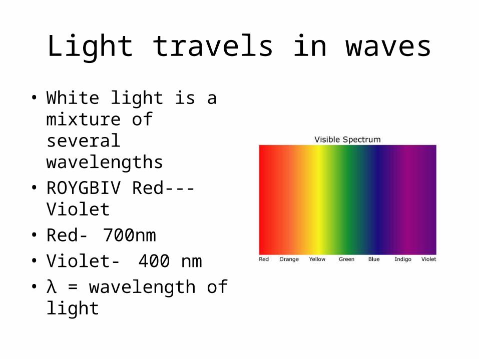
Light travels in waves
• White light is a mixture of several wavelengths
• ROYGBIV Red---Violet• Red- 700nm• Violet- 400 nm• λ = wavelength of light

Refractive index• Refractive indices:• Air-• Vacuum 1 (exactly) • Air @ STP 1.0002926 • Gases @ 0 °C and 1 atm• Air 1.000293
[1]
• Helium 1.000036 • Water 1.333 • Ethyl alcohol (ethanol) 1.361 • Diamond 2.419• Amber 1.55• Sodium chloride 1.50 • Other materials • Pyrex (a borosilicate glass) 1.470 [
• Ruby 1.760• Glycerol 1.4729 • Cubic zirconia 2.15 - 2.18• Diamond 2.419 • Gallium(III) arsenide 3.927• Silicon 4.01

Field of View
• Actual diameter of microscope image at a certain mag.
• As magnification increases, field of view _______.

Depth of field
• A measure of the thickness of the focal plane of an image
• As magnification increases, depth of field _______________.

Depth of field in Photography
• Shallow depth of field prevents an entire object from being in focus

Depth of field
• Can be exploited for identifying layers in a substance

Quiz Wednesday
• Microscope care and maintenance (how to keep from breaking them)
• Microscpe anatomy (labelling of parts)
• Microscope principles (wavelength, magnification, etc.)

Microscope anatomyParts
1. Headpiece
2. Eyepieces
3. Nosepiece/turret
4. Objective leses
5. Stage clips
6. Stage
7. Condenser
8. Substage filter holder/iris diapragm lever
9. Mirror
10. Base
11. Condenser adjustment knob
12. Fine focus knob
13. Coarse focus knob
14. Arm

Phase-contrast

Electron Microscopes• Use electrons instead of light• Use magnets instead of glass
lenses• electron wavelengths are
much shorter than those of light
• TEM- sends electrons through a specimen
• SEM- specimen spraypainted with gold

TEM

SEM
• Only looks at surfaces• Generates 3-D image• Often color-retouched

Fluorescence microscopes look at a single wavelength of light at a time

Green Fluorescent Protein• discovered in 1960s by Dr. Frank
Johnson and colleagues
• closely related to jellyfish aequorin
• absorption max = 470nm
• emission max = 508nm
• 238 amino acids, 27kDa
• “beta can” conformation: 11 antiparallel beta sheets, 4 alpha helices, and a centered chromophore
• amino acid substitutions result in several variants, including YFP, BFP, and CFP
40 Å
30 Å
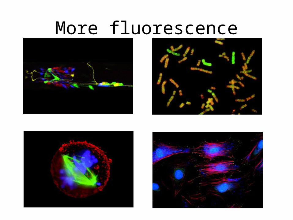
More fluorescence

• If you reach your fingers into the area under the microscope stage without looking, which of the following is most likely to be damaged?
• A. The base of the microscope• B. The coarse adjustment knob.• C. The fine focus adjustment knob.• D. The blue frosted glass filter.• E. The mirror

• When carrying a microscope, your principal grip should be on its:
• A. tube. • B. nosepiece. • C. stage.• D. arm. • E. coarse adjustment knob.

Iris diaphragms are most easily damaged by: A. being touched by someone’s fingers. B. too high a light intensity. C. having too little oil on their leaves. D. too low a light intensity. E. Not being wiped clean before every use.•