Methods for Detection and Quantification of Aflatoxins - InTech
22
See discussions, stats, and author profiles for this publication at: https://www.researchgate.net/publication/260085918 Instrumentation and Control to Improve the Crop Yield Chapter · January 2014 DOI: 10.1007/978-3-319-03880-3_13 CITATIONS 2 READS 254 10 authors, including: Some of the authors of this publication are also working on these related projects: Desarrollo integral de la cadena de valor de xoconostle en Guanajuato View project Obtención de imágenes, caracterización gravimétrica y perfil fenólico en dos estados de madurez fenológica en Opuntia joconostle var. Cuaresmeño. View project Maria Acosta-Navarrete Universidad Tecnológica del Suroeste de Guanajuato 11 PUBLICATIONS 8 CITATIONS SEE PROFILE José Alfredo Padilla-Medina Instituto Tecnológico de Celaya 54 PUBLICATIONS 220 CITATIONS SEE PROFILE Enrique Botello-Alvarez Instituto Tecnológico de Celaya 53 PUBLICATIONS 407 CITATIONS SEE PROFILE Juan Prado-Olivarez Instituto Tecnológico de Celaya 22 PUBLICATIONS 485 CITATIONS SEE PROFILE All content following this page was uploaded by Jesus Roberto Millan-Almaraz on 09 February 2014. The user has requested enhancement of the downloaded file.
Transcript of Methods for Detection and Quantification of Aflatoxins - InTech

See discussions, stats, and author profiles for this publication at: https://www.researchgate.net/publication/260085918
Instrumentation and Control to Improve the Crop Yield
Chapter · January 2014
DOI: 10.1007/978-3-319-03880-3_13
CITATIONS
2READS
254
10 authors, including:
Some of the authors of this publication are also working on these related projects:
Desarrollo integral de la cadena de valor de xoconostle en Guanajuato View project
Obtención de imágenes, caracterización gravimétrica y perfil fenólico en dos estados de madurez fenológica en Opuntia joconostle
var. Cuaresmeño. View project
Maria Acosta-Navarrete
Universidad Tecnológica del Suroeste de Guanajuato
11 PUBLICATIONS 8 CITATIONS
SEE PROFILE
José Alfredo Padilla-Medina
Instituto Tecnológico de Celaya
54 PUBLICATIONS 220 CITATIONS
SEE PROFILE
Enrique Botello-Alvarez
Instituto Tecnológico de Celaya
53 PUBLICATIONS 407 CITATIONS
SEE PROFILE
Juan Prado-Olivarez
Instituto Tecnológico de Celaya
22 PUBLICATIONS 485 CITATIONS
SEE PROFILE
All content following this page was uploaded by Jesus Roberto Millan-Almaraz on 09 February 2014.
The user has requested enhancement of the downloaded file.
7
Methods for Detection and Quantification of Aflatoxins
Alejandro Espinosa-Calderón, Luis Miguel Contreras-Medina,
Rafael Francisco Muñoz-Huerta, Jesús Roberto Millán-Almaraz,
Ramón Gerardo Guevara González and Irineo Torres-Pacheco C.A. Ingeniería de Biosistemas, División de Estudios de Posgrado, Facultad de Ingeniería,
Universidad Autónoma de Querétaro, Querétaro, Qro., México
1. Introduction
Mycotoxins are fungal toxic metabolites which naturally contaminate food and feed. aflatoxins (AFs), a kind of mycotoxins, are the main toxic secondary metabolites of some Aspergillus moulds such as Aspergillus flavus, Aspergillus parasiticus and the rare Aspergillus nomius (Ali et al., 2005, Alcaide-Molina et al., 2009). Such toxins can be separated into aflatoxins B1, B2, G1, B2a and G2a. Its order of toxicity is B1 > G1 > B2 > G2. Letters ‘B’ and ‘G’ refer to its blue and green fluorescence colors produced by these compounds under UV light. Numbers 1 and 2 indicate major and minor compounds, respectively (Weidenbörner, 2001; Hussein & Brasel, 2001). A. flavus only produces B aflatoxins, while A. parasiticus and A. nomius also produce G aflatoxins (Alcaide-Molina et al., 2009). Aflatoxins are produced on various grains and nuts, e.g., corn, sorghum, cottonseed, peanuts, pistachio nuts, copra, cereals, fruits, oilseeds, dried fruits, cocoa, spices and beer in the field and during storage. AFs occur mainly in hot and humid regions where high temperature and humidity are optimal for moulds growth and toxins production (Ventura et al., 2004; Zollner & Mayer-Helm, 2006). Its presence is enhanced by factors as stress or damage to the crop due to drought before harvest, insect activity, soil type and inadequate storage conditions (Alcaide-Molina et al., 2009). Aflatoxins, when ingested, inhaled or adsorbed through the skin, have carcinogenic, hepatotoxic, teratogenic and mutagenic effects in human and animals (rats, ferrets, ducks, trout, dogs, turkeys, cattle and pigs) (Anwar-Ul_Haq & Iqbal, 2004) even at very small concentrations. When aflatoxins B1 is ingested by cows, it is transformed into its hydroxylated product, AFs M1 and M2. Such aflatoxins is secreted in the milk and is relatively stable during milk pasteurization, storage, and preparation of various dairy products (Stroka & Anklam, 2002). Among the more than 300 known mycotoxins, aflatoxins represent the main threat worldwide. After 1975 there has been an increased concern about the possibility of the presence of carcinogenic mold metabolites, particularly aflatoxins in food and animal feed products. Although aflatoxins are regulated in more than 80 countries, their legislation is not yet completely harmonized at the international level (Cucci et al., 2007). Several
www.intechopen.com

Aflatoxins – Detection, Measurement and Control
110
institutions around the world have classified and regulated aflatoxins in food. The European Union (EU) has the most rigorous regulations concerning mycotoxins in food. The limits of AFB1 and total AF in foods are 5 and 10 µg/kg, respectively, in more than 75 countries around the world whilst they are 2 and 4 µg/kg in the European Union (EU) (Herzallah, 2009). The maximum residue levels for total AFs and also for the most toxic of them (AFB1) according to the EU Commission Regulations are 2 and 4 g/kg, respectively. The maximum legal limit for AFM1 in milk is set at 0.05µg/kg (50 ppt) for all EU Member States, and 25 ppt for baby food (Cucci et al., 2007). The European Committee Regulations (ECR) has established the maximum acceptable level of AFB1 in cereals, peanuts and dried fruits for direct human consumption in 4ng/g for total aflatoxins (AFB1, AFG1, AFB2, AFG2) and 2ng/g for AFB1 alone (Ricci et al., 2007). The International Agency for Research on Cancer (IARC) classified aflatoxins as Group 1 of human carcinogens (Alcaide-Molina et al., 2009). In USA, the U.S. Department of Agriculture and the U.S. Food and Drug Administration (FDA) have established an "actionable" level of 15-20 ppb of AFs in animal feed products. Because of such facts, several methodologies for detection and quantification of AFs have been developed. The principal immunochemical based assay is the widespread enzyme linked immunosorbent assay (ELISA). Other methodologies base their performance upon electrochemical and optical principles such as: chromatography, UV-absorption, spectrometry, fluorescence and immunochemical assay tests. The aforementioned methods require well equipped laboratories, trained personnel, harmful solvents and several hours to complete an assay. Novel methods for detection of aflatoxins try to avoid these disadvantages. Among such novel methods, it can be found: biosensors, electrokinetics, electrochemical transduction, amperometric detection, and adsorptive stripping voltammetry. Each of the aforementioned methodologies has its own advantages and limitations according to sensitivity, easiness of use and cost-effectiveness. The objective of this chapter is to provide a general overview of the different methodologies to detect and quantify aflatoxins in the food analysis field.
2. Electrochemicals techniques
Aflatoxins can be measured by the use of electricity and electrochemical immunosensors. These immunosensors consist of a pair of electrodes (measuring and reference), implemented by using the screen-printing technique. The measuring electrode is coated with specific antibodies which will retain interest aflatoxins in the sample, whereas the other electrode (reference) is commonly made of a combination of Ag / AgCl. The measurement procedure is similar to that carried out by the ELISA test (Enzyme Linked immunoabsorbent Assay). ELISA process is done by taking a sample of the substance to be measured and mixed with a known portion of conjugated aflatoxins with a special enzyme in a microtiter plate hole, and then it is inserted the measuring electrode. In this way, free aflatoxins in the sample compete for fill the places available (antibodies) in the measuring electrode. After some stabilization time, the measuring electrode is removed from the sample, washed with a buffer solution that removes all traces of the sample and leaves intact the electrode coating with aflatoxins that were captured but are not conjugate. After cleaning procedure, the electrode is introduced in a substrate solution that reacts with enzymes in aflatoxins conjugate, changing the electrical conductivity of the substrate depending on the amount of labeled aflatoxins antibodies attached to the electrode. Thus,
www.intechopen.com

Methods for Detection and Quantification of Aflatoxins
111
the greater will see the effect of aflatoxins marked, the lower the concentration of free aflatoxins in the sample. However, the electrodes developed by Tan et al. (2009), were coated with conjugate aflatoxins instead of being coated with specific antibody, whereas the sample was mixed with the antibody. In this manner, some antibodies will be captured by free aflatoxins in the sample and some others by those attached to the electrode. Following that, the electrode is washed and it is placed into a solution with antibodies conjugated with alkaline phosphatase enzyme that binds to the antibodies that are bound to conjugate aflatoxins onto the electrode. After that, the electrode is immersed in the substrate solution in order that antibodies conjugate react and cause a change in electrical conductivity. Some methods have been reported the use of simple electrodes (Rameil et al., 2010; Tan et al., 2009), while others have made use of multiple electrodes (Neagu et al., 2009; Piermarini et al., 2007), where the latter has shown to have advantages over the first in that: it is more user friendly; it is possible to carry out many experiments in parallel with different samples; and it reduces the time required for new procedures (Piermarini et al., 2007). In order to measure the electrical conductivity in the electrodes there are different techniques, such as intermittent pulse amperometric (IPA), potentiometry, or linear sweep voltage (LSV). The intermittent pulse amperometric technique involves the application of a periodic pulse of some duration fixed voltage across the electrodes coated and reference measurement, while the measured current varies depending on the conductivity of the substance. Moreover, in the technique of potentiometry, the measuring electrode coated is immersed in substrate solution without contaminating aflatoxins until a stable electrical potential is obtained, called the potential base. This potential varies depending on the amount of aflatoxins contained in the sample. In the linear sweep voltage technique, the sample is fed with a voltage which changes linearly, with a fixed slope. The ability of these techniques to detect aflatoxins depends on many factors, including the type of substrate solution that is used, as is the case reported by Rameil et al. (2010), where it was shown that the use of 3 - (4-hydroxyphenyl ) propionic acid (p-HPPA), being a little toxic substance and does not require the use of organic solvents, can increase the conductivity of the substrate in potentiometry to measure aflatoxins M1 in milk. Another factor is the concentration of antibodies in the lining of the electrode, since the higher concentration of these, it can be reached higher peak current than in IPA technique, although the relationship between antibody concentration and electric current conducted is linear in a certain range, such as Tan et al. (2009) work suggests, where the linear range extends from a dilution of 1:30000 to 1:10000 of antibody against aflatoxins B1 found in rice, being the latter dilution which gave the best results. Another point to consider is the detection limit, defined as the maximum decrease in signal equal to three times the standard deviation measured in the absence of aflatoxins to be determined. Detection limits down to 1 pg/ml have been obtained in the measurement of aflatoxins M1 in milk (Neagu et al., 2009); meanwhile, detection of aflatoxins B1 in rice has reached the limit 0.06 ng/ml (Tan et al., 2009). There are also other measurement devices, as the case of piezoelectric immunosensors. Piezoelectricity is the property possessed by certain materials in which either generates a potential difference from applied mechanical deformation or vice versa (Webster 1999), so that materials that have this feature can resonate at certain frequencies. One of the most common piezoelectric materials is quartz crystal, used by Jin et al. (2009) as a sensor for
www.intechopen.com
Aflatoxins – Detection, Measurement and Control
112
measuring aflatoxins B1 in milk. In this case, the crystal was treated to bind aflatoxins AFB1-BSA conjugate to the material for later subjecting to a similar procedure as mentioned by Tan et al. (2009), differing from this one in that the antibodies attached to conjugate aflatoxins attached to crystal, were marked with gold nanoparticles coated with antibody detector first. The concentration of aflatoxins will be reflected in this case as a change in the resonant frequency of the crystal, as reported by Jin et al. (2009) for the case of aflatoxins B1, where there is a linear relationship between the frequency of resonance and the logarithm of the concentration of aflatoxins.
3. Chromatography
Chromatography is one of the most popular methods to analyze mycotoxins such as aflatoxins. The most common techniques of chromatography are Gas chromatography (GC), liquid chromatography (LC), High performance liquid chromatography (HPLC) and Thin-layer chromatography (TLC). From these methods, LC and HPLC are the most used. In many cases, they are followed by fluorescence detections stage (Cavaliere et al., 2006). LC, TLC and HPLC are the most used quantitative methods in research and routine analysis of aflatoxins (Vosough et al., 2010); these techniques offer excellent sensitivities but they frequently require skilled operators, extensive sample pretreatment and expensive equipment (Sapsford et al., 2006).
3.1 Liquid chromatography
At the beginning the only separative method was GC, nevertheless, it is restricted to a small set of biological molecules for instance. Those should not be volatiles or should be derivatizated (Roux et al., 2011). LC is other separative method which offers good sensitivity, high dynamic range, versatility and soft ionization conditions that permit access to the molecular mass of intact biological molecules. LC is usually coupled to fluorescence detection stage (FLD), UV absorption and amperometric detection (Elizalde-González, 1998) with pre-column derivatization or post-column derivatization. Extraction and clean up procedures for aflatoxins analysis typically rely on solid phase extraction (SPE) with different absorbent materials. A particular case of SPE is immunoaffinity columns. Improvements have been done, creating techniques based on LC, such as: TLC and Reversed-phase high performance liquid chromatography (RP-HPLC) (Elizalde-González, 1998). LC coupled with fluorescence stage use the aflatoxins fluorescence properties to quantify them. So that, by improve this property it can be obtained better sensibility for aflatoxin detection. The most common techniques to improve fluorescence properties are the use of pre-column derivatization with trifluoretic acid and post-column derivatization with iodine or bromine (Elizalde-González, 1998). Other studies have been done in order to obtain enhancement of the fluorescence emissions of aflatoxins. Franco et al. (1998) collected emission data for AFQ1, AFM1, AFP1 in solvents usually used for their chromatography separation in absence and in presence of different cyclodextrins. Such experiment was made in order to be applied principally in liquid chromatography.
3.2 Thin-Layer Chromatography (TLC)
Thin-layer chromatography is widely used in laboratories throughout the world for food analysis and quality control. Applications of TLC have been reported in areas of food composition, intentional additives, adulterants, contaminants, etc. TLC has been used to
www.intechopen.com
Methods for Detection and Quantification of Aflatoxins
113
analyze agricultural products and plants. It has advantages as: simplicity of operation; availability of many sensitive and selective reagents for detection and confirmation without interference of the mobile phase; ability to repeat detection and quantification; and cost effectiveness analysis, because many samples can be analyzed on a single plate with low solvent usage, and the time that TLC employs to analyze the sample is less that LC method (Sherma, 2000; Fuch et al., 2010). The most important differences between TLC and HPTLC are: the different particular size of stationary phase; the care used to apply the samples; and the way to process the obtained data (Fuch et al., 2010). Diprossimo et al. (1996) present a work where show that TLC was superior to the methods of BF (Best food) CB-RCS-Mod (modified CB method-Rapid Modification of the Cottonseed Method) in terms of less fluorescence interferences, better solvent efficiency, and lower detection levels. Results obtained using TLC method compared to HPLC and enzyme-linked immunosorbent assay (ELISA) was found to agree among method but TLC was least expensive (Schaafsma et al., 1998). Papers that use TLC methods to detect and quantify aflatoxins use sample clean-up based on immunoaffinity columns. Therefore, they avoid interfering compounds and allow visual quantification of aflatoxins at concentrations of less than 1 ng/g (Stroka et al., 2000). Immnunoaffinity procedures provide very clean extracts because the sample is cleaned of interference substances. It also permits an easy aflatoxins determination, since they are applicable for automated sample clean-up (Stroka et al., 2002). Because of the advantages of this method, researches have been focused on them to develop new techniques to improve the methodologies for quantification of aflatoxins.
3.3 High Performance Liquid Chromatography (HPLC)
As aforementioned, HPLC is one of the most common methods to detect and quantify aflatoxins in food. It has been used jointly with techniques such as UV absorption, fluorescence, mass spectrometry and amperometric detectors. Elizalde-González et al. (1998) analyzed aflatoxins B1,B2, G1 and G2 based on HPLC and amperometric detection, and report that it is possible to detect 5 ng of all four aflatoxins. This proposed method is recommended for detection and quantification of the less toxic aflatoxin B2, which is presented in grains. Quinto et al. (2009) proposed a new method for determine aflatoxins B1, B2, G1, and G2 in cereal foods. This method is based on solid phase microextraction coupled with HPLC and a post-column photochemical derivatization to improve the fluorescence of analytes and fluorescence detection. Such method is fast compared with the complete analytical process that uses Immunoaffinity column. However, its sensibility is below the legal limits. Vosugh et al. (2009) present a work that uses HPLC in conjunction with diode array detector (DAD) and a second order iterative algorithm called parallel factor analysis (PARAFAC). Such method is used for quantifing aflatoxins B1, B2, G1, and G2 in pistachio nuts, this work also use a solid phase extraction stage as a clean-up procedure. Manneta et al. (2005) presents a new method with fluorescence detection using pyridinium hydrobromide perbromide as a post-column derivatization agent to determine aflatoxin M1 in milk and cheese. The detection limits obtained were of 1 ng/kg for milk and 5 ng/kg for cheese that are 50-fold lower than the maximum residue level (MRL) for AFM1 in milk and 40-fold than MRL for AFM1 in cheese set by various European countries. An interesting application of HPLC is the combination of immobilized enzyme reactor (IMER) in on-line high performance liquid chromatography. This combination allows the selectivity, rapidity and non-destructive, reproducibility of this chromatographic system to
www.intechopen.com
Aflatoxins – Detection, Measurement and Control
114
be combined with the specification and sensitivity for an enzymatic reaction (Girelli & Mattei, 2005). Derivatization with a fluorophore enhances the natural fluorescence of aflatoxins and improves detectability. The pre-column approach uses the formation of the corresponding hemiacetals using trifluoroacetic acid (TFA), while the post-column one utilizes either bromination by an electrochemical cellor in addition of bromide, or pyridinium hydrobromide perbromide, for the mobile phase and the formation of an iodine derivative. Even though the optical devices have dominated the traditional methods for HPLC, the present trend is to use mass detectors in the different HPLC types and configurations. This is because of the universal, selective and sensitive detection they provide (Alcaide-Molina, 2009). There are several techniques that use chromatography for aflatoxin analysis in food (principally in milk, cheese, corn, peanuts, nuts). Commonly the quantification of the aflatoxins is made by a fluorescence detector that takes advantage of fluorescence properties of aflatoxins under determined wavelength. As a result, researchers have been focused on improving these fluorescence properties to develop more sensitive methods than the commonly used so far. Currently techniques such as pre-column derivatization and post-column derivatization are commonly used to improve aflatoxins fluorescence properties. They also have a clean-up stage to obtain a more pure sample, permiting a better quantification. Some of the common methods used in the clean-up stage are: immunoaffinity column and solid phase extraction.
3.4 Electrokinetics
HPLC is a method for detection of aflatoxins which often is enhanced by other techniques,
resulting on alternative chromatographic methods. Accomplishing techniques related to
electrokinetics are: Micellar electrokinetic chromatography (MEKC), reversed flow micellar
electrokinetic chromatography (RFMEKC), and capillary electrokinetic chromatography
(CEKC) with multiphoton excited fluorescence (MPE) detection, among others (Gilbert &
Vargas, 2003).
Electrokinetics consists on an interfacial double layer of charges effect in heterogeneous
fluids (Rathore and Guttman, 2003). Such effect generates the motion of the fluid due to an
external force. This external force may be of different natures, but it is called electrophoresis
when the force is an electric field; and capillary osmosis when the force is a chemical
potential gradient and the motion of liquid happens in a porous body.
Capillary electrophoresis is a technique that although not been widely available as an
alternative in many laboratories which routinely conduct HPLC, it has the advantage that it
avoids the use of organic solvents. aflatoxin B1 can be determined by capillary
electrophoresis (CEKC) with laser-induced fluorescence (LIF) detection (Maragos & Greer,
1997) after a clean-up process comparable to that required for HPLC, and with a very similar
sensitivity to it. Besides, Electrophoresis does not require derivatization of aflatoxins, being
that an advantage over HPLC. Sensitivity on CEKC can be further improved by using
multiphoton excitation. Detection at levels 104 better than previously achieved by capillary
separation in less than 90 seconds can be reached, which demonstrates the potential of this
technique (Wei et al., 2000).
Micellar electrokinetic chromatography (MEKC) is conducted in polyacrylamide-coated capillaries under almost complete suppression of electroosmotic flow (Janini et al., 1996).
www.intechopen.com
Methods for Detection and Quantification of Aflatoxins
115
When small amounts of organic solvents are used in the buffer system good separation of aflatoxins are achieved. Nonetheless, it has been probed only with standard buffers (Gilbert & Vargas, 2003).
4. Fluorescence
All the aflatoxins have a maximum absorption around 360 nm (Akbas and Ozdemir, 2006). Letters ‘B’ and ‘G’ of the aflatoxins refer to its blue (425nm) and green-blue (450nm) fluorescence colours produced by these compounds under Ultra Violet (UV) light. AFB1 is the most common aflatoxin; it is followed by the AFB2. AFG is fairly rare. The fluorescence emission of the G toxin is more than 10 times greater than that for the B toxin (Alcaide-Molina et al., 2009). Different techniques for detection of AFs related to fluorescence are exposed bellow.
4.1 Black light test
The black light test is a method which correctly identifies negative AFs samples with minimum expenditure of time and money. It consists on the illumination of the sample with a UV lamp. Tests should be made in a darkened area for best contrast. Fluorescence may be bright or dim, depending on the amount of fluorescing agent present. Polished metal surfaces reflect blue light, thus, users must be careful distinguishing fluorescence from such reflection. It is highly recommended to use safety goggles when working with the black light test. These goggles eliminate blue haze resulting from eye fluorescence caused by reflected longwave UV radiation. However, fluorescence does not happen exclusively when aflatoxins are present. There are other substances in food that fluoresce under long wave UV radiation. Fungi as Aspergillus niger, various Penicillium species, Aspergillus repens and other species do not produce aflatoxins, but may produce fluorescent harmless metabolites. Then, it can be said that fluorescence is not a specific indication of the presence of aflatoxins, although it may indicate that conditions have been favourable for growth of toxic molds (B-100 Series Ultraviolet Lamps, UVP). Furthermore, fluorescence is not stable. It disappears in 4 to 6 weeks of continuous exposure to visible or UV radiation although the toxin remains. Therefore, fresh samples must be taken. Hence, the reliability of the method depends on the size of the sample taken for analysis and how it is taken. A sample must be large enough to be representative of the entire lot and must be taken from all parts of the lot (B-100 Series Ultraviolet Lamps, UVP). The black light test is commonly applied on animal feed. However, it is only a preliminary confirmatory test; it does not give a quantitative indication. Thus confirmatory and quantitative measurements are needed to be applied to those samples that reacted positively to the black light test. Non-fluorescing samples need not be subjected to this. A quantitative screening test which commonly follows the black light test is small chromatographic column (mini-column) (B-100 Series Ultraviolet Lamps, UVP). After the quantitative test a judgment can be made as to whether or not accept a lot.
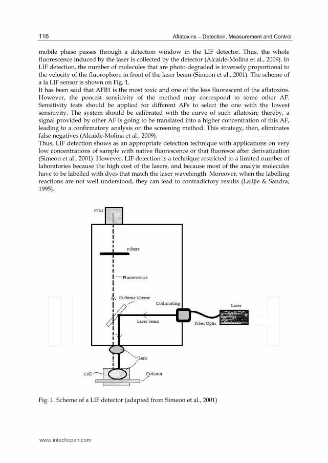
4.2 Laser-Induced Fluorescence (LIF) screening method
LIF detection technique was pioneered by Yeung (Novotny & Ishii, 1985). This screening method consists on a mobile phase which contains an eluted sample of aflatoxins. Such
www.intechopen.com
Aflatoxins – Detection, Measurement and Control
116
mobile phase passes through a detection window in the LIF detector. Thus, the whole fluorescence induced by the laser is collected by the detector (Alcaide-Molina et al., 2009). In LIF detection, the number of molecules that are photo-degraded is inversely proportional to the velocity of the fluorophore in front of the laser beam (Simeon et al., 2001). The scheme of a la LIF sensor is shown on Fig. 1. It has been said that AFB1 is the most toxic and one of the less fluorescent of the aflatoxins. However, the poorest sensitivity of the method may correspond to some other AF. Sensitivity tests should be applied for different AFs to select the one with the lowest sensitivity. The system should be calibrated with the curve of such aflatoxin; thereby, a signal provided by other AF is going to be translated into a higher concentration of this AF, leading to a confirmatory analysis on the screening method. This strategy, then, eliminates false negatives (Alcaide-Molina et al., 2009). Thus, LIF detection shows as an appropriate detection technique with applications on very low concentrations of sample with native fluorescence or that fluoresce after derivatization (Simeon et al., 2001). However, LIF detection is a technique restricted to a limited number of laboratories because the high cost of the lasers, and because most of the analyte molecules have to be labelled with dyes that match the laser wavelength. Moreover, when the labelling reactions are not well understood, they can lead to contradictory results (Lalljie & Sandra, 1995).
Fig. 1. Scheme of a LIF detector (adapted from Simeon et al., 2001)
www.intechopen.com
Methods for Detection and Quantification of Aflatoxins
117
4.3 High-Performance Liquid Chromatography (HPLC) with LIF
Fluorescence detection and electrochemical detection are the two sensitive detection means
most commonly used for quantitative studies in HPLC. This happens because the sensitivity
levels of those hybrid techniques are much better than the ones observed with conventional
fluorescence. It has been demonstrated the usefulness of LIF for sensitive detection in HPLC
and micro high-performance liquid chromatography (µHPLC) in sensing very low
concentrations of substances that can be excited in the near-UV range (325 nm) after
labelling at nanomolar concentrations (Folestad et al., 1985; Diebold et al., 1979). Thus, LIF-
HPLC method has become very popular and an essential detection technique in capillary
electrophoresis (CE). Its sensitivity has been increased by the use of photoactivation devices
(Reif & Metzger, 1995). Its popularity is due to its capability to detect substances at lower
ranges than the micromolar (Bayle et al., 2004). For more information about HPLC refer to
section 3.
It has been said that in LIF detection, the number of molecules that are photo-degraded is
inversely proportional to the velocity of the fluorophore in front of the laser beam. On the
other side, the sensitivity of detection in HPLC depends on the inner diameter of the
capillary connected to the output of the column. Therefore, at a constant flow-rate, the
sensitivity depends on the velocity of the fluorophore in front of the laser beam of the LIF,
and the solid angle of fluorescence collection by the optical arrangement (Simeon et al.,
1999). As a result, the union of LIF and HPLC offers a good compromise between sensitivity
and dead volumes (Simeon et al., 2001).
In flow injection experiments with LIF-HPLC systems, at a given diameter, the detector
signal will increase when increasing flow-rates if photochemical degradation is a limiting
factor (Simeon et al., 2001). Conversely, if the flow-rate is fixed, an increase in diameter is
expected to lead to a quadratic increase in the detector volume, generating also a quadratic
increase in the number of detectable molecules. Then can be said that if a larger volume is
irradiated at a larger capillary diameter, the efficiency of fluorescence collection is less
important than in the case of smaller capillaries (Simeon et al., 2001).
4.4 Photomultipliers (PTM)
Since Fluorescence systems have a wide sensitivity, they are a useful tool to measure AFM1 in milk, which legal limit is very low (about 50 ppt). These systems are suitable for preliminary screening at the earlier stages of the industrial process, and make it possible to discard contaminated milk stocks before their inclusion in the production chain (Cucci et al., 2007). PMTs are highly sensitive photomultipliers based flow through detection system suited for ultra low fluorescence, chemiluminescence or bioluminescence measurements (PMT-FL, FIAlab Instruments). Their photon counting photon counting sensor has a blue-green (280–630 nm) spectral response with a peak of quantum efficiency at 400 nm and ultra-low dark counts. The high sensitivity of these devices reaches parts per trillion, permitting measurements of extremely low fluorescence signals. These devices may work with an internal excitation lamp, a LED source or an SMA terminated fiber optic cable for use with an external lamp. They also count with removable emission and excitation filters, allowing placing the most suitable emission filter for selecting the spectral region of interest. The output of the PTMs is expressed in photo-counts, and corresponds to the entire signal integrated in the transmission spectral band of the emission filter. Therefore, the signal acquired from a sample can also include a background contribution due to the solvent. In
www.intechopen.com
Aflatoxins – Detection, Measurement and Control
118
principle, the latter can contribute to the actual fluorescence of the substance under analysis with a spurious signal of intrinsic fluorescence or Raman, depending on the excitation wavelength (Cucci et al., 2007). The use of cyclodextrin (CD) as fluorescence enhancer for aflatoxins detection is widely reported in the literature (Zhilong, G. & Zhujun, 1997; Dall'asta et al., 2003), nevertheless, an increased error bar affects measurements due to the CD scattering effects. The signal-to-noise ratio of these fluorescence measurements strongly depends on the type of cuvette used for containing the liquid sample. The cell geometry and its constituting material give rise to different effects, such as multiple reflections and stray-light. Small sample volumes and darkened walls are mandatory to achieve a better signal-to-noise ratio. Plastic cuvettes without the use of an additional fluorescence enhancer are not useful for the implementation of an early-warning system. Conversely, quartz cuvettes perform very well (Cucci et al., 2007). Then, PTMs are compact and easy-to-handle sensors for the rapid detection of low concentrations of AFM1 in liquid solutions without the need for pre-concentration of the sample. They can be used as quick “threshold indications” and as an “early warning system”, so as to rapidly single out risk/alarm situations (Cucci et al., 2007).
5. Ultra violet absorption
It has been said that all the aflatoxins have a maximum absorption around 360 nm with a molar absorptivity of about 20,000 cm2 /mol (Akbas & Ozdemir, 2006). But, even though aflatoxins could be detected by UV absorption, the sensitivity of such systems is not sufficient to detect these compounds at the parts per billion (ppb) levels required for food analyses (Alcaide-Molina et al., 2009). The detection limit of UV sensors reaches micromolar ranges (Couderc et al., 1998). This is why fluorescence (FL) techniques have become more popular for AFs detection. For overcoming the named limitation, UV absorption technique is usually combined with HPLC systems. Experimental results indicate that the detection limit of aflatoxins is enhanced by the proper method of extraction and clean-up process (Göbel & Lusky, 2004; Ali et al., 2005). For example, the selected clean-up and extraction procedures should minimize the interfering substances and matrix effect on the elution and separation of aflatoxins (Akiyama et al., 2001). Such important factors, correctly applied, may be of great importance to help the less sophisticated laboratories with HPLC instruments equipped with UV detector to detect aflatoxins with a precision that complies with the international guidelines and regulations. Then, even though, HPLC-UV systems still are less sensitive than HPLC-FL systems, especially at low AF levels (Herzallah, 2009), HPLC-UV systems indicate to be accurate, precise, and consequently, reliable enough for determination of aflatoxins in food, with low duration and running cost.
6. Spectrometry
6.1 Ion mobility spectrometry
The Ion mobility spectrometry is a technique that is used in the characterization of chemicals on the basis of speed acquired by the gas-phase ions in an electric field. This technique has been used to determine the concentration of aflatoxins, as evidenced by the work of Sheibani
www.intechopen.com
Methods for Detection and Quantification of Aflatoxins
119
et al. (2008) in which are detected and quantified the concentration of aflatoxins B1 and B2 in pistachio. It has certain advantages in common with the FT-NIR, and low detection limit, fast response, simplicity, portability, low cost. To detect aflatoxins in a sample, this is evaporated and mixed with a carrier gas. Then it is
entered into the Ion Mobility Spectrometer (IMS) where the mixture is ionized and passed
through an electric field gradient, where ions of different substances will travel at different
speeds. The study by Sheibani et al. (2008) shows that using this technique is impossible to
quantify as low as 0.25 ng.
6.2 Fourier Transform Near Infrared (FT-NIR) spectrometry
This technique has been underutilized for the detection of aflatoxins due to calibration
requirements required against standard reference chemical processes (Tripathi & Mishra,
2009). Despite of the aforementioned limitations, this technique has some advantages, such
as: fast and easy equipment operation, good accuracy, precision, performing nondestructive
analyzes, prediction of chemical and physical sample from a single spectrum parameters
from a single spectrum enabling several components to be determined simultaneously
based on the use of multivariate calibrations.
It basically consists of measuring the absorbance of the sample to light whose wavelength
varies in the range known as the Near Infrared (NIR). In the work of Tripathi & Mishra
(2009) it is found that for the correct quantification of aflatoxins B1 in chili powder
network readings were taken in the range of 6900.3 - 4998.8 cm-1 and also in the range of
4902.3 - 3999.8 cm-1, excluding the water absorption bands (5155 and 7000 cm-1). Good
results were obtained with respect to chemical techniques such as High Performance
Liquid Chromatography (HPLC) and Thin Layer Chromatography (TLC), although its
detection range is between 15 to 500 mg / kg which is slightly high compared to these
techniques.
7. Biosensors
The term biosensors refers generally to a small, portable and analytical device based on the
combination of recognition biomolecules with an appropriate transducer, and able of
detecting chemical or biological materials selectively and with a high sensitivity (Paddle,
1996). Its principle of detection is the specific binding of the analyte of interest to the
complementary biorecongnition element immobilized on a suitable support medium. When
the analyte binds the element, there happens a specific interaction which results in a change
of one or more physico-chemical properties. Such properties may be: pH, electron transfer,
mass, or heat transfer that are detected and can be measured by a transducer. Depending of
the method of signal transduction, biosensors can be divided into different groups:
electrochemical, optical, thermometric, piezo-electric or magnetic. In the case of aflatoxin
detection, electrochemical and optical are the most commonly used (Velasco-Garcia &
Mottram, 2002). Until 1996 only few biosensors for toxins were recorded and most of them
were based on ELISA. The goal of the more recent studies is to simplify and expedite the
method of detection while maintenance and improvement of sensitivity is attempted
(Sapsford et al., 2006).
A method that has gained popularity is the use of antibodies to clean-up samples prior to measurement by LC of HPLC. Carlson et al. (2000) present an immune-affinity fluorometric
www.intechopen.com
Aflatoxins – Detection, Measurement and Control
120
biosensor where the sample was filtered through a column containing sepharosa beads to which the polyclonal aflatoxin-specific antibodies were joined. The beads with attached aflatoxins were subsequently rinsed to remove any impurities and interference. Posterior, an eluant solution was passed through the beads causing antibodies to release the bound aflatoxins. The analyte was collected and placed in a fluorometer. This system consists essentially of two subsystems a fluidics subsystem in charge of mechanical-handling and processing and an electro-optical system that add a miniature fluorometer. Sapsfor et al. (2006) present a system to detect and quantify foodborne contaminants using
an array biosensor. It is capable of measuring large pathogens such as the bacteria
Campylobacter jejuni and small toxins (mycotoxins ochratoxin A, fumonisin B, aflatoxin B1
and deoxynivalenol). The system is capable of multiple detections of aflatoxins in a short
time.
Aflatoxins have inhibitory effect on acetylcholinesterase (AchE) and their detection is
coupled with the decrease in the activity of AchE which is measured using a choline oxidase
amperometric biosensor (Nayak et al., 2009). Amperometric methods allow the detection of
low aflatoxin concentration that cannot be detected by classical spectrophotometry because
of the omission of the dilution step used in classical method.
Wang et al. (2009) present an implementation of Long range surface Plasmon – enhanced
fluorescence spectroscopy (SPFS) in an immunoassay based biosensor for the highly
sensitive detection of AFM1 in milk samples (LRSP). Here fluoropore-labeled molecules
captured on the sensor are exited with surface plasmons (SPs) and the emitted fluorescence
light is measured. The system takes the advantage of the electromagnetic intensity
improvement occurring upon the resonance excitation of SPs that increase the intensity of
fluorescence signal. This technique is based on surface Plasmon resonance which is
becoming popular for the detection of chemical and biological species.
Others tendencies are the use of nanotechnology to detect aflatoxins such as the paper
presented by Xiulan et al. (2004) where colloidal gold particles and antibodies were
combined and used to develop an immunochromatographic (IC) method for aflatoxin B1
analysis. The result of this was that the analysis could be completed in less than 10 minutes
and the lower test limit was 2.5ng/ml for aflatoxin B1. Such limit was increased in two times
of ELISA.
When aflatoxins are consumed by cattle, they are transformed into their hydroxylated
product, AFM1 that is known for its hepatotoxic and carcinogenic effects. To date, aflatoxins
are regulated in many countries because of the milk intake in infants is high and when they
are young the vulnerability to toxins is higher. Because of this, it is necessary to monitor
AFM1 in milk at ultra low level, so that, analytical methods with high detectability and
analytical throughput are required. Kanungo et al. (2011) present a novel approach where a
highly sensitive microplate sandwich ELISA was developed and integrated with Magnetic
nanoparticles (MNPs) which could detect ultra trace amount of AFM1 in milk. Sandwich-
type immunoassay is an effective bioassay due to the high specificity and sensitivity. MNPs
were used as an affinity capture column wherein immobilized antibodies on their surface
could capture AFM1 from milk sample.
According to the aforementioned, the new trends could be the use of nanoparticles in combinations of the commonly used techniques such as LC, HPLC, TLC and immunoassay techniques. These combinations are to improve the detection at ultra low level of compounds in order to diminish the risk that this kind of mycotoxins causes to
www.intechopen.com
Methods for Detection and Quantification of Aflatoxins
121
humankind. For doing this it is necessary to use methods that combine simplicity with high detectability.
8. Adsorptive stripping voltammetry
Adsorptive Stripping Voltammetry is a method based on accumulation and reduction of AFB1 and AFB2 species on the surface of hanging mercury drop electrode (HMDE). Such electrode offers both sensitivity and selectivity. The pioneers on this method applied to detection of aflatoxins are Hajian and Ensafi (2009), for more information refer to their article. Voltammetry is an electro-analytical method. It obtains information about the sample by measuring a current while the potential is varied (Komorsky et al., 1992). The voltammetry used in the experiment of Hajian and Ensafi had three-electrodes containing hanging mercury drop electrode as a working electrode, a carbon rod as an auxiliary electrode and an Ag/AgCl (3.0 M KCl) reference electrode. This method was proved only on AFs B1 and B2, where both aflatoxins were found to adsorb and undergo irreversible reduction reaction at the working mercury electrode (Rodriguez et al., 2005). Adsorptive Stripping Voltammetry is an electrochemical method which has no or very low dependence on pH. This dependence displayed only for B1 in the pH range of 5.0 to 6.0 (Sun et al., 2005). As it is expected in adsorption processes, by increasing accumulation time, the peak currents for both of the aflatoxins are increased and then leveled off because of the saturation of electrode surface (Hajian & Ensafi, 2009). Therefore, an accumulation time of 60 seconds is recommended for improving sensitivity. It is also recommended to use the extraction and clean-up method for aflatoxins that was used by Garden and Strachan (2001). Such extraction and clean-up method try to obtain the highest yield of aflatoxins with the minimum matrix effect. This method uses single standard addition method by spiking 10 ng / ml of standard aflatoxin followed with general procedure for voltammetric analysis. The total determination of aflatoxins is based on the next formula reported by Hajian and Ensafi (2009):
'
20ng P
Aflatoxin Cml P
(1)
Where: P’ is peak current of sample (nA), P is peak current of standard aflatoxin (after subtract from P’) (nA), C is the concentration of aflatoxin spiked in the cell (ng/ ml) and 20 is a factor value after the sample weight, volume of methanol/water used in the extraction and preparation of injection sample have been considered (Hajian & Ensafi, 2009). Adsorptive Stripping Voltammetry is a suitable method for determination of total aflatoxins (B1 and B2) in food. This method has some advantages such as high sensitivity, extended linear dynamic range, simplicity and speed (Hajian & Ensafi, 2009). The reliability of this method for determination of total aflatoxins is comparable to HPLC.
9. Miscellaneous methods
Different techniques have tried to offer new options for screening aflatoxins. Screening consists on rapid and/or in situ detection. There are two main difficulties for an effective
www.intechopen.com
Aflatoxins – Detection, Measurement and Control
122
screening method: the necessity of a very high sensitivity, which in fact is a necessity of any technique; and the demand of preliminary sample preparations. Some of these techniques, which are commented ahead, present a lack of applications because of their practical inconveniences or because they have not been proved yet with real samples (Gilbert & Vargas, 2003). Optical-fiber: Modular separation based on a fiber-optic sensor (Dickens & Sepaniak, 2000) has been tested in buffers, showing enough sensitivity (0.005 ng/ml for detection of aflatoxin B1). Unfortunately, it is limited to handling only liquid matrices. Electrochemical transduction: The interaction of the aflatoxin M1 with bilayer lipid membranes can be sensed electrochemically (Andreou & Nikolelis, 1997; Andreou et al., 1997) reaching a good specificity and speed of response. But, its principal negative factor is its detection limit 750 ng/ml, which is very unpractical. Flow injection monitoring: Stabilized systems of filter-supported membranes are capable of achieving significantly improved sensitivity (Andreou & Nikolelis, 1998). These membranes have been proposed for use in detecting aflatoxin M1 in cheese (Siontorou et al., 2000). Single strand DNA oligomers have been incorporated into the membranes to control surface electrostatic properties. This incorporation led to achievement a sensitivity much closer to regulatory limits, and with the ability to analyze four cheese samples per minute. Even though this technique appears to be a good option for in situ testing, it does not have yet many applications (Gilbert & Vargas, 2003).
9. Conclusion
Different methods for detection and quantification of aflatoxins have been discussed along this document. Through the researching made for this document, it has been found that the most popular methods are: ELISA, electrochemical immunosensors, chromatography and fluorescence. Even though ELISA is the most common and widespread technique, it has the disadvantage of requiring well equipped laboratories, trained personnel, harmful solvents and several hours to complete an assay. The detection and quantification of aflatoxins by using electrochemical immunosensor has proven to be efficient, easy to use and able to detect very low levels of these substances. Chromatography is a method which needs immunoaffinity columns and phase solid extraction need to be used to clean-up the sample, and also pre-column and post-column derivatization to enhance the aflatoxins fluorescence properties. So that, by improving these characteristics, it is possible to obtain a better quantification and sensibility. Fluorescence detection is a very good alternative to the conventional techniques used today. It has a very high sensitivity, especially when is combined with other techniques as HPLC. Some fluorescence techniques are used even in portable sensors, resulting on in situ measurements. Techniques such as FT-NIR spectrometer and IMS have proven to be quick, inexpensive and user-friendly, however, the FT-NIR technique shows lack of sensitivity when detecting low concentrations of aflatoxins. New techniques in this field are being developed in order to give a rapid and/or in situ detection of these toxins. Some examples of these new techniques are: optical-fiber, electrochemical transduction, low injection monitoring and biosensors. All of these, except for the biosensors, still present a lack of applications because of their practical inconveniences. The biosensors have been designed to overcome the drawbacks that the common tools employed to detect and quantify aflatoxins presents. They use the inherent fluorescence property that aflatoxins have to improve the detection, that in combination
www.intechopen.com
Methods for Detection and Quantification of Aflatoxins
123
with optical and immunochemical techniques used to clean-up the samples achieve a better quantification. Due to the risk that the aflatoxins represent to humans, the researchers all over the word are looking for methods to detect and quantify them. Apparently, the measurement of aflatoxins in the future tends to be the combination of optical, immunchemical, and fluorescence techniques.
10. Acknowledgements
Authors give thanks to Consejo Nacional de Ciencia y Tecnología (CONACyT), in Mexico, for its financial support through the scholarships with Registration Numbers: 239421 (AEC), 201401 (LMCM), 209021 (RFMH) and 207684 (JRMA). We also express our gratitude to CONACYT for funding the project CB-2008-01.000000000106133.
11. References
B-100 Series Ultra Violet Lamps, Ultra Violet Products (UVP). Nuffield Road, Cambridge,
UK.
PMT-FL fluorometer, FIAlab Instruments. Bellevue WA, USA.
Akbas, M. and Ozdemir, M. (2006). Effect of different ozone treatments on aflatoxin
degradation and hysicochemical properties of pistachios. Journal of the Science of
Food and Agriculture, 86(13):2099–2104.
Akiyama, H., Goda, Y., Tanaka, T., and Toyoda, M. (2001). Determination of aflatoxins B1,
B2, G1 and G2 in spices using a multifunctional column clean-up. Journal of
Chromatography A, 932(1-2):153–157.
Alcaide-Molina, M., Ruiz-Jiménez, J., Mata-Granados, J., and Luque de Castro, M. (2009).
High through-put aflatoxin determination in plant material by automated solid-
phase extraction on-line coupled to laser-induced fluorescence screening and
determination by liquid chromatography-triple quadrupole mass spectrometry.
Journal of Chromatography A, 1216(7):1115–1125.
Ali, N., Hashim, N., Saad, B., Safan, K., Nakajima, M., and Yoshizawa, T. (2005).
Evaluation of a method to determine the natural occurrence of aflatoxins in
commercial traditional herbal medicines from Malaysia and Indonesia. Food and
chemical toxicology, 43(12):1763–1772.
Andreou, V. and Nikolelis, D. (1997). Electro-chemical transduction of interactions of
aflatoxin M1 with bilayer lipid membranes (BLMs) for the construction of one-
shot sensors. Sensors and Actuators B: Chemical, 41(1-3):213–216.
Andreou, V. and Nikolelis, D. (1998). Flow injection monitoring of aflatoxin M1 in milk
and milk preparations using filter-supported bilayer lipid membranes. Analytical
chemistry, 70(11):2366–2371.
Bayle, C., Causs´e, E., and Couderc, F. (2004). Determination of aminothiols in body
fluids, cells, and tissues by capillary electrophoresis. Electrophoresis, 25(10-
11):1457–1472.
www.intechopen.com
Aflatoxins – Detection, Measurement and Control
124
Carlson, M., Bargeron, C., Benson, R., Fraser, A., Phillips, T., Velky, J., Groopman, J.,
Strickland, P., and Ko, H. (2000). An automated, handheld biosensor for aflatoxin.
Biosensors and Bioelectronics, 14(10-11):841–848.
Cavaliere, C., Foglia, P., Pastorini, E., Samperi, R., and Lagana, A. (2006). Liquid
chromatography/tandem mass spectrometric confirmatory method for
determining aflatoxin M1 in cow milk:: Comparison between electrospray and
atmospheric pressure photoionization sources. Journalof Chromatography A,
1101(1-2):69–78.
Couderc, F., Caussé, E., and Bayle, C. (1998). Drug analysis by capillary electrophoresis
and laser-induced fluorescence. Electrophoresis, 19(16-17):2777–2790.
Cucci, C., Mignani, A., Dall’Asta, C., Pela, R., and Dossena, A. (2007). A portable
fluorometer for the rapid screening of M1 aflatoxin. Sensors and Actuators B:
Chemical, 126(2):467–472.
Dallasta, C., Ingletto, G., Corradini, R., Galaverna, G., and Marchelli, R. (2003).
Fluorescence enhancement of aflatoxins using native and substituted
cyclodextrins. Journal of Inclusion Phenomena and Macrocyclic Chemistry,
45(3):257–263.
Dickens, J. and Sepaniak, M. (2000). Modular separation-based fiber-optic sensors for
remote in situ monitoring. Journal of Environmental Monitoring, 2(1):11–16.
Diebold, G., Karny, N., and Zare, R. (1979). Determination of zearalenone in corn by laser
fluorimetry. Analytical Chemistry, 51(1):67–69.
DiProssimo, V. and Malek, E. (1996). Comparison of three methods for determining
aflatoxins in melon seeds. Journal of AOAC International, 79(6):1330–1335.
Eiceman, G. and Karpas, Z. (2005). Ion mobility spectrometry. Number v. 1. CRC Press.
Elizalde-Gonzalez, M., Mattusch, J., and Wennrich, R. (1998). Stability and determination
of aflatoxins by high-performance liquid chromatography with amperometric
detection. Journal of Chromatography A, 828(1-2):439–444.
Franco, C., Fente, C., Vazquez, B., Cepeda, A., Mahuzier, G., and Prognon, P. (1998).
Interaction between cyclodextrins and aflatoxins Q1, M1 and P1:: Fluorescence
and chromatographic studies. Journal of Chromatography A, 815(1):21–29.
Folestad, S., Galle, B., and Josefsson, B. (1985). Small-bore LC/laser fluorescence. Journal
of chromatographic science, 23(6):273– 278.
Fuchs, B. et al. (2010). Lipid Analysis by Thin-Layer Chromatography-A Review of the
current State. Journal of Chromatography A.
Gilbert, J. and Vargas, E. (2003). Advances in sampling and analysis for aflatoxins in food
and animal feed. Toxin Reviews, 22(2-3):381–422.
Girelli, A. and Mattei, E. (2005). Application of immobilized enzyme reactor in on-line
high performance liquid chromatography: a review. Journal of Chromatography
B, 819(1):3–16.
Göbel, R. and Lusky, K. (2004). Simultaneous determination of aflatoxins, ochratoxin A,
and zearalenone in grains by new immunoaffinity column/liquid
chromatography. Journal of AOAC International, 87(2):411–416.
www.intechopen.com
Methods for Detection and Quantification of Aflatoxins
125
Hajian, R. and Ensafi, A. (2009). Determination of aflatoxins B1 and B2 by adsorptive
cathodic stripping voltammetry in ground-nut. Food Chemistry, 115(3):1034–
1037.
Herzallah, S. (2009). Determination of aflatoxins in eggs, milk, meat and meat products
using HPLC fluorescent and UV detectors. Food Chemistry, 114(3):1141–1146.
Hussein, H. and Brasel, J. (2001). Toxicity, metabolism, and impact of mycotoxins on
humans and animals. Toxicology, 167(2):101–134.
Kanungo et al., 2010] Kanungo, L., Pal, S., and Bhand, S. (2010). Miniaturised hybrid
immunoassay for high sensitivity analysis of aflatoxin M1 in milk. Biosensors and
Bioelectronics.
Kok, W. (1994). Derivatization reactions for the determination of aflatoxins by liquid
chromatography with fluorescence detection. Journal of Chromatography B:
Biomedical Sciences and Applications, 659(1-2):127–137.
Janini, G., Muschik, G., and Issaq, H. (1996). Micellar electrokinetic chromatography in
zero-electroosmotic flow environment. Journal of Chromatography B: Biomedical
Sciences and Applications, 683(1):29–35.
Jin, X., Jin, X., Chen, L., Jiang, J., Shen, G., and Yu, R. (2009). Piezoelectric immunosensor
with gold nanoparticles enhanced competitive immunoreaction technique for
quantification of aflatoxin B1. Biosensors and Bioelectronics, 24(8):2580–2585.
Komorsky-Lovric, S., L. M. and Branica, M. (1992). Peak current–frequency relationship in
adsorptive stripping square-wave voltammetry. Journal of Electroanalytical
Chemistry, 335(1-2):297–308.
Lalljie, S. and Sandra, P. (1995). Practical and quantitative aspects in the analysis of FITC
and DTAF amino acid derivatives by capillary electrophoresis and LIF detection.
Chromatographia, 40(9):519–526.
Manetta, A., Di Giuseppe, L., Giammarco, M., Fusaro, I., Simonella, A., Gramenzi, A., and
Formigoni, A. (2005). High-performance liquid chromatography with post-
column derivatisation and fluorescence detection for sensitive determination of
aflatoxin M1 in milk and cheese. Journal of Chromatography A, 1083(1-2):219–
222.
Nayak, M., Kotian, A., Marathe, S., and Chakravortty, D. (2009). Detection of
microorganisms using biosensors–A smarter way towards detection techniques.
Biosensors and Bioelectronics, 25(4):661–667.
Neagu, D., Perrino, S., Micheli, L., Palleschi, G., and Moscone, D. (2009). aflatoxin M1
determination and stability study in milk samples using a screen-printed 96-well
electrochemical microplate. International Dairy Journal, 19(12):753–758.
Novotny, M. and Ishii, D. (1985). Microcolumn separations: columns, instrumentation,
and ancillary techniques. Elsevier Science Ltd.
Paddle, B. (1996). Biosensors for chemical and biological agents of defence interest.
Biosensors and Bioelectronics, 11(11):1079–1113.
Piermarini, S., Micheli, L., Ammida, N., Palleschi, G., and Moscone, D. (2007).
Electrochemical immunosensor array using a 96-well screen-printed microplate
for aflatoxin B1 detection. Biosensors and Bioelectronics, 22(7):1434–1440.
www.intechopen.com
Aflatoxins – Detection, Measurement and Control
126
Quinto, M., Spadaccino, G., Palermo, C., and Centonze, D. (2009). Determination of
aflatoxins in cereal flours by solid-phase microextraction coupled with liquid
chromatography and post-column photochemical derivatization-fluorescence
detection. Journal of Chromatography A, 1216(49):8636–8641.
Rameil, S., Schubert, P., Grundmann, P., Dietrich, R., and Märtlbauer, E. (2010). Use of 3-
(4-hydroxyphenyl) propionic acid as electron donating compound in a
potentiometric aflatoxin M1-immunosensor. Analytica chimica acta, 661(1):122–
127.
Rastogi, S., Das, M., and Khanna, S. (2001). Quantitative determination of aflatoxin B1-
oxime by column liquid chromatography with ultraviolet detection. Journal of
Chromatography A, 933(1-2):91–97.
Rathore, A. and Guttman, A. (2003). Electrokinetic phenomena: principles and
applications in analytical chemistry and microchip technology. CRC Press.
Reif, K. and Metzger, W. (1995). Determination of aflatoxins in medicinal herbs and plant
extracts. Journal of Chromatography A, 692(1-2):131–136.
Ricci, F., Volpe, G., Micheli, L., and Palleschi, G. (2007). A review on novel developments
and applications of immunosensors in food analysis. Analytica chimica acta,
605(2):111–129.
Rodriguez, J., Berzas, J., Castaneda, G., and Rodriguez, N. (2005). Voltammetric
determination of Imatinib (Gleevec) and its main metabolite using square-wave
and adsorptive stripping square-wave techniques in urine samples. Talanta,
66(1):202–209.
Roux, A., Lison, D., Junot, C., and Heilier, J. (2010). Applications of liquid
chromatography coupled to mass spectrometry-based metabolomics in clinical
chemistry and toxicology: A review. Clinical Biochemistry.
Sapsford, K., Ngundi, M., Moore, M., Lassman, M., Shriver-Lake, L., Taitt, C., and Ligler,
F. (2006a). Rapid detection of food-borne contaminants using an array biosensor.
Sensors and Actuators B: Chemical, 113(2):599–607.
Sapsford, K., Taitt, C., Fertig, S., Moore, M., Lassman, M., Maragos, C., and Shriver-Lake,
L. (2006b). Indirect competitive immunoassay for detection of aflatoxin B1 in corn
and nut products using the array biosensor. Biosensors and Bioelectronics,
21(12):2298–2305.
Schaafsma, A., Nicol, R., Savard, M., Sinha, R., Reid, L., and Rottinghaus, G. (1998).
Analysis of Fusarium toxins in maize and wheat using thin layer
chromatography. Mycopathologia, 142:107–113.
Sheibani, A., Tabrizchi, M., and Ghaziaskar, H. (2008). Determination of aflatoxins B1 and
B2 using ion mobility spectrometry. Talanta, 75(1):233–238.
Sherma, J. (2000). Thin-layer chromatography in food and agricultural analysis. Journal of
Chromatography A, 880(1-2):129–147.
Simeon, N., Chatelut, E., Canal, P., Nertz, M., and Couderc, F. (1999). Anthracycline
analysis by capillary electrophoresis: Application to the analysis of
daunorubicine in Kaposi sarcoma tumor. Journal of Chromatography A, 853(1-
2):449–454.
www.intechopen.com
Methods for Detection and Quantification of Aflatoxins
127
Simeon, N., Myers, R., Bayle, C., Nertz, M., Stewart, J., and Couderc, F. (2001). Some
applications of near-ultraviolet laser-induced fluorescence detection in
nanomolar-and subnanomolar-range high-performance liquid chromatography
or micro-high-performance liquid chromatography. Journal of Chromatography
A, 913(1-2):253–259.
Siontorou, C., Andreou, V., Nikolelis, D., and Krull, U. (2000). Flow Injection Monitoring
of aflatoxin M1 in Cheese Using Filter-Supported Bilayer Lipid Membranes with
Incorporated DNA. Electroanalysis, 12(10):747–751.
Stroka, J., Otterdijk, R., and Anklam, E. (2000). Immunoaffinity column clean-up prior to
thin-layer chromatography for the determination of aflatoxins in various food
matrices. Journal of Chromatography A, 904(2):251–256.
Stroka, J. and Anklam, E. (2002). New strategies for the screening and determination of
aflatoxins and the detection of aflatoxin-producing moulds in food and feed.
TrAC Trends in Analytical Chemistry, 21(2):90–95.
Sun, N., Mo, W., Shen, Z., and Hu, B. (2005). Adsorptive stripping voltammetric technique
for the rapid determination of tobramycin on the hanging mercury electrode.
Journal of pharmaceutical and biomedical analysis, 38(2):256–262.
Tan, Y., Chu, X., Shen, G., and Yu, R. (2009). A signal-amplified electrochemical
immunosensor for aflatoxin B1 determination in rice. Analytical biochemistry,
387(1):82–86.
Tripathi, S. and Mishra, H. (2009). A rapid FT-NIR method for estimation of aflatoxin B1
in red chili powder. Food Control, 20(9):840–846.
Velasco-Garcia, M. and Mottram, T. (2003). Biosensor technology addressing agricultural
problems. Biosystems engineering, 84(1):1–12.
Ventura, M., Gomez, A., Anaya, I., Diaz, J., Broto, F., Agut, M., and Comellas, L. (2004).
Determination of aflatoxins B1, G1, B2 and G2 in medicinal herbs by liquid
chromatography-tandem mass spectrometry. Journal of Chromatography A,
1048(1):25–29.
Vosough, M., Bayat, M., and Salemi, A. (2010). Matrix-free analysis of aflatoxins in
pistachio nuts using parallel factor modeling of liquid chromatography diode-
array detection data. Analytica chimica acta, 663(1):11–18.
Wang, Y., Dost´alek, J., and Knoll, W. (2009). Long range surface plasmon-enhanced
fluorescence spectroscopy for the detection of aflatoxin M1 in milk. Biosensors
and Bioelectronics, 24(7):2264–2267.
Webster, J. (1999). The measurement, instrumentation, and sensors handbook. The
electrical engineering handbook series. CRC Press.
Wei, J., Okerberg, E., Dunlap, J., Ly, C., and Jason, B. (2000). Determination of biological
toxins using capillary electrokinetic chromatography with multiphoton-excited
fluorescence. Analytical chemistry, 72(6):1360–1363.
Weidenbörner, M. (2001). Encyclopedia of food mycotoxins. Springer Verlag.
Xiulan, S., Xiaolian, Z., Jian, T., Zhou, J., and Chu, F. (2005). Preparation of gold-labeled
antibody probe and its use in immunochromatography assay for detection of
aflatoxin B1. International journal of food microbiology, 99(2):185–194.
www.intechopen.com

Aflatoxins – Detection, Measurement and Control
128
Yaacob, M., Yusoff, M., Rahim, A., and Ahmad, R. (2008). Square wave cathodic stripping
voltammetric technique for determination of aflatoxin B1 in ground nut sample.
The Malaysian Journal of Analytical Sciences, 12(1):132–141.
Zhilong, G. and Zhujun, Z. (1997). Cyclodextrin-based optosensor for determination of
tryptophan. Microchimica Acta, 126(3):325–328.
Zollner, P. and Mayer-Helm, B. (2006). Trace mycotoxin analysis in complex biological
and food matrices by liquid chromatography-atmospheric pressure ionisation
mass spectrometry. Journal of Chromatography A, 1136(2):123–169.
www.intechopen.com
Aflatoxins - Detection, Measurement and ControlEdited by Dr Irineo Torres-Pacheco
ISBN 978-953-307-711-6Hard cover, 364 pagesPublisher InTechPublished online 21, October, 2011Published in print edition October, 2011
InTech EuropeUniversity Campus STeP Ri Slavka Krautzeka 83/A 51000 Rijeka, Croatia Phone: +385 (51) 770 447 Fax: +385 (51) 686 166www.intechopen.com
InTech ChinaUnit 405, Office Block, Hotel Equatorial Shanghai No.65, Yan An Road (West), Shanghai, 200040, China
Phone: +86-21-62489820 Fax: +86-21-62489821
This book is divided into three sections. The section called Aflatoxin Contamination discusses the importancethat this subject has for a country like the case of China and mentions examples that illustrate the ubiquity ofaflatoxins in various commodities The section Measurement and Analysis, describes the concept ofmeasurement and analysis of aflatoxins from a historical prespective, the legal, and the state of the art inmethodologies and techniques. Finally the section entitled Approaches for Prevention and Control of Aflatoxinson Crops and on Different Foods, describes actions to prevent and mitigate the genotoxic effect of one of themost conspicuous aflatoxins, AFB1. In turn, it points out interventions to reduce identified aflatoxin-inducedillness at agricultural, dietary and strategies that can control aflatoxin. Besides the preventive management,several approaches have been employed, including physical, chemical biological treatments and solventextraction to detoxify AF in contaminated feeds and feedstuffs.
How to referenceIn order to correctly reference this scholarly work, feel free to copy and paste the following:
Alejandro Espinosa-Caldero ́n, Luis Miguel Contreras-Medina, Rafael Francisco Mun ̃oz-Huerta, Jesu ́s RobertoMilla ́n-Almaraz, Ramo ́n Gerardo Guevara Gonza ́lez and Irineo Torres-Pacheco (2011). Methods for Detectionand Quantification of Aflatoxins, Aflatoxins - Detection, Measurement and Control, Dr Irineo Torres-Pacheco(Ed.), ISBN: 978-953-307-711-6, InTech, Available from: http://www.intechopen.com/books/aflatoxins-detection-measurement-and-control/methods-for-detection-and-quantification-of-aflatoxins
View publication statsView publication stats