Metabolism in iron - sc19.weebly.com
29
Metabolism of iron Prof. Mamoun Ahram Hematopoietic-lymphatic system
Transcript of Metabolism in iron - sc19.weebly.com
Metabolism of iron
Prof. Mamoun AhramHematopoietic-lymphatic system
Resources
This lecture
Yiannikourides and Latunde-Dada. A Short Review of Iron Metabolism and Pathophysiology of Iron Disorders. Medicines 2019, 6, 85. https://www.mdpi.com/2305-6320/6/3/85
Lippincott’s Biochemistry, 7th edition
The Medical Biochemistry page, Iron and Copper Metabolism https://themedicalbiochemistrypage.org/iron-and-copper-homeostasis/
Fleming and Ponka, Iron Overload in Human Disease, N Engl J Med 2012;366:348-59, https://www.nejm.org/doi/full/10.1056/nejmra1004967
Brissot and Loréal, Iron metabolism and related genetic diseases: A cleared land, keeping mysteries, Journal of Hepatology 2016 vol. 64 j 505–515, https://www.sciencedirect.com/science/article/pii/S0168827815007424?via%3Dihub
Importance of iron
Within the body, iron exists in two oxidation states: ferrous (Fe2+) or, the highly insoluble, ferric (Fe3+).
It is also the prosthetic group of a number of enzymes such as redox cytochromes and the P450 class of detoxifying cytochromes.
Iron is important for metabolism and oxygen transport.
Yet…
Iron can be potentially toxic due its ability to form free radicals.Solution: iron is not free.

What is life cycle of iron in the body?
Start
200 bill./day
Well-nourished people: 3-4g Iron is mainly used for hemoglobin
synthesis (~70% of all iron).
The released iron is
scavenged by macrophages in
the reticuloendothelial system.
Additional iron (300 mg) is
channeled to other cellular
proteins (myoglobin and
cytochromes).
Small amounts are lostLevels are maintained by
dietary absorption.
Tf-bound
1 mg a day for men and 1.5–2
mg a day for women with
regular menstrual periods Hepatocytes and in Kupffer cells
(reticuloendothelial cells)

Iron absorption
State of iron
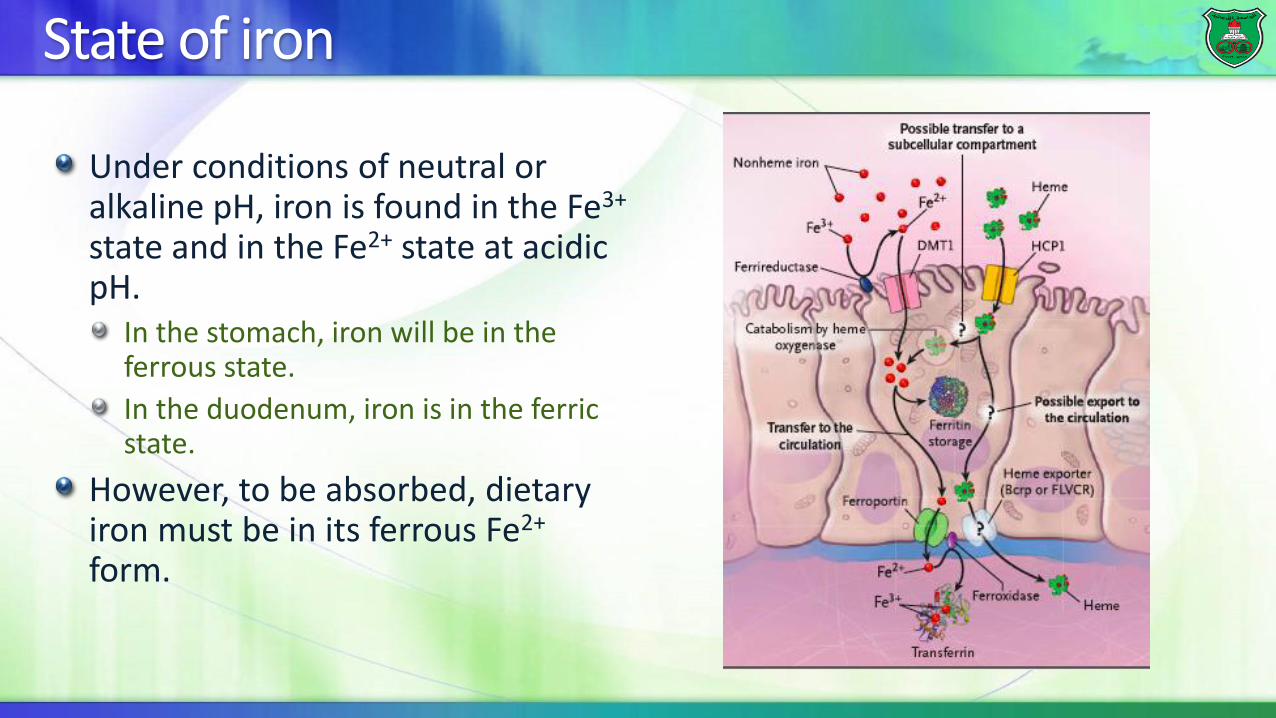
Under conditions of neutral or alkaline pH, iron is found in the Fe3+
state and in the Fe2+ state at acidic pH.
In the stomach, iron will be in the ferrous state.
In the duodenum, iron is in the ferric state.
However, to be absorbed, dietary iron must be in its ferrous Fe2+
form.
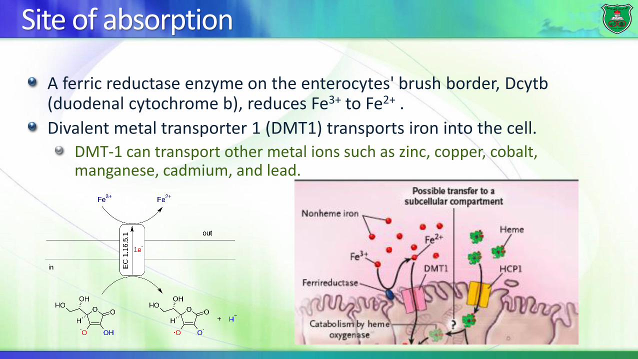
Site of absorption
A ferric reductase enzyme on the enterocytes' brush border, Dcytb(duodenal cytochrome b), reduces Fe3+ to Fe2+ .
Divalent metal transporter 1 (DMT1) transports iron into the cell.DMT-1 can transport other metal ions such as zinc, copper, cobalt, manganese, cadmium, and lead.

Heme as a source of iron
Iron can also be obtained from ingested heme.
Heme is absorbed by a receptor called heme-carrier protein 1 (HCP-1) and iron is released by heme oxygenase-1 (HO-1).
In other cells such as macrophages, heme oxygenase also extracts iron from heme.
Heme oxygenase
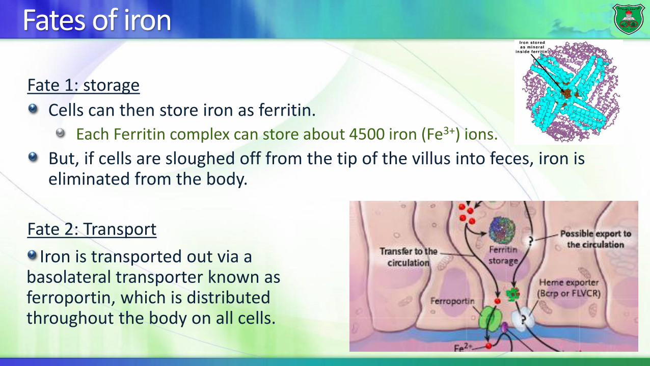
Fates of iron
Fate 1: storage
Cells can then store iron as ferritin.Each Ferritin complex can store about 4500 iron (Fe3+) ions.
But, if cells are sloughed off from the tip of the villus into feces, iron is eliminated from the body.
Fate 2: Transport
Iron is transported out via a basolateral transporter known as ferroportin, which is distributed throughout the body on all cells.
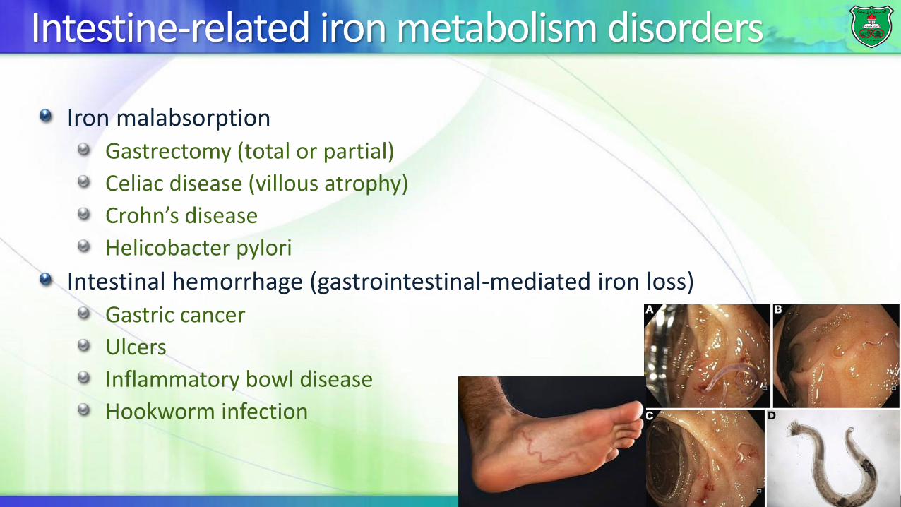
Intestine-related iron metabolism disorders
Iron malabsorptionGastrectomy (total or partial)
Celiac disease (villous atrophy)
Crohn’s disease
Helicobacter pylori
Intestinal hemorrhage (gastrointestinal-mediated iron loss)Gastric cancer
Ulcers
Inflammatory bowl disease
Hookworm infection
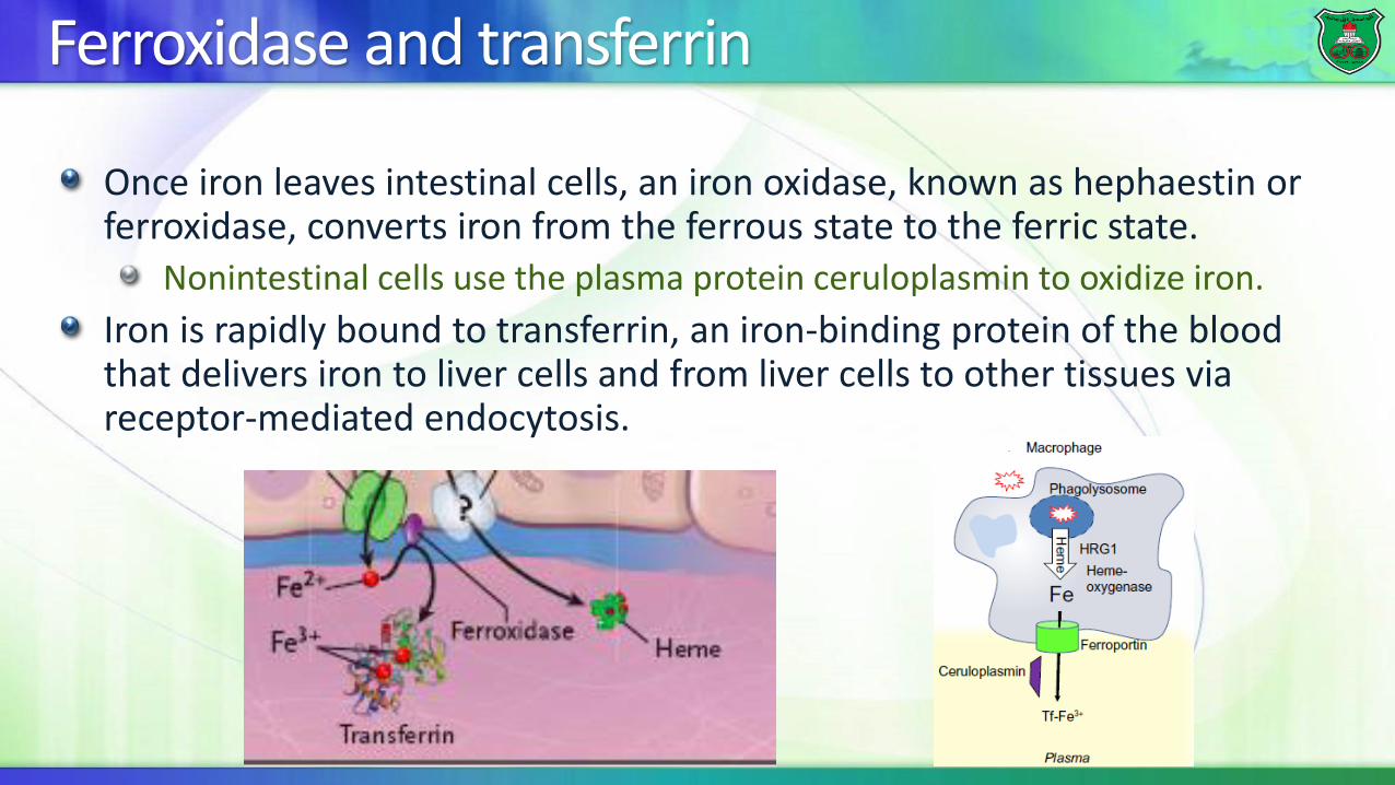
Ferroxidase and transferrin
Once iron leaves intestinal cells, an iron oxidase, known as hephaestin or ferroxidase, converts iron from the ferrous state to the ferric state.
Nonintestinal cells use the plasma protein ceruloplasmin to oxidize iron.
Iron is rapidly bound to transferrin, an iron-binding protein of the blood that delivers iron to liver cells and from liver cells to other tissues via receptor-mediated endocytosis.
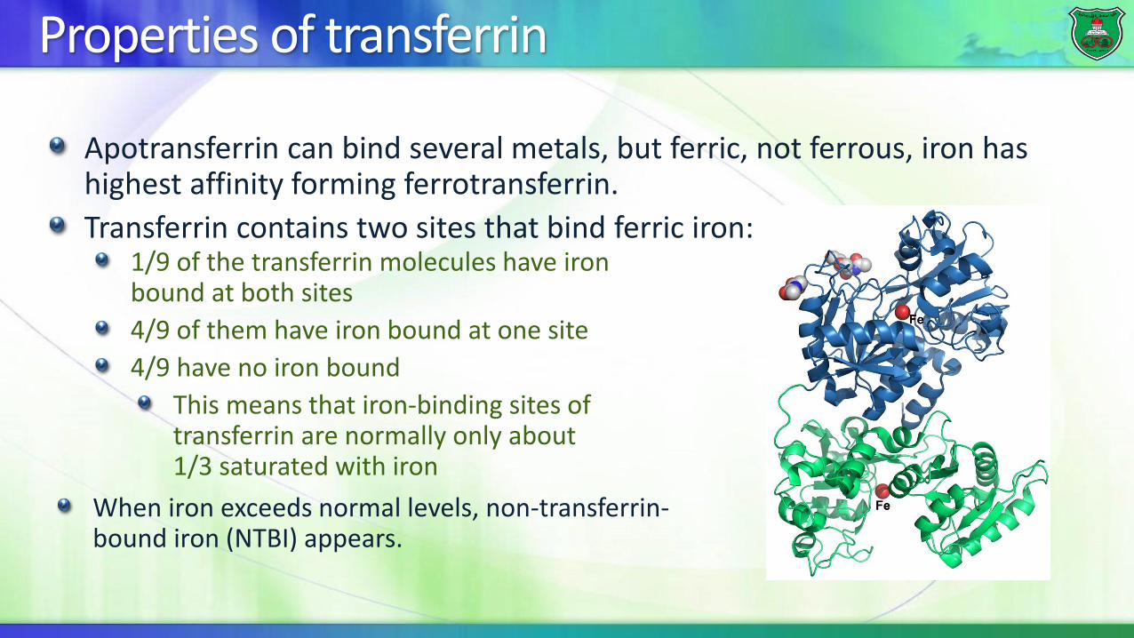
Properties of transferrin
Apotransferrin can bind several metals, but ferric, not ferrous, iron has highest affinity forming ferrotransferrin.
Transferrin contains two sites that bind ferric iron:1/9 of the transferrin molecules have iron bound at both sites
4/9 of them have iron bound at one site
4/9 have no iron bound
This means that iron-binding sites of transferrin are normally only about 1/3 saturated with iron
When iron exceeds normal levels, non-transferrin-bound iron (NTBI) appears.
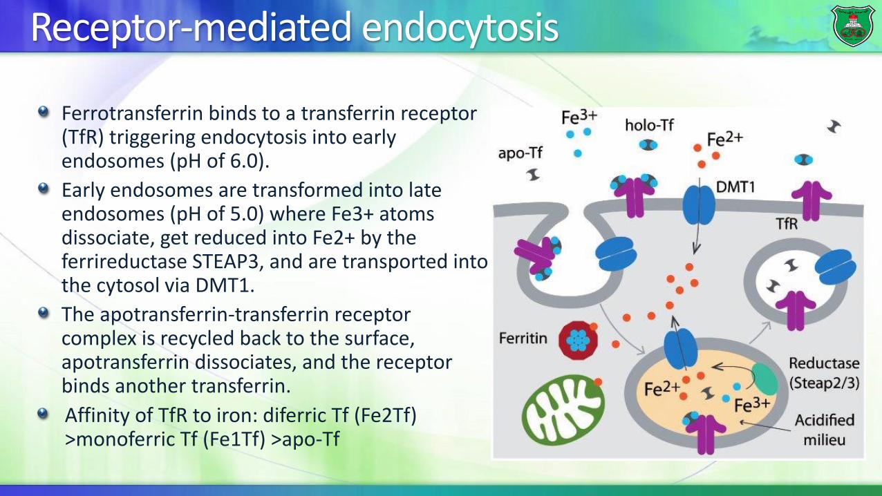
Receptor-mediated endocytosis
Ferrotransferrin binds to a transferrin receptor (TfR) triggering endocytosis into early endosomes (pH of 6.0).
Early endosomes are transformed into late endosomes (pH of 5.0) where Fe3+ atoms dissociate, get reduced into Fe2+ by the ferrireductase STEAP3, and are transported into the cytosol via DMT1.
The apotransferrin-transferrin receptor complex is recycled back to the surface, apotransferrin dissociates, and the receptor binds another transferrin.
Affinity of TfR to iron: diferric Tf (Fe2Tf) >monoferric Tf (Fe1Tf) >apo-Tf

Regulation of protein function
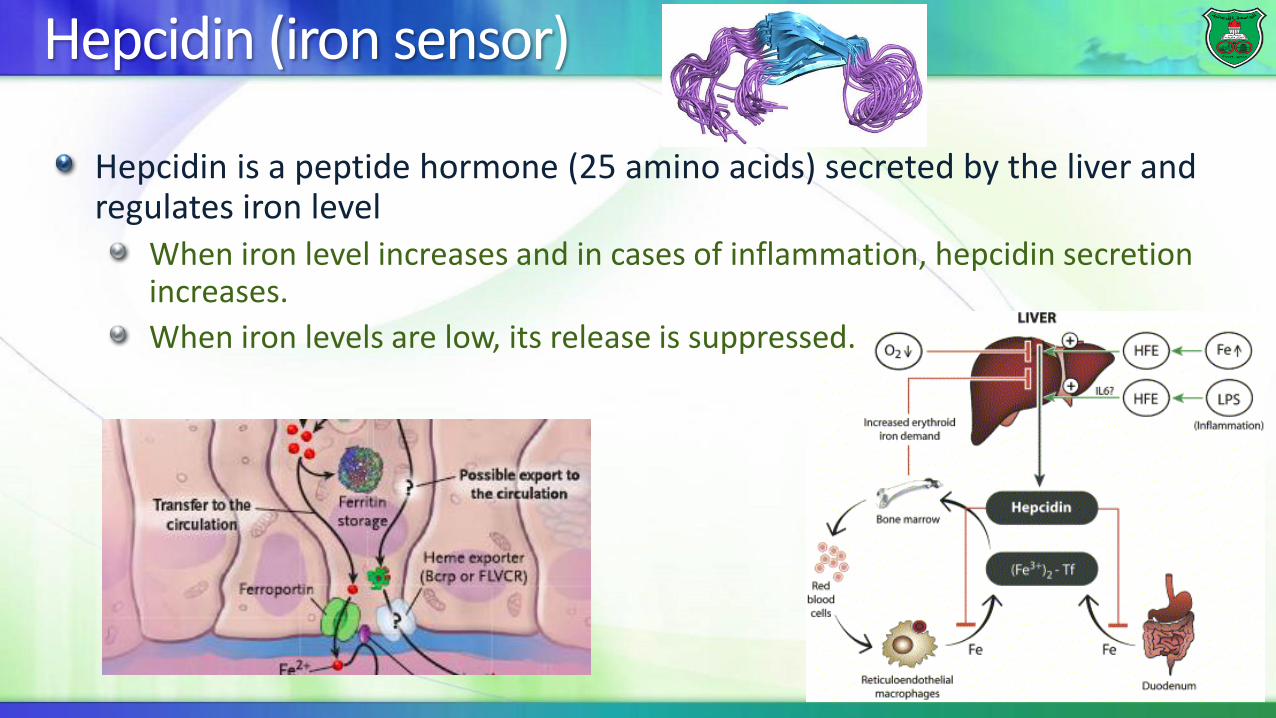
Hepcidin (iron sensor)
Hepcidin is a peptide hormone (25 amino acids) secreted by the liver and regulates iron level
When iron level increases and in cases of inflammation, hepcidin secretion increases.
When iron levels are low, its release is suppressed.

How does hepcidin reduce iron levels in the body?
Hepcidin binds to the basolateral iron transporter ferroportin inducing ferroportin internalization and degradation.
This results in higher iron storage.
Hepcidin also inhibits the presentation of the iron transporters (e.g. DMT1) in intestinal membranes decreasing iron absorption
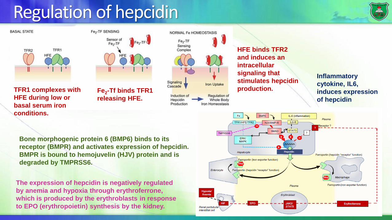
Regulation of hepcidin
The expression of hepcidin is negatively regulated
by anemia and hypoxia through erythroferrone,
which is produced by the erythroblasts in response
to EPO (erythropoietin) synthesis by the kidney.
TFR1 complexes with
HFE during low or
basal serum iron
conditions.
Fe2-Tf binds TFR1
releasing HFE.
HFE binds TFR2
and induces an
intracellular
signaling that
stimulates hepcidin
production.
Bone morphogenic protein 6 (BMP6) binds to its
receptor (BMPR) and activates expression of hepcidin.
BMPR is bound to hemojuvelin (HJV) protein and is
degraded by TMPRSS6.
Inflammatory
cytokine, IL6,
induces expression
of hepcidin

Post-transcriptionl regulation of expression
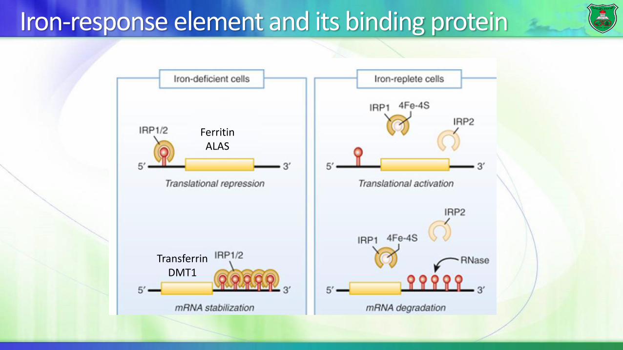
FerritinALAS
TransferrinDMT1
Iron-response element and its binding protein

Iron-related diseases
Hereditary hemochromatosis (HH)Iron-deficiency anemia

Hereditary hemochromatosis
It is a group of disorders in iron metabolism that is characterized by excess iron absorption, saturation of iron-binding proteins and deposition of hemosiderin in the tissues.
more commonly in males than in females (why?)
The primary cause of hemochromatosis is the inheritance of an autosomal recessive allele designated as HFE (type I or primary HH) , but four other genes that regulate the hepcidin–ferroportin axis can also be involved.
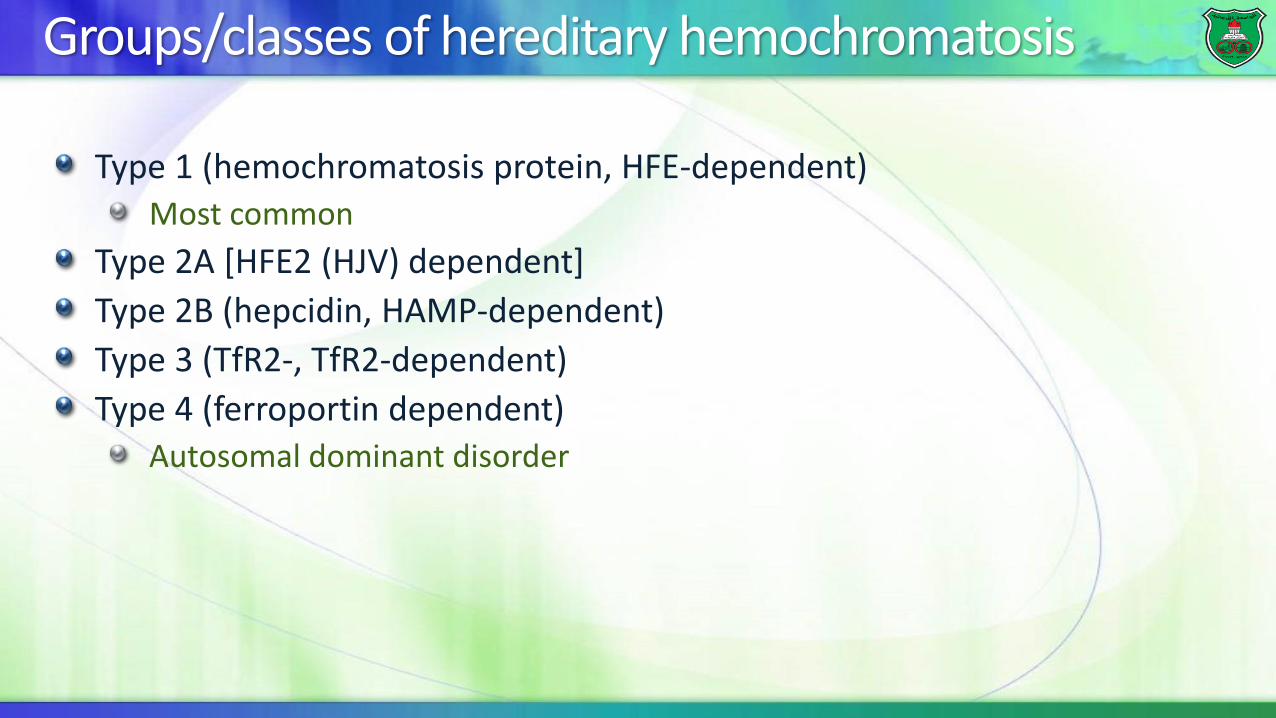
Groups/classes of hereditary hemochromatosis
Type 1 (hemochromatosis protein, HFE-dependent)Most common
Type 2A [HFE2 (HJV) dependent]
Type 2B (hepcidin, HAMP-dependent)
Type 3 (TfR2-, TfR2-dependent)
Type 4 (ferroportin dependent)Autosomal dominant disorder

Hemosiderin
The normal total body iron stores may range from 2 to 6 gm, but persons with hemochromatosis have much greater stores.
The total iron stores of affected persons may exceed 50 gm.
If the capacity for storage of iron in ferritin is exceeded, iron is stored as water-insoluble deposits known as hemosiderin, mainly in macrophages.
Excess hemosiderin leads to cellular dysfunction and damage.
Affected organs and conditions
• Liver (hepatic fibrosis)
• Pancreas (diabetes mellitus)
• Joints (arthropathy)
• Skin (pigmentation)
• Heart (cardiomyopathy)
• Gonadotrophin-secreting cells
(hypogonadotrophic hypogonadism)

Regulation of transferrin receptor
HFE is a major histocompatibility complex (MHC) class-1 gene.
Normal HFE complexes with TfR1 reducing iron transfer into cells.
Mutated HFE (e.g. C282Y) has reduced presence on membrane and/or lack of interaction with Tfr1, loss of inhibition of transferrin receptor, and, therefore increased iron uptake and storage.
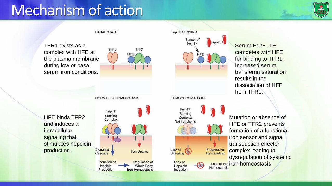
Mechanism of action
Mutation or absence of
HFE or TFR2 prevents
formation of a functional
iron sensor and signal
transduction effector
complex leading to
dysregulation of systemic
iron homeostasis
TFR1 exists as a
complex with HFE at
the plasma membrane
during low or basal
serum iron conditions.
Serum Fe2+ -TF
competes with HFE
for binding to TFR1.
Increased serum
transferrin saturation
results in the
dissociation of HFE
from TFR1.
HFE binds TFR2
and induces a
intracellular
signaling that
stimulates hepcidin
production.
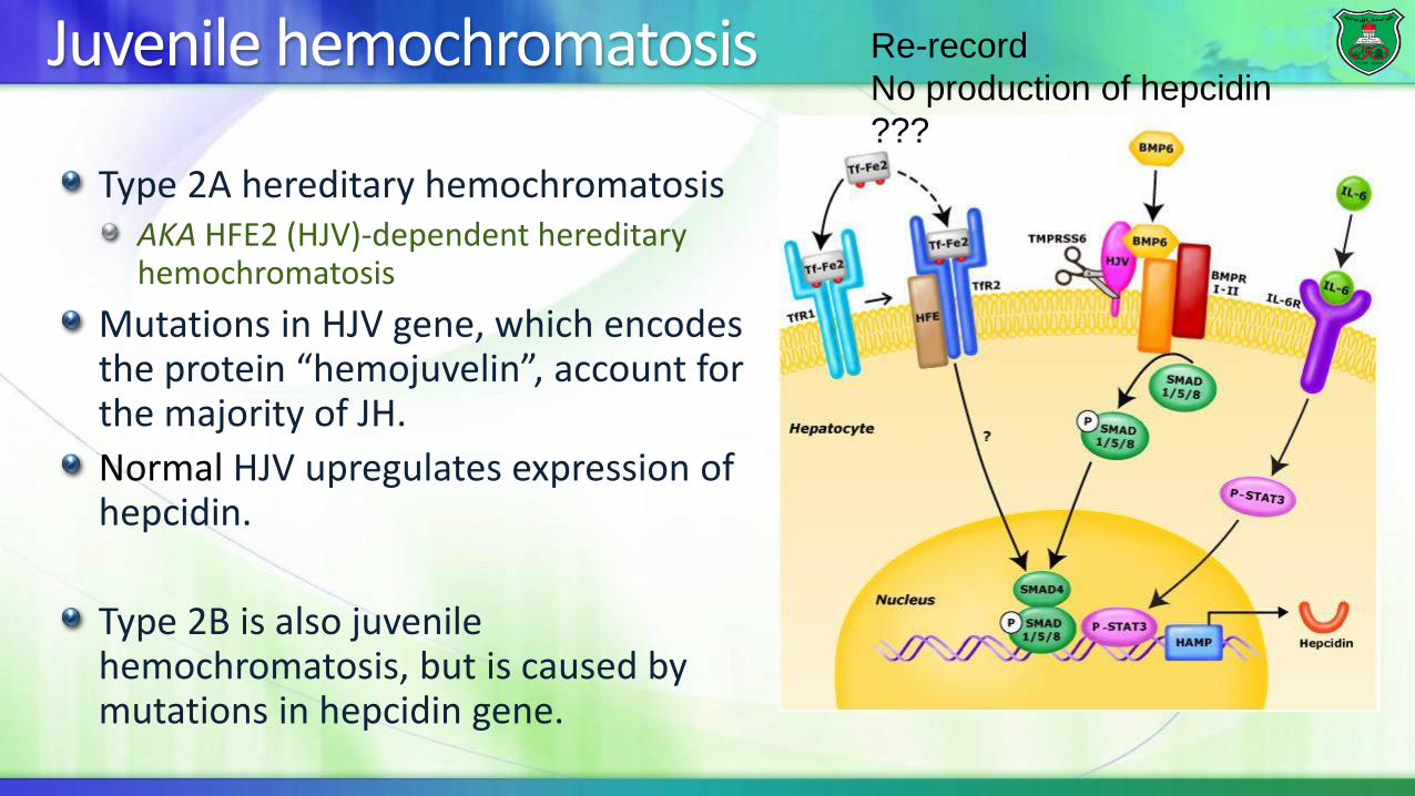
Juvenile hemochromatosis
Type 2A hereditary hemochromatosisAKA HFE2 (HJV)-dependent hereditary hemochromatosis
Mutations in HJV gene, which encodes the protein “hemojuvelin”, account for the majority of JH.
Normal HJV upregulates expression of hepcidin.
Type 2B is also juvenile hemochromatosis, but is caused by mutations in hepcidin gene.
Re-record
No production of hepcidin
???

Iron-deficiency anemia
Anemias are characterized by a deficiency in the number of mature erythrocytes in the circulation, lowering the oxygen-carrying capacity of the blood, causing tissue hypoxia, and clinical symptoms such as fatigue, weakness, increased cardiac output, as well as increased morbidity and mortality.
Cells cannot synthesize
DNA and, hence, cannot
divide and megaloblasts
accumulate.
Folate is not regenerated

Anemia of chronic disease
Causes: chronic kidney disease, chronic infections and chronic inflammatory diseases
Inflammatory cytokines → increased hepcidin production by hepatocytes →downregulation of ferroportin expression in major iron-exporting cells such as macrophages, duodenal enterocytes, and hepatocytes → decreased enteric iron absorption and, perhaps more importantly, to increased iron retention within splenic macrophages and hepatocytes.
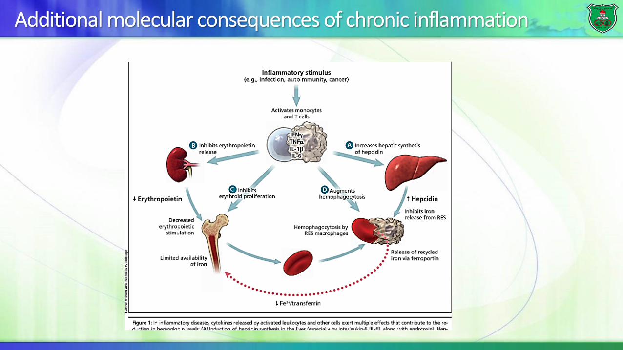
Additional molecular consequences of chronic inflammation