Menstrual cycle-dependent changes of Toll-like receptors ...
8
Human Reproduction Vol.22, No.2 pp. 586–593, 2007 doi:10.1093/humrep/del388 Advance Access publication October 16, 2006. 586 © The Author 2006. Published by Oxford University Press on behalf of the European Society of Human Reproduction and Embryology. All rights reserved. For Permissions, please email: [email protected] Menstrual cycle-dependent changes of Toll-like receptors in endometrium R.Aflatoonian 1 , E.Tuckerman 2 , S.L.Elliott 1 , C.Bruce 1 , A.Aflatoonian 3 , T.C.Li 2 and A.Fazeli 1,4 1 Academic Unit of Reproductive and Developmental Medicine, 2 Biomedical Research Unit, The University of Sheffield, Jessop Wing, Sheffield, UK and 3 Research and Clinical Center for Infertility, Shahid Sadoughi University of Medical Sciences, Yazd, Iran 4 To whom correspondence should be addressed at: Academic Unit of Reproductive and Developmental Medicine, The University of Sheffield, Level 4, Jessop Wing, Tree Root Walk, Sheffield, S10 2SF, UK. E-mail: [email protected] BACKGROUND: Rapid innate immune defences against infection usually involve the recognition of invading patho- gens by specific pattern recognition receptors recently attributed to the family of Toll-like receptors (TLRs). Reports from our laboratory and others have demonstrated the existence of TLRs 1–6 in the female reproductive tract. How- ever, little has been done to identify TLRs 7–10 in the female reproductive tract, particularly in the uterus. Also little information exists regarding variation in TLRs in the female reproductive tract during the menstrual cycle. METHOD: The distribution of TLR7–10 protein was detected by immunostaining in timed endometrial biopsies from normal women. RT–PCR was used to show the existence of TLR1–10 genes in endometrial tissue and real-time PCR analysis to investigate the relative expression of these genes during the menstrual cycle in normal human endometrium. RESULTS: TLR7–10 proteins were detected in endometrial epithelium and stroma. TLR1–10 genes were expressed in human endometrial tissue, and the mean relative expression of TLR2–6, 9 and 10 genes was signif- icantly higher during the secretory phase compared with other phases of the menstrual cycle. CONCLUSIONS: TLR7–10 localization is not limited to endometrial epithelium but is also present in the stroma of the endometrial tis- sue. Endometrial TLR2–6, 9 and 10 genes are cyclically expressed during the menstrual cycle. Key words: endometrium/menstrual cycle/Toll-like receptors Introduction Sexually transmitted diseases (STDs) are a major worldwide health problem that compromise reproductive fecundity as well as cut short the lives of millions of men, women and children (Cates, 1986; Piot et al., 1988). Despite advances in the manage- ment of these infections, only limited success has been achieved in curtailing the morbidity and mortality associated with them. The commoner STDs with the largest health and socio-economic impact include infections by the herpes simplex virus type 2 (HSV-2), Chlamydia trachomatis, the gonococcus and human immunodeficiency viruses (HIVs). In addition, maternal genital tract carriage of the group B streptococcus and bacterial vagino- sis seems to be associated with premature birth and neonatal mortality and morbidity (Martius and Eschenbach, 1990). Because of this growing global health problem, prevention and effective treatment methodologies need to be developed. Under- standing the mechanisms that regulate the female reproductive tract immune system holds therapeutic promise. If the immuno- logical host defence mechanisms against these infections are clarified, effective vaccines can be developed. Rapid innate immune defences against infection usually involve the recognition of invading pathogens by specific pat- tern recognition receptors recently attributed to the family of Toll-like receptors (TLRs) (Medzhitov and Janeway, 2000; Janeway and Medzhitov, 2002). TLRs are expressed by cells involved in the first line of host defence, including neutrophils, macrophages, dendritic cells, dermal endothelial cells and mucosal epithelial cells. Collectively, TLRs function to alert the immune system to the presence of micro-organisms. Differ- ent members of the TLR family are expressed on different cell organelles and appear to mediate signal transduction to a range of antigenic stimuli by engaging with specific ligands leading to the production of various proinflammatory cytokines, chem- okines and effector molecules, depending on the cell type that is activated. The members of the TLR family, of which at least 11 have been identified, recognize distinct pathogen-associated molecular patterns (PAMPs) produced by various bacterial, fungal and viral pathogens. TLR2 forms heterodimers with TLRs 1 and 6 and recognizes a broad range of microbial prod- ucts from Gram-positive bacteria (peptidoglycan) (Schwandner et al., 1999), fungi (zymosan) (Underhill et al., 1999) and syn- thetic lipoproteins (Pam3Cys-Ser-(Lys)4) (Takeuchi et al., 2002). TLR3 recognizes double-stranded RNA of viral or cellular origin (Alexopoulou et al., 2001; Kariko et al., 2004). The major component of the outer membrane of Gram-negative bacteria, lipopolysaccharide (LPS), is recognized by TLR4 in by guest on November 4, 2010 humrep.oxfordjournals.org Downloaded from
Transcript of Menstrual cycle-dependent changes of Toll-like receptors ...

Human Reproduction Vol.22, No.2 pp. 586–593, 2007 doi:10.1093/humrep/del388
Advance Access publication October 16, 2006.
586 © The Author 2006. Published by Oxford University Press on behalf of the European Society of Human Reproduction and Embryology. All rights reserved.For Permissions, please email: [email protected]
Menstrual cycle-dependent changes of Toll-like receptors in endometrium
R.Aflatoonian1, E.Tuckerman2, S.L.Elliott1, C.Bruce1, A.Aflatoonian3, T.C.Li2 and A.Fazeli1,4
1Academic Unit of Reproductive and Developmental Medicine, 2Biomedical Research Unit, The University of Sheffield, Jessop Wing, Sheffield, UK and 3Research and Clinical Center for Infertility, Shahid Sadoughi University of Medical Sciences, Yazd, Iran4To whom correspondence should be addressed at: Academic Unit of Reproductive and Developmental Medicine, The University of Sheffield, Level 4, Jessop Wing, Tree Root Walk, Sheffield, S10 2SF, UK. E-mail: [email protected]
BACKGROUND: Rapid innate immune defences against infection usually involve the recognition of invading patho-gens by specific pattern recognition receptors recently attributed to the family of Toll-like receptors (TLRs). Reportsfrom our laboratory and others have demonstrated the existence of TLRs 1–6 in the female reproductive tract. How-ever, little has been done to identify TLRs 7–10 in the female reproductive tract, particularly in the uterus. Also littleinformation exists regarding variation in TLRs in the female reproductive tract during the menstrual cycle.METHOD: The distribution of TLR7–10 protein was detected by immunostaining in timed endometrial biopsiesfrom normal women. RT–PCR was used to show the existence of TLR1–10 genes in endometrial tissue and real-timePCR analysis to investigate the relative expression of these genes during the menstrual cycle in normal humanendometrium. RESULTS: TLR7–10 proteins were detected in endometrial epithelium and stroma. TLR1–10 geneswere expressed in human endometrial tissue, and the mean relative expression of TLR2–6, 9 and 10 genes was signif-icantly higher during the secretory phase compared with other phases of the menstrual cycle. CONCLUSIONS:TLR7–10 localization is not limited to endometrial epithelium but is also present in the stroma of the endometrial tis-sue. Endometrial TLR2–6, 9 and 10 genes are cyclically expressed during the menstrual cycle.
Key words: endometrium/menstrual cycle/Toll-like receptors
Introduction
Sexually transmitted diseases (STDs) are a major worldwidehealth problem that compromise reproductive fecundity as wellas cut short the lives of millions of men, women and children(Cates, 1986; Piot et al., 1988). Despite advances in the manage-ment of these infections, only limited success has been achievedin curtailing the morbidity and mortality associated with them.The commoner STDs with the largest health and socio-economicimpact include infections by the herpes simplex virus type 2(HSV-2), Chlamydia trachomatis, the gonococcus and humanimmunodeficiency viruses (HIVs). In addition, maternal genitaltract carriage of the group B streptococcus and bacterial vagino-sis seems to be associated with premature birth and neonatalmortality and morbidity (Martius and Eschenbach, 1990).Because of this growing global health problem, prevention andeffective treatment methodologies need to be developed. Under-standing the mechanisms that regulate the female reproductivetract immune system holds therapeutic promise. If the immuno-logical host defence mechanisms against these infections areclarified, effective vaccines can be developed.
Rapid innate immune defences against infection usuallyinvolve the recognition of invading pathogens by specific pat-tern recognition receptors recently attributed to the family of
Toll-like receptors (TLRs) (Medzhitov and Janeway, 2000;Janeway and Medzhitov, 2002). TLRs are expressed by cellsinvolved in the first line of host defence, including neutrophils,macrophages, dendritic cells, dermal endothelial cells andmucosal epithelial cells. Collectively, TLRs function to alertthe immune system to the presence of micro-organisms. Differ-ent members of the TLR family are expressed on different cellorganelles and appear to mediate signal transduction to a rangeof antigenic stimuli by engaging with specific ligands leadingto the production of various proinflammatory cytokines, chem-okines and effector molecules, depending on the cell type thatis activated. The members of the TLR family, of which at least11 have been identified, recognize distinct pathogen-associatedmolecular patterns (PAMPs) produced by various bacterial,fungal and viral pathogens. TLR2 forms heterodimers withTLRs 1 and 6 and recognizes a broad range of microbial prod-ucts from Gram-positive bacteria (peptidoglycan) (Schwandneret al., 1999), fungi (zymosan) (Underhill et al., 1999) and syn-thetic lipoproteins (Pam3Cys-Ser-(Lys)4) (Takeuchi et al.,2002). TLR3 recognizes double-stranded RNA of viral or cellularorigin (Alexopoulou et al., 2001; Kariko et al., 2004). Themajor component of the outer membrane of Gram-negativebacteria, lipopolysaccharide (LPS), is recognized by TLR4 in
by guest on Novem
ber 4, 2010hum
rep.oxfordjournals.orgD
ownloaded from

TLR expression during the menstrual cycle
587
association with CD14 and MD-2 (Akashi et al., 2001; da SilvaCorreia et al., 2001; Nagai et al., 2002). TLR5 recognizes bac-terial flagellin (Hayashi et al., 2001), TLRs 7 and 8 recognizesingle-stranded RNA (ssRNA) and synthetic nucleotide deriva-tives (Hemmi et al., 2002; Heil et al., 2004), and TLR9 recog-nizes non-methylated CpG containing DNA (Hemmi et al.,2000). No specific ligand has yet been identified for TLR10(Chuang and Ulevitch, 2001). Studies in mice have shownTLR11 binds to a profilin-like protein from Toxoplasma gondii(Yarovinsky et al., 2005; Yarovinsky and Sher, 2006).
Several reports exist on the determination and characteriza-tion of TLRs in different tissues and organs (Bsibsi et al.,2002; Zarember and Godowski, 2002; Backhed and Hornef,2003; Basu and Fenton, 2004). However, little has been doneto identify TLRs in the human female. Recently, we demon-strated the in vivo distribution of TLRs 1–6 in the humanfemale reproductive tract using immunohistochemical tech-niques (Fazeli et al., 2005). With the exception of TLR4, allother TLRs studied were uniformly distributed throughout thetract. TLR7–10 mRNA has been shown to be expressed inhuman uterine tissue (Nishimura and Naito, 2005). TLRs havebeen predominantly described in the epithelial cells of thehuman endometrium. Primary human uterine epithelial cellcultures and the uterine epithelial cell line ECC-1 expressTLR1–9 genes (Schaefer et al., 2004, 2005). However, in con-trast to the studies utilizing cultured cells, Young et al. (2004)could not detect the expression of TLR7, 8 and 10 genes inendometrial tissue samples. No information exists regardingthe potential immunohistochemical localization of TLR7–10 inthe female reproductive tract and in particular in theendometrium in vivo. In the present investigation, we report thegene expression and the in vivo localization of TLR7–10 mole-cules in healthy human endometrial tissue biopsies.
Although reports from our laboratory and others have dem-onstrated the existence of TLRs in the female reproductivetract, little information exists regarding variation in TLRpresence in the female reproductive tract during the men-strual cycle. Recently, Jorgenson et al. (2005) demonstratedthe cycle-dependent expression of TLR3 in primary endome-trial epithelial tissue. One can hypothesize that other TLRmolecules may have a cycle-dependent expression in theendometrial tissue as well. We tested this hypothesis, andhere, we report the alteration in the expression of TLR1–10genes during the menstrual cycle in normal human endometrialtissue.
Materials and methods
Tissue collection for immunostaining and genomic investigations
This investigation was approved by the Local Ethics Committee, andwritten informed consent was obtained prior to the collection of tissuesamples. For immunohistochemical investigations, tissue sampleswere obtained from six fertile women, and for genomic studies,endometrial biopsies were obtained from 21 fertile women. All thewomen taking part in the investigation had regular cycles, showed noevidence of any pathological uterine disorder and had not used oralcontraception or an intrauterine device in the previous 3 months.Biopsies were obtained in the operating theater between 1 and 29 days
after the last menstrual period (LMP). The mean age of the womentaking part in the study was 35 (range 24–40) years, and each had hadat least one previous successful pregnancy. Endometrial biopsies forimmunocytochemistry were immediately snap-frozen and stored inliquid nitrogen until processed. Cryosections were cut at 5 mm andstored at –70°C until use. For genomic studies, endometrial biopsieswere immediately placed in RNAlater (Ambion, Huntingdon, UK)followed by immediate immersion in liquid nitrogen until processed.
Antibodies and peptides
Antibodies and peptides used in the experiments were obtained fromSanta Cruz Biotechnology (CA, USA). These were goat polyclonalantibodies specific for N-terminal domains of TLRs 7 and 9 (cataloguenumber sc13207 and sc13212, respectively), goat polyclonal antibodyspecific for V-terminal domains of TLR10 (catalogue numbersc23577) and rabbit polyclonal specific for D-terminal domains ofTLR8 (catalogue number sc13212-R). Blocking peptides specific forthe respective antibodies were used to detect non-specific staining.
Immunostaining
Cryosections were removed from –70°C freezer, fixed in 4% parafor-maldehyde for 15 min, washed twice in phosphate-buffered saline(PBS) for 5 min and then immersed in methanol at –20°C for 4 minfollowed by 2 min in acetone at –20°C before being finally washed inPBS. Cryopreserved slides were timed according to LMP and mor-phology and divided into three groups (menstrual, proliferative orsecretory phase). Slides were stained using a Vectorstain Elite ABCperoxidase kit, according to the manufacturer’s instructions (VectorLaboratories, Peterborough, UK). To avoid the non-specific bindingof biotin, we used an avidin/biotin blocking kit (Vector Laboratories).Briefly, slides were blocked for 1 h at room temperature in PBS con-taining 0.2% appropriate serum and 250 μl/ml of avidin. The blockwas removed, and slides were incubated overnight at 4°C in primaryantibody at an appropriate dilution using antibody diluent media(Dakocytomation, Ely, UK) containing 250 μl/ml of biotin. Bindingwas visualized by incubation with peroxidase substrate 3-amino-9-ethylcarbazole (AEC) (Vector Laboratories) for 10 min, washed indistilled water for 3 min and counterstained in 10% haematoxylin for10 min. Slides were washed in tap water for 2 min and mounted withAquamount (VWR).
Optimum staining was achieved by incubating tissue sections with10 μg/ml of the specific TLR antibody. Negative control sectionswere obtained by blocking of primary antibody with its specificpeptide. Immunostained sections were examined using an OlympusBH2 microscope (Olympus, London, UK).
RNA isolation, cDNA production and quantitative PCR
Tissues were removed from RNAlater and homogenized in 3 ml ofTRI reagent (Sigma, Pool, UK) using an Ultra Turrax homogenizer(VWR) for ∼2 min. Total RNA was extracted using TRI reagentstandard protocol supplied by the manufacturer. Total RNA wastreated with DNase I (DNA-free Kit; Ambion) to remove genomicDNA contamination from samples. First-strand cDNA synthesis wasperformed using oligo dT primers and the Superscript II reverse tran-scriptase system (Invitrogen, Paisley, UK).
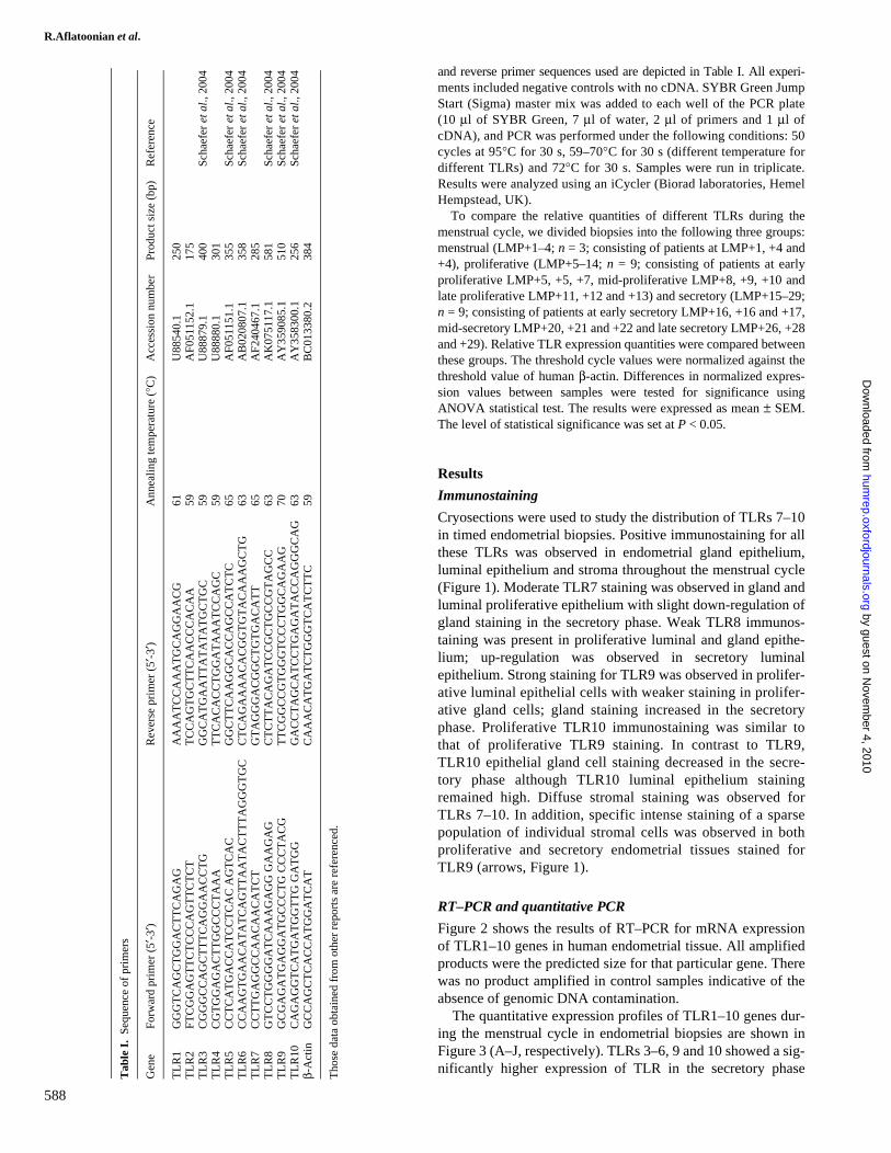
Reverse-transcription polymerase chain reaction (RT–PCR) wasperformed using the prepared cDNA, primers for TLRs 1–10 (Table I)and Platinum Blue PCR Super Mix (Invitrogen) under the followingconditions: 40 cycles at 95°C for 30 s, 59–70°C for 1 min (differenttemperature for different TLRs) and 72°C for 2 min.
Quantitative real-time PCR was performed using the preparedcDNA and primers for TLRs 1–10 and human β-actin. The forward
by guest on Novem
ber 4, 2010hum
rep.oxfordjournals.orgD
ownloaded from
R.Aflatoonian et al.
588
and reverse primer sequences used are depicted in Table I. All experi-ments included negative controls with no cDNA. SYBR Green JumpStart (Sigma) master mix was added to each well of the PCR plate(10 μl of SYBR Green, 7 μl of water, 2 μl of primers and 1 μl ofcDNA), and PCR was performed under the following conditions: 50cycles at 95°C for 30 s, 59–70°C for 30 s (different temperature fordifferent TLRs) and 72°C for 30 s. Samples were run in triplicate.Results were analyzed using an iCycler (Biorad laboratories, HemelHempstead, UK).
To compare the relative quantities of different TLRs during themenstrual cycle, we divided biopsies into the following three groups:menstrual (LMP+1–4; n = 3; consisting of patients at LMP+1, +4 and+4), proliferative (LMP+5–14; n = 9; consisting of patients at earlyproliferative LMP+5, +5, +7, mid-proliferative LMP+8, +9, +10 andlate proliferative LMP+11, +12 and +13) and secretory (LMP+15–29;n = 9; consisting of patients at early secretory LMP+16, +16 and +17,mid-secretory LMP+20, +21 and +22 and late secretory LMP+26, +28and +29). Relative TLR expression quantities were compared betweenthese groups. The threshold cycle values were normalized against thethreshold value of human β-actin. Differences in normalized expres-sion values between samples were tested for significance usingANOVA statistical test. The results were expressed as mean ± SEM.The level of statistical significance was set at P < 0.05.
Results
Immunostaining
Cryosections were used to study the distribution of TLRs 7–10in timed endometrial biopsies. Positive immunostaining for allthese TLRs was observed in endometrial gland epithelium,luminal epithelium and stroma throughout the menstrual cycle(Figure 1). Moderate TLR7 staining was observed in gland andluminal proliferative epithelium with slight down-regulation ofgland staining in the secretory phase. Weak TLR8 immunos-taining was present in proliferative luminal and gland epithe-lium; up-regulation was observed in secretory luminalepithelium. Strong staining for TLR9 was observed in prolifer-ative luminal epithelial cells with weaker staining in prolifer-ative gland cells; gland staining increased in the secretoryphase. Proliferative TLR10 immunostaining was similar tothat of proliferative TLR9 staining. In contrast to TLR9,TLR10 epithelial gland cell staining decreased in the secre-tory phase although TLR10 luminal epithelium stainingremained high. Diffuse stromal staining was observed forTLRs 7–10. In addition, specific intense staining of a sparsepopulation of individual stromal cells was observed in bothproliferative and secretory endometrial tissues stained forTLR9 (arrows, Figure 1).
RT–PCR and quantitative PCR
Figure 2 shows the results of RT–PCR for mRNA expressionof TLR1–10 genes in human endometrial tissue. All amplifiedproducts were the predicted size for that particular gene. Therewas no product amplified in control samples indicative of theabsence of genomic DNA contamination.
The quantitative expression profiles of TLR1–10 genes dur-ing the menstrual cycle in endometrial biopsies are shown inFigure 3 (A–J, respectively). TLRs 3–6, 9 and 10 showed a sig-nificantly higher expression of TLR in the secretory phaseT
able
I.
Sequ
ence
of
prim
ers
Tho
se d
ata
obta
ined
fro
m o
ther
rep
orts
are
ref
eren
ced.
Gen
eF
orw
ard
prim
er (
5′-3
′)R
ever
se p
rim
er (
5′-3
′)A
nnea
ling
tem
pera
ture
(°C
)A
cces
sion
num
ber
Prod
uct s
ize
(bp)
Ref
eren
ce
TL
R1
GG
GT
CA
GC
TG
GA
CT
TC
AG
AG
AA
AA
TC
CA
AA
TG
CA
GG
AA
CG
61U
8854
0.1
250
TL
R2
FTC
GG
AG
TT
CT
CC
CA
GT
TC
TC
TT
CC
AG
TG
CT
TC
AA
CC
CA
CA
A59
AF0
5115
2.1
175
TL
R3
CG
GG
CC
AG
CT
TT
CA
GG
AA
CC
TG
GG
CA
TG
AA
TT
AT
AT
AT
GC
TG
C59
U88
879.
140
0Sc
haef
er e
t al.,
200
4T
LR
4C
GT
GG
AG
AC
TT
GG
CC
CT
AA
AT
TC
AC
AC
CT
GG
AT
AA
AT
CC
AG
C59
U88
880.
130
1T
LR
5C
CT
CA
TG
AC
CA
TC
CT
CA
C A
GT
CA
CG
GC
TT
CA
AG
GC
AC
CA
GC
CA
TC
TC
65A
F051
151.
135
5Sc
haef
er e
t al.,
200
4T
LR
6C
CA
AG
TG
AA
CA
TA
TC
AG
TT
AA
TA
CT
TT
AG
GG
TG
CC
TC
AG
AA
AA
CA
CG
GT
GT
AC
AA
AG
CT
G63
AB
0208
07.1
358
Scha
efer
et a
l., 2
004
TL
R7
CC
TT
GA
GG
CC
AA
CA
AC
AT
CT
GT
AG
GG
AC
GG
CT
GT
GA
CA
TT
65A
F240
467.
128
5T
LR
8G
TC
CT
GG
GG
AT
CA
AA
GA
GG
GA
AG
AG
CT
CT
TA
CA
GA
TC
CG
CT
GC
CG
TA
GC
C63
AK
0751
17.1
581
Scha
efer
et a
l., 2
004
TL
R9
GC
GA
GA
TG
AG
GA
TG
CC
CT
G C
CC
TA
CG
TT
CG
GC
CG
TG
GG
TC
CC
TG
GC
AG
AA
G70
AY
3590
85.1
510
Scha
efer
et a
l., 2
004
TL
R10
CA
GA
GG
TC
AT
GA
TG
GT
TG
GA
TG
GG
AC
CT
AG
CA
TC
CT
GA
GA
TA
CC
AG
GG
CA
G63
AY
3583
00.1
256
Scha
efer
et a
l., 2
004
β-A
ctin
GC
CA
GC
TC
AC
CA
TG
GA
TC
AT
CA
AA
CA
TG
AT
CT
GG
GT
CA
TC
TT
C59
BC
0133
80.2
384
by guest on Novem
ber 4, 2010hum
rep.oxfordjournals.orgD
ownloaded from

TLR expression during the menstrual cycle
589
Figure 1. Immunohistochemical staining of Toll-like receptor (TLR) 7–10 expression in human endometrium during secretory and proliferativephases of the menstrual cycle. Positive staining is red and negative staining blue. Insets show blocking of the anti-TLR7–10 antibodies with itsspecific peptides. Arrows depict cells in endometrial stroma with intensive staining with TLR9 antibody. Bar = 50 μm.
by guest on Novem
ber 4, 2010hum
rep.oxfordjournals.orgD
ownloaded from

R.Aflatoonian et al.
590
compared with the proliferative and menstrual phases of thecycle. Only, TLR2 expression was higher in the secretoryphase compared with the proliferative phase but similar tomenstrual phase. No significant difference was observed in therelative expression of genes for TLR1, 7 and 8 genes duringthe menstrual cycle.
Discussion
Uterine endometrial epithelial cells are the first layer of uterinedefence against pathogens ascending the female reproductivetract. Thus, as detectors of non-self entities, TLRs may beexpected to be present in this tissue. In the present investiga-tion, we found the presence of TLR1–10 mRNA in humanendometrial biopsies. Our results are in agreement with anotherreport regarding the presence of TLRs 1–6 and 9 in endome-trial samples (Young et al., 2004); however, in contrast to thatreport, we also detected the endometrial expression of TLRs 7,8 and 10. None of the negative controls taken during our PCRsshowed an amplified product (Figure 2), thus confirming theabsence of genomic DNA contamination in our test PCR sam-ples. The expression of all TLR molecules except TLR10 hasbeen shown in endometrial cell lines (ECC-1) (Schaefer et al.,2004) and primary uterine epithelial cell cultures (Schaeferet al., 2005). The only significant difference between ourinvestigations and that of Young et al. is that the TLR primersused for the amplification of TLR molecules in our study aredifferent from those used previously. The immunohistochemi-cal staining results of antibodies against TLRs 7–10 and theirblocking with its specific peptides confirm the results of ourgene expression studies regarding the presence of TLRs 7–10in endometrial tissue.
Within the TLR family, TLRs 7–9 appear to be phylogeneti-cally closely related to each other (Du et al., 2000) and form afunctional subgroup that recognizes viral PAMPs in endosomalor lysosomal compartments (Heil et al., 2004). This is consist-ent with the fact that viral nucleic acid would be most likelydetected by TLRs within an infected cell. For example, ssRNAviruses would reach the endosome through receptor-mediateduptake of a viral particle. There is evidence accumulating that,like TLR3, these TLRs can also respond to ‘self’ nucleic acid,which has been found to be immunostimulatory and may act asa ‘danger signal’ depending on its compartmentalization (Heil
et al., 2004). Hence, it may be more correct to think of TLRs7–9 as detectors of the abnormal localization of nucleic acidrather than as structures or motifs absent from the host (Dieboldet al., 2004). It is reported that TLR9 recognizes unmethylateddeoxycytidyl-phosphate-deoxyguanosine (CpG) dinucleotidesthat are common in bacterial and some viral nucleic acids(Hemmi et al., 2000; Bauer et al., 2001). Initially, TLRs 7 and8 were shown to detect small antiviral compounds known asimidazoquinolines (Hemmi et al., 2002; Jurk et al., 2002).These were guanosine-based antiviral drugs. This indicatedthat the natural ligands for TLRs 7 and 8 could be viral nucleicacids. It has recently been reported that mouse TLR7 andhuman TLR8 (but not human TLR7) could recognize syntheticGU-rich ssRNA (Diebold et al., 2004; Lund et al., 2004).
Previously, we have reported the localization of TLRs 1–6in various sections of the female reproductive tract usingimmunohistochemistry (Fazeli et al., 2005). Here, we providefurther information regarding the localization of TLRs 7–10 inthe endometrial tissue. For nearly all TLR molecules studied inthe present investigation, the staining was not limited to epithe-lial cells and glands. It was also present in the stroma of theendometrium. No difference was found between proliferative-and secretory-stage endometrium staining with antibodies fordifferent TLR molecules except slight increase in the stromalstaining of TLR9. However, immunohistochemical staining isa qualitative technique and as such is not ideal for quantitativeanalysis. The specificity of staining for each TLR moleculewas verified by blocking the staining using specific peptidesfor the respective antibody. Several other studies using immu-nohistochemistry have demonstrated the presence of TLR7[tonsils (Mansson et al., 2006)] and TLR9 [liver (Martin-Armas,2006), conjunctiva (Bonini et al., 2005) and gut (Rumio et al.,2004)] in different human tissues.
The endometrial environment is under the control of sexhormones during the menstrual cycle. The sex hormones notonly regulate the anatomical and histological characteristics ofendometrium (Beier and Beier-Hellwig, 1998; Classen-Linkeet al., 1998) but are involved in the influx and localization ofimmune cells in the endometrium (Spornitz, 1992; Yeamanet al., 1997; von Rango, et al., 2001). For example, uterine nat-ural killer (uNK) cells are found in the human uterus in largenumbers spread throughout the endometrium with increasingnumbers as the menstrual cycle progresses (Hunt, 1994; Givan
Figure 2. The expression of Toll-like receptor (TLR) 1–10 genes in human endometrial tissue. Each pair of primers produced a specific productwith the specific predicted size in the test (T) samples. C = control samples.
by guest on Novem
ber 4, 2010hum
rep.oxfordjournals.orgD
ownloaded from

TLR expression during the menstrual cycle
591
Figure 3. Mean ± SEM of normalized expression values for Toll-like receptor (TLR) 1–10 genes in endometrial biopsies during different phasesof menstrual cycle (A–J, respectively). The level of statistical significance was set at P < 0.05. Different letters denote significant differences.
0
50
100
Menstrual Proliferative Secretory
oisserpxe dezilamro
Nn
A
0
50
100
150
Menstrual Proliferative Secretory
oisserpxe dez il amro
Nn
Ba, b
a
b
0
100
200
300
Menstrual Proliferative Secretory
oisserpxe dezilamro
Nn
C
a
b
a
0
100
200
Menstrual Proliferative Secretory
oisserpxe dezilamro
Nn
D
a
a
b
0
500
1000
Menstrual Proliferative Secretory
oisserpxe de zilam ro
Nn
Eb
aa
0
100
200
300
Menstrual Proliferative Secretory
oisserpxe dez ilamr o
Nn
F
a
a
b
0
100
200
300
Menstrual Proliferative Secretory
oisserpxe dezilamro
Nn
G
0
500
1000
1500
Menstrual Proliferative Secretory
oisserpxe dezilamro
Nn
H
J
0
200
400
600
800
Menstrual Proliferative Secretory
oisserpxe dez ilamro
Nn
I
a a
b
0
50
100
150
Menstrual Proliferative Secretory
n ois se rpxe d ezi lam roN
a
a
b
by guest on Novem
ber 4, 2010hum
rep.oxfordjournals.orgD
ownloaded from

R.Aflatoonian et al.
592
et al., 1997). uNK cells mediate interferon-gamma productionin the endometrium and are believed to be involved in thedevelopment of spiral arteries during early pregnancy and thecontrol of trophoblast invasion. A recent article has shown thatuNK cells express TLRs 2–4 (Eriksson et al., 2006) and thattheir response to TLR agonists is dependent on other cellswithin the endometrium. TLRs may therefore play a role inimplantation other than the control of pathogens. Defensins, orcationic peptides, represent an important component of innateimmune system at mucosal surfaces, including the femalereproductive tract (Gallo et al., 2006; Lehrer and Ganz, 2002).Several of these broad-spectrum natural antimicrobial peptidesare expressed in urogenital tissues, and their expression seemsto be regulated by cycle-associated changes in sex hormones.For example, human intestinal defensin-5 (HD-5) mRNA isexpressed in the vagina, ectocervix and variably in theendocervix, endometrium and Fallopian tube (Quayle et al.,1998). The endometrial expression of HD-5 mRNA has beenreported to be higher during the early secretory phase of thecycle. The secreted HD-5 peptide in cervicovaginal lavage wasalso highest during the secretory phase of the menstrual cycle.The levels of other antimicrobials such as lactoferrin and lys-ozyme are also affected by oestradiol concentrations and thestage of the oestrous cycle (Cohen et al., 1984; Walmer et al.,1992). The adaptive immune system is also influenced by thealtering levels of sex hormones during the menstrual cycle.Antigen presentation has been shown to be suppressed inresponse to increasing concentrations of oestrogen. Further tothis, receptors for oestrogen have been found on both CD8 andCD4 T cells, so it is likely that their actions are also modified bychanging concentrations of oestrogen. The actions of cytotoxicT cells appear to be down-regulated during days 14–28 of themenstrual cycle, when concentrations of progesterone and oes-trogen are high (Beagley and Gockel, 2003). IgA and IgG havebeen shown to alter during the menstrual cycle, with the highesttotal levels of immunoglobulin occurring during menses, whenoestrogen and progesterone are low and lowest at ovulation,when oestrogen is high and progesterone is low. However,responses vary in different parts of the reproductive tract withrising oestradiol increasing IgA in uterine secretions but sup-pressing levels in cervical mucus (Beagley and Gockel, 2003).
The present investigation clearly demonstrates alterationsin relative expression of TLR2–6, 9 and 10 genes in theendometrial tissue during the menstrual cycle. Although theseTLR molecules are expressed throughout the cycle, it seemsthe lowest amount of these genes is expressed during men-strual and proliferative stages of the cycle. The oestrogen lev-els are higher at the proliferative phase of the cycle comparedwith the secretory phase. At the same time, progesteronelevel is relatively higher at the secretory phase compared withthe proliferative phase of the cycle. This may indicate aninhibitory effect of the oestrogen and/or a supporting influ-ence of progesterone on the expression of TLR molecules inthe endometrium. Further research should be focused towardsunderstanding the regulation of expression of TLR moleculesby sex hormones.
In agreement with our findings, Jorgenson et al. (2005)have recently demonstrated the cycle-dependent expression of
TLR3 in primary endometrial epithelial tissue. AlthoughLesmeister et al. (2005) showed that in vitro treatment ofendometrial epithelial cell lines with 17beta-oestradiol did notaffect TLR3 mRNA or protein expression, treatment with17beta-oestradiol did suppress cytokine and chemokine pro-duction resulting from TLR3 stimulation with poly I:C, sug-gesting that 17beta-oestradiol modulates TLR3 function.
In conclusion, we report the presence and localization ofTLRs 7–10 for the first time in human endometrial biopsies. Ourinvestigations indicate that the expression of TLRs 2–6, 9 and 10is altered in the endometrium during the menstrual cycle. Furtherinvestigations should be directed towards understanding theunderlying mechanism leading to changes in TLR expression inendometrium during the menstrual cycle as well as the signifi-cance of these cycle-dependent changes in mediating innate andadaptive immune responses in the female reproductive tract.
AcknowledgementsWe thank Dr E. Sostaic for her critical discussion during the course ofthe experiments and preparation of the manuscript and Dr M. Aarabifor statistical support.
References
Akashi S, Nagai Y, Ogata H, Oikawa M, Fukase K, Kusumoto S, Kawasaki K,Nishijima M, Hayashi S, Kimoto M et al. (2001) Human MD-2 confers onmouse Toll-like receptor 4 species-specific lipopolysaccharide recognition.Int Immunol 13(12),1595–1599.
Alexopoulou L, Holt AC, Medzhitov R and Flavell RA (2001) Recognition ofdouble-stranded RNA and activation of NF-kappaB by Toll-like receptor 3.Nature 413(6857),732–738.
Backhed F and Hornef M (2003) Toll-like receptor 4-mediated signaling byepithelial surfaces necessity or threat? Microbes Infect 5(11),951–959.
Basu S and Fenton MJ (2004) Toll-like receptors function and roles in lungdisease. Am J Physiol Lung Cell Mol Physiol 286(5),L887–L892.
Bauer S, Kirschning CJ, Hacker H, Redecke V, Hausmann S, Akira S, Wagner Hand Lipford GB (2001) Human TLR9 confers responsiveness to bacterialDNA via species-specific CpG motif recognition. Proc Natl Acad Sci USA98(16),9237–9242.
Beagley KW and Gockel CM (2003) Regulation of innate and adaptive immu-nity by the female sex hormones oestradiol and progesterone. FEMS Immu-nol Med Microbiol 38(1),13–22.
Beier HM and Beier-Hellwig K (1998) Molecular and cellular aspects ofendometrial receptivity. Hum Reprod Update 4,448–458.
Bonini S, Micera A, Iovieno A and Lambiase A (2005) Expression of Toll-likereceptors in healthy and allergic conjunctiva. Ophthalmology 112(9),1528;discussion 1548–1549.
Bsibsi M, Ravid R, Gveric D and van Noort JM (2002) Broad expression ofToll-like receptors in the human central nervous system. J Neuropathol ExpNeurol 61(11),1013–1021.
Cates W Jr (1986) Priorities for sexually transmitted diseases in the late 1980sand beyond. Sex Transm Dis 13(2),114–117.
Chuang T and Ulevitch RJ (2001) Identification of hTLR10: a novel humanToll-like receptor preferentially expressed in immune cells. Biochim Bio-phys Acta 1518(1–2),157–161.
Classen-Linke I, Alfer J, Hey S, Krusche CA, Kusche M and Beier HM(1998) Marker molecules of human endometrial differentiation can behormonally regulated under in-vitro conditions as in-vivo. Hum ReprodUpdate 4,539–549.
Cohen MS, Black JR, Proctor RA and Sparling PF (1984) Host defences andthe vaginal mucosa. A re-evaluation. Scand J Urol Nephrol Suppl 86,13–22.
Diebold SS, Kaisho T, Hemmi H, Akira S and Reis e Sousa C (2004) Innateantiviral responses by means of TLR7-mediated recognition of single-stranded RNA. Science 303(5663),1529–1531.
Du X, Poltorak A, Wei Y and Beutler B (2000) Three novel mammalian toll-like receptors: gene structure, expression, and evolution. Eur Cytokine Netw11(3),362–371.
by guest on Novem
ber 4, 2010hum
rep.oxfordjournals.orgD
ownloaded from

TLR expression during the menstrual cycle
593
Eriksson M, Meadows SK, Basu S, Mselle TF, Wira CR and Sentman CL(2006) TLRs mediate IFN-(gamma) production by human uterine NK cellsin endometrium. J Immunol 176(10),6219–6224.
Fazeli A, Bruce C and Anumba DO (2005) Characterization of Toll-likereceptors in the female reproductive tract in humans. Hum Reprod20(5),1372–1378.
Gallo SA, Wang W, Rawat SS, Jung G, Waring AJ, Cole AM, Lu H, Yan X,Daly NL and Craik DJ, et al. (2006) Theta-defensins prevent HIV-1env-mediated fusion by binding gp41 and blocking 6-helix bundle formation.J Biol Chem 281(27),18787–18792.
Givan AL, White HD, Stern JE, Colby E, Gosselin EJ, Guyre PM and Wira CR(1997) Flow cytometric analysis of leukocytes in the human female repro-ductive tract: comparison of fallopian tube, uterus, cervix, and vagina. Am JReprod Immunol 38(5),350–359.
Hayashi F, Smith KD, Ozinsky A, Hawn TR, Yi EC, Goodlett DR, Eng JK,Akira S, Underhill DM and Aderem A (2001) The innate immune responseto bacterial flagellin is mediated by Toll-like receptor 5. Nature410(6832),1099–1103.
Heil F, Hemmi H, Hochrein H, Ampenberger F, Kirschning C, Akira S,Lipford G, Wagner H and Bauer S (2004) Species-specific recognition ofsingle-stranded RNA via toll-like receptor 7 and 8. Science303(5663),1526–1529.
Hemmi H, Takeuchi O, Kawai T, Kaisho T, Sato S, Sanjo H, Matsumoto M,Hoshino K, Wagner H, Takeda K et al. (2000) A Toll-like receptor recog-nizes bacterial DNA. Nature 408(6813),740–745.
Hemmi H, Kaisho T, Takeuchi O, Sato S, Sanjo H, Hoshino K, Horiuchi T,Tomizawa H, Takeda K and Akira S (2002) Small anti-viral compoundsactivate immune cells via the TLR7 MyD88-dependent signaling pathway.Nat Immunol 3(2),196–200.
Hunt JS (1994) Immunologically relevant cells in the uterus. Biol Reprod50(3),461–466.
Janeway CA, Jr. and Medzhitov R (2002) Innate immune recognition. AnnuRev Immunol 20,197–216.
Jorgenson RL, Young SL, Lesmeister MJ, Lyddon TD and Misfeldt ML(2005) Human endometrial epithelial cells cyclically express Toll-likereceptor 3 (TLR3) and exhibit TLR3-dependent responses to dsRNA. HumImmunol 66(5),469–482.
Jurk M, Heil F, Vollmer J, Schetter C, Krieg AM, Wagner H, Lipford G andBauer S (2002) Human TLR7 or TLR8 independently confer responsivenessto the antiviral compound R-848. Nat Immunol 3(6),499.
Kariko K, Ni H, Capodici J, Lamphier M and Weissman D (2004) mRNA isan endogenous ligand for Toll-like receptor 3. J Biol Chem279(13),12542–12550.
Lehrer RI and Ganz T (2002) Defensins of vertebrate animals. Curr Opin Immunol14(1),96–102.
Lesmeister MJ, Jorgenson RL, Young SL and Misfeldt ML (2005) 17Beta-estradiol suppresses TLR3-induced cytokine and chemokine production inendometrial epithelial cells. Reprod Biol Endocrinol 3,74.
Lund JM, Alexopoulou L, Sato A, Karow M, Adams NC, Gale NW, Iwasaki Aand Flavell RA (2004) Recognition of single-stranded RNA viruses by Toll-like receptor 7. Proc Natl Acad Sci USA 101(15),5598–5603.
Mansson A, Adner M and Cardell LO (2006) Toll-like receptors in cellularsubsets of human tonsil T cells: altered expression during recurrent tonsilli-tis. Respir Res 7(1),36.
Martin-Armas M, Simon-Santamaria J, Pettersen I, Moens U, Smedsrod B andSveinbjornsson B (2006) Toll-like receptor 9 (TLR9) is present in murineliver sinusoidal endothelial cells (LSECs) and mediates the effect of CpG-oligonucleotides. J Hepatol 44(5),939–946.
Martius J and Eschenbach DA (1990) The role of bacterial vaginosis as a causeof amniotic fluid infection, chorioamnionitis and prematurity—a review.Arch Gynecol Obstet 247(1),1–13.
Medzhitov R and Janeway C Jr (2000) Innate immunity. N Engl J Med343(5),338–344.
Nagai Y, Akashi S, Nagafuku M, Ogata M, Iwakura Y, Akira S, Kitamura T,Kosugi A, Kimoto M and Miyake K (2002) Essential role of MD-2 in LPSresponsiveness and TLR4 distribution. Nat Immunol 3(7),667–672.
Nishimura M and Naito S (2005) Tissue-specific mRNA expression profiles ofhuman toll-like receptors and related genes. Biol Pharm Bull 28(5),886–892.
Piot P, Plummer FA, Mhalu FS, Lamboray JL, Chin J and Mann JM (1988)AIDS: an international perspective. Science 239(4840),573–579.
Quayle AJ, Porter EM, Nussbaum AA, Wang YM, Brabec C, Yip KP and Mok SC(1998) Gene expression, immunolocalization, and secretion of humandefensin-5 in human female reproductive tract. Am J Pathol 152(5),1247–1258.
von Rango U, Classen-Linke I, Kertschanska S, Kemp B and Beier HM (2001)Effects of trophoblast invasion on the distribution of leukocytes in uterineand tubal implantation sites. Fertil Steril 76,116–124.
Rumio C, Besusso D, Palazzo M, Selleri S, Sfondrini L, Dubini F, Menard Sand Balsari A (2004) Degranulation of paneth cells via toll-like receptor 9.Am J Pathol 165(2),373–381.
Schaefer TM, Desouza K, Fahey JV, Beagley KW and Wira CR (2004)Toll-like receptor (TLR) expression and TLR-mediated cytokine/chemokineproduction by human uterine epithelial cells. Immunology 112(3),428–436.
Schaefer TM, Fahey JV, Wright JA and Wira CR (2005) Innate immunity inthe human female reproductive tract: antiviral response of uterine epithelialcells to the TLR3 agonist poly(I: C). J Immunol 174(2),992–1002.
Schwandner R, Dziarski R, Wesche H, Rothe M and Kirschning CJ (1999)Peptidoglycan- and lipoteichoic acid-induced cell activation is mediated bytoll-like receptor 2. J Biol Chem 274(25),17406–17409.
da Silva Correia J, Soldau K, Christen U, Tobias PS and Ulevitch RJ (2001)Lipopolysaccharide is in close proximity to each of the proteins in its mem-brane receptor complex: transfer from CD14 to TLR4 and MD-2. J BiolChem 276(24),21129–21135.
Spornitz UM (1992) The functional morphology of the human endometriumand decidua. Adv Anat Embryol Cell Biol 124,1–99.
Takeuchi O, Sato S, Horiuchi T, Hoshino K, Takeda K, Dong Z, Modlin RLand Akira S (2002) Cutting edge role of Toll-like receptor 1 in mediatingimmune response to microbial lipoproteins. J Immunol 169(1),10–14.
Underhill DM, Ozinsky A, Hajjar AM, Stevens A, Wilson CB, Bassetti Mand Aderem A (1999) The Toll-like receptor 2 is recruited to macrophagephagosomes and discriminates between pathogens. Nature 401(6755),811–815.
Walmer DK, Wrona MA, Hughes CL and Nelson KG (1992) Lactoferrinexpression in the mouse reproductive tract during the natural estrous cycle:correlation with circulating estradiol and progesterone. Endocrinology131(3),1458–1466.
Yarovinsky F and Sher A (2006) Toll-like receptor recognition of Toxoplasmagondii. Int J Parasitol 36(3),255–259.
Yarovinsky F, Zhang D, Andersen JF, Bannenberg GL, Serhan CN, HaydenMS, Hieny S, Sutterwala FS, Flavell RA, Ghosh S et al. (2005) TLR11 acti-vation of dendritic cells by a protozoan profilin-like protein. Science308(5728),1626–1629.
Yeaman GR, Guyre PM, Fanger MW, Collins JE, White HD, Rathbun W,Orndorff KA, Gonzalez J, Stern JE and Wira CR (1997) Unique CD8+ Tcell-rich lymphoid aggregates in human uterine endometrium. J Leukoc Biol61(4),427–435.
Young SL, Lyddon TD, Jorgenson RL and Misfeldt ML (2004) Expression ofToll-like receptors in human endometrial epithelial cells and cell lines. Am JReprod Immunol 52(1),67–73.
Zarember KA and Godowski PJ (2002) Tissue expression of human Toll-likereceptors and differential regulation of Toll-like receptor mRNAs in leuko-cytes in response to microbes, their products, and cytokines. J Immunol168(2),554–561.
Submitted on May 30, 2006; resubmitted on August 29, 2006; accepted onSeptember 11, 2006
by guest on Novem
ber 4, 2010hum
rep.oxfordjournals.orgD
ownloaded from