Polymorphism in Toll-Like Receptors and Motility in ...
22
cancers Article Polymorphism in Toll-Like Receptors and Helicobacter Pylori Motility in Autoimmune Atrophic Gastritis and Gastric Cancer Valli De Re 1, * , Ombretta Repetto 1 , Mariangela De Zorzi 1 , Mariateresa Casarotto 1 , Massimo Tedeschi 1 , Paolo Giuffrida 2 , Marco Vincenzo Lenti 2 , Raffaella Magris 3 , Gianmaria Miolo 4 , Cinzia Mazzon 5 , Giorgio Zanette 5 , Lara Alessandrini 6,7 , Vincenzo Canzonieri 6,8 , Laura Caggiari 1 , Stefania Zanussi 1 , Agostino Steffan 1 , Antonio Di Sabatino 2 and Renato Cannizzaro 3 1 Immunopatologia e Biomarcatori Oncologici/Bio-proteomics facility, Centro di Riferimento Oncologico di Aviano (CRO), IRCCS, 33081 Aviano, Italy; [email protected] (O.R.); [email protected] (M.D.Z.); [email protected] (M.C.); [email protected] (M.T.); [email protected] (L.C.); [email protected] (S.Z.); asteff[email protected] (A.S.) 2 First Department of Internal Medicine, San Matteo Hospital Foundation, University of Pavia, 27100 Pavia, Italy; paolo.giuff[email protected] (P.G.); [email protected] (M.V.L.); [email protected] (A.D.S.) 3 Gastroenterologia Oncologica Sperimentale, Centro di Riferimento Oncologico di Aviano (CRO), IRCCS, 33081 Aviano, Italy; raff[email protected] (R.M.); [email protected] (R.C.) 4 Preventive Medical Oncology, Centro di Riferimento Oncologico di Aviano (CRO), IRCCS, 33081 Aviano, Italy; [email protected] 5 SSD Endocrinologia e malattie del metabolismo, Azienda per l’Assistenza Sanitaria 5 Friuli Occidentale, 33170 Pordenone, Italy; [email protected] (C.M.); [email protected] (G.Z.) 6 Pathology Dept CRO Aviano, IRCCS, National Cancer Institute, 33081 Aviano, Italy; [email protected] (L.A.); [email protected] (V.C.) 7 Department of Medicine (DIMED), Pathology, Azienda Ospedaliera di Padova, 35121 Padova, Italy 8 Department of Medical, Surgical and Health Sciences, University of Trieste, 34127 Trieste, Italy * Correspondence: [email protected]; Tel.: +39-0434-659-672 Received: 26 February 2019; Accepted: 7 May 2019; Published: 10 May 2019 Abstract: Autoimmune atrophic gastritis (AAG) is associated with an increased risk of certain types of gastric cancer (GC). Helicobacter pylori (H. pylori) infection may have a role in the induction and/or maintenance of AAG and GC. Toll-like receptors (TLR) are essential for H. pylori recognition and subsequent innate and adaptive immunity responses. This study therefore aimed to characterize TLR polymorphisms, and features of bacterial flagellin A in samples from patients with AAG (n = 67), GC (n = 114) and healthy donors (HD; n = 97). TLR5 rs5744174 C/C genotype was associated with GC, lower IgG anti H. pylori response and a higher H. pylori flagellin A abundance and motility. In a subset of patients with AAG, H. pylori strains showed a reduction of the flagellin A abundance and a moderate motility compared with strains from GC patients, a prerequisite for active colonization of the deeper layers of the mucosa, host immune response and inflammation. TLR9 rs5743836 T allele showed an association with serum gastrin G17. In conclusion, our study suggests that alterations of flaA protein, moderate motility in H. pylori and two polymorphisms in TLR5 and TLR9 may favor the onset of AAG and GC, at least in a subset of patients. These findings corroborate the function of pathogen–host cell interactions and responses, likely influencing the pathogenetic process. Keywords: autoimmune gastritis; flagellin A; Helicobacter pylori; toll-like receptor 5 (TLR5); toll-like receptor 9 (TLR9) Cancers 2019, 11, 648; doi:10.3390/cancers11050648 www.mdpi.com/journal/cancers
Transcript of Polymorphism in Toll-Like Receptors and Motility in ...

cancers
Article
Polymorphism in Toll-Like Receptors andHelicobacter Pylori Motility in Autoimmune AtrophicGastritis and Gastric Cancer
Valli De Re 1,* , Ombretta Repetto 1 , Mariangela De Zorzi 1 , Mariateresa Casarotto 1 ,Massimo Tedeschi 1, Paolo Giuffrida 2 , Marco Vincenzo Lenti 2 , Raffaella Magris 3 ,Gianmaria Miolo 4, Cinzia Mazzon 5, Giorgio Zanette 5, Lara Alessandrini 6,7,Vincenzo Canzonieri 6,8 , Laura Caggiari 1 , Stefania Zanussi 1 , Agostino Steffan 1,Antonio Di Sabatino 2 and Renato Cannizzaro 3
1 Immunopatologia e Biomarcatori Oncologici/Bio-proteomics facility, Centro di Riferimento Oncologico diAviano (CRO), IRCCS, 33081 Aviano, Italy; [email protected] (O.R.); [email protected] (M.D.Z.);[email protected] (M.C.); [email protected] (M.T.); [email protected] (L.C.); [email protected] (S.Z.);[email protected] (A.S.)
2 First Department of Internal Medicine, San Matteo Hospital Foundation, University of Pavia, 27100 Pavia,Italy; [email protected] (P.G.); [email protected] (M.V.L.);[email protected] (A.D.S.)
3 Gastroenterologia Oncologica Sperimentale, Centro di Riferimento Oncologico di Aviano (CRO), IRCCS,33081 Aviano, Italy; [email protected] (R.M.); [email protected] (R.C.)
4 Preventive Medical Oncology, Centro di Riferimento Oncologico di Aviano (CRO), IRCCS, 33081 Aviano,Italy; [email protected]
5 SSD Endocrinologia e malattie del metabolismo, Azienda per l’Assistenza Sanitaria 5 Friuli Occidentale,33170 Pordenone, Italy; [email protected] (C.M.); [email protected] (G.Z.)
6 Pathology Dept CRO Aviano, IRCCS, National Cancer Institute, 33081 Aviano, Italy;[email protected] (L.A.); [email protected] (V.C.)
7 Department of Medicine (DIMED), Pathology, Azienda Ospedaliera di Padova, 35121 Padova, Italy8 Department of Medical, Surgical and Health Sciences, University of Trieste, 34127 Trieste, Italy* Correspondence: [email protected]; Tel.: +39-0434-659-672
Received: 26 February 2019; Accepted: 7 May 2019; Published: 10 May 2019�����������������
Abstract: Autoimmune atrophic gastritis (AAG) is associated with an increased risk of certain typesof gastric cancer (GC). Helicobacter pylori (H. pylori) infection may have a role in the induction and/ormaintenance of AAG and GC. Toll-like receptors (TLR) are essential for H. pylori recognition andsubsequent innate and adaptive immunity responses. This study therefore aimed to characterize TLRpolymorphisms, and features of bacterial flagellin A in samples from patients with AAG (n = 67),GC (n = 114) and healthy donors (HD; n = 97). TLR5 rs5744174 C/C genotype was associated withGC, lower IgG anti H. pylori response and a higher H. pylori flagellin A abundance and motility. In asubset of patients with AAG, H. pylori strains showed a reduction of the flagellin A abundance and amoderate motility compared with strains from GC patients, a prerequisite for active colonization ofthe deeper layers of the mucosa, host immune response and inflammation. TLR9 rs5743836 T alleleshowed an association with serum gastrin G17. In conclusion, our study suggests that alterations offlaA protein, moderate motility in H. pylori and two polymorphisms in TLR5 and TLR9 may favorthe onset of AAG and GC, at least in a subset of patients. These findings corroborate the function ofpathogen–host cell interactions and responses, likely influencing the pathogenetic process.
Keywords: autoimmune gastritis; flagellin A; Helicobacter pylori; toll-like receptor 5 (TLR5); toll-likereceptor 9 (TLR9)
Cancers 2019, 11, 648; doi:10.3390/cancers11050648 www.mdpi.com/journal/cancers

Cancers 2019, 11, 648 2 of 22
1. Introduction
Autoimmune atrophic gastritis (AAG) is a relatively common form of gastritis [1] characterizedby an immune response directed toward self-parietal cells [2]. AAG induces hypo-/a-chlorhydrialeading to vitamin B12 deficiency and pernicious anemia over time. In the late stages of the disease, animportant reduction of the oxyntic glands accompanied by a reduction of the inflammatory reactionand the development of pseudo/hyperplastic polyps and/or intestinal metaplasia (IM) are present.Both the polyps and IM are associated with the development of gastric cancer (GC). AAG patients areknown to be at increased risk of GC compared to the general population, although cohort studies ofpatients with AAG are limited. A recent study, although presenting limitations, reports an annualincidence of GC of about 1.4% in Americans with a biopsy-proven AAG on endoscopic gastric biopsybetween January 2010 and November 2015; an incidence that was higher when compared with theestimated 0.07% annual-incidence of GC in the United States [3].
Traditionally, AAG has been considered as a disease affecting predominantly elderly femaleindividuals of Northern European descent, but subsequent studies demonstrated that people of anyage, gender, and ethnicity could be affected [3]. Similarly to other autoimmune diseases, AAG ismore prevalent in women (F:M ratio 3:1) with an increasing incidence over the last years. To date,AAG pathogenesis remains poorly understood, although some pathways related to Helicobacter pylori(H. pylori)-driven gastritis, [4] and antigen presentation associated with the loss of tolerance have beenreported. Recently we demonstrated a significant decrease in flagellin A (FlaA) protein expressionin H. pylori strains isolated from patients with AAG compared to those isolated from patients withGC [5,6], the consequence of that was deeper in the present study.
Multifactorial etiology plays a role in GC development, although the genetic and immunologicalcomponents still need to be fully explained. In most cases, especially of intestinal GC type, GC isassociated with chronic H. pylori infection and represents the final stage of a multistep carcinogenicprocess including non-atrophic gastritis, chronic atrophic gastritis, IM, dysplasia and GC. Recently,although incidence trend studies suggest an evolution in GC carcinogenesis, with H. pylori infectionno longer being the sole etiological driver [7]. Hence, GC incidence rate may increase in the next fewyears, mitigating the previously described predilection of male sex.
Although AAG and GC share a presumed immune-mediated pathogenesis as well as a possibleassociation with H. pylori infection mechanism differences that distinguish or relate these two disordersremain to be elucidated. Starting from this premise, our study aimed to compare selected genetic toll-likereceptor (TLR) polymorphisms functionally related with microbioma and host immune response andbacterial flagellin A (FlaA) characteristics in patients with AAG, GC and healthy donors (HD).
2. Results
2.1. Design of the Study and Patient’s Characteristics
Based on the diagnosis, participants were divided into three sets: GC, AAG and HD. The mainsteps of the study were: (1) the characterization of selected TLR polymorphisms associated with GC,AAG and HD, (2) the association of discriminating TLR5 and TLR9 polymorphisms with serum levelof pro-inflammatory H. pylori—induced Il-8 and IL-18 cytokines, with the serum gastrin G-17 andpepsinogen (PG)I for diagnosis of premalignant gastric lesions and risk factors for GC development (3)the characterization of H. pylori isolated from AAG and GC regarding the flagellin flaA abundanceand sequence information, the bacteria motility rate and the presence of the virulent CagA gene, (4)the potential association of data obtained with the TLR5 and TLR9 polymorphisms. Study design isschematically shown in Figure 1.
Cancers 2019, 11, 648 3 of 22Cancers 2019, 11, x 3 of 22
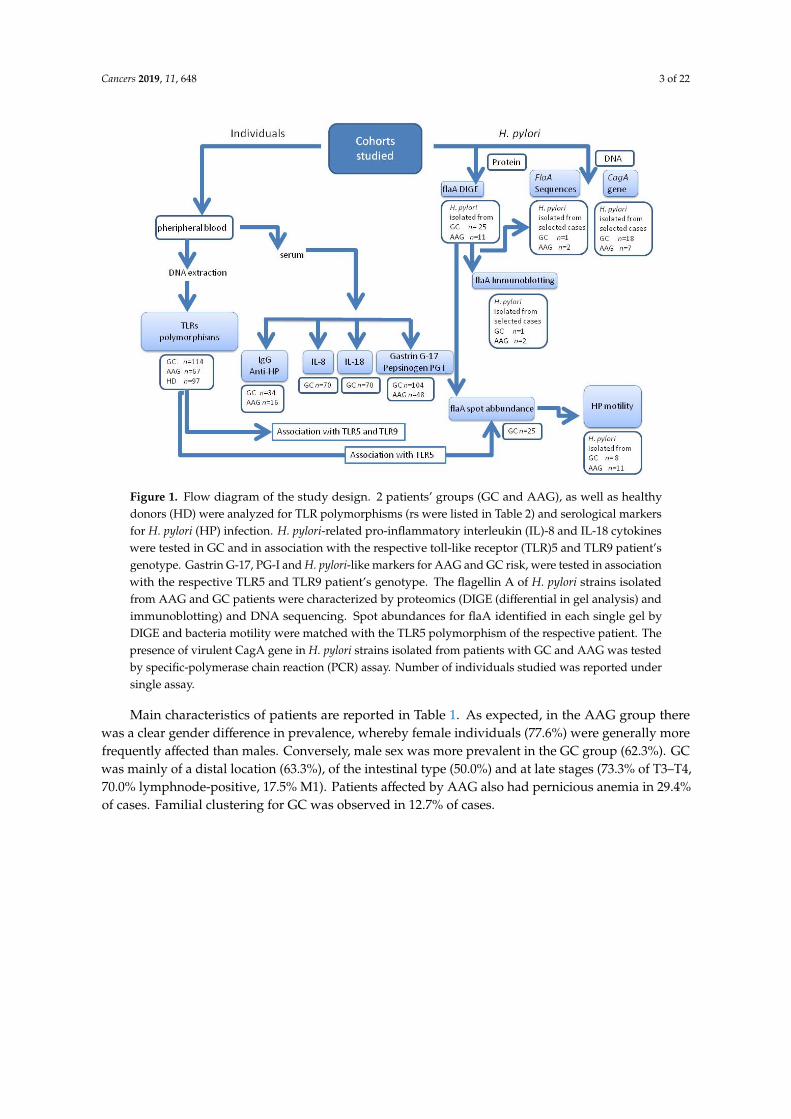
Figure 1. Flow diagram of the study design. 2 patients’ groups (GC and AAG), as well as healthy donors (HD) were analyzed for TLR polymorphisms (rs were listed in Table 2) and serological markers for H. pylori (HP) infection. H. pylori-related pro-inflammatory interleukin (IL)-8 and IL-18 cytokines were tested in GC and in association with the respective toll-like receptor (TLR)5 and TLR9 patient’s genotype. Gastrin G-17, PG-I and H. pylori-like markers for AAG and GC risk, were tested in association with the respective TLR5 and TLR9 patient’s genotype. The flagellin A of H. pylori strains isolated from AAG and GC patients were characterized by proteomics (DIGE (differential in gel analysis) and immunoblotting) and DNA sequencing. Spot abundances for flaA identified in each single gel by DIGE and bacteria motility were matched with the TLR5 polymorphism of the respective patient. The presence of virulent CagA gene in H. pylori strains isolated from patients with GC and AAG was tested by specific-polymerase chain reaction (PCR) assay. Number of individuals studied was reported under single assay.
Main characteristics of patients are reported in Table 1. As expected, in the AAG group there was a clear gender difference in prevalence, whereby female individuals (77.6%) were generally more frequently affected than males. Conversely, male sex was more prevalent in the GC group (62.3%). GC was mainly of a distal location (63.3%), of the intestinal type (50.0%) and at late stages (73.3% of T3–T4, 70.0% lymphnode-positive, 17.5% M1). Patients affected by AAG also had pernicious anemia in 29.4% of cases. Familial clustering for GC was observed in 12.7% of cases.
Figure 1. Flow diagram of the study design. 2 patients’ groups (GC and AAG), as well as healthydonors (HD) were analyzed for TLR polymorphisms (rs were listed in Table 2) and serological markersfor H. pylori (HP) infection. H. pylori-related pro-inflammatory interleukin (IL)-8 and IL-18 cytokineswere tested in GC and in association with the respective toll-like receptor (TLR)5 and TLR9 patient’sgenotype. Gastrin G-17, PG-I and H. pylori-like markers for AAG and GC risk, were tested in associationwith the respective TLR5 and TLR9 patient’s genotype. The flagellin A of H. pylori strains isolatedfrom AAG and GC patients were characterized by proteomics (DIGE (differential in gel analysis) andimmunoblotting) and DNA sequencing. Spot abundances for flaA identified in each single gel byDIGE and bacteria motility were matched with the TLR5 polymorphism of the respective patient. Thepresence of virulent CagA gene in H. pylori strains isolated from patients with GC and AAG was testedby specific-polymerase chain reaction (PCR) assay. Number of individuals studied was reported undersingle assay.
Main characteristics of patients are reported in Table 1. As expected, in the AAG group therewas a clear gender difference in prevalence, whereby female individuals (77.6%) were generally morefrequently affected than males. Conversely, male sex was more prevalent in the GC group (62.3%). GCwas mainly of a distal location (63.3%), of the intestinal type (50.0%) and at late stages (73.3% of T3–T4,70.0% lymphnode-positive, 17.5% M1). Patients affected by AAG also had pernicious anemia in 29.4%of cases. Familial clustering for GC was observed in 12.7% of cases.

Cancers 2019, 11, 648 4 of 22
Table 1. Patient (GC n = 114, AAG n = 67) and healthy donors (HD n = 97) characteristics.
Demographic features
Age at Inclusion Mean ± SEM (Range) p
GC 61.45 ± 1.04 (19–85) GC vs. HD, p < 0.001
AAG 54.59 ± 1.79 (31–70) GC vs. AAG, p = 0.019
HD 42.03 ± 1.66 (24–64) AAG vs. HD, p < 0.001
Gender Male
GC 71 (62.3%) GC vs. HD
AAG 15 (22.4%) GC vs. AAG, p < 0.001
HD 55 (56.7%) AAG vs. HD, p < 0.001
H. pylori status
IgG Anti-H. PyloriAntibodies positive
GC 34 (56.7%) GC vs. HD, p < 0.001
AAG 16 (40.0%) GC vs. AAG, p = 0.021
HD 18 (18.6%) AAG vs. HD
Missed data 101
Pepsinogen
Pepsinogen I (ng/mL) Mean ± SD
GC 153.85 ± 137 GC vs. HD, p < 0.001
AAG 37.29 ± 75 GC vs. AAG, p = 0.001
HD 73.7 ± 89 AAG vs. HD
Missed data 50
Pepsinogen II (ng/mL) Mean ± SD
GC 22.58 ± 19 GC vs. HD, p < 0.001
AAG 12.13 ± 6 GC vs. AAG, p = 0.007
HD 7.2 ± 5 AAG vs. HD, p < 0.001
Missed data 50
PG I/PG II Ratio Mean ± SD
GC 6.96 ± 4.17 GC vs. HD
AAG 2.43 ± 3.95 GC vs. AAG
HD 9.57 ± 2.78 AAG vs. HD, p < 0.001
Gastrin
Gastrin G17 (pg/mL) Mean ± SD
GC 20.47 ± 21 GC vs. HD, p = 0.003
AAG 431.8 ± 504 GC vs. AAG, p < 0.001
HD 7.7 ± 29 AAG vs. HD, p < 0.001
Missed data 50
Tumor characteristics(n = 60 GC)
GC Lauren Classification
Intestinal 30 (50.0%)
Diffuse 17 (28.3%)
Mixed 10 (16.7%)
unspecified 3 (5.0%)
GC Anatomical Site
Proximal 21 (35.0%)
Distal 38 (63.3%)
unspecified 1 (1.7%)
Clinical Stage
T T1–T2 (26.7%) T3–T4 (73.3%)
N no (30.0%) yes (70.0%)
M no (82.5%) yes (17.5%)
Surgery
Total gastrectomy yes (40.7%)
Partial gastrectomy yes (44.4%)
No surgery yes (18.8%)
Autoimmune characteristics(n = 47 AAG)
Pernicious Anemia yes (29.4%)
Familiarity for GC yes (12.7%)

Cancers 2019, 11, 648 5 of 22
2.2. Individual TLR-Polymorphisms Showed an Association of TLR5 rs5744174 Genotype-C/T and TLR9rs5743836 Allele-T in AAG Patients Compared to Healthy Donors
Allelic and genotype frequencies were estimated for each selected TLR based on literature fortheir potential influence on H. pylori infection and/or GC susceptibility (Table 2).
Table 2. Characteristics of selected Toll like receptors (TLR).
Gene ChromosomalPosition Locus SNP ID Change Nucleotide AA Reference
TLR1 4p14 exon 4 rs4833095 T > C N248S [8]
TLR2 4q32 5’UTR -196 to -174del(rs111200466) 22-bp ins/del — [9]
exon 3 rs3804099 T > C N199N [10]
TLR4 9q33.1 exon 3 rs4986790 A > G D299G [9,11]
exon 3 rs4986791 C > T T399I [9,11]
TLR5 1q41 Exon 4 rs5744174 T > C F616L [12]
TLR8 Xp22.2 5´UTR rs3764880 A > G — [13]
TLR9 3p21.3 5´UTR rs5743836 T > C — [14]
The distribution of genotypes for all the TLR polymorphism was consistent with Hardy–Weinbergequilibrium. TLR8 gene is located on the X-chromosome and therefore its frequency is higher in AAGsince female-prevalence is higher in AAG (77.6%) than in GC cases (38.1%. p < 0.0001). The distributionof genotype and allele frequencies in AAG, HD and GC of the TLR polymorphisms are shown inTable S1. Allele and genotype frequencies observed in HD are consistent with literature (CaucasiansHapMap information-Phase II CEU- for TLR1, TLR2, TLR4, TLR5, TLR8, TLR9, TLR2-196 to -174del). No significant differences were found in the distribution of allele and genotype frequencies ofall SNPs between patients with GC and HD (regression analysis). Conversely, the most significantpolymorphism based on Akaike’s information criterion (AIC) using genetic inheritance model ofassociation (SNPstats tool), was an over dominant model for the TLR5 rs5744174 polymorphism(T/T + C/C) in HD (61%) compared to AAG (45%) (p = 0.021 corrected by sex and age), and a higherfrequency of the recessive C/C genotype in GC (20%) compared to AAG (10%, p = 0.03 adjusted by sexand age, Table S1). In addition, AAG demonstrated a decrease in TLR9 rs5743836 allele C frequency(9%) compared to GC (17%) (p = 0.04, Table S1).
2.3. Targeted H. pylori Flagellin Protein Identification and Quantification by LC-MS/MS Spectrometry
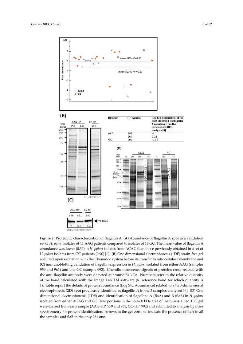
In a previous study [6], by two-dimensional fluorescence difference gel electrophoresis (DIGE) weidentified a reduction in H. pylori flagellin A (flaA) protein abundance in the H. pylori isolates fromAAG patients compared to those of GC. In the present study, we added H. pylori isolates from sevenfurther AAG patients and repeated the analysis. The abundance of flaA of H. pylori isolates from AAGsamples confirmed the selective reduction of the flaA in H. pylori isolates of AAG (n = 11, median 0.37)compared to those of GC (n = 18, median 0.90) (Figure 2A). Among these H. pylori isolates we selectedtwo samples from AAG either due to their high (AAG-HP sample 961, Log St Abund. = 0.24), or low(AAG-HP sample 959, Log St Abund. = −1) flagellin A content and one sample of H. pylori isolate froma GC with a low flagellin A content (GC-HP sample 992, Log St Abund. = −0.79). Protein fluorescentone-dimensional electrophoresis (1DE) sample load was shown in Figure 2B. After immunoblotting(the whole blot is displayed in Figure S1), a band at ~54 kDa in chemiluminescence, correspondingto the weight of flagellin (flaA: UniProtKB entry P0A0S1, 53284 Da; flaB: UniProtKB entry Q07911,53882 Da), was visible in all the three lanes (Figure 2C). The apparent discrepancy obtained betweenthe immunoblotting data for flaA abundance band and the DIGE analysis may come from the affinityand specificity of the antibody which recognizes all types of flagellins and the ability of 2D-DIGE todiscriminate among post-translational forms of the proteins.
Cancers 2019, 11, 648 6 of 22
Cancers 2019, 11, x 6 of 22
Figure 2. Proteomic characterization of flagellin A. (A) Abundance of flagellin A spot in a validation set of H. pylori isolates of 11 AAG patients compared to isolates of 18 GC. The mean value of flagellin A abundance was lower (0.37) in H. pylori isolates from ACAG than those previously obtained in a set of H. pylori isolates from GC patients (0.90) [6]. (B) One dimensional electrophoresis (1DE) strain-free gel acquired upon excitation with the Chemidoc system before its transfer to nitrocellulose membrane and (C) immunoblotting validation of flagellin expression in H. pylori isolated from either AAG (samples 959 and 961) and one GC (sample 992). Chemiluminescence signals of proteins cross-reacted with the anti-flagellin antibody were detected at around 54 kDa. Numbers refer to the relative quantity of the band calculated with the Image Lab TM software (R, reference band for which quantity is 1). Table report the details of protein abundance (Log Std Abundance) related to a two-dimensional
Figure 2. Proteomic characterization of flagellin A. (A) Abundance of flagellin A spot in a validationset of H. pylori isolates of 11 AAG patients compared to isolates of 18 GC. The mean value of flagellin Aabundance was lower (0.37) in H. pylori isolates from ACAG than those previously obtained in a set ofH. pylori isolates from GC patients (0.90) [6]. (B) One dimensional electrophoresis (1DE) strain-free gelacquired upon excitation with the Chemidoc system before its transfer to nitrocellulose membrane and(C) immunoblotting validation of flagellin expression in H. pylori isolated from either AAG (samples959 and 961) and one GC (sample 992). Chemiluminescence signals of proteins cross-reacted withthe anti-flagellin antibody were detected at around 54 kDa. Numbers refer to the relative quantityof the band calculated with the Image Lab TM software (R, reference band for which quantity is1). Table report the details of protein abundance (Log Std Abundance) related to a two-dimensionalelectrophoresis (2D) spot previously identified as flagellin A in the 3 samples analysed [6]. (D) Onedimensional electrophoresis (1DE) and identification of flagellins A (flaA) and B (flaB) in H. pyloriisolated from either ACAG and GC. Two portions in the ~50–60 kDa area of the blue-stained 1DE gelwere excised from each sample (AAG-HP: 959 and 961; GC-HP: 992) and submitted to analysis by massspectrometry for protein identification. Arrows in the gel portions indicate the presence of flaA in allthe samples and flaB in the only 961 one.

Cancers 2019, 11, 648 7 of 22
Identification of the protein components present in the band of about 54 kDa was performed byusing liquid cromatography (LC) mass (MS)/MS spectrometry and extraction of peptides present in the1DE gel in a range of ~50–60 kDa. Flagellin A peptides were identified in the sample 959 (gel portionsnr 2), 961 (gel portions nr 3 and 4), and 992 (gel portions nr 5 and 6); in addition, peptides of flagellintype B were also detected in sample 961 (Figure 2D and Table S2). Different flaA entries were foundafter database searches with Mascot software: the SwissProt 2::FLAA_HELPJ was found in all thesamples, while the NCBInr 1::gi|261839570, 1::gi|317180372 and 1::gi|188527549 were found in the onlysample 961, characterized by higher flaA (Table S2).
2.4. DNA Sequencing of H. pylori Flagellin A
The sequence alignment of H. pylori flagellin FlaA of samples 961, 959 and 992, compared to areference H. pylori FlaA gene; sample FlaA; WP_000885488.1 demonstrated a high degree of similarityin their nucleotide sequences (97% overall identity) (Figure 3A). The alignment emphasized the FlaAconservation and compared to the reference flaA showed three mutations (C296R; G315S; Q502H),which are present in all three samples, and a fourth mutation (A178T) present in sample AAG 961only. All the mutations were external to the D1 domain known for the TLR5 binding site (indicatedas interface for TLR5 in the Figure 3A,B). However, we cannot exclude that the Q502H mutation,present in the C-terminal D0 region, could have an effect on the stabilization of a flagellin-TLR5 dimericsignaling complex [15] or to be sensed by the intracellular NAIP5/NLCR4 inflammasome receptor [16].Moreover, in two rodent models of gastritis, the C296R mutation had been described to affect structureand function of the protein FlaA [17]. The sample 961 showed the A179T, in the vicinity of the knownglycosylated 181T residue [18]. In vitro H. pylori FlaA protein had been reported to lack the TLR5activation [19].

Cancers 2019, 11, 648 8 of 22Cancers 2019, 11, x 8 of 22
Interface B TLR5
Interface A TLR5
ND0 ND1
N= N-terminal, C= Carbossi terminal
D2-D3
CD1
CD0
(A)
(B)
Figure 3. Multiple sequence alignment of flagellin A and schematic structure of flaA-TLR5 interaction. (A) The strains of H. pylori used as sources of DNA for FlaA sequencing were previously isolated from patients with GC and AAG. Two strains represent H. pylori isolated from the biopsy of AAG patient (sample 961 and 959), and one H. pylori strain isolated from the biopsy of a patient with GC (sample 992). Raw sequences of DNA samples were analyzed and compared to a reference sequence (FlaA) by using multiple BLAST CLUSTAL alignment analysis. (B) Schematic diagram of flagellin structure with respect to possible interactions with TLR5. D0 domain of flagellin is needed for the dimerization of TLR5 and it is recognized by both the C-terminal part of the TLR5 extracellular domain and the intracellular NOD-like receptor (NLRC4) of the inflammasome complex. Conserved residues in the D1 domain (indicated with blue and red lines in the figure) interface primarly with the groove TLR5 binding site.
Figure 3. Multiple sequence alignment of flagellin A and schematic structure of flaA-TLR5 interaction.(A) The strains of H. pylori used as sources of DNA for FlaA sequencing were previously isolated frompatients with GC and AAG. Two strains represent H. pylori isolated from the biopsy of AAG patient(sample 961 and 959), and one H. pylori strain isolated from the biopsy of a patient with GC (sample992). Raw sequences of DNA samples were analyzed and compared to a reference sequence (FlaA)by using multiple BLAST CLUSTAL alignment analysis. (B) Schematic diagram of flagellin structurewith respect to possible interactions with TLR5. D0 domain of flagellin is needed for the dimerizationof TLR5 and it is recognized by both the C-terminal part of the TLR5 extracellular domain and theintracellular NOD-like receptor (NLRC4) of the inflammasome complex. Conserved residues in theD1 domain (indicated with blue and red lines in the figure) interface primarly with the groove TLR5binding site.

Cancers 2019, 11, 648 9 of 22
2.5. Motility Assays of H. pylori Isolates
H. pylori motility was investigated by assaying the ability of the H. pylori isolates from AAG andGC patients to spread on soft agar plates (Figure 4A). The area of spreading of H. pylori isolates from11 AAG (hollow circle) and from 8 GC patients (filled squares) (the Y axis, Figure 4B) were plottedwith the FlaA abundance obtained using the respective H. pylori isolate (The X axis, Figure 4B). Linearregression formula (Y = 0.12 + 0.42x) reflected populations of H. pylori from which abundance of theFlaA protein directly correlate with the area of H. pylori spread. Patients carrying the TLR5 rs5744174C/C genotype showed an increased area of spreading, compared with T/T homozigotes (ANCOVA test,covariate FlaA abundance, p = 0.039, Figure 4C).
Cancers 2019, 11, x 9 of 22
2.5. Motility Assays of H. Pylori Isolates
H. pylori motility was investigated by assaying the ability of the H. pylori isolates from AAG and GC patients to spread on soft agar plates (Figure 4A). The area of spreading of H. pylori isolates from 11 AAG (hollow circle) and from 8 GC patients (filled squares) (the Y axis, Figure 4B) were plotted with the FlaA abundance obtained using the respective H. pylori isolate (The X axis, Figure 4B). Linear regression formula (Y = 0.12 + 0.42x) reflected populations of H. pylori from which abundance of the FlaA protein directly correlate with the area of H. pylori spread. Patients carrying the TLR5 rs5744174 C/C genotype showed an increased area of spreading, compared with T/T homozigotes (ANCOVA test, covariate FlaA abundance, p = 0.039, Figure 4C).
Figure 4. Motility of H. pylori (A) Bacteria were plated in soft agar and photographed after incubation for 7 days at the same multiplicity. Control: HPJ99, motile H. pylori strain J99 (ATCC700824) from patient with duodenal ulcer (B) Passing badlock regression plots for area of spreading surrounding each H. pylory colony (mm, mean of triplicate experiment) versus FlaA abundance. H. pylori were recovered from 11 AAG (hollow circle) and from 8 GC patients (filled squares). Dashed line represents the optimal regression line; solid line represents the best fit by linear regression including AAG and GC (n = 19) cases (Y = 0.12 + 0.42x). (C) Analysis of area of spreading versus TLR5 genotype corrected by FlaA abundance covariate.
2.6. Identification of the CagA Virulent Gene in H. Pylori Isolates
CagA gene was identified in all the H. pylori isolates of 18 GC patients (18/18, 100%) but in 4 of AAG patients (4/7, 57%). H. pylori isolates of AAG patients showed a CagA-positive gene in 3/4 (75%) carriers with the TLR5 C/T-TLR9 allele-T haplotype, and in 1/3 case (33%) of carriers having the TLR5 T/T-TLR9 allele-C haplotype.
n
Y x
p=0.039
Figure 4. Motility of H. pylori (A) Bacteria were plated in soft agar and photographed after incubationfor 7 days at the same multiplicity. Control: HPJ99, motile H. pylori strain J99 (ATCC700824) frompatient with duodenal ulcer (B) Passing badlock regression plots for area of spreading surroundingeach H. pylory colony (mm, mean of triplicate experiment) versus FlaA abundance. H. pylori wererecovered from 11 AAG (hollow circle) and from 8 GC patients (filled squares). Dashed line representsthe optimal regression line; solid line represents the best fit by linear regression including AAG and GC(n = 19) cases (Y = 0.12 + 0.42x). (C) Analysis of area of spreading versus TLR5 genotype corrected byFlaA abundance covariate.
2.6. Identification of the CagA Virulent Gene in H. pylori Isolates
CagA gene was identified in all the H. pylori isolates of 18 GC patients (18/18, 100%) but in 4 ofAAG patients (4/7, 57%). H. pylori isolates of AAG patients showed a CagA-positive gene in 3/4 (75%)carriers with the TLR5 C/T-TLR9 allele-T haplotype, and in 1/3 case (33%) of carriers having the TLR5T/T-TLR9 allele-C haplotype.

Cancers 2019, 11, 648 10 of 22
2.7. Patients Having TLR5 rs5744174 T Allele Show a Tendency to Have a Higher H. pylori Antibody Titer andH. pylori Strains with a Lower Flagellin an Abundance
To further investigate the association of TLR5 rs5744174 polymorphism with H. pylori infection,we tested the impact of TLR5 genotype stratification on 1) IgG anti-H. pylori titer and 2) flagellinA abundance. Of the total of 104 cases tested for H. pylori infection 50 (34 GC and 16 AAG) wereH. pylori-positive. H. pylori-positive cases in GC carrying the TLR5 T/T genotype (53.6%) were higherthan in AAG (35.3%, Figure 5) and both in the respective groups showed a tendency to have anincreased IgG anti-H. pylori titer as compared with carriers of C/C and C/T genotype, although theresult did not attain a statistical significance (Figure 5)
Cancers 2019, 11, x 10 of 22
2.7. Patients Having TLR5 rs5744174 T Allele Show a Tendency to Have a Higher H. Pylori Antibody Titer and H. Pylori Strains with a Lower Flagellin an Abundance
To further investigate the association of TLR5 rs5744174 polymorphism with H. pylori infection, we tested the impact of TLR5 genotype stratification on 1) IgG anti-H. pylori titer and 2) flagellin A abundance. Of the total of 104 cases tested for H. pylori infection 50 (34 GC and 16 AAG) were H. pylori-positive. H. pylori-positive cases in GC carrying the TLR5 T/T genotype (53.6%) were higher than in AAG (35.3%, Figure 5) and both in the respective groups showed a tendency to have an increased IgG anti-H. pylori titer as compared with carriers of C/C and C/T genotype, although the result did not attain a statistical significance (Figure 5)
In addition, the result of ANOVA test showed a significant variation between abundance of the spot, corresponding to the flaA protein in DIGE and GC groups stratified by different TLR5 genotypes (Figure 5B). In particular patients having the TLR5 T/T genotype showed a lower mean of the flagellin A abundance (−0.1771, SD ± 0.41) compared to those having the C/C (0.335 SD ± 0.017, p=0.04) and the C/T (0.260 SD ± 0.08, p=0.02) genotype. The number of AAG patients with FlaA abundance data available were too low to perform the analysis.
AAG H. pylori-positive GC H. pylori-positive
TLR5 (rs5744174)C/C C/T T/T
AAGH. pylori-positiveH. pylori-negative
1 (50%)1
9 (47%)10
6 (35.3%)11
GCH. pylori-positiveH. pylori-negative
8 (23.5%)6
11 (45.8%)13
15 (53.6%)13
(A)
(B) p=0.04p=0.02
Figure 5. H. pylori IgG antibody titer and flaA abundance were associated with TLR5 genotype.(A) Figure illustrates the mean of H. pylori IgG titer stratified based on the presence of TLR5 rs5744174genotype. Samples tested n = 100, 60 were GC and 40 AAG. IgG anti-H. pylori titer showed a slighttendency to increase in the genotype T/T, and in particular in the GC group of patients. (B) The meanof flagellin A abundance in H. pylori strain isolates is lower in the isolates of patients carrying the TLR5rs5744174 genotype T/T.
In addition, the result of ANOVA test showed a significant variation between abundance of thespot, corresponding to the flaA protein in DIGE and GC groups stratified by different TLR5 genotypes

Cancers 2019, 11, 648 11 of 22
(Figure 5B). In particular patients having the TLR5 T/T genotype showed a lower mean of the flagellinA abundance (−0.1771, SD ± 0.41) compared to those having the C/C (0.335 SD ± 0.017, p = 0.04) andthe C/T (0.260 SD ± 0.08, p = 0.02) genotype. The number of AAG patients with FlaA abundance dataavailable were too low to perform the analysis.
2.8. Evaluation of the Serum Level of Inflammation-Related IL-8 and IL-18 Cytokines
TLR activation results in the release of various chemokines and cytokines. We focused on the effectof TLR5 and TLR9 polymorphisms on the pro-inflammatory cytokines interleukin (IL)-8 and IL-18,sharing a common relation to H. pylori infection. TLR5 polymorphism showed no significant relationwith neither IL-8 nor with IL-18 production in GC patients. Conversely, these results showed higheramounts of secreted serum IL-8 in patients having the TLR9 rs5743836 T/T genotype (4.21 ± 3.18 pg/mL,mean ± SD) compared to those having the C/T or C/C genotype (1.63 ± 1.58 pg/mL, mean ± SD)(p = 0.015; Figure 6A). A similar effect, which, however, did not reach a statistically significant difference,was observed for the IL-18 (Figure 6B).
Cancers 2019, 11, x 11 of 22
Figure 5. H. pylori IgG antibody titer and flaA abundance were associated with TLR5 genotype. (A) Figure illustrates the mean of H. pylori IgG titer stratified based on the presence of TLR5 rs5744174 genotype. Samples tested n = 100, 60 were GC and 40 AAG. IgG anti-H. pylori titer showed a slight tendency to increase in the genotype T/T, and in particular in the GC group of patients. (B) The mean of flagellin A abundance in H. pylori strain isolates is lower in the isolates of patients carrying the TLR5 rs5744174 genotype T/T.
2.8. Evaluation of the Serum Level of Inflammation-Related IL-8 and IL-18 Cytokines
TLR activation results in the release of various chemokines and cytokines. We focused on the effect of TLR5 and TLR9 polymorphisms on the pro-inflammatory cytokines interleukin (IL)-8 and IL-18, sharing a common relation to H. pylori infection. TLR5 polymorphism showed no significant relation with neither IL-8 nor with IL-18 production in GC patients. Conversely, these results showed higher amounts of secreted serum IL-8 in patients having the TLR9 rs5743836 T/T genotype (4.21 ± 3.18 pg/mL, mean ± SD) compared to those having the C/T or C/C genotype (1.63 ± 1.58 pg/mL, mean ± SD) (p = 0.015; Figure 6A). A similar effect, which, however, did not reach a statistically significant difference, was observed for the IL-18 (Figure 6B).
Figure 6. Cytokines, gastrin G17 and pepsinogen PGI association with TLR9 polymorphism. (A) IL-8and (B) IL-18 cytokines titer were tested using a Luminex MagPlex based assay and result stratifiedbased on the carriers TLR9 rs5743836 polymorphism. A significant increase in the mean IL-8 titer wasfound associated with the presence of the TLR9 T/T genotype (A). Samples tested were n = 70 GC,samples duplicated with a CV > 15% were excluded from the analysis and they are not reported in thefigure. The mean of IL-18 levels between the 2 TLR9 genotypes (C/T and C/C vs. T/T) did not reach astatistical difference (B). TLR9 T/T genotype was found associated to highest mean level of gastrin G17in the AAG group (C) but not in GC (E). TLR9 T/T genotype was found associated with a mean slightlyhigher PGI level in AAG (D) and in GC (F), although in both the groups the difference did not reach asignificant difference. Compared to AAG, the mean of PG1 level is higher in GC.

Cancers 2019, 11, 648 12 of 22
2.9. Association of the Serum Level of Atrophic Markers Gastrin G17 and PGI with TLR9 rs5743836 T Allele
Elevated gastrin G17 titer is a diagnostic marker of AAG and in association with low PGI level(<48 pg/mL), well correlate with advanced stage of metaplasia in AAG, a condition considered to be atrisk for GC development [20]. We focused if TLR5 or TLR9 polymorphisms were associated with higherlevel of the released serum AAG marker gastrin G17 and/or a lower level of PGI. Results showed highermean of secreted gastrin G17 level in AAG patients (Figure 6C), but not in GC (Figure 6E), having theTLR9 rs5743836 T/T (mean 376.52 SD ± 339; C/T or C/C genotype mean 137.52, SD ± 124; p = 0.02).Inboth the AAG and GC patients, the difference between TLR9 genotype and PGI titer was slightly higherin the patients with the T/T genotype (mean 34.75 SD ± 52 in AAG, Figure 6D; 161.53 SD ± 161 in GC,Figure 6F, respectively) compared to those with the T/C or C/C genotype (18.42 SD ± 14.56 in AAG;153.74 SD ± 113 in GC), but in these last cases the difference did not reach a statistical significance.
3. Discussion
Alterations in the immune response of patients with AAG may predispose to GC, and a linkbetween AAG and GC has been reported in many studies. AAG is the outcome of a pathologicalCD4 T cell-mediated autoimmune response directed against the gastric H+/K+-ATPase and severalepidemiological data point to a relation between immunogenetic background of the host and AAGdevelopment. A role for H. pylori as an antigen trigger for the genesis of AAG trough a molecularmimicry between H. pylori antigens and gastric H/K-ATPase was evoked, although evidence to supportthat this pathogen is needed for disease development still require more extensive investigation [21,22].
In the present study, compared with HD, we found that the TLR5 rs5744174 C-allele and thereduction of H. pylori motility were associated with AAG. Furthermore, results showed a TLR5discrepancy between AAG and GC with a predominance of TLR5 rs5744174 in heterozygosis (C/T) inAAG, and in homozygosis (C/C) in GC suggesting a possible role of TLR5 C-allele in increasing therisk for gastric diseases. In addition, the lower frequency of TLR9 C-allele in AAG, largely of femalesex, compared to GC, suggested an association with a protective sexual dimorphism related to TLR9in AAG.
The TLR5 receptor specifically binds bacterial flagellin, the principal component of flagella, abacterial appendage necessary for its locomotion. Activation of TLR5, located on the cell membranesurface, by flagellin results in activation of the NF-kB pathway, which is linked to cancer, inflammatoryand autoimmune diseases. Due to the high importance of TLR5 in induction of both innate andadaptive immunity flagellin is now used as an adjuvant of different vaccine preparations and ananti-cancer agent in several clinical trials, as well as a potential agent to reduce radiation toxicity.However, besides having beneficial actions, it is also increasingly clear that a paradoxical effect ofTLR5 expression may be present in different autoimmune and inflammatory diseases (e.g., acute lunginflammation, inflammatory bowel disease, liver injury) [23,24] as well as in specific cancers, as in thecase of GC [25,26].
On the contrary to other flagellated bacteria, H. pylori flagellin, like those of H. pylori strainsisolated from the biopsies of our patients (Figure 3), presented a peculiar binding interface for TLR5(D1 and D0 domains, Figure 3) did not consent the traditional TLR5 activation [19], but nonethelessmay produce an inflammatory complex [27]. Indeed, it was demonstrate that the peculiar D0 domainof H. pylori flagellin, interacting with the C-terminal region of the TLR5 including the TLR5 rs5744174polymorphism and playing a role in TLR5 dimerization [15], may be recognized by both the TLR5 andthe intracellular Nod-like receptors, NLRC4, an element of inflammasome complex. However, the H.pylori flagellin- TLR5/NLCR4 recognition failed to elicit the caspase-1 activation necessary to processespro-IL-18 into IL-18-mature form of secretion and to initiate pyroptosis cell-mediated death [27,28]. Inspite of that, both TLR5 and NLRC4 receptors contribute to flagellin-induced antibody production(adaptive immunity) [29], since TLR5 may present peptides to flagellin-specific CD4+ T-cells also inthe absence of conventional TLR signaling [30]. In our series, the different TLR5 genotypes we foundshowed a similar serum IL-8 and IL-18 cytokine level among patients, while the TLR5 C-allele was

Cancers 2019, 11, 648 13 of 22
associated to a reduction in the H. pylori-induced antibody production (Figure 5A). Thus, these resultsindicate that in our patients TLR5 in the presence of H. pylori flagellin should produce an inflammasomecomplex that however did not culminate in the release of IL-18 involved in resistance to infectiouspathogens, and that moreover the presence of TLR5 C-allele, particularly when homozygous, mightreduce the host adaptive immune response (anti H. pylori antibody), thus favoring H. pylori persistenceand tissue damage-associated inflammasome activation. In accordance with this, H. pylori flagellinby influencing the inflammatory milieu trough TLR5 might have a role in gastric cancerogenesis, ahypothesis supported by the demonstration that inflammasome-derived exosomes from GC cellswere able to directly activate NF-kB signaling pathway promoting GC [31] and as a consequence ofthe inflammation, the disruption of the epithelial polarity and integrity of the gastric cells shouldfavor the insertion of H. pylori into the gastric epithelial cells [32], a phenomenon also associated withGC development.
Previously we found in H. pylori strains isolated from AAG patients a significant decrease in theflaA protein compared to strains isolated from both GC and from patients with duodenal ulcer [33].Herein, we confirmed that H. pylori strains of AAG showed a reduction in the flaA abundance(Figure 2A). Furthermore, we also demonstrated a moderate reduction in motility of those H pyloristrains showing a reduction in the FlaA abundance or with a mutation in the FlaA sequence (sample661). Presence of TLR5 T-allele also correlate with the reduction of flaA abundance and H. pylorimotility (Figure 4). Several studies have demonstrated that alteration in the flaA expression as well asits glycosilation modulated the motility of H. pylori [34,35]. Phenotypic and molecular characterizationfurther demonstrated that H. pylori strains showing the highest motility exhibited a better capabilityof colonization and cancerogenesis (CagA phosphorylation and NF-kB activation) [35,36]. Indeed,H. pylori needs to invade and proliferate in the epithelial cells to induce GC development [37]. Thus,the reduction in motility, although moderate, showed in H. pylori strains isolated from patients withAAG, also more evident in patients carrying the TLR5 T-allele, should protect patients by reducing theefficiency of H. pylori colonization and the maintenance of infection in the gastric epithelium. However,modulation of the motility is a result of several factors, not only flaA expression, as at least in onecase of H. pylori with a relative high abundance of flaA expression isolated from an AAG individual(sample 961) we found a reduction in the motility in the in vitro assay (Figure 4). In this last case,however, a mutation in the flagellin A sequence near a site of glycosilation was found. In addition,H. pylori chronic infection could be reduced by the hypo-achlorhydria that characterizes the gastricmilieu in AAG patients [38]. H. pylori adhesion to gastric cells is essential to inject the bacterial CagAoncoprotein into the host cells [36]. We found the CagA gene in the H. pylori strains isolated from allthe GC cases while only in 50% of H. pylori isolated from AAG patients (Figure 4) and that the CagAgene was present less frequently in H. pylori isolated from AAG carrying the suggested protective TLR5rs5744174 genotype-T/T. Therefore, moderate motility and a low frequency of CagA gene in H. pyloristrain isolated from patients with AAG and TLR5 T/T genotype, both allowed the distinction betweenAAG and GC, becoming in these way potentially protective factors towards GC development in thesepeculiar subset of AAG patients (Figure 4). The reason for flaA and motility reduction in H. pyloriisolates from AAG patients remains to be elucidated since the regulatory network that modulatesthe expression of flagellar genes and bacterial motility is complex and not fully understood. In theintestinal tract, TLR-mediated flagellin antibody response was found to originate from antigen onlywhen a tissue damage condition was present [39] and the formation of the flaA-anibody complexundergoing rapid degradation via Fc receptor-mediated pahogytosis [40]. Therefore, we hypothesizedthat the higher H. pylori-antibody level we found in patients having the TLR5 T/T genotype could inpart contribute to the protective reduction of flagellin A expression in some patients. Alternatively,H. pylori strain-gene specificity due to the high variation of the H. pylori genome, may regulate theflagellin gene expression and most likely induce severe disease. Further studies should be performedto elucidate this point.

Cancers 2019, 11, 648 14 of 22
In addition, the interaction between flagellin from other bacteria than H. pylori and TLR5 mighthave a role in gastric diseases. In vitro and in vivo studies showed that homozygous TLR5 rs5744174C/C carriage is associated with a reduced traditional TLR5 responsiveness to flagellin of overall gastriccommensal bacteria [41]. Accordingly, T/T carriage was associated with higher secretion of severalantimicrobial factors against most infections resulting in a better protective genotype, compared to theC/C genotype [42]. In line with this hypothesis, it has been speculated that TLR5 rs5744174 variantmay be informative not only for H. pylori but also for commensal bacteria whose contribution toimmunomodulation is only emerging, suggesting that survival of gastric microbioma via TLR5 mightnot only be dependent on the species specific stimulatory potential of flagellin but also on TLR5genotype [41]. As a consequence the resultant positive selection of the rs5744174 C-polymorphismamong the human evolutionary changes in individuals constantly exposed to high levels of pathogenshas been suggested to be likely to reduce NF-κB activation, which perpetuated a pro-inflammatorystatus and tissue damage in the host [41]. The TLR5 rs5744174 C/C genotype was also associatedwith increased risk for some cancers, including GC [26] and a lower survival in colorectal cancer byinfluencing cytokine response to intestinal bacteria [26] However, NF-κB paradoxically may also beinvolved in autoimmune suppression, by promoting the development and the immunosuppressivefunction of regulatory T cells, which are also crucial for central and peripheral tolerance, althoughthis factor alone was not enough [43]. Intriguingly, over the recent decades in Western societies,concurrently to the overall reduction of infectious diseases and the consolidation of the TLR5 C-variantin the human genome, epidemiological data provided evidence of a steady rise of autoimmunediseases [44].
In the present study, we also found a decrease in the TLR9 rs5743836 minor allele frequency (i.e.,-1237 C-variant) in AAG, compared to GC, suggesting a protective role of the TLR9 C-variant towardsAAG development. The rs5743836 variant-C, located at the promoter region of the TLR9 gene createsIL-6 and NF-kB responding elements, so conferring an increased TLR9 transcription activity comparedto the T-variant in response to various stimulants, like induced NF-kB-mediated inflammatory factors.To our knowledge, this is the first study regarding the TLR9 rs5743836 polymorphism in AAG. Aclear sex difference in prevalence for most autoimmune diseases, including AAG, whereby femaleindividuals are generally more frequently affected than male [45]. Recently, in healthy individuals,serum testosterone levels was found to negatively correlate with the expression of TLR9 suggestingthat this effect may have a protective role against autoimmune disease development [46]. TLR9 wasshown over-expressed in hormone-regulated cancers (e.g., breast, ovary and prostate cancers) where itshowed by contrast a relationship with a poor prognosis; in these cases the increasing expression ofestrogen receptor (ERα) have been reported to lead to the decreased expression and function of TLR9with a beneficial effect against tumor invasion [47].
TLR9 expression in B cells was shown important for autoantibody production and activationof autoreactive B cells in the periphery and treatment by using TLR9 inhibitors was demonstratedto improve the clinical outcome of some autoimmune disorders (e.g., rheumatoid arthritis, systemiclupus erythematosus) [48]. Regarding H. pylori infection, in literature TLR9 activation resulted in apro-inflammatory response [49], but during the acute phase of H. pylori infection, TLR9 showed anopposite role by seeming to promote anti-inflammatory signaling in order to favor the establishmentof a persistent infection and, thus, acting as a suppressor of H. pylori-induced gastritis [50,51]. TLR9expression also differs in healthy individuals and in patients infected with the H. pylori; TLR9 waslocated in the apical compartment of the gastric epithelium in healthy individuals, while its expressionwas located in the basolateral compartment in individuals having a H. pylori infection [32]. Authorsproposed that the inflammatory microenvironment resulting from chronic H. pylori infection containsmore gastric cells without polarity that activate TLR9 to further promote the inflammatory cascadesand then eventually the development of GC. Accordingly, TLR9 was found up-regulated in GC tissueand absent in epithelium with intestinal metaplasia and dysplasia [32] and TLR9 C-allele, inducing ahigher TLR9 expression, was found associated with GC [14], as well as in our series, in high GC risk
Cancers 2019, 11, 648 15 of 22
areas, like Colombia [52], in relatives of GC patients in West of Scotland [14]. Cag T4SS system wasmandatory to actively transport H. pylori DNA into the host cells and for TLR9 engagement [52]. In ourseries CagA gene was present in all GC cases but only in 50% of AAG cases supporting the active roleof CagA gene and TLR9 activation in particular in GC. Moreover, we found an association betweenTLR9 rs5743836 C-allele with a lower PGI level, indicative of advanced stage of atrophy in AAG [20],with an increased TLR9 expression (Figure 6) and in GC with an increase in the pro-inflammatorycytokines, IL-8 (Figure 6), which is known to be induced by H. pylori infection in GC [14]. Overallthese data suggested that TLR9 rs5743836 C-allele was less frequent in AAG, particularly of femalesex, since TLR9 express a sex hormone or receptor that may modulate the TLR9 expression andthat expression/activation of the TLR9 could result in a detrimental effect in GC development. Arecent study showing that (i) expression of TLR9 and TCR inducible costimulatory receptor (ICOS)ligand (ICOS-L) in plasmacytoid dendritic cells (pDCs) infiltrating the GC tumor showed a strongimmunosuppressive function and (ii) that the percentage of these cells increased in patients withH. pylori infection, more in the late GC stages than in the early stages and in patients with a lowerrelapse-free survival [53], support our results. As TLR9 is located in the cytoplasmic compartment, it isbelieved that ligand(s) may gain access to the TLR9 by using a receptor-mediated delivery (e.g., H.pylori T4SS system, B-cell receptor immune complexes, KIR3DL receptor).
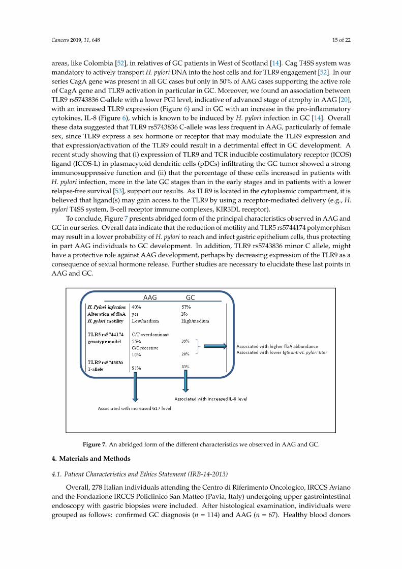
To conclude, Figure 7 presents abridged form of the principal characteristics observed in AAG andGC in our series. Overall data indicate that the reduction of motility and TLR5 rs5744174 polymorphismmay result in a lower probability of H. pylori to reach and infect gastric epithelium cells, thus protectingin part AAG individuals to GC development. In addition, TLR9 rs5743836 minor C allele, mighthave a protective role against AAG development, perhaps by decreasing expression of the TLR9 as aconsequence of sexual hormone release. Further studies are necessary to elucidate these last points inAAG and GC.
Cancers 2019, 11, x 15 of 22
expression also differs in healthy individuals and in patients infected with the H. pylori; TLR9 was located in the apical compartment of the gastric epithelium in healthy individuals, while its expression was located in the basolateral compartment in individuals having a H. pylori infection [32]. Authors proposed that the inflammatory microenvironment resulting from chronic H. pylori infection contains more gastric cells without polarity that activate TLR9 to further promote the inflammatory cascades and then eventually the development of GC. Accordingly, TLR9 was found up-regulated in GC tissue and absent in epithelium with intestinal metaplasia and dysplasia [32] and TLR9 C-allele, inducing a higher TLR9 expression, was found associated with GC [14], as well as in our series, in high GC risk areas, like Colombia [52], in relatives of GC patients in West of Scotland [14]. Cag T4SS system was mandatory to actively transport H. pylori DNA into the host cells and for TLR9 engagement [52]. In our series CagA gene was present in all GC cases but only in 50% of AAG cases supporting the active role of CagA gene and TLR9 activation in particular in GC. Moreover, we found an association between TLR9 rs5743836 C-allele with a lower PGI level, indicative of advanced stage of atrophy in AAG [20], with an increased TLR9 expression (Figure 6) and in GC with an increase in the pro-inflammatory cytokines, IL-8 (Figure 6), which is known to be induced by H. pylori infection in GC [14]. Overall these data suggested that TLR9 rs5743836 C-allele was less frequent in AAG, particularly of female sex, since TLR9 express a sex hormone or receptor that may modulate the TLR9 expression and that expression/activation of the TLR9 could result in a detrimental effect in GC development. A recent study showing that (i) expression of TLR9 and TCR inducible costimulatory receptor (ICOS) ligand (ICOS-L) in plasmacytoid dendritic cells (pDCs) infiltrating the GC tumor showed a strong immunosuppressive function and (ii) that the percentage of these cells increased in patients with H. pylori infection, more in the late GC stages than in the early stages and in patients with a lower relapse-free survival [53], support our results. As TLR9 is located in the cytoplasmic compartment, it is believed that ligand(s) may gain access to the TLR9 by using a receptor-mediated delivery (e.g., H. pylori T4SS system, B-cell receptor immune complexes, KIR3DL receptor).
To conclude, Figure 7 presents abridged form of the principal characteristics observed in AAG and GC in our series. Overall data indicate that the reduction of motility and TLR5 rs5744174 polymorphism may result in a lower probability of H. pylori to reach and infect gastric epithelium cells, thus protecting in part AAG individuals to GC development. In addition, TLR9 rs5743836 minor C allele, might have a protective role against AAG development, perhaps by decreasing expression of the TLR9 as a consequence of sexual hormone release. Further studies are necessary to elucidate these last points in AAG and GC.
Figure 7. An abridged form of the different characteristics we observed in AAG and GC. Figure 7. An abridged form of the different characteristics we observed in AAG and GC.
4. Materials and Methods
4.1. Patient Characteristics and Ethics Statement (IRB-14-2013)
Overall, 278 Italian individuals attending the Centro di Riferimento Oncologico, IRCCS Avianoand the Fondazione IRCCS Policlinico San Matteo (Pavia, Italy) undergoing upper gastrointestinalendoscopy with gastric biopsies were included. After histological examination, individuals weregrouped as follows: confirmed GC diagnosis (n = 114) and AAG (n = 67). Healthy blood donors

Cancers 2019, 11, 648 16 of 22
(HD) without either GC or AAG (n = 97) were included as negative control. HD were individualswith functional dyspepsia, normal gastric histology and a negative family history of GC (n = 44) orblood sample donation from healthy individuals collected by the CRO biobanking for DNA research(n = 53). Histopathologic diagnosis of GC and AAG was confirmed by experienced gastrointestinalpathologists. GC was classified according to the Lauren classification [54] and the disease stage wasassessed according to the TNM criteria, 7th edition [55]. Diagnosis of AAG was based on both theupdated Sydney-Houston criteria (stomach corpus and fundus atrophy with antrum sparing) and theanti-parietal cell antibody positivity (PCA) [2]. Overall clinical, pathological and laboratory features ofpatients with AAG and GC, and those of HD were reported in Table 1. GC were Caucasian patients: 71men and 43 women with a mean age of 61.45 ± 1.04 years (range 19 to 85 years). AAG patients were: 15men and 52 women with a mean age of 54.59 ± 1.79 years (range 31 to 70 years). The healthy controlswere: 55 men and 42 women with a mean age of 42.03 ± 1.66 years (range 24 to 64 years). Amongthese, 197 (60 GC, 40 AAG and 97 HD) were screened for pepsinogen (PGI and PGII), gastrin G17 andH. pylori infection using the gastropanel kit (Biohit HealthCare, Milan, Italy) [20]. Significant value forpositive IgG anti-H. pylori antibody titer is ≥30 U/mL. Parietal cell antibody (PCA) titer was determinedin all patients through immunofluorescence (IF) technique. A titer greater of 1:40 was considered assignificant. In case of PCA negativity detected through IF, but with histopathological lesions consistentwith AAG, PCA were assessed again with an ELISA technique, which is more accurate [56,57]. OnlyPCA positive patients were included in this study. Pernicious anemia with vitamin B12 deficiency orfolate deficiency were diagnosed according to laboratory tests (hemoglobin <13 g/dL in men, <12 g/dLin women, serum vitamin B12 < 240 pg/mL, serum folate < 3 ng/mL) and occurred in 70.6% of AAGcases. The study was approved by the Internal review board (CRO no. 14). Informed consent wasobtained for all subjects.
4.2. Selection of TLRs Genetic Variants
Eight TLR polymorphisms were selected based on literature for their potential influence on H.pylori infection and/or GC susceptibility. TLR2 and TLR4 have a key role in recognition of variousstructural components of bacterial outer membrane [9–11]. TLR1 and TLR6, represent coreceptorsof TLR2 and associated with risk for atrophic gastritis [8]. TLR4 polymorphic variants were linkedto a decreased responsiveness to H. pylori lipopolysaccharide and a risk factor for precancerouslesions; although other studies indicated TLR2 rather than TLR4 as the dominant innate immunereceptor for the recognition of gastrointestinal Helicobacter species. Murine models indicated a role forTLR2, TLR9, and TLR8 in the recognition of H. pylori by dendritic cells, which are recruited duringinflammation and have the ability to traverse the epithelial tight junctions to sample luminal bacterialantigens for presentation. TLR5 recognizes bacterial flagellin and a still unknown bacterial protein [58].TLR3, 7, 8 and 9 are expressed in the endosomal membrane and can recognize DNA and RNA ofinvading microorganisms [59]. Some TLR8 polymorphisms has been associated with exacerbatedgastric inflammation and were found more frequently in Asian (85%) than in Caucasian patients(20–30%) in agreement with the higher frequency of H. pylori infection in Asian populations thanin American or European. TLR9 recognizes the DNA of cancer-associated CagA+ type IV secretionsystem (T4SS) of H. pylori so interfering with the cellular persistence of the bacteria [52]. Table 2 listedthe selected TLR1, TLR2, TLR4, TLR5, TLR8 and TLR9 genetic variants used in the present study.
4.3. Analysis of TLR Polymorphisms
Genomic DNA was extracted from whole blood by using the EZ1 Qiagen blood kit (Qiagen Inc.,Valencia, CA, USA). Allele-specific polymerase chain reaction (PCR) was used to detect TLR2-196to -174del and TLR4 polymorphisms [60]. The primer sequences and the sizes of PCR products areshown on Table 3. The TLR1 (rs4833095), TLR2 (rs3804099), TLR8 (rs3764880) and TLR9 (rs5743836)SNPs genotyping were performed by allelic discrimination using TaqMan SNP-genotyping Assays(Applied Biosystems/Thermo Fisher Scientific, Waltham, MA, USA). Genotyping was carried out on an

Cancers 2019, 11, 648 17 of 22
ABI 7900HT Fast Real-Time PCR System (Applied Biosystems, Foster City, CA, USA) with standardconditions and according to the manufacturer’s protocol. We used a home-made High-ResolutionMelting method (HRM) to discriminate the TLR5 rs5744174 genotype. PCR-HRM analysis wasperformed using a CFX96 Touch Real-Time PCR Detection System (Bio-Rad, Hercules CA, USA) in afinal volume of 20 µL of a reaction mixture containing: 30 ng of genomic DNA template and 0.7 µM ofeach PCR primer. The HRM-PCR protocol included 1 cycle at 98 ◦C for 3 min, followed by 40 cyclesat 95 ◦C for 10 s, 62 ◦C for 10 s, and 72 ◦C for 20 s. HRM curve acquisition (60 ◦C to 95 ◦C, with anincrement of 0.1 ◦C/10 s) and analysis were performed on CFX Real-time PCR instrument (Bio-RadCFX Manager software). Melting curves were normalized to relative fluorescence units (RFU) in aspecified melt region (77.5 ◦C to 82.5 ◦C). Three pre-defined genotype samples and a negative controlwithout DNA were used as controls.
Table 3. TLR primers.
Primers ID Sequence (bp) Size Products References
TLR2-196, -174 FOR: CTCGGAGGCAGCGAGAAA 286/264 [60]REV: CTGGGCCGTGCAAAGAAG
TLR4-EXON-3FOR: TCTGCTCTAGAGGGCCTGTGC
632 (designed by the authors)REV: TCCTGGAAAGAATTGCCAGCCA
TLR5-5744174FOR: TGTCACTATAGCTGGGCCTC
104 (designed by the authors)REV: CCTCTTCATCACAACCTTCCG
TLRs: Toll-like receptors; bp: base pair.
4.4. Anti-Flagellin Antibody and Liquid MS/MS Spectrometry Validation
In a previous study comparative 2D-DIGE analysis of H. pylori strains isolated from patients withAAG (AAG-HP) and GC (GC-HP) revealed a significant decrease in expression of the flaA protein in H.pylori strain isolated from AAG patients [6]. In the present study we want to further validate this databy adding some samples (Figure 2A) and by using immunoblot and liquid mass spectrometry analyses.Analyses focused on two samples of AAG-HP characterized by a high (sample 961, Log St Abund. =
0.24), or a low (sample 959, Log St Abund. = −1) flagellin A content. A sample of GC-HP showing a lowflaA content was used as reference control (sample 992, Log St Abund. =−0.79). Ten µg of proteins werefractionated on 12% Criterion TGX Stain-Free gels and, after gel image acquisition with the Chemidocsystem (Bio-Rad), electrotransferred onto nitrocellulose membranes. Membranes were incubated withthe monoclonal antibody anti-flagellin (1:12000; ABIN235293, Antibodies Online, Aachen, Germany).Antibody-bound proteins were detected by enhanced chemiluminescence using the Chemidoc systemafter incubation with ECL HRP-conjugated secondary antibodies (1:10,000 dilution, Santa-Cruz, Dallas,TX, USA) and reaction with ClarityTM Western ECL Substrate (Bio-Rad). The image of the gel acquiredbefore its transfer was used as control for equal protein loading among samples.
Protein extracts from AAG- and GC-HP (50 µg per lane) were separated by 1DE, and images ofblue-stained gel were acquired with the Chemidoc system. A total of two gel portions in the MWrange between 50 and 60 kDa containing proteins cross-reacting with the flagellin antibody (Figure 2D,rectangle and numbered lanes) were excised, reduced by incubation with 10 mM dithiothreitol (1 hat 57 ◦C), and alkylated with 55 mM iodoacetamide (45 min at room temperature). Samples werefurther washed with NH4HCO3, dehydrated, trypsin digested and processed for LC-MS/MS analysesusing a LTQ XL-Orbitrap ETD equipped with a HPLC NanoEasy-PROXEON (Thermo Fisher Scientific,Waltham, MA, USA). Database searches were done with the MASCOT search engine version 2.3 againstSwissProt and NCBInr (Matrix Science, London, UK), and the presence of flagellin was searched amongfirst 30 report hits.

Cancers 2019, 11, 648 18 of 22
4.5. Flagellin A Nucleotide Sequencing
The nucleotide sequence of flaA encoding a protein of 510 AA with a predicted molecular mass of53.2 kDa. Genomic DNA for flaA sequencing was extracted from H. pylori strains isolated from AAGand GC patients as previously reported [6]. The following samples were chosen based on the highvariance in the flaA expression: sample 961 from AAG, with both FlaA and FlaB expression, sample 959from AAG, with only FlaA expression, and sample 992 from a GC showing lower abundance of FlaAwhen compared with other H. pylori strains isolated from GC patients (Figure 2). After alignment of 30non-redundant H. pylori flaA gene sequences(https://www.ncbi.nlm.nih.gov/), a conserved region waschosen and the Primer3Plus software was used to design the oligonucleotide primers for amplificationand sequencing (http://bioinfo.ut.ee/primer3-0.4.0/). Sequences of primers were reported in Table S3.Template DNA for cycle sequencing was of 1521 bp and obtained through amplification for 30 cyclesas follows: 94 ◦C for 30 s, 48 ◦C for 30 s, and 72 ◦C for 2 min with an initial denaturing step of 95◦C for 6 min. The PCR products were purified by the addition of 3 µL of Clean up Reagent (Abbott,Des Plaines, IL, USA), 37 ◦C for 15 min and 80 ◦C for 15 min. Cycle sequencing was performed usingDyeDeoxy Terminator Cycle Sequencing kit 3.1 from Applied Biosystems. PCR products were purifiedusing the Centrisep spin columns (Princeton Separations, Adelphia, NJ, USA) and run on an ABI Prism3130XL automated DNA Sequencer (Applied Biosystems, Foster City, CA, USA). Raw sequences ofDNA samples were analyzed and compared to a reference FlaA aminoacid sequence from H. pylorigenome (NCBI WP_000885488) using multiple BLAST CLUSTALW alignment analysis.
4.6. Motility Assays of H. pylori Isolates
For the motility assay H. pylori samples isolated from 11 patients with AAG and used for theflaA abundance analysis were chosen and compared with other H. pylori strains isolated from eightGC patients. Briefly, 3 uL of H. pylori suspensions grown at the same exponential rate and at a 108
colony-forming units (CFU)/mL were spotted onto soft agar plates containing 4.3% Brucella Broth(Difco BD, Saprks, MS, USA), 0.4% of Bacto agar (Difco) and 10% horse serum and incubated forseven days at 37 ◦C under microaerophilic conditions. The motility assay was conducted in threeindependent experiments and the mean diameter of the migration was then calculated and comparedto the reference J99 H. pylori strain.
4.7. Identification of the CagA Gene
H. pylori genomic DNA was extracted from H. pylori isolates from 18 GC patients and from 7AAG as previously reported [6] and tested for the presence of the CagA virulent gene. Primers specificfor CagA gene [61] were used for the PCR amplification and products were then analyzed on 8%polyacrylamide gel electrophoresis.
4.8. IL-8 and IL-18 Cytokines Assay
A serum sample was collected from all participants. Cytokines were assayed using a Luminexbased MAG multiplex assay (Luminex 200 Xponent, Austin, TX, USA). Two cytokines (IL-8 and IL-18)from a custom Human High Sensitivity panel (ThermoFisher Scientific) were used in GC. Serumsamples were thawed at 4 ◦C and then centrifuged at 1400× g to remove any protein aggregates thatmay potentially obstruct the measurement. The supernatant was then transferred to a fresh tube andwas diluted 1:2 in assay buffer. A 10-point standard curve with serial dilutions of 1:4 was generatedusing reconstituted stock standards supplied by the manufacturer; quality controls supplied by themanufacturer were also used to determine assay accuracy (Luminex 200 performance and verificationand calibration kits). The data was generated using the Xponent software, which calculated averagevalues against a 5-parameter logistic standard curve corrected by background readings. Plasmasamples were assayed in duplicate.

Cancers 2019, 11, 648 19 of 22
4.9. Statistical Analysis
Continuous variables are presented as the mean ± SD and were analyzed by independent t-testand Anova test among variable groups. Genetic data was assessed using regression analysis (SNPStatssoftware; http://bioinfo.iconcologia.net/SNPstats) in patients and controls, and adjusted for sex. Forbinary responses, the logistic regression analysis was performed to obtain genotype frequencies,odd-rations (OR) and 95% confidence intervals (CI). Multiple logistic regression “enter” model, includevariables which meet a preset cutoff for significance (p < 0.10) were employed to obtain odds ratios(ORs), 95% confidence intervals (CIs) and p-values. Five inheritance models are fitted which correspondto different parameterizations of the genotypes: co-dominant, dominant, recessive, over-dominant,and additive. Most frequent genotypes are automatically selected as reference category. The selectionof the best model for a specific polymorphism was calculated using the Akaike’s information criterion(AIC). Statistical significance was set at p < 0.05.
5. Conclusions
In conclusion, our study suggests that a moderate motility in H. pylori and two polymorphisms inTLR5 and TLR9, may represent critical determinants in AAG and in GC development, at least in a subsetof patients. Therefore, understanding of the complex regulatory pathways of flagella, H. pylori motility,and TLR-mediated immune response could clarify gastric autoimmune and malignant conditions.
Supplementary Materials: The following are available online at http://www.mdpi.com/2072-6694/11/5/648/s1,Table S1: Genotype and allele frequencies of the TLR gene polymorphisms in AAG compared to (A) HD and to (B)GC, Table S2: List of flagellins (A and/or B) identified in Helicobacter pylori isolates, Table S3: Primers used inthe amplification and sequencing PCR of H. pylori flaA gene, Figure S1: Proteomic characterization of flagellinA. The whole immunoblotting from which Figure 2C comes is shown, and arrows indicate signal of proteinscross-reacting with the anti-flagellin antibody at around 54 kDa in H. pylori isolated from either AAG (samples 959and 961) and on GC (sample 992).
Author Contributions: Conceptualization and methodology: V.D.R.; Acquisition of data: O.R., M.D.Z., M.T.C.,M.T., P.G., R.M., L.A., R.C., V.C., C.M., G.Z.; Analysis and interpretation of data: V.D.R., O.R., L.C., M.V.L., S.Z.,G.M.; Original draft preparation: V.D.R., O.R., P.G., M.V.L.; Review and editing of the manuscript for importantintellectual content: All authors; Statistical analysis: V.D.R.; Funding acquisition: A.S., A.D.S., R.C.; Supervision:V.D.R.
Funding: This research work was supported by the CRO 5X1000_2010_MdS, Italy.
Acknowledgments: We thank Andrew Sturgeon for having proofread the paper.
Conflicts of Interest: The authors declare no conflict of interest.
References
1. Jacobson, D.L.; Gange, S.J.; Rose, N.R.; Graham, N.M. Epidemiology and estimated population burdenof selected autoimmune diseases in the United States. Clin. Immunol. Immunopathol. 1997, 84, 223–243.[CrossRef]
2. Di Sabatino, A.; Lenti, M.V.; Giuffrida, P.; Vanoli, A.; Corazza, G.R. New insights into immunemechanismsunderlying autoimmune diseases of the gastrointestinal tract. Autoimmun. Rev. 2015, 14,1161–1169. [CrossRef] [PubMed]
3. Mahmud, N.; Stashek, K.; Katona, B.W.; Tondon, R.; Shroff, S.G.; Roses, R.; Furth, E.E.; Metz, D.C. Theincidenceof neoplasia in patients with autoimmune metaplastic atrophic gastritis: A renewed call forsurveillance.Ann. Gastroenterol. 2019, 32, 67–72. [PubMed]
4. Neumann, W.L.; Coss, E.; Rugge, M.; Genta, R.M. Autoimmune atrophic gastritis—Pathogenesis,pathologyand management. Nat. Rev. Gastroenterol. Hepatol. 2013, 10, 529–541. [CrossRef]
5. De Re, V.; Repetto, O.; Zanussi, S.; Casarotto, M.; Caggiari, L.; Canzonieri, V.; Cannizzaro, R. Protein signaturecharacterizing Helicobacter pylori strains of patients with autoimmune atrophic gastritis, duodenal ulcer andgastric cancer. Infect. Agents Cancer 2017, 12, 22. [CrossRef]

Cancers 2019, 11, 648 20 of 22
6. Repetto, O.; Zanussi, S.; Casarotto, M.; Canzonieri, V.; De Paoli, P.; Cannizzaro, R.; De Re, V. Differential 655proteomics of Helicobacter pylori associated with autoimmune atrophic gastritis. Mol. Med. 2014, 20, 57–71.[CrossRef] [PubMed]
7. Song, M.; Rabkin, C.S.; Camargo, M.C. Gastric Cancer: An Evolving Disease. Curr. Treat. Options Gastroenterol.2018, 16, 561–569. [CrossRef]
8. Mayerle, J.; den Hoed, C.M.; Schurmann, C.; Stolk, L.; Homuth, G.; Peters, M.J.; Capelle, L.G.; Zimmermann, K.;Rivadeneira, F.; Gruska, S.; et al. Identification of genetic lociassociated with Helicobacter pylori serologicstatus. JAMA 2013, 309, 1912–1920. [CrossRef]
9. Castano-Rodriguez, N.; Kaakoush, N.O.; Goh, K.L.; Fock, K.M.; Mitchell, H.M. The role of TLR2, TLR4andCD14 genetic polymorphisms in gastric carcinogenesis: A case-control study and meta-analysis. PLoSONE 2013, 8, e60327. [CrossRef] [PubMed]
10. Wang, X.; Li, J.; Xie, W.; Zhang, W.; Chang, Y. Toll-like receptor 2 gene polymorphisms and cancersusceptibility:A meta-analysis. Neoplasma 2013, 60, 459–467. [CrossRef]
11. Achyut, B.R.; Ghoshal, U.C.; Moorchung, N.; Mittal, B. Association of Toll-like receptor-4 (Asp299Gly andThr399Ileu) gene polymorphisms with gastritis and precancerous lesions. Hum. Immunol. 2007, 68, 901–907.[CrossRef]
12. Zeng, H.M.; Pan, K.F.; Zhang, Y.; Zhang, L.; Ma, J.L.; Zhou, T.; Su, H.J.; Li, W.Q.; Li, J.Y.; Gerhard, M.; et al.Genetic variants of toll-like receptor 2 and 5, helicobacter pylori infection, and risk of gastric cancer anditsprecursors in a chinese population. Cancer Epidemiol. Biomark. Prev. 2011, 20, 2594–2602. [CrossRef]
13. Gantier, M.P.; Irving, A.T.; Kaparakis-Liaskos, M.; Xu, D.; Evans, V.A.; Cameron, P.U.; Bourne, J.A.;Ferrero, R.L.; John, M.; Behlke, M.A.; et al. Genetic modulation of TLR8 response following bacterialphagocytosis. Hum. Mutat. 2010, 31, 1069–1079. [CrossRef] [PubMed]
14. Ng, M.T.; Van’t Hof, R.; Crockett, J.C.; Hope, M.E.; Berry, S.; Thomson, J.; McLean, M.H.; McColl, K.E.;El-Omar, E.M.; Hold, G.L. Increase in NF-kappaB binding affinity of the variant C allele of the toll-likereceptor 9 -1237T/C polymorphism is associated with Helicobacter pylori-induced gastric disease. Infect.Immun. 2010, 78, 1345–1352. [CrossRef] [PubMed]
15. Lu, Y.; Swartz, J.R. Functional properties of flagellin as a stimulator of innate immunity. Sci. Rep. 2016, 6,18379. [CrossRef]
16. Matusiak, M.; Van, O.N.; Vande, W.L.; Sirard, J.C.; Kanneganti, T.D.; Lamkanfi, M. Flagellin-inducedNLRC4phosphorylation primes the inflammasome for activation by NAIP5. Proc. Natl. Acad. Sci. USA 2015, 112,1541–1546. [CrossRef]
17. Franco, A.T.; Friedman, D.B.; Nagy, T.A.; Romero-Gallo, J.; Krishna, U.; Kendall, A.; Israel, D.A.; Tegtmeyer, N.;Washington, M.K.; Peek, R.M., Jr. Delineation of a carcinogenic Helicobacter pylori proteome. Mol. Cell.Proteom. 2009, 8, 1947–1958. [CrossRef]
18. Schirm, M.; Soo, E.C.; Aubry, A.J.; Austin, J.; Thibault, P.; Logan, S.M. Structural, genetic andfunctionalcharacterization of the flagellin glycosylation process in Helicobacter pylori. Mol. Microbiol.2003, 48, 1579–1592. [CrossRef] [PubMed]
19. Song, W.S.; Jeon, Y.J.; Namgung, B.; Hong, M.; Yoon, S.I. A conserved TLR5 binding and activation hotspoton flagellin. Sci. Rep. 2017, 7, 40878. [CrossRef]
20. De Re, V.; Orzes, E.; Canzonieri, V.; Maiero, S.; Fornasarig, M.; Alessandrini, L.; Cervo, S.; Steffan, A.;Zanette, G.; Mazzon, C.; et al. Pepsinogens to Distinguish Patients with Gastric Intestinal Metaplasia andHelicobacter pylori Infection Among Populations at Risk for Gastric Cancer. Clin. Transl. Gastroenterol. 2016, 7,e183. [CrossRef]
21. Amedei, A.; Bergman, M.P.; Appelmelk, B.J.; Azzurri, A.; Benagiano, M.; Tamburini, C.; van der Zee, R.;Telford, J.L.; Vandenbroucke-Grauls, C.M.; D’Elios, M.M.; et al. Molecular mimicry between Helicobacterpylori antigens and H+, K+—adenosine triphosphatase in human gastric autoimmunity. J. Exp. Med. 2003,198, 1147–1156. [CrossRef] [PubMed]
22. Toh, B.H.; Chan, J.; Kyaw, T.; Alderuccio, F. Cutting edge issues in autoimmune gastritis. Clin. Rev. AllergyImmunol. 2012, 42, 269–278. [CrossRef]
23. Elshabrawy, H.A.; Essani, A.E.; Szekanecz, Z.; Fox, D.A.; Shahrara, S. TLRs, future potential therapeutictargetsfor RA. Autoimmun. Rev. 2017, 16, 103–113. [CrossRef]
24. Wu, Y.W.; Tang, W.; Zuo, J.P. Toll-like receptors: Potential targets for lupus treatment. Acta Pharmacol. Sin.2015, 36, 1395–1407. [CrossRef] [PubMed]

Cancers 2019, 11, 648 21 of 22
25. Song, E.J.; Kang, M.J.; Kim, Y.S.; Kim, S.M.; Lee, S.E.; Kim, C.H.; Kim, D.J.; Park, J.H. Flagellin promotes theproliferation of gastric cancer cells via the Toll-like receptor 5. Int. J. Mol. Med. 2011, 28, 115–119.
26. Xu, T.; Fu, D.; Ren, Y.; Dai, Y.; Lin, J.; Tang, L.; Ji, J. Genetic variations of TLR5 gene interacted with Helicobacterpylori infection among carcinogenesis of gastric cancer. Oncotarget 2017, 8, 31016–31022. [CrossRef] [PubMed]
27. Engler, D.B.; Leonardi, I.; Hartung, M.L.; Kyburz, A.; Spath, S.; Becher, B.; Rogler, G.; Muller, A. Helicobacterpylori-specific protection against inflammatory bowel disease requires the NLRP3 inflammasomeand IL-18.Inflamm. Bowel. Dis. 2015, 21, 854–861. [CrossRef] [PubMed]
28. Tenthorey, J.L.; Kofoed, E.M.; Daugherty, M.D.; Malik, H.S.; Vance, R.E. Molecular basis for specific recognitionof bacterial ligands by NAIP/NLRC4 inflammasomes. Mol. Cell. 2014, 54, 17–29. [CrossRef] [PubMed]
29. Vijay-Kumar, M.; Carvalho, F.A.; Aitken, J.D.; Fifadara, N.H.; Gewirtz, A.T. TLR5 or NLRC4 is necessary andsufficient for promotion of humoral immunity by flagellin. Eur. J. Immunol. 2010, 40, 3528–3534. [CrossRef]
30. Letran, S.E.; Lee, S.J.; Atif, S.M.; Flores-Langarica, A.; Uematsu, S.; Akira, S.; Cunningham, A.F.; McSorley, S.J.TLR5-deficient mice lack basal inflammatory and metabolic defects but exhibit impaired CD4 T cell responsesto a flagellated pathogen. J. Immunol. 2011, 186, 5406–5412. [CrossRef]
31. Wu, L.; Zhang, X.; Zhang, B.; Shi, H.; Yuan, X.; Sun, Y.; Pan, Z.; Qian, H.; Xu, W. Exosomes derived fromgastric cancer cells activate NF-kappaB pathway in macrophages to promote cancer progression. Tumour.Biol. 2016, 37, 12169–12180. [CrossRef] [PubMed]
32. Schmausser, B.; Andrulis, M.; Endrich, S.; Muller-Hermelink, H.K.; Eck, M. Toll-like receptors TLR4, TLR5and TLR9 on gastric carcinoma cells: An implication for interaction with Helicobacter pylori. Int. J. Med.Microbiol. 2005, 295, 179–185. [CrossRef]
33. Asakura, H.; Churin, Y.; Bauer, B.; Boettcher, J.P.; Bartfeld, S.; Hashii, N.; Kawasaki, N.; Mollenkopf, H.J.;Jungblut, P.R.; Brinkmann, V.; et al. Helicobacter pylori HP0518 affects flagellin glycosylation to alter bacterialmotility. Mol. Microbiol. 2010, 78, 1130–1144. [CrossRef] [PubMed]
34. Gu, H. Role of Flagella in the Pathogenesis of Helicobacter pylori. Curr. Microbiol. 2017, 74, 863–869. [CrossRef]35. Kao, C.Y.; Sheu, B.S.; Sheu, S.M.; Yang, H.B.; Chang, W.L.; Cheng, H.C.; Wu, J.J. Higher motility
enhancesbacterial density and inflammatory response in dyspeptic patients infected with Helicobacterpylori. Helicobacter 2012, 17, 411–416. [CrossRef]
36. Kao, C.Y.; Sheu, B.S.; Wu, J.J. Helicobacter pylori infection: An overview of bacterial virulence factors andpathogenesis. Biomed. J. 2016, 39, 14–23. [CrossRef]
37. Jeong, S.; Choi, E.; Petersen, C.P.; Roland, J.T.; Federico, A.; Ippolito, R.; D’Armiento, F.P.; Nardone, G.;Nagano, O.; Saya, H.; et al. Distinct metaplastic and inflammatory phenotypes in autoimmuneandadenocarcinoma-associated chronic atrophic gastritis. United European. Gastroenterol. J. 2017, 5,37–44.
38. Sanders, C.J.; Yu, Y.; Moore, D.A., III; Williams, I.R.; Gewirtz, A.T. Humoral immune response to flagellinrequires T cells and activation of innate immunity. J. Immunol. 2006, 177, 2810–2818. [CrossRef]
39. Cullender, T.C.; Chassaing, B.; Janzon, A.; Kumar, K.; Muller, C.E.; Werner, J.J.; Angenent, L.T.; Bell, M.E.;Hay, A.G.; Peterson, D.A.; et al. Innate and adaptive immunity interact to quench microbiome flagellarmotility in the gut. Cell Host Microbe 2013, 14, 571–581. [CrossRef]
40. Klimosch, S.N.; Forsti, A.; Eckert, J.; Knezevic, J.; Bevier, M.; von Schönfels, W.; Heits, N.; Walter, J.; Hinz, S.;Lascorz, J.; et al. Functional TLR5 genetic variants affect human colorectal cancer survival. Cancer Res. 2013,73, 7232–7242. [CrossRef] [PubMed]
41. Dhiman, N.; Ovsyannikova, I.G.; Vierkant, R.A.; Ryan, J.E.; Pankratz, V.S.; Jacobson, R.M.; Poland, G.A.Associations between SNPs in toll-like receptors and related intracellular signaling molecules and immune681 responses to measles vaccine: Preliminary results. Vaccine 2008, 26, 1731–1736. [CrossRef]
42. Grossman, S.R.; Andersen, K.G.; Shlyakhter, I.; Tabrizi, S.; Winnicki, S.; Yen, A.; Park, D.J.; Griesemer, D.;Karlsson, E.K.; Wong, S.H.; et al. Identifying recent adaptations in large-scale genomic data. Cell 2013, 152,703–713. [CrossRef]
43. Sun, S.C.; Chang, J.H.; Jin, J. Regulation of nuclear factor-kappaB in autoimmunity. Trends Immunol. 2013, 34,282–289. [CrossRef]
44. Lerner, A.; Jeremias, P.; Matthias, T. The World Incidence and Prevalence of Autoimmune Diseases isIncreasing. Int. J. Celiac Disease 2015, 3, 151–155. [CrossRef]
45. Ngo, S.T.; Steyn, F.J.; McCombe, P.A. Gender differences in autoimmune disease. Front. Neuroendocrinol.2014, 35, 347–369. [CrossRef]

Cancers 2019, 11, 648 22 of 22
46. Webb, K.; Butler, G.; Ciurtin, C.; Peckham, H.; Radziszewska, A.; Wedderburn, L.R.; Ioannou, Y. Sexdifferences in autoimmune disease: Testosterone is associated with a decrease in expression of key anti-viralgenes during puberty, which may decrease the risk of autoimmunity in males. ESPE Abstracts 2018, 89,FC14.6.
47. Berger, R.; Fiegl, H.; Goebel, G.; Obexer, P.; Ausserlechner, M.; Doppler, W.; Hauser-Kronberger, C.;Reitsamer, R.; Egle, D.; Reimer, D.; et al. Toll-like receptor 9 expression in breast and ovarian cancer isassociated 722 with poorly differentiated tumors. Cancer Sci. 2010, 101, 1059–1066. [CrossRef] [PubMed]
48. Krieg, A.M. Therapeutic potential of Toll-like receptor 9 activation. Nat. Rev. Drug Discov. 2006, 5, 471–484.[CrossRef]
49. Rad, R.; Ballhorn, W.; Voland, P.; Eisenacher, K.; Mages, J.; Rad, L.; Ferstl, R.; Lang, R.; Wagner, H.;Schmid, R.M.; et al. Extracellular and intracellular pattern recognition receptors cooperate in the recognitionof Helicobacter pylori. Gastroenterology 2009, 136, 2247–2257. [CrossRef]
50. Otani, K.; Tanigawa, T.; Watanabe, T.; Nadatani, Y.; Sogawa, M.; Yamagami, H.; Shiba, M.; Watanabe, K.;Tominaga, K.; Fujiwara, Y.; et al. Toll-like receptor 9 signaling has anti-inflammatory effects on the earlyphase of Helicobacter pylori-induced gastritis. Biochem. Biophys. Res. Commun. 2012, 426, 342–349. [CrossRef][PubMed]
51. Varga, M.G.; Peek, R.M. DNA Transfer and Toll-like Receptor Modulation by Helicobacter pylori. Curr. Top.Microbiol. Immunol. 2017, 400, 169–193.
52. Varga, M.G.; Shaffer, C.L.; Sierra, J.C.; Suarez, G.; Piazuelo, M.B.; Whitaker, M.E.; Romero-Gallo, J.;Krishna, U.S.; Delgado, A.; Gomez, M.A.; et al. Pathogenic Helicobacter pylori strains translocate DNAand activate TLR9 via the cancer-associated cag type IV secretion system. Oncogene 2016, 35, 6262–6269.[CrossRef]
53. Nagase, H.; Takeoka, T.; Urakawa, S.; Morimoto-Okazawa, A.; Kawashima, A.; Iwahori, K.; Takiguchi, S.;Nishikawa, H.; Sato, E.; Sakaguchi, S.; et al. ICOS(+) Foxp3(+) TILs in gastric cancer are prognostic markersand effector regulatory T cells associated with Helicobacter pylori. Int. J. Cancer 2017, 140, 686–695. [CrossRef]
54. Lauren, P. The two histological main type of gastric carcinoma: Diffuse and so-called intestinal-typecarcinoma.Acta Pathol. Microbiol. Scand. 1965, 64, 31–49. [CrossRef]
55. Sobin, L.H.; Gospodarowicz, M.; Wittekind, C. TNM Classification of Malignant Tumours, 7th ed.;Wiley-Blackwell: Hoboken, NJ, USA, 2009.
56. Di Sabatino, A.; Biagi, F.; Lenzi, M.; Frulloni, L.; Lenti, M.V.; Giuffrida, P.; Corazza, G.R. Clinicalusefulnessof serum antibodies as biomarkers of gastrointestinal and liver diseases. Dig. Liver Dis. 2017, 49, 947–956.[CrossRef]
57. Lenti, M.V.; Miceli, E.; Padula, D.; Di Sabatino, A.; Corazza, G.R. The challenging diagnosis ofautoimmuneatrophic gastritis. Scand. J. Gastroenterol. 2017, 52, 471–472.
58. Forstneric, V.; Ivicak-Kocjan, K.; Plaper, T.; Jerala, R.; Bencina, M. The role of the C-terminal D0 domainofflagellin in activation of Toll like receptor 5. PLoS Pathog. 2017, 13, e1006574. [CrossRef]
59. Chi, H.; Flavell, R.A. Innate recognition of non-self nucleic acids. Genome Biol. 2008, 9, 211–219. [CrossRef]60. De Re, V.; De Zorzi, M.; Caggiari, L.; Lauletta, G.; Tornesello, M.L.; Fognani, E.; Miorin, M.; Racanelli, V.;
Quartuccio, L.; Gragnani, L.; et al. HCV-related liver and lymphoproliferative diseases: Associationwithpolymorphisms of IL28B and TLR2. Oncotarget 2016, 7, 37487–37497. [CrossRef]
61. Tomasini, M.L.; Zanussi, S.; Sozzi, M.; Tedeschi, R.; Basaglia, G.; De, P.P. Heterogeneity of cag genotypesinHelicobacter pylori isolates from human biopsy specimens. J. Clin. Microbiol. 2003, 41, 976–980. [CrossRef]
© 2019 by the authors. Licensee MDPI, Basel, Switzerland. This article is an open accessarticle distributed under the terms and conditions of the Creative Commons Attribution(CC BY) license (http://creativecommons.org/licenses/by/4.0/).