Melanocytic tumors: pitfalls and how to deal with themepathologies.com/sem/ssp11_0905/BV_01.pdf ·...
14
The 11th Congress of the Syrian Society of Pathology Melanocytic tumors: pitfalls and how to deal with them ? B. Vergier (department of Pathology, CHU Bordeaux)
Transcript of Melanocytic tumors: pitfalls and how to deal with themepathologies.com/sem/ssp11_0905/BV_01.pdf ·...

The 11th Congress of the Syrian Society of Pathology
Melanocytic tumors: pitfalls and how to deal with them ?
B. Vergier (department of Pathology, CHU Bordeaux)
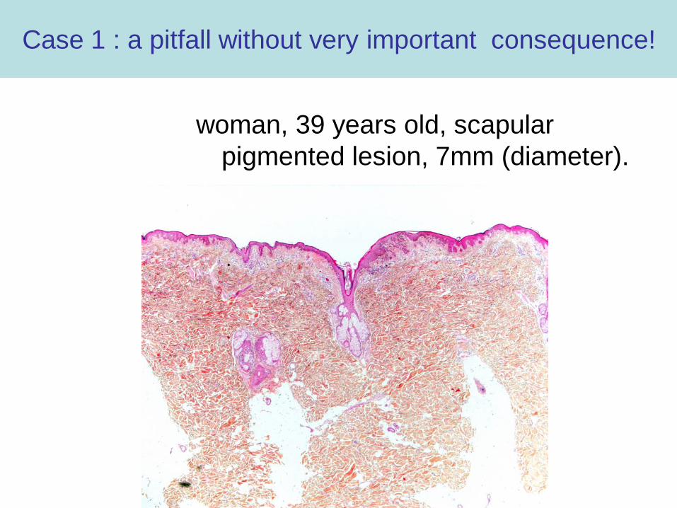
Case 1 : a pitfall without very important consequence!
woman, 39 years old, scapular pigmented lesion, 7mm (diameter).


to summarize…
• 39-yr old woman• atypical intraepidermal
melanocytic proliferation
Questions:
- Is this lesion atypical enough to propose a diagnosis of in situ Superficial Spreading (SSM) Melanoma?
- What are the consequences of such a diagnosis?

How to analyze an atypical intraepidermal melanocytic proliferation??
• First step: analyze clinical context– age? (< or > 40yrs old?)– is the lesion clinically
atypical?– does the patient present
a dysplastic nevus syndrome?

• Second step: histologically is there any reason to explain intra-epidermal atypia ?– Signs of traumatism or
irritation?• Signs of previous
treatment (biopsy, cryotherapy, laser…)?
How to analyze an atypical intraepidermal melanocytic proliferation??
Tronnier experiment:-Half nevus traumatized by scotch tape-Half nevus got sunburnt (UV=2DEM)After 1 week histological aspect of in situ SSM which regressed after 3 weeks

• Second step (twice): histologically is there any reason to explain intra-epidermal atypia ?
Melanocytic stimulation by sun?due to pregnancy?due to recent melanoma?
How to analyze an atypical intraepidermal melanocytic proliferation??

• Third step:could this lesion be a specific well-known, benign entity? - Reed nevus- Spitz intraepidermal nevus
How to analyze an atypical intraepidermal melanocytic proliferation??
Be sure of spitzoid cytology for a patient older than 40 yrs !!

Step4: Weigh up the pros and cons for malignancy
• Malignant?
age 39yrs oldno irritation or traumatismno previous melanomapolymorphous architecture with
intraepidermal ascent of cells+/- moderate atypical melanocyteslymphocytic dermal inflammation
• Benign?

How to conclude this diagnosis?
• In this case: either – in situ SSM – or very atypical intra-epidermal melanocytic proliferation– or in Britain: MIN (melanocytic intraepidermal neoplasm)
• Whatever your conclusion it is essential to be certain that the surgical excision is complete with narrow margins (5mm)
• Because in this case the patient is cured
• In case of doubtful intra-epidermal lesion (younger patient, moderate atypia)– Prefer descriptive diagnosis– But if you are worried be sure that the excision is complete with 5 mm
margins– So be very precise with your clinician

because the consequences for patient are not very important
if the lesion is completely removed !
Case 1: atypical intra-epidermal melanocytic lesion
a minor pitfall!