MEDICAL MICROBIOLOGY - Kuala Lumpur Hospital
20
114 MEDICAL MICROBIOLOGY Head of Unit DR SALMAH BINTI IDRIS Phone 03-26155590 1. INTRODUCTION Medical Microbiology Unit provides diagnostic and consultative services, training, research and development to support screening, diagnosis and treatment monitoring of infectious diseases caused by bacteria, fungi, parasites viruses and one type of infectious protein called prion. Services are also provided to support non-infectious diseases such as autoimmune disease, allergy and immunodeficiency. Comprehensive services are provided either in house or referred to other facilities within government agencies or outsourced to other non-Ministry of Health or private laboratories both locally and internationally. The Medical Microbiology Unit works in close collaboration with Infection Control Unit, Infectious Disease Physician and Pharmacist for the prevention, control and management of hospital acquired infection and antimicrobial resistance. For activities related to prevention and control of vaccine preventable diseases and other communicable infectious diseases, collaboration is with other relevant department and units such as Medical Department, Occupational and Safety Unit and Public Health Unit. 2. LIST OF SERVICES Microbiology Unit provides the following services: 2.1 Diagnostic microbiological services provided by various laboratories namely bacteriology, mycology, parasitology, general serology, immunology and virology. Microbiology Laboratory Diagnostic Services offered are as follows: Direct detection of bacteria, viruses and fungi in clinical specimens by microscopic examination of stained or unstained smears Isolation, identification and sensitivity testing of significant isolates of bacteria and fungi Utilisation of immunological methods for antibody or antigen detection
Transcript of MEDICAL MICROBIOLOGY - Kuala Lumpur Hospital
114
MEDICAL MICROBIOLOGY
Head of Unit DR SALMAH BINTI IDRIS
Phone 03-26155590
1. INTRODUCTION
Medical Microbiology Unit provides diagnostic and consultative services, training, research and development to support screening, diagnosis and treatment monitoring of infectious diseases caused by bacteria, fungi, parasites viruses and one type of infectious protein called prion. Services are also provided to support non-infectious diseases such as autoimmune disease, allergy and immunodeficiency. Comprehensive services are provided either in house or referred to other facilities within government agencies or outsourced to other non-Ministry of Health or private laboratories both locally and internationally. The Medical Microbiology Unit works in close collaboration with Infection Control Unit, Infectious Disease Physician and Pharmacist for the prevention, control and management of hospital acquired infection and antimicrobial resistance. For activities related to prevention and control of vaccine preventable diseases and other communicable infectious diseases, collaboration is with other relevant department and units such as Medical Department, Occupational and Safety Unit and Public Health Unit.
2. LIST OF SERVICES
Microbiology Unit provides the following services: 2.1 Diagnostic microbiological services provided by various laboratories namely bacteriology, mycology, parasitology, general serology, immunology and virology.
Microbiology Laboratory Diagnostic Services offered are as follows:
Direct detection of bacteria, viruses and fungi in clinical specimens by
microscopic examination of stained or unstained smears
Isolation, identification and sensitivity testing of significant isolates of bacteria
and fungi
Utilisation of immunological methods for antibody or antigen detection
115
Viral genome detection and/or viral load determination using nucleic acid testing
such polymerase chain reaction (PCR) both manual and automated.
2.2 Participation in hospital wide infection and antibiotic stewardship activities related to surveillance, control and prevention of healthcare-associated infections 2.3 Provision of microbiologic studies of the hospital environment and sterility testing for compliance to MSQH standards for prevention and control of infection. 2.4 Consultative services to clinicians and other health care providers, contribution to
development of relevant policy, clinical care guidelines and hospital infection and
antibiotic control related documentation or activity
2.5 Training for technical, scientific, undergraduate and post graduate medical
personnel.
3.0 LABORATORY CONSULTATION
In addition to overseeing the diagnostic tests the Clinical Microbiologist, Medical Officer
and Microbiology Scientific Officer also provide consultative services to clinicians to
determine the most appropriate tests, specimens and timing of collection to meet
particular diagnostic needs.
The choice of most appropriate test(s) and interpretation of result(s) will require a
certain amount of clinical information to be provided on the request form, thus,
Clinicians should consider consultation when:
3.1 the laboratory frequently requests either more clinical information or
additional specimens or both
3.2 It is unclear whether routine procedures can provide the information desired
3.3 It is not known whether any specific viral infections are associated with an
unusual syndrome.
3.4 It is a medico legal case
3.5 If the test result are repeatedly unsatisfactory for whatever reason or not in
tandem with the patient’s clinical state.
116
4. REQUEST FORMS
4.1 All specimens must be accompanied with designated test request form, which is
the PER-PAT 301 form. PER-PAT 301 form still need until LIS is fully utilized.
4.2 Forms must be filled LEGIBLY and COMPLETELY with the following
information:
Patient’s details : Name, Identity Card number, Sex, Age,
Clinic/Ward / Hospital name, Bed number
Patient’s Clinical and Test details: Relevant clinical information
pertaining to the requested test, diagnosis, tests required, type of
sample, time and date of sampling. For culture request, important
information is antimicrobial treatment already given or planned for the
patient management. For non culture request, it is important to
document disease onset so as to guide the appropriate test to be
done.
Requesting doctors details : Name in print, Official stamp,
signature, telephone number. Consultant signature and official stamp
are required for expansive specialised and test outsourced non-MOH
laboratory.
4.3 Incompletely filled forms may result in sample being rejected or may cause delay
in testing. For medico-legal reasons, if patients name or unique identification is
not confirmed or consent not obtained, the request may be withheld or rejected
e.g. ‘Unknown’ instead of a name.
117
5. SPECIMEN COLLECTION AND HANDLING
5.1 General guidelines 5.1.1 The quality of laboratory results depends greatly on the proper collection and handling of the specimen as well as obtaining satisfactory material for examination 5.1.2 The clinical specimen must be material from the actual infection site and must be collected with minimum contamination from adjacent tissues, organs or secretions. 5.1.3 A sufficient quantity of specimen must be obtained in order to perform the examination required 5.1.4 Appropriate collection devices, specimen containers and culture media must be used to ensure optimal recovery of microorganisms. Please refer to the list of tests for further details. 5.1.5 Ideally the specimen must be collected before the commencement of antimicrobial therapy 5.1.6 The specimen container must be properly labelled, placed in a biohazard plastic bag and accompanied by a completed laboratory request form 5.1.7 Specimens are best transported immediately to the laboratory. 5.2 Specific collection guidelines 5.2.1 Bacteriology 5.2.1.1 Autopsy material
Blood i) Aspirate 10ml of the right heart blood either through skin and chest wall or
(through unopened heart) from right ventricle after removal of sternum into a set of blood culture broths or sterile tube.
ii) Avoid contamination with bacteria from the water faucet and with the enteric bacteria. A block of splenic tissue may be submitted in lieu of a blood culture.
Tissue i) Best collected before the body is being handled at an earlier stage.
Decontaminate the skin or sear surface of heart or other organ before inserting needle or cutting out tissue block.
ii) Collect the tissue and placed in sterile container. Large piece is preferred (because aseptic collection is difficult). In the laboratory, 1 cm cube will be
118
aseptically cut from the suspicious area including some normal tissue for processing.
5.2.1.2 Blood cultures and bone marrow aspirate
An automated blood culture system with different types of bottles according to age and tests is available: Adults : Aerobic and anaerobic culture bottle Volume : 5 – 10 mL into each bottle Pediatric : Paediatric blood culture bottle Volume : 2 -3 mls
For Fungal C&S : Use aerobic bottle (To be incubated for 14 days ) For TB Blood Culture: Use Myco F Lytic bottle (request from Microbiology Laboratory).
For bone marrow aspirate, 1-2 ml of aspirate is required and to be inoculated directly into the appropriate culture bottles. Method of collection: 1. Disinfect the culture bottle with 70% isopropyl alcohol. 2. Palpate for the vein first. 3. Before venepuncture, the skin must be carefully disinfect with 70% alcohol. 4. Swab concentrically starting at the centre with skin disinfectant. Allow the skin disinfectant to dry before blood collection (do not palpate vein at this point). 5. Collect the blood. 6. After venepuncture remove disinfectant from the skin with alcohol Note: Acute sepsis : 2 – 3 sets from separate sites, all within 10 minutes Endocarditis : 3 sets at 3 intervals at least 30 minutes apart In the suspicion of catheter related bacteraemia, blood must be drawn from both the line and peripheral vein and submitted concurrently.
119
5.2.1.3 A. Blood Film for Malarial Parasite (BFMP)
i) Clean new glass slides with absolute alcohol ii) Select the third finger from the thumb (big toe can be used for infants).
Clean the finger with 70% alcohol swab. Dry the finger with cotton towel. iii) With a sterile lancet, puncture the ball of the finger using quick rolling
action. iv) By applying gentle pressure to the finger, express the first drop of blood
and wipe it away with dry cotton wool. How to prepare thick blood film
i) Apply gentle pressure to the finger and collect a single drop of blood on the surface of clean slide.
ii) Using the corner of another glass slide as a spreader, quickly spread the blood to make an even, thick film. The blood is spread in a circular motion with 3 – 6 movements, and spread over 20mm diameter.
iii) Label the slide with patient’s registration number and date of collection with grease pencil
iv) Place the blood film in a slide tray to air dry at room temperature. How to prepare thin blood film
i) With another gentle pressure to the finger collect a small drop of blood onto a new slide about 5 mm away from the edge of the slide.
ii) Rest the blood slide on a firm, flat surface. Use another slide as a spreader. Touch the drop of blood with a spreader and allow the blood to run along its edge. Keep the spreader at an angle of 30 – 45 ˚ and in steady movement, firmly push the spreader forward to prepare a thin smear.
iii) Label the slide with patient’s registration number and date of collection with grease pencil
iv) Place the blood film in a slide tray to air dry at room temperature. B. Blood Film for Microfilaria Parasite (BFMF) Preparation of smear
i) Collect a big drop of blood by pricking a finger. Blood collection must be done after 9.00 pm.
ii) Make an oval thick blood film on a clean glass slide. iii) Dry it in a horizontal position, taking care to protect it from dust and pests. iv) Send immediately to the laboratory after the smear dried off.
120
Note : For both thick and thin smear, a good smear is one that on drying still shows the hands of a watch place beneath it or one can read newsprint through it. Because of the potential for drug resistance in some of the Plasmodium species, particularly P. falciparum, it is important that every positive smear be assessed and the parasitemia reported. In cases where the patient is hospitalized, repeat BFMP should be performed at 24, 48, and 72 h after initiating therapy. 5.2.1.4 Cerebrospinal Fluid (CSF) A. Lumbar puncture 1. Disinfect site with 2% iodine tincture. 2. Insert needle with a stylet at L3 – L4, L4 – L5 or L5 – S1 interspace. Upon reaching the subarachnoid space, remove the stylet and collect 1 – 2 ml of fluid into sterile bijou bottle. Note : always send the most turbid tube to the lab. B. Ommaya reservoir fluid 1. Clean the Ommaya reservoir site with antiseptic solution and alcohol 2. Remove Ommaya fluid via the Ommaya reservoir unit and place into bijou bottle Note: CSF for microscopic examination, cell count, bacterial antigen test can be collected into a single screw capped sterile container. C. Brain abscess and CNS biopsy sample 1. Aspirate material or biopsy sample collected from lesions must be placed in a sterile bottle or syringe and send immediately to the laboratory for anaerobic culture. 2. Do not add formalin *5.2.1.5 Chlamydia trachomatis direct fluorescence smear Suitable specimens : Endocervical swab (female) Urethral swab (male) Eye swab (baby) i) Firmly role the swab onto the wells of the glass slide. Do not smear the swab
onto the wells (distortion of cells may occur) ii) Examine the slide macroscopically for adequate cell coverage. iii) Allow the slide to air dry for 5 – 10 minutes
121
iv) Label and send the slide immediately to the lab Note: Specimen with insufficient numbers of epithelial cells (<10 cells) is inadequate and cannot be interpreted. 5.2.1.6 Genital samples A. Endocervical swab
i) This is the best specimen for the diagnosis of gonorrhoea and puerperal sepsis
ii) Under direct vision, gently compress cervix with blades of the speculum and use rotating motion with swab, obtain exudates from the endocervical canal
iii) Inoculate the swab into Amies transport media. B. Urethral discharge
i) Wipe the urethral with a sterile gauze or swab. ii) Collect the exudate with a sterile swab and inoculate into Amies transport
media iii) If discharge cannot be obtained by ‘milking’ the urethra, use a sterile swab to
collect material from about 2 cm inside the urethra iv) Place the swab into Amies transport media.
5.2.1.6 Helicobacter pylori culture Specimen : Gastric biopsy
i) Place biopsy material in a sterile, bullet container with approximately 1 ml of sterile normal saline
ii) Transport immediately to the laboratory. Specimen must be processed within 2 hours of collection.
5.2.1.7 Indwelling devices The acceptable devices are : IV catheters for semi-Quantitative culture (Maki method) : central, CVP, Hickman,s, Broviac, peripheral, arterial, umbilical, hyperalimentation, Swan-ganz. The specimen should be accompanied by blood drawn from peripheral vein. Foley’s tube is not acceptable for culture
122
i. Cleanse the skin around the catheter site with alcohol. ii. Aseptically remove catheter and clip 5 cm of the distal tip of the catheter directly into a sterile screw-cap container iii. Transport directly to the lab to prevent drying 5.2.1.8 Mycobacterium : Acid – fast bacilli stains and culture Acceptable specimens: Respiratory secretions, urine, CSF, body fluids, whole blood and tissue biopsies. Swab specimens are not acceptable.
i. Collect respiratory secretion, urine and tissue biopsies in sterile Falcon tube (available from the general distribution counter)
ii. Collect a minimum of 3 early morning sputum or urine specimens in successive 24 hours period
iii. Place whole blood, body fluids and CSF into a commercial TB blood culture bottle (available from the laboratory on request)
5.2.1.9 Sterile body fluid C&S (excluding CSF, urine and blood)
i. Clean needle puncture site with skin disinfectant solution ii. Aseptically perform percutaneous aspiration to obtain pleural, pericardial,
peritoneal or synovial fluid. iii) Collect fluid into a sterile screw-cap container. iv) For anaerobic culture send sample immediately in the syringe.
5.2.1.10 Pus / swab / tissue
i) Send pus if available, in a sterile screw-cap container or syringe ii) Swab is an inferior substitute and should be avoided, if sent, in an Amies
Transport Medium. iii) Send all tissues for culture in sterile screw-cap container. Do not add
formalin to the specimen. Note :
i) A dry swab may fail to yield organisms in smear and culture ii) Surface swabs of deeply infected lesions (eg ; sinus tracts from
osteomylitis, pressure sores) usually grow surface contaminants like coliforms and pseudomonads.
123
5.2.1.11 Respiratory specimens Upper Respiratory 1. Nasal swab Submitted primarily for detection of MRSA carriers
i) Wet a sterile swab in sterile Normal Saline and insert the swab into the the nasal cavity until resistance is met at the level of turbinates (approximately 1 inch into the nose)
ii) Gently rotate the swab against the nasal mucosa iii) Repeat the process on the other side with the same swab
2. Nasopharyngeal swab / aspirate This is done to isolate Corynebacterium diphteriae for the diagnosis of diphteria and to isolate Bordetella pertussis for the diagnosis of pertussis.
i) Carefully insert a flexible wire calcium alginate-tipped swab through the nose into the posterior nasopharynx
ii) Rotate the swab (keep the swab near the septum and floor of the nose) If Nasopharyngeal aspirate
i) Gently pass a sterile catheter through one nostril as far as the nasopharynx. ii) Attached a sterile syringe to the catheter and aspirate a specimen of
mucopus iii) Put into sterile container and send immediately to the laboratory
3. Throat swab Submitted primarily for the detection of Group A Streptococci, Bordetella pertussis , Corynebacterium diphteria and Neisseria gonorrhea
i) Depress tongue gently with tongue depressor. ii) Extend sterile swab between the tonsillar pillars and behind the uvula (avoid
touching the cheeks, tongue, uvula, or lips. iii) Sweep the swab back and forth across the posterior pharynx, tonsillar areas,
and any inflamed or ulcerated areas to obtain sample. Lower respiratory tract 1. Sputum
124
i) Collect early morning specimen after rinsing the mouth and gargling with water
ii) Instruct the patient to cough deeply and expectorate sputum only and not saliva into the sterile screw-cap container
2. Tracheal aspirate/ bronchial washing / broncho alveolar lavage / lung aspirate /lung biopsy Tracheostomy is followed by colonization within 24 hours of insertion of the tube. Results must be correlated with clinical findings such as fever of infiltrate on chest x-ray. 5.2.1.12 A Stool Culture and Sensitivity / Clostridium difficile C&S / C. difficile toxin
i) Collect faeces into a sterile / clean wide-mouth screw-capped plastic container
ii) If the faeces is liquid, the container may be filled to one-third full (excessive amount will result in spillage when opened)
Note: 1. Rectal swab is poor second best alternative to faeces. If it is not possible to
obtain faeces, collect a specimen by inserting a cotton swab into the rectum and send in a suitable transport medium.
2. For stool clearance culture in cases of typhoid and cholera, stool should be sent upon completion of therapy
3. Clostridium difficile toxin test a. Collect a max` imum of 3 stool specimens with 18-24 hour intervals
in between collection for. If 2 or more specimens are collected on the same day, only one will be tested and reported.
b. Collect at least 3 to 5 ml of faeces. Faeces should be loose/watery to conform to the shape of the container. Formed faeces will be rejected unless the requisition indicates the patient may have pseudomembranous colitis.
c. Rectal swabs are not an acceptable alternative and will not be tested for toxin.
125
Stool Ova and Cysts/ Cryptosporidium and Isospora / E. histolytica
i) Collect faeces into a clean wide-mouth container with a tight fitting lid to prevent accidental spillage and maintain moisture.
ii) The specimen should not be contaminated with water and urine. iii) Place in the plastic bag, label properly, accompanied by a request
form and send immediately. Note: Stool specimens containing barium and antibiotics are not acceptable. Specimen should be collected 5 – 10 days after these substances are given to the patient. For patients treated with mineral oil, bismuth, anti-malarial agents and non- absorbable anti-diarrhoeal preparation, specimen collection should be delayed at least for 2 weeks. It is recommended that normal examination for stool parasites before therapy include three specimens submitted on separate days. Collect the specimen every other day or the series of 3 specimens within no more than 10 days. 5.2.1.13 Urine A. Midstream urine Male
iii) Cleanse the glands and penis with soap and water. iv) Rinse area with wet gauze pads v) While holding the foreskin retracted, begin voiding. vi) After several millilitres has passed, collect a midstream portion without
stopping the flow of urine. Female
i) Thoroughly cleanse the urethral area with soap and water. ii) Rinse area with wet gauze. iii) While holding the labia apart, begin voiding. iv) After several millilitres has passed, collect a midstream portion without
stopping the flow of urine. B. Suprapubic aspirate (SPA) Note: SPA is useful in determining urinary infection in adults in which the results from routine procedures are equivocal but the diagnosis is critical. Also useful for paediatric patients when clean-catch urine is difficult to obtain.
126
i) Before SPA, the patient should force fluids until bladder is full. ii) Shave and disinfect the suprapubic skin overlying the urinary bladder. iii) Aspirate urine from the bladder by using a needle aspiration technique.
C. Catheter urine Catheters are placed in patients who are unable to pass urine
i) Clean the catheter collection port with 70% alcohol wipe ii) Using sterile technique, puncture the collection port with a needle attached
to a syringe. Do not collect the urine from the bag. iii) Aspirate urine and place it in sterile container. iv) Send specimen immediately to the laboratory
5.2.2 Mycology 5.2.2.1 Skin, nails and hair
Clean cutaneous and scalp lesions with 70% alcohol prior to sampling as this will improve the chances of detecting fungus on microscopic examination, as well as reducing the likelihood of bacterial contamination of cultures. Prior cleaning is essential if ointments, creams or powders have been applied to the lesion.
Skin, nails and hairs specimens should be collected into folded squares of paper or directly onto agar plate. Skin Material should be collected from cutaneous lesions by scraping outwards from the margin of the lesions with the edge of a glass microscope slide or a blunt scalpel
Hair i) Specimen from the scalp should include hair roots, the contents of
plugged follicles and skin scales ii) Hairs should be plucked from the scalp with forceps or the scalp is
brushed with a plastic hairbrush and collected onto an agar plate.
127
Nail i) Nail specimens should be taken from any discoloured, dystrophic or brittle
parts of the nail. ii) Specimen should be cut as far back as possible from the edge of the nail
and should include the full thickness of the nail. 5.2.2.2 Mouth and vagina
i) Swabs from the buccal mucosa should be moistened with sterile water prior to taking the sample and sent in Amies transport media
ii) For vaginal infections, swabs should be taken from discharge in the vagina and from the lateral vaginal walls. Swabs to be sent to the laboratory in Amies transport media.
5.2.2.3 Ear
Scrapings of material from the ear canal are to be preferred, although swabs can also be used.
5.2.2.4 Ocular specimens
i) Material from patients with suspected fungal infection of the cornea (Keratomycosis) should be collected by scraping the ulcer. The entire base of the ulcer, as well as the edges should be scraped. Swabs are not suitable for sampling corneal lesions
ii) The material is collected directly onto agar plates for culture and to glass slide for microscopic examination.
5.2.2.5 Blood
Blood culture for fungal is collected aseptically as in culture for aerobic bacterial culture but using the available commercial fungal bottle.
The request for fungal culture should be indicated clearly on the request form and a total of two weeks incubation will be carried out.
5.2.2.6 Cerebrospinal fluid
CSF specimens (3-5mls) should be collected in a sterile container for microscopy and culture.
128
5.2.2.7 Bone marrow
This specimen is helpful for making the diagnosis in a number of deep fungal infection, including histoplasmosis and cryptococcosis. 3 – 5 ml of aspirated material should be collected and transferred into the commercial fungal culture bottle.
5.2.2.8 Pus
i) Pus from undrained subcutaneous abscesses or sinus tract should be collected with a sterile needle and syringe
ii) If grains are visible in the pus (as in mycetoma), these must be collected. In mycetoma, if the crusts at the opening of the sinus tracts are lifted, grains can often be found in the pus underneath.
5.2.2.9 Tissue
i) If possible, material should be obtained from both the middle and edge of the lesions
ii) Small cutaneous, subcutaneous of mucosal lesions can often be excised completely
iii) Tissue specimens should be placed in a sterile container without formalin.
5.3 Specimens for serological tests
These comprise of tests in bacteriology, virology, parasitology and immunology. Method of blood collection: i) Draw 3 – 5 ml of blood into a plain tube without anticoagulants ii) Clot at ambient temperature iii) Despatch to the laboratory within 4 hours of collection for serum
separation by centrifugation. iv) If delays in delivery of more than 4 hours is anticipated, centrifuge blood
at 3,000 rpm for 10 min, pack serum properly with dry ice/ wet ice and send to lab via courier service.
Note: Haemolysed, icteric or lipaemic specimens invalidate certain tests. If such specimens are received, the samples will be rejected to ensure that results are of clinical value.
129
5.4. ‘Specialized’ Virology Test:
i) ImmunoFlourescence Antibody (IFA) a) Respiratory test specimens.
i) Sputum: -Ask patient to cough deeply and spit directly into a sterile container. Expectorate should be the secretions from the bronchi and not just saliva.
ii) Tracheal aspirate / Nasopharyngeal aspirate and
Bronchial alveolar lavage (BAL): -Place the specimen obtained via the above methods into a sterile container.
b) Herpes Simplex I & II virus, Varicella Zoster virus and
Enterovirus test specimens. i) Lesion – samples are suitable from lesion area of oral,
genital, skin cervix and cornea.
Specimens are to be collected very carefully as to avoid any contamination of the sampling site and during slide preparation procedure. Moisten swab with sterile water or saline before collecting samples and prepare the slides at the bedside.
Vesicular lesion: Open a vesicular lesion with a sterile swab. For oral, genital and skin lesion use a sterile needle and a sterile swab for sampling. Vesicular fluid may be collected in syringes, and these are sent to the laboratory.
Ulcerative lesion: Remove any pus from the lesion with a sterile swab for sampling. Corneal lesions must be scrapped at the edge to collect infected cells.
Dried lesion: Lift the crust from the dried lesion with a sterile needle and place sample in container.
*Slide preparation procedure: i. Prepare for minimum of 5 smears on the Daflon coated slide
which is the most preferred type of slide for IFAT. ii. If using normal slide, please make 3 smears on it, each the
size of a five cent coin, and circle the smear area on the under surface of the slide with a coloured pen.
iii. Firmly roll the swab onto the wells of the slide. iv. Air dry the slides for 5-10 min. v. Label and Place in slide box and dispatch to laboratory.
130
Note: Specimens from vesicular lesions are preferred to samples from ulcerative and dried lesions because the viral titer decreases as the lesion ulcerates, crusts and heals.
ii) Special Diagnosis Test for Transplant
a) Immunoblot (ELISA-based tests): 1) Blood – Collect 3ml-5ml blood in plain tube. Clot at room
temperature for 10 min. dispatch to Virology Lab within 4 hours.
2) Serum – follow procedure as for blood collection above,
and spin the collected blood by centrifuge at 3,000 rpm for 10 min. and send the serum to lab within 4 hours.
iii) Viral Genome Detection (PCR) a) Blood:
Collect 3-5ml of blood into plain tube for HCV genotyping, HCV RNA, HBV DNA and BK virus quantitation.
Use EDTA container for CMV DNA and HIV RNA quantitation. Do not exceed the marked blood level on the EDTA container.
Send directly to Virology Lab within 4 hours after collection in ice-box containing wet ice.
b) CSF:
Collect a minimum of 0.3ml of CSF into a sterile screw capped container.
c) Respiratory specimen
Collect lower respiratory tract specimens (deep cough sputum, bronchoalveolar lavage, tracheal aspirate, pleural fluid into screw capped sterile container.
ONLY if lower respiratory tract specimens is not possible, upper respiratory tract specimens (URTSs) may be taken:
-COMBINED nasopharyngeal and oropharyngeal swabs (NP/OP swabs), nasopharyngeal aspirate/wash collected in viral transport media
Send the specimens in ice to the laboratory ASAP. If
delay is anticipated, keep specimens at 4°C. DO NOT
FREEZE.
131
Ensure triple packaging if the suspected pathogen is a risk group 3 pathogen and transport in ice
d) Tissue Biopsy:
If possible, sample should consist of both the middle and the edge section of the tissue.
Small sample approximately of 0.3cm size is appropriate.
Place tissue in an empty sterile container and moisten with viral transport media. DO NOT add Formalin into the specimen.
e) Vesicle fluids:
Collect a minimum of 0.3ml of sample using a sterile needle by puncturing the lesion.
Place specimen into an empty sterile Bijoux bottle.
f) Bone Marrow:
Collect a min. of 0.3ml of the aspirated sample.
Place specimen into an empty sterile Bijoux bottle. Note:
Where indicated, non-blood samples for molecular virology testing , must be accompanied with 3 ml of blood sample in plain tube for parallel serological or other testing.
Send all collected non-blood samples to the pre-analytical unit within 2 hours of collection.
5.5 Medicolegal cases
Specific guidelines i) Specimens should be sealed and send directly to the microbiology
laboratory ii) Specimens should be sent to the laboratory by a designated personnel iii) Chain of custody should be maintained at all times and record book
should accompany the sample(s).
Sample collection of various tests should follow the guidelines as of normal microbiological requirements and the specific headings are referred.
132
6.0 SERVICES AFTER OFFICE HOUR
a) Blood for Malaria Parasite and Culture for bacteria and fungi is available 24
hours and 7 days a week.
b) Samples from Urgent and Transplant cases will be handled and processed
after office hours and on public holidays. Some routine virology services will
also be available on public holidays on a need basis such as in the case of an
outbreak, or in the event of a long holiday season.
c) Blood sample from needle stick injury cases will be processed if both the
sample from the source and affected health care worker/victim is received
concurrently. If only a sample form the health care worker/victim is received,
the sample will be processed if the status of the source is known to be
positive. If the infectivity of the source is not known and only the health care
worker/victim sample is received, sample will be processed the next working
day.
d) Specimen for routine virology tests received during after office hours and on
public holidays will be kept in the pre analytical unit refrigerator and will be
processed on the next available working day.
7.0 REPORTING OF RESULT
Reporting of Result
For preliminary result will be informed to respective ward / location via telephone
call
For all results will be validated by a Clinical Microbiologist /Scientific Officer
before they are reported.
For external location, results will be sending through post mail / email.
133
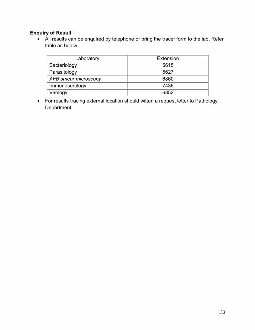
Enquiry of Result
All results can be enquired by telephone or bring the tracer form to the lab. Refer
table as below.
For results tracing external location should witten a request letter to Pathology
Department.
Laboratory Extension
Bacteriology 5615
Parasitology 5627
AFB smear microscopy 6860
Immunoserology 7436
Virology 6852