Medical Emergencies in Diagnostic Imaging. Goal The RT student will be able to recognize...
58
Medical Emergencies in Diagnostic Imaging
-
Upload
david-reeves -
Category
Documents
-
view
218 -
download
2
Transcript of Medical Emergencies in Diagnostic Imaging. Goal The RT student will be able to recognize...

Medical Emergencies in Diagnostic Imaging
Goal
The RT student will be able to recognize life-threatening emergencies and initiate appropriate medical action.

Objectives
After completing this lesson the student will be able to: List the visible symptoms of shock. List the visible symptoms of an anaphylactic reaction. List the observable symptoms of diabetic ketoacidosis,
hypoglycemia, hyperosmolar coma and describe the actions the RT must take if he observes these symptoms in his patient.
List the early symptoms of cerebral vascular accident and describe the action the RT should take if these symptoms are observed.
List the symptoms of respiratory failure and describe the action that an RT must take if this emergency occurs in his department.
List the symptoms of cardiac failure and describe the actions that the RT must take if this emergency occurs
List the symptoms of mechanical airway obstruction and describe the action an RT should take if this emergency occurs.
List the emergency action that the RT must take if a patient is having a convulsion or is fainting.

Medical emergency?
The abnormal physiologic reactions, especially of patients whose physical condition is poor, that, occur quickly, with little or no warning, and often life threatening are called medical emergencies.

Common medical emergencies
The most common medical emergencies in x-ray departments are:
Shock Anaphylaxis Diabetic reactions Cerebral vascular accidents Cardiac failure Respiratory failure Fainting Convulsions

What is the RT’s action?
The RT’s first action is:Call the hospital/departmental emergency
team, the physician /radiologist conducting the procedure, and colleagues for assistance.
Then obtain the emergency trolley/crash cart immediately.
Emergency trolley/crash cart is a trolley that contains medications and equipment
needed when a patient’s condition becomes suddenly critical.

Shock?
Shock is a physiologic reaction to illness or trauma, in which, there is a disturbance of blood flow to
the vital organs, or a decreased ability of the body tissues
to use oxygen and other nutrients needed to maintain them in a healthy state.
It can occur quickly and without warning.
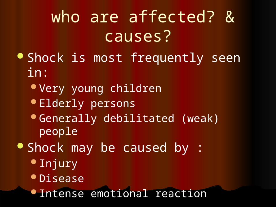
who are affected? & causes?
Shock is most frequently seen in:Very young childrenElderly personsGenerally debilitated (weak) people
Shock may be caused by :Injury DiseaseIntense emotional reaction

Signs & symptoms of shock
Increased temperatureWeak, thready pulseRapid heartbeatRapid shallow respirationHypotensionSkin pallorCyanosisIncreased thirst

Development of signs & symptoms
In the early stages, because of an inadequate supply of oxygen to the brain, the patient will display signs of:RestlessnessConfusionAnxiety
Later, (if allowed to progress), the patient will become Apathetic (droopy, unconcerned)Confused (puzzled, bewildered)Comatose (exhausted)
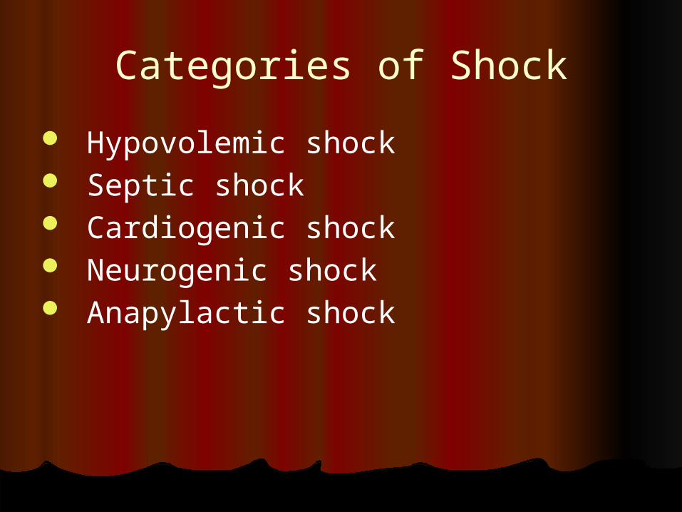
Categories of Shock
Hypovolemic shock Septic shock Cardiogenic shock Neurogenic shock Anapylactic shock
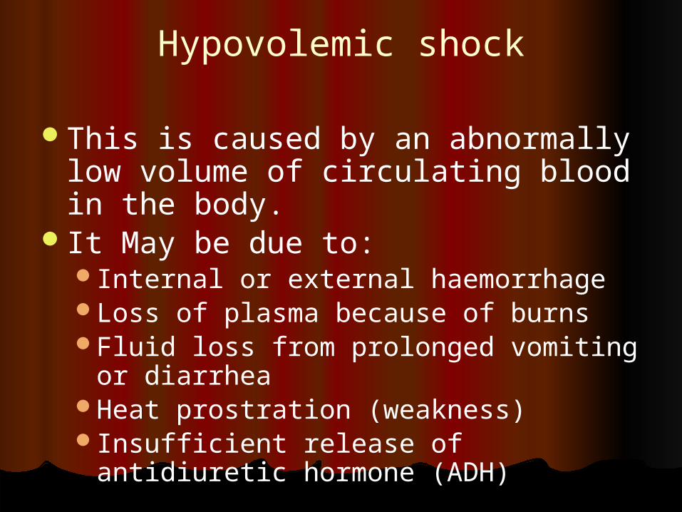
Hypovolemic shock
This is caused by an abnormally low volume of circulating blood in the body.
It May be due to:Internal or external haemorrhageLoss of plasma because of burnsFluid loss from prolonged vomiting or
diarrheaHeat prostration (weakness)Insufficient release of antidiuretic
hormone (ADH)

Signs & symptoms
Restlessness; thirst; cold, clammy skin Pallor, sweating Falling blood pressure; weak, thready
pulse Rapid respirations Extreme weakness; lethargy Cold extremities Semiconsciousness, coma Systolic blood pressure lower than 60 mm
Hg Oliguria to anuria

Action to take Place the patient in a flat, supine position and allow him to
rest. Notify the physician & call for assistance Make certain that the patient is able to breath without
obstruction (release any tight clothing and clear the airway) Note any visible discharge of bodily fluids (blood, vomitus,
faeces, urine) and wipe them away. Keep any blood out of patient’s view. If there is loss of blood from open wound apply pressure to
stop it. Be prepared to assist with administration of oxygen, IV
fluids or medications. Keep the patient warm and dry. Check blood pressure, pulse, and respirations every 10
minutes. Observe the pt’s skin colour and body temperature Do not offer food or fluids Do not leave the patient unattended.

Septic shock
A shock caused by severe systemic infections and bacteremia (bacterial endotoxins released in the bloodstream).
Symptoms progress somewhat differently from those of other types of shock.

Signs & symptoms
In early stages, the skin is warm, dry, and flushed.
Urine output may be normal or excessive. The patient may have chills. As the shock progresses, there may be an abrupt
personality change or a decrease in the level of consciousness.
There is an increase in pulse and respiration and a decrease in urinary output.
The skin becomes cold and clammy. Seizures, circulatory collapse, and
cardiorespiratory failure will follow if the course is not reversed.

Cardiogenic shock
A shock caused by a failure of the heart to pump an adequate amount of blood to the vital organs.
This causes inadequate tissue perfusion. The onset of cardiogenic shock is sudden
and often occurs in patients hospitalized for acute myocardial infarction, cardiac tamponade (excessive pressure on the heart), or pulmonary embolus.
It may follow cardiac surgery.
Signs & symptoms
Restlessness, anxiety, falling blood pressure, and falling pulse pressure.
Weak, rapid pulse Shallow, labored respirationsDecreased urinary outputCool, clammy skinPossible semiconsciousness or coma

Action to be taken Summon emergency assistance and place the
emergency cart ready. Notify the physician in charge of the patient. Place the patient in a semi-Fowlers position or a
position of comfort. Keep the patient warm and quiet. Take the vital signs every 5 to 10 minutes. Do not give the patient anything to eat or drink. Do not leave the patient alone. Be prepared to assist with oxygen and
intravenous fluids, and medication administration.
Be prepared to begin CPR.

Neurogenic shock
A shock occurs when concussion (limited period of unconsciousness), spinal cord injury, psychic trauma, or spinal anesthesia causes abnormal dilatation of the peripheral blood vessels.
This dilatation in turn causes a fall in blood pressure as blood pools in the veins. This leads to reduced cardiac output and shock.
Signs & symptoms
Hypertension and bradycardia Warm, dry, skin and subnormal body
temperature. Initial alertness unless the patient is
unconscious because of head injury. Initially good, but deteriorating, tissue
perfusion. Visible signs of poor tissue perfusion –
coolness of extremities and diminishing peripheral pulse.

Action to take
Notify the physician in charge of the patient.
Summon assistance and stay with the patient.
Keep the patient flat, and monitor vital signs every 10 minutes.
Do not move the patient if there is a possible spinal injury.
Prepare to assist with oxygen, intravenous fluid, and medication administration.

Anaphylactic shock Anaphylactic shock is the result of an
exaggerated hypersensitivity reaction (allergic reaction) to an antigen that was previously encountered by the body’s immune system.
When this occurs, vasodilator substances (histamine and histaminelike compounds) which may produce massive vasodilatation and peripheral pooling of blood, are released in the body.
This reaction is accompanied by contraction of nonvascular smooth muscles, particularly the smooth muscles of the respiratory system.
This reaction can produce shock, respiratory failure and death within minutes following exposure to the agent that produces the reaction.
This is the type of shock seen most often in radiology departments.

Common causes of anaphylaxis
Drugs Iodinated contrast agents Chemotherapeutic agents Anesthetics Certain foods Insect venoms

Early signs & symptoms
Itching at the site of injection and/or around the eyes and nose.
Sneezing and coughingApprehensiveness; a feeling of doomNausea, vomiting, and diarrhea
(usually related to food)

Late symptoms
Angioneurotic edema of the face, hands, and other body parts
Urticaria (an itchy rash resulting from the release of histamine)
Chocking, wheezing, or dyspnea and cyanosis
Hypotension, weak rapid pulse and dilated pupils

Precautions & Actions to take
Keep the emergency trolley ready and correctly prepared whenever an iodinated contrast medium is being administered.
Before starting any procedure that involves the use of iodinated contrast medium, ask the patient the following questions.
“Are you allergic to any food or medicine?” “Which ones?”
“Do you have asthma or hay fever?” “have you ever had an x-ray examination that
involved the use of contrast medium?”. “If so, did you have a reaction during or following that examination?”

If the answer for any question is positive, the radiologist should be informed for necessary precautions
Never leave a patient who is receiving an iodinated contrast agent unattended.
If he complains itching, if swelling or redness of the skin is noted, or if the patient seems unduly anxious notify the radiologist.
Monitor the vital signs and observe for respiratory distress.
If the patient is in anaphylactic shock, call the emergency team
Keep the patient in semi Fowler’s position or sitting position if possible.
Prepare to assist with the administration of oxygen, intravenous fluids, and medications.
Medications given for anaphylaxis
Epinephrine (Adrenaline)DiphenhydramineHydrocortisoneAminophylline
If the patient stops breathing start pulmonary resuscitation.
If the patient becomes breathless and pulseless, administer Cardiopulmonary resuscitation (CPR)

Diabetic emergencies
Diabetic mellitus(DM) is a chronic disease involving a disorder of carbohydrate, protein, and fat metabolism, which also affects the structure and function of the blood vessels.
The underlying cause is a disturbance in the production, action, or utilization of insulin, a hormone normally secreted by the islands of langerhans located in the pancreas.
Medical treatment consists of diet therapy, insulin injections, or use of oral hypoglycemic drugs.

Types of diabetes and diagnosis
Type 1 ;- Insulin-dependent form:- There is no production of insulin and therefore depend on outside sources of insulin for the entire life.
Type 2 :- Noninsulin-dependent form:- The production of insulin is less than necessary or the insulin does not have the desired effect on the body. They are treated with diet control and drugs that increase the carbohydrate metabolism.
DM is diagnosed by laboratory measurement of blood glucose levels.
A normal adult blood glucose level should range from 80 to 115 mg/dl.
Complications of DM
Hypoglycemia Diabetic ketoacidosis Nonketotic hyperosmolar coma
Hypoglycemia
Hypoglycemia or insulin reaction occurs when patients who have diabetes mellitus have an excess amount of insulin in their blood stream, an increased rate of glucose utilization, or an inadequate diet to utilize the insulin.
A patient who has DM may come to the imaging department after he has taken insulin or some other hypoglycemic agent, but before his body has had sufficient nourishment to utilize the medication. The result may be a hypoglycemic reaction. The onset of symptoms is rapid, and immediate action is necessary in order to prevent coma.

Signs & symptoms
Shaking, nervousness, and irritability Dizziness and hunger; may complain of
headache Profuse perspiration; cold, clammy skin Blurred vision Tremor, numbness of lips or tongue,
slurred speech Impaired motor function; convulsions Diminishing level of consciousness; quick
lapse into coma

Actions to take
Notify the Radiologist Administer some type of sugar
immediately Call for help Do not leave the patient unattended Monitor vital signs if the patient is unconscious prepare to
assist with administration of oxygen, intravenous fluids, and medication
usually in this type of coma, 20 to 50 % glucose in solution is administered intravenously.

Diabetic ketoacidosis
When a patient has insufficient insulin available to metabolize the glucose that is present, his body begins to mobilize fatty acids, and the result is an acidotic state called diabetic ketoacidosis.
In this condition, acid and ketone bodies accumulate in the blood. If this accumulation is not corrected quickly, the patient will become comatose and may die.

Signs & symptoms
Weakness, drowsiness, and dull headache
Sweet odor to the breadth, hypotension
Warm, dry skin; parched tongue; dry mucous membranes; extreme thirst
General weakness, lethargy, and fatigue
Flushed face, deep and rapid respirations
Tachycardia, weak, thread pulse and, ultimately, coma

Actions to take
Check patient chart to identify him as a diabetic.
Stop treatment/examination Notify the physician Call for assistance Do not leave the patient unattended Monitor vital signs Give fluids by mouth if possible Prepare to assist with administration of
intravenous fluids, and oxygen

Hyperosmolar coma
Hyperosmolar coma(hyperglycemic, nonketoic coma) is a complication of diabetes mellitus that usually occurs in the elderly diabetic patient.
It is frequently mistaken for a stroke or drunkeness and is extremely serious, life-threatening problem
Factors that cause this condition are diagnostic procedures that require changes in diet,
especially fasting for long hours, hyperglycemic-inducing agents and resistance to insulin.
The blood glucose level in patients with this problem is grater than 600 mg/dl; there is little or no ketosis and the plasma is hyperosmolar.

Signs & symptoms
Extreme patient dehydration; dry skin; sunken eyes
Increased body temperature; polyuria; extreme thirst
Muscle twitching; difficult, slurred speech
Mental confusion; convulsionComa

Actions to take
Stop treatmentNotify the physicianCall for assistanceDo not leave the patient unattendedMonitor vital signsGive fluids by mouth if possiblePrepare to assist with administration
of intravenous fluids, and oxygen
Respiratory failure, cardiac arrest, airway obstruction
Respiratory failure or severe respiratory dysfunction may result from airway obstruction caused by the patient’s position, the tongue, a foreign object, vomitus lodged in
the throat, disease, drug overdose, injury, or coma.
Whatever the cause, gas exchange is no longer adequate to maintain normal arterial blood gases.
Symptoms of a partially obstructed airway
Labored, noisy breathing Wheezing Use of accessory muscles of the neck,
abdomen and chest for breathing Neck-vein distention Anxiety Cyanosis of the lips and nail beds Productive cough with pink-tinged, frothy
sputum

………………….continued
If the patient lapses into complete respiratory failure, his pulse will continue to beat for a brief period of time. However, the pulse becomes weak and then ceases. Chest movement stops and eventually cardiac arrest will result.

Action to take
1. Clear and open the airway Check the larynx and trachea to make certain
that the patient’s tongue, epiglottis, or a foreign body is not blocking the airway.
Tilt the head by placing one hand on the patient’s forehead and applying firm backward pressure with the palm to tilt the head back.
Keep the fingers of the other hand under the lower jaw near the chin and lift so that the chin is brought forward.
The lips should remain apart
If the patient does not resume breathing, rescue breathing must be begun.

2. Rescue Breathing
Move the patient to a supine position Check the carotid pulse Squeeze the nostrils together Cover his mouth tightly with yours Inflate the patient’s lungs by giving two full breaths in
succession into his mouth. Allow the patient time to exhale these breaths as you
inhale between each. Recheck the carotid pulse If present continue pulmonary resuscitation by breathing
into patient’s mouth at the rate of 12 breaths per minute. Check the carotid pulse each minute.
If the pulse is absent cardiac compression must be started immediately.

3. Application of External cardiac compression
External cardiac compression is effective only if the patient is lying on a firm surface.
Take an adequate amount of time to determine pulselessness (5 to 10 seconds).
Performing cardiac compression on a person whose heart is functioning is extremely dangerous.
Once compressions have started, do not interrupt them for more than 7 seconds at a time.
Place the heel of one hand in the midline of the sternum above the xiphoid process.
Put the other hand on the first.

For children, land marks are the same , but only one hand is used to prevent excessive pressure.
For adult cardiac compression, the lower half of the sternum is compressed.
Compress the sternum 1 ½ to 2 inches directly downward and then release the compression completely.
Do not apply pressure on the rib cage itself. Keep your elbows straight and give 15 compressions in a
smooth, even rhythm. Then inflate the patient’s lungs two more times. Next give 15 more compressions, then two more
inflations. This rhythm must be maintained until help arrives.
Following the initial cycles of compressions and ventilations, pause to reassess the pulse and breathlessness.
If the patient remains breathless and pulseless, continue the cycle of two ventilations and 15 compressions, maintaining 80 to 100 external chest compressions per minute.

Cerebral Vascular Accident (Stroke)
Cerebral vascular accidents (CVA) are caused by occlusion or rupture of the cerebral arteries directly into the brain tissue or into the subarachnoid space. This is commonly called a stroke. Strokes vary in severity from a mild transischemic attack (TIA) to severe life threatening situations.

Signs& symptoms
Possible severe headache Muscle weakness or flaccidity of face or
extremities. Eye deviation, usually one sided, may
loose vision Dizziness or stupor Difficult speech (dysphasia) or no speech
(aphasia0. Ataxia May complain of stiff neck. Nausea or vomiting may occur.

Actions
Call for emergency aid, do not leave patient alone.
Put patient in resting position with head slightly elevated.
Monitor vital signs every 10 minutes. Report to the physician Prepare to administer intravenous
medications, fluids, and oxygen Prepare to administer CPR if the patient
becomes breathless or plseless.

Fainting
Fainting is caused by an insufficiency in the supply of blood to the brain. The possible causes are:
Heart diseaseHungerPoor ventilationFatigueEmotional shock

Signs and symptomsPallor, dizziness, and possibly nauseaCold, clammy skin
ActionsHave the patient lie down, if possiblePosition his head so it is level with or
some what lower than his body.Summ)on medical assistance.

Convulsive seizures
Convulsive seizures are associated with many physical disorders, including; uremia, eclampsia, tetanus, infections characterized by high body temperature, poisoning, and increased intracranial pressure caused by a brain
tumour Epilepsy is the most common cause of convulsive
seizures. Children are more susceptible than adults to
seizures of all types.

Classifications of seizures
Grand mal or generalized seizures:- The patients whole body convulses,
and he loses consciousness for a period of minutes.
Partial seizures :- one focal point is affected
Petit mal or absence seizures.

Signs and symptoms (of Grand mal or generalized
seizures) May utter a sharp cry as air is rapidly
exhaled. Muscles become rigid, and eyes open wide
(tonic phase) May exhibit jerky body movements and
rapid, irregular respirations (clonic phase) May vomit May froth, and may have blood streaked
saliva caused by biting his lips or tongue May exhibit urinary incontinence. Usually falls into a deep sleep.

Action Prevent the patient from injuring himself during a
seizure. Do not attempt to insert hard objects into the
mouth. Do not place your fingers into the patient’s mouth Stay with the patient Protect him from hitting his head or limbs against
hard objects. Restrain him gently. Call for help After the seizure, position the patient to prevent
chocking or aspiration of secretion and vomitus Turn the patient to his side or to a prone position. Prepare to assist in oxygen administration If possible remove dentures or foreign objects
from the mouth. Note and report to the physician.

summary