Median and Ulnar Nerves Traumatic Injuries Rehabilitation
19
15 Median and Ulnar Nerves Traumatic Injuries Rehabilitation Rafael Inácio Barbosa, Marisa de Cássia Registro Fonseca, Valéria Meirelles Carril Elui, Nilton Mazzer and Cláudio Henrique Barbieri University of São Paulo Brazil 1. Introduction Peripheral nerves are structures that suffer injuries similar to those seen in other tissues, resulting in important motor and sensory disabilities. It is estimated that the incidence of traumatic lesions is as high as 500.000 cases per year in some countries, where 2,8% of the patients become permanently disabled due to prolonged nerve regeneration time (Noble et al., 1998; Rodrígues et al., 2004) Injuries to the peripheral nerve system can cause significant motor and sensory changes, which are classified, by Seddon, as neuropraxis, axonotmesis, and neurotmesis (Fonseca et al., 2006; Lundborg, 2000; Novak & Mackinnon, 2005). The causes of peripheral nerve system injuries include cutting wounds, firearm lesions, injuries due to temperature changes, prolonged or acute compressions, mechanical traction, infectious and toxic causes. There are also different injuries mechanisms such as laceration, avulsion, section, stretching, compression and crushing. These injuries can damage the tissue integrity, causing important dysfunctions in the innervated structures of the damaged nerve, with consequent changes in the nerve pathway and axonal transport (Dahlin, 2004; Marcolino et al., 2008; Sulaiman & Gordon, 2000). 2. Median and ulnar nerve injuries The traumatic transaction of median or ulnar nerve in the hand usually results in impairment of function and represents a major problem for the patient. Traffic accidents and glass injury are common causes of fracture or tendon and nerves lacerations in young people (Fonseca et al., 2006). Median nerve injury can cause palsy disfunction in thenar muscles and sensitive alteration of thumb, 2nd and 3rd fingers and radial portion of anular finger. At wrist level can be affected the following muscles: abductor pollicis brevis, superficial portion of brevis flexor of the thumb, opponents and 1 st and 2 nd lumbricals, and can cause the fingers claw. When more proximal lesions occurs (arm, elbow or cervical area) extrinsic muscles are also involved as: flexor pollicis longus, radial portion of profundus fingers www.intechopen.com
Transcript of Median and Ulnar Nerves Traumatic Injuries Rehabilitation

15
Median and Ulnar Nerves Traumatic Injuries Rehabilitation
Rafael Inácio Barbosa, Marisa de Cássia Registro Fonseca, Valéria Meirelles Carril Elui, Nilton Mazzer and Cláudio Henrique Barbieri
University of São Paulo Brazil
1. Introduction
Peripheral nerves are structures that suffer injuries similar to those seen in other tissues,
resulting in important motor and sensory disabilities. It is estimated that the incidence of
traumatic lesions is as high as 500.000 cases per year in some countries, where 2,8% of the
patients become permanently disabled due to prolonged nerve regeneration time (Noble et
al., 1998; Rodrígues et al., 2004)
Injuries to the peripheral nerve system can cause significant motor and sensory changes,
which are classified, by Seddon, as neuropraxis, axonotmesis, and neurotmesis (Fonseca et
al., 2006; Lundborg, 2000; Novak & Mackinnon, 2005).
The causes of peripheral nerve system injuries include cutting wounds, firearm lesions,
injuries due to temperature changes, prolonged or acute compressions, mechanical traction,
infectious and toxic causes. There are also different injuries mechanisms such as laceration,
avulsion, section, stretching, compression and crushing. These injuries can damage the
tissue integrity, causing important dysfunctions in the innervated structures of the damaged
nerve, with consequent changes in the nerve pathway and axonal transport (Dahlin, 2004;
Marcolino et al., 2008; Sulaiman & Gordon, 2000).
2. Median and ulnar nerve injuries
The traumatic transaction of median or ulnar nerve in the hand usually results in
impairment of function and represents a major problem for the patient. Traffic accidents and
glass injury are common causes of fracture or tendon and nerves lacerations in young
people (Fonseca et al., 2006).
Median nerve injury can cause palsy disfunction in thenar muscles and sensitive alteration
of thumb, 2nd and 3rd fingers and radial portion of anular finger.
At wrist level can be affected the following muscles: abductor pollicis brevis, superficial
portion of brevis flexor of the thumb, opponents and 1st and 2nd lumbricals, and can cause
the fingers claw. When more proximal lesions occurs (arm, elbow or cervical area) extrinsic
muscles are also involved as: flexor pollicis longus, radial portion of profundus fingers
www.intechopen.com

Basic Principles of Peripheral Nerve Disorders 262
flexors, superficiallis fingers flexors, pronators, flexor radiallis carpi and palmar longus.
Such alterations can lead to a manipulative dysfunction of small and greater objects. (Colli et
al., 2003).
Ulnar nerve injuries cause palsy and hypotrophy in intrinsic hand muscles, palmar and dorsal interosseous, ulnar fingers lumbricals, hypothenar eminency, thumb adutor and thumb flexor brevis profundus, which results in a deformity characterized as ulnar claw hand (Figure 1). A typical deformity at 5th finger in hyperabduction can also be present what usually happens because of the imbalance between intrinsic and extrinsic muscles. Hypoesthesia or anesthesia can be present at the 4th and 5th fingers. In proximal lesions, the muscles ulnar carpi flexor and profundus flexor of 4th and 5th fingers are affected. The most incapacity in that case is the reduction in grip strength. This is mainly attributed to failure in fingers abduction, damaging circumduction of a object in the act of prehension. The inefficiency action of the adductor muscles of the thumb also hinders the pinch execution (Pereira et al., 2003).
Fig. 1. Ulnar claw hand in patient with ulnar nerve injury.
3. Physical and functional assessment
Through standardized assessment and analysis of physical disability, therapists and surgeons seek to determine the quality of results after surgery or to schedule and monitor the rehabilitation process in any disease, such as a traumatic nerve injury or compression syndrome, for example, thereby allowing, comparisons between different groups of patients (Amadio, 2001; Gianini, 2007; Macdermid, 2011). New protocols have been developed and validated regarding evaluation items related to symptoms, dysfunction, disability and quality of life related to a disease, based on the World Health Organization concept. (Padua et al., 2007).
An early accurate diagnosis in all peripheral nerve injury is essential to determine the prognosis and treatment plan, which could be surgical or conservative.
In some cases are necessary complementary exams like images searching for nerve
structures pathological alterations. To evaluate only the anatomy of the nervous structures,
www.intechopen.com

Median and Ulnar Nerves Traumatic Injuries Rehabilitation 263
sometimes resulting in false-negative or false-positive diagnosis. In order to have a more
accurate diagnosis and obtain more reliable information about the location, severity and
prognosis of peripheral nerve injury, is fundamental to perform an electroneuromyography
exam. This exam is a type of electrodiagnostic that investigate the existence of any
alterations in the motor unit or in its components.
Sensory and motor hand assessment after a complex hand injury are made by several
methods and tools (Aulicino, 2002; Bell-krotoski & Buford, 1997; Byl et al., 2002;
Dannenbaum et al., 2002; Davis et al., 1999; Fess, 1995, 2011; Hagander et al., 2000; Jerosch-
Herold, 2005; Lundborg & Rosén, 2007; Macey et al., 1995; Novak, 2001; Patel & Bassin, 1999;
Polatkan et al., 1998; Rosén, 1996; Rosén & Lundborg, 2000; Rosén & Lundborg, 2001;
Roséntal et al., 2000).
The Semmes-Weinstein monofilaments (Figure 2) are objective and semi-quantitative
measurement instruments for assessment the skin peripheral innervations. It is considered a
test of sensory threshold that evaluate the group of slowly adapting fibers. It is easy to
apply, providing the mapping of sensory dermatomes, and can be used with reliability and
repeatability. (Bell-Krotoski, 2002; Bell-Krotoski, 2011).
The two points discrimination test (2PD) (Figure 2) evaluates the density of reinnervation of
large myelinated fibers of the skin receptors, through a pressure-specific sensory device
(Aszmann & Dellon, 1998). This test correlates with nerve conduction velocity, although this
depends on several factors such as age (Kaneko et al., 2005) and should be accompanied by a
description of how the test was performed to quantify the tactile discrimination, in association
of others tests (Jerosch-Herold, 2000; Jerosch-Herold, 2003; Lundborg & Rosén, 2004).
Fig. 2. Sensation assessment: Semmes-Weinstein monofilaments (A) and two points discrimination test (B)
The prehension and pinch muscle strength are evaluated using the Jamar™ and Pinch
Gauge™ dynamometer. The nominal value of isometric force is measured in kilograms, and
the examined limb position follows the norms established by the American Association of
Hand Surgery and the American Association of Hand Therapists (Abdalla & Brandão, 2005).
Manual muscle testing is also useful in motor nerve recovery evaluation (Macdermid, 2005).
www.intechopen.com

Basic Principles of Peripheral Nerve Disorders 264
Fig. 3. The Jamar™ (A) and Pinch Gauge™ (B) dynamometers.
Nerve repair is a specific situation that needs a specific available scale relating activity and participation allied with motor, sensation and discomfort dysfunction (Macdermid, 2005).
Rosén et al. (1996) in their study highlighted four aspects in the recovery of hand function
after a nerve injury, the more effective tests and its correlation with function. Through the
calculating of data collection from various evaluation items in median or ulnar nerve injury
in adults, an index called Rosén Score was validated (Rosén, 2000, 2003). It comprises
several items divided into three areas: sensory, motor and pain/discomfort. These are
related to pain sensitivity, motor function, muscle strength, function and identification of
shapes and textures.
These include mapping of sensory threshold that is accomplished through the use of the technique of esthesiometry on key points of sensory dermatomes related to nerves evaluated. The assessment of tactile gnosis is made by the Weber Disk Discriminator™ (D2P), the shape and texture identification through the STI-test™ (Figure 4) (Rosén et al., 1998, 2000, 2003).
Fig. 4. The STI-test™, developed and validated for the identification of shapes and textures (A), Some itens off Sollerman test to evaluate the sensory integration motor function (B and C).
For the motor area, maximal isometric grip and pinch of the fingers are evaluated with the
use of isometric grip strength using the Jamar™ and Pinch Gauge™ and functional manual
muscle test is applied for palmar abduction, radial abduction of the second digit and
adduction and abduction fifth digit (Brandsma et al., 1995).
The pain and cold discomfort are analyzed using a specific scale. To evaluate the sensory
integration and motor function are applied four issues from Sollerman test (Figure 4)
www.intechopen.com

Median and Ulnar Nerves Traumatic Injuries Rehabilitation 265
(Sollerman & Ejeskär, 1995). Thus, through this index is possible to monitor the progress of
each patient after a specific rehabilitation process.
The esthesiometry test and identification of texture and shape test (STI-test™) have
psychometric properties evaluated and quantified and are considered tests with
standardized criteria (Rosén & Lundborg, 1998; Rosén, 2003; Jerosch-Herold, 2005).
The assessment of disability, progression, symptom relief and functional improvement due
to disease or trauma remains a challenge. Several tools have been developed, either for
dysfunction or for specific body segment analysis (Amadio, 2001, Heras-Palou et al., 2003;
Macdermid, 2002, 2011a, 2011b).
The DASH questionnaire (Disabilities of the Arm, Shoulder and Hand) was developed in a
multidisciplinary effort, based on questionnaires previously tested and is clinically useful
for the entire upper limb in relation to their function. It is used for evaluation of single or
multiple disorders. It is a disability questionnaire with 30 items related to activities of daily
living, social integration, work and leisure. This questionnaire evaluates symptoms and
physical function, with five response options for each item, totalizing 100 points. The higher
the value, the greater the dysfunction (Beaton et al., 2001). This questionnaire is validated
for several countries (Padua et al. 2003; Macdermid et al., 2004; Orfale et al., 2005;
Themistocleous et al.,2006).
The evaluation process starts in the first visit but need to be repeated by times. It is crucial
because can give the therapist the actual status of the regeneration process and prognosis
but more than that, helps the therapist to educate the patient in a way he/she can
understand what is happening and can occurs, give them a feedback, motivation and also a
evidence bases for the therapist to change the treatment plan. It is a long rehabilitation
period and the patient education is one of the keys for success and the focus must be in
nerve regeneration process and brain interaction bringing the patient into his treatment and
responsible for his rehabilitation.
4. Rehabilitation after peripheral nerve repair in the hand
The traumatic transection of median or ulnar nerve in the hand usually results in function
impairment and represents a major problem for the patient. It can cause different levels of
motor and sensitive dysfunction, as protective sensation, tactil discrimination, pain,
disestesia, cold intolerance and uncoordinated grip strength. (Novak, 2001; Lundborg, 2000;
Lundborg & Rosén, 2007). This kind of injury is common in the upper extremity of young
male (Noble et al. , 1998).
The use of exercises post-immobilization period aim recover the motion and muscle
function lost during the phase of immobilization. For example, with a low median and/or
ulnar nerve repair, usually the wrist is positioned in flexion during the immobilization
period and the patient may have restricted wrist flexion when permitted to begin exercises.
Exercises are directed at gradually recovering of wrist extension and all fingers movement,
generally starting with active range of motion (ROM). Passive and active-assisted ROM
exercises are introduced depending on the patient´s progress as well as on specific
precautions relevant to the individual cases.
www.intechopen.com

Basic Principles of Peripheral Nerve Disorders 266
In recovery phase, before an evidence of muscle reinnervation, passive exercises are
important to maintain joint ROM and muscle-tendon length.
The motor retraining begins at the earliest evidence of muscle reinervation and progressive
resistive exercise is also used to increase strength and endurance in muscle. Key exercises
for median nerve injury involve the tenar intrinsic muscles and finger abduction an
adduction exercises are key with ulnar nerve injury and also the intrinsic plus exercise.
The use of splints in peripheral-nerve injury to the hand, follow some principles like: to keep
the denervated muscles from remaining in an overstreched position; to prevent a joint
stiffness; the development of strong movement substitution patterns and to maximize
functional use of the hand (Colditz, 2002).
The goal in splinting a low lesion of ulnar nerve is to prevent a overstretching of the
denervated intrinsic muscles of ring and little fingers. Any splint that blocks the MP joints in
slight flexion prevent de claw deformity by forcing the extrinsic extensor to transmit force
into the dorsal hood mechanism of the finger (Figure 5). High ulnar palsy lesions are
commonly a result of trauma at or above de elbow and cause the palsy in flexor digitorum
profundi associated with a absence of the all intrinsic muscles of the ring and little fingers.
For this reason, clawing in the high ulnar nerve lesion is rarely present (Colditz, 2002).
Fig. 5. Examples of splints for ulnar claw hand, whit blocks hyperextension of the metacarpophalangeal joints and allows full flexion off all fingers joints.
The deformity of the median nerve injury occurs with the flattening of the thenar eminence,
with the thumb next to the palm of the hand, resulting in loss of opposition and palmar
abduction. The goal of splinting is the maintenance of the first space, placing the thumb in
palmar abduction and the indicator in opposition that could be indicated for night time (A)
and promote function use of the hand during the day time (B) (Figure 6).
www.intechopen.com

Median and Ulnar Nerves Traumatic Injuries Rehabilitation 267
Fig. 6. Example of splint for median nerve injury (A) and median/ulnar nerve injury (B).
The number and type of regenerate nervous fibers as well the new connections after reparation or nerve reconstruction are quite different as original. The same stimuli will generate confusing sensorial impulses, sometimes painful or hard to interpret (Dellon, 1982, 1997). In consequence of axonal growth to other directions than original and due to remapping of cortical representation, the hand “talks another language to the brain”, being necessary a time for sensory re-education in order to regain functional sensation as described, Dellon (1982) and Callahan (1990).
According to these programs the stimulation are started only when some return of sensitivity of the hand happens, usually several months after suture (Dellon, 1997). However, when evaluating recovery of tactile gnosis, which is the ability to discriminate objects, the result is disappointing (Fonseca et al. 2003; Rosén & Lundborg, 2001). One reason for these bad results is the long absence of sensitivity that allows a disfunctional reorganization and change in the cortical map of the hand in the brain.
Sensory re-education is a process of reprogramming the brain trough a new learning process with progressive challenges, exploring the aid of vision trough exercises with opened and closed eyes. (Lundborg, 2000; Lundborg & Rosén, 2007). The proposed alternative sensory stimuli feed the somatosensory cortex and is essential to preserve the cortical map of the hand and to facilitate sensory recovery. (Rosén & Lundborg, 2003).
Changes in cerebral cortex starts early after the lesion resulting in overlapping of adjacent cortical areas in response to absence of stimuli in injury nerve representation area. (Lundborg, 2000). In the early post-operative phase, mechanoreceptors in the hand, as well the cerebral cortex are intact, but functional properties of the communication system and peripheral nerve are lost (Lundborg, 1988). So, in case of absence of peripheral stimuli, a week is sufficient to alter neighboring cortical areas (Lundborg, 2000).
Several studies describe physiologic changes after peripheral nerve injury and its consequences in short and long term showed that relearning process is facilitated by sensory re-education programs (Lundborg, 2000; Dellon, 1982, 1997; Rosén & Lundborg, 2000).
Monkey experiments demonstrated that tasks executed by hands or even by the observation of other actions performed can activate pre-motor cortex neurons (Di Pellegrino et al, 1992; Di Pellegrino, Wise, 1993; Rizzolatti et al., 2001). Another study in humans through cortical image reveled that tactile hand stimulation activates areas of somatosensorial cortex (Hansson et al., 2004).
www.intechopen.com

Basic Principles of Peripheral Nerve Disorders 268
The observation of a tactile stimuli in the hand through mirror can hypothetically active neurons in somatosensorial cortex, so early re-education helps to preserve cortical representation and reduce or inhibit cortical “bad” reorganization that could occur without interventions (Lundborg & Richard, 2003; Merzenich & Jenkins, 1993; Rosén et al., 2003; Pons et al., 1991; Buccino et al., 2004; Rizzolatti & Craighero, 2004; Rizzolatti et al., 1998).
Rosén and Lundborg (1999), reports a case using the concept of artificial sensation based in substitution touch from hearing. They used a tactile glove with microphones over the fingertips which were introduced as the patient could move his hand, with five weeks postoperatively. The microphone captured the sound produced by the manipulation of objects and then was amplified for the patient to ”hear” what the injury hand feels (Rosen & Lundborg, 2003). With the same goal of preserving the cortical map, case studies were performed with the use of mirror, which was established in the fourth week after surgery, replacing the visual stimulus by touch. A mirror was placed vertically in front of the patient to reflect the full innervated hand, thus the patient would receive the stimuli with the perception that the sensitivity of the damaged hand remains intact (Rosen & Lundborg, 2005).
Besides wide literature involving new rehabilitation and surgical concepts, there is still not a single technique that ensures the full recovery of tactile discrimination of the hand of an adult after a peripheral nerve injury (Lundborg & Rosén, 2007). Therefore, new strategies for sensory re-education could be adapted to the sensory and functional recovery after repair (Lundborg & Richard, 2003).
Methods such as the mirror and the sensory glove allow sensory reeducation is started early, before some innervation is noticed. Both studies showed favorable results for early realization of stimuli to keep the cortical areas and accelerate the return of sensitivity, although further investigations are needed with larger groups of individuals (Rosen & Lundborg, 2003, 2005).
4.1 Therapeutic modalities
The use of therapeutic modalities for peripheral nerve system regeneration is currently investigated. Low-power laser (Barbosa et al., 2010a, 2010b; Marcolino et al., 2010), ultrasound (Monte Raso et al., 2005) and electric stimulation (Mendonça et al., 2003) have been used for accelerating regenerative processes in order to achieve early functional recovery.
Low-power laser has been used in several clinical and experimental research studies on peripheral nerve system injuries because it promotes microcirculation stimulus through paralysis of pre-capillary sphincters, induction of arteriolar and capillary vasodilatation, and vascular neoformation, thus leading to an increase in blood flow in the irradiated area. This procedure promotes changes in enzymatic reactions by inhibiting both synthesis of prostaglandins and release of autacoids. Low-power laser has also been employed for healing different types of tissues, because it stimulates the production of adenosine triphosphate (ATP), which enhances the cells’ mitotic activity (Karu et al., 1995, 2004; Khullar et al., 1995; Kitchen & Partridge, 1991; Manteifel et al., 1997; Schindl et al., 1999). Several studies using different methodologies to assess the use of low-power laser for treating peripheral nerve system injuries are currently being carried out. The use of different laser models depends on variables such as wavelength (632–904 nm), energy, density,
www.intechopen.com

Median and Ulnar Nerves Traumatic Injuries Rehabilitation 269
duration, mechanism, type of injury and its treatment. Several parameters, such as wavelength, energy density, laser pulse and potency, have been used to stimulate regeneration and accelerate functional recovery of peripheral nerves (Belchior et al., 2009; Mohammed et al., 2007; Rochkind et al., 1987; Reis et al., 2009; Walsh et al., 2000).
In general, studies on laser therapy using continuous emissions had positive outcomes for peripheral nerve regeneration. However, Bagis et al. (2003) observed no benefit from using low-power laser for nerve injuries.
The interaction between laser and molecules depends on several physical parameters and is evident in the relationship between wavelength and biological response. The activation pathways proposed for low-level laser therapy (LLLT) take into account its action on the chromophores located in the mitochondria and the cell membrane. Red light has a preferred share in the mitochondria and infrared chromophores in the cell membrane (Amat et al., 2006). Therefore, the therapeutic effects are specific, which suggests that there is the possibility of using wavelengths defined with the aim of increasing a particular biological response.
The biological action of laser radiation in the visible region of light, and its clinical application,
is based on three reactions: (1) photodynamic action on membranes, accompanied by
intracellular calcium increase and cell stimulation; (2) photoreactivation of Cu-Zn superoxide
dismutase (SOD); and (3) photolysis of the metal complexes of nitric oxide with release of this
vasodilator. It was postulated that these three effects underlie the indirect bactericidal,
regenerative, and vasodilatory actions of laser radiation (Vladimirov et al., 2004). It can be
considered that the improvement in motor response obtained with a wavelength of 660 nm
can be related to the phenomenon of photoreactivation of cellular superoxide dismutase (Cu-
Zn-SOD), observed with the helium–neon (He–Ne) laser in wound healing. Radiation of
exudates with an He–Ne laser also suppressed luminescence, the laser light thus acting as
catalase or superoxide dismutase. It would be natural to suggest that the activity of catalase or
superoxide dismutase in exudates was initially reduced under some conditions and that laser
radiation reactivated one of those enzymes (Romm et al., 1986). It should be noted that both
enzymes absorb at the He–Ne laser wavelength of 633 nm.
Another well studied activity, which might be related to the results, is associated with the
production of ATP. In animal cells the sodium–potassium (Na+–K+ gradient controls cell
volume, drives the active transport of sugars and amino acids, and renders nerve and
muscle cells electrically excitable. The fact that more than one-third of the ATP consumed by
an animal at rest is used to operate this pump underscores the importance of this
mechanism (Pedersen & Carafoli, 1987). It must be considered that cytochrome-c oxidase is
the photoreceptor in the red region of the spectrum and is responsible for activating the
synthesis of ATP and, consequently, cell metabolism (Manteifel & Karu, 2005). The ability of
the cell to have a greater energy intake during the repair process might be related to the
better response observed in the group treated with laser 660 nm, since the mitochondria
selectively absorb that wavelength. Visible wavelengths (632.8 nm) are reported to increase
the activity of Na+–K+ ATPase in erythrocytes (Kilanczyk et al., 2002). In cells that do have
mitochondria, the operation of the Na+–K+ ATPase without ATP due to irradiation in
concrete cellular metabolic states will lead to an increase in cellular ATP concentration, and,
therefore, ATP synthesis will stop. This hypothesis is supported by the experimental
observation that the substance that blocks the Na+–K+ ATPase stops mitochondrial
www.intechopen.com
Basic Principles of Peripheral Nerve Disorders 270
respiration by increasing cellular ATP concentration (Karu et al., 2004). The authors also
mention that nitric oxide is associated with stimulation of mitochondria biogenesis,
increased microcirculation and apoptosis. Bolognani et al. (1992) found that myosin ATPase
previously inactivated by carbon dioxide (CO2) gas could be partially reactivated after
irradiation with He-Ne (632.8 nm). In this context, it is suggested that increased
mitochondrial ATP might have promoted a more restorative response in the peripheral
nerve, thus enabling better functional recovery.
Morphological changes in the mitochondria of lymphocytes were also observed after
radiation with red laser, as well as the proliferation of mononuclear cells, responses that
might be beneficial in the process of tissue repair (Gulsoy et al., 2006; Karu, 1992).
For all effects presented, the use of low-power laser should be considered in case of injuries of the peripheral nervous system.
Is well known nowadays that physical agents like electricity, magnetic field and ultrasound
may positively influence the outcome of the healing process of different tissues like skin,
bone, muscles and tendons and peripheral nerves (Brighton, 1981; Mendonça et al., 2003;
Pomeranz et al., 1984).
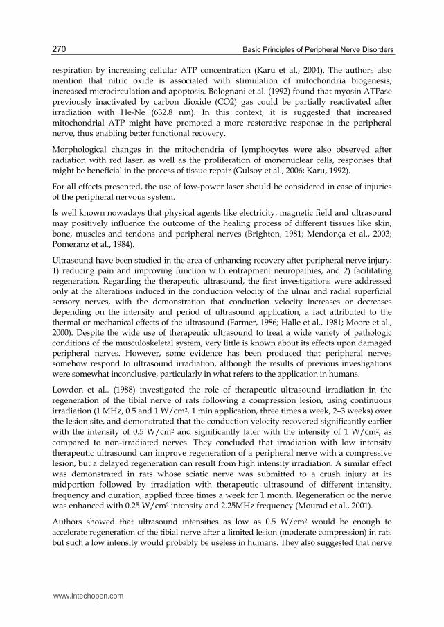
Ultrasound have been studied in the area of enhancing recovery after peripheral nerve injury: 1) reducing pain and improving function with entrapment neuropathies, and 2) facilitating regeneration. Regarding the therapeutic ultrasound, the first investigations were addressed only at the alterations induced in the conduction velocity of the ulnar and radial superficial sensory nerves, with the demonstration that conduction velocity increases or decreases depending on the intensity and period of ultrasound application, a fact attributed to the thermal or mechanical effects of the ultrasound (Farmer, 1986; Halle et al., 1981; Moore et al., 2000). Despite the wide use of therapeutic ultrasound to treat a wide variety of pathologic conditions of the musculoskeletal system, very little is known about its effects upon damaged peripheral nerves. However, some evidence has been produced that peripheral nerves somehow respond to ultrasound irradiation, although the results of previous investigations were somewhat inconclusive, particularly in what refers to the application in humans.
Lowdon et al.. (1988) investigated the role of therapeutic ultrasound irradiation in the
regeneration of the tibial nerve of rats following a compression lesion, using continuous
irradiation (1 MHz, 0.5 and 1 W/cm2, 1 min application, three times a week, 2–3 weeks) over
the lesion site, and demonstrated that the conduction velocity recovered significantly earlier
with the intensity of 0.5 W/cm2 and significantly later with the intensity of 1 W/cm2, as
compared to non-irradiated nerves. They concluded that irradiation with low intensity
therapeutic ultrasound can improve regeneration of a peripheral nerve with a compressive
lesion, but a delayed regeneration can result from high intensity irradiation. A similar effect
was demonstrated in rats whose sciatic nerve was submitted to a crush injury at its
midportion followed by irradiation with therapeutic ultrasound of different intensity,
frequency and duration, applied three times a week for 1 month. Regeneration of the nerve
was enhanced with 0.25 W/cm2 intensity and 2.25MHz frequency (Mourad et al., 2001).
Authors showed that ultrasound intensities as low as 0.5 W/cm2 would be enough to
accelerate regeneration of the tibial nerve after a limited lesion (moderate compression) in rats
but such a low intensity would probably be useless in humans. They also suggested that nerve
www.intechopen.com

Median and Ulnar Nerves Traumatic Injuries Rehabilitation 271
reaction to ultrasound would be different in damaged and intact nerves, the former being
more sensitive and susceptible to the induced thermal conduction, probably the actual agent of
regeneration. They were unable to suggest any other mechanism of action of the ultrasound.
There are some evidences that regeneration of the peripheral nerves can be accelerated by electric stimulation and a number of experimental studies have shown that the first signs of regeneration begin to appear by the third postoperative week and continue to happen for up to 90 days. Authors are unanimous to state that such low intensity has beneficial effect upon peripheral nerves regeneration. Although neither the intensity suggested nor the material used to make the electrodes vary from one to another. There is a controversy regarding to current intensity. Some authors used a very low current of up to 1.5 mA (Beveridge and Politis, 1988; Kerns et al., 1987, 1991; Politis et al., 1988a, 1988b; Pomeranz et al., 1984; Shen and Zhu, 1995), while others used 10 mA (McDevitt et al., 1987; Roman et al., 1987; Pomeranz & Campbell, 1993) or higher (Kerns et al., 1986, with 10 mA/cm2). One study used rat femoral nerve model supported a continuous electrical stimulation proximal to the site of repair for accelerating axonal growth (Al-Majed et al., 2000).
5. Conclusion
Despite advances in surgical techniques over time, several cellular events and favorable clinical status should be linked and coordinated so that nerve regeneration occurs with success. In clinical practice, it’s observed that the recovery of motor and sensory function still represents a challenge to reconstructive surgery and rehabilitation.
Regarding the hand sensation recovery, various sensorial re-education strategies have been introduced in the rehabilitation process with the aim of enhance patient capacity to reinterpret altered sensory stimuli due to injury sustained in the hand.
Rehabilitation is based on exercise therapy, splints and neuroplasticity principles.
This concept aim to facilitate sensory integration with the cortex area and promotes an interaction between tactile, visual and auditive stimuli, therefore represents an important tool in order to optimize sensory re-education strategies and maximize preservation of the hand’s cortical map representation in the early phase following injury.
Furthermore, the use of therapeutic modalities for peripheral nerve system regeneration is currently investigated. Low-power laser, ultra-sound, and electric stimulation have been used for accelerating regenerative processes in order to achieve early functional recovery.
6. Acknowledgment
This project had financial support from the FAEPA – Hospital das Clínicas da Faculdade de Medicina de Ribeirão Preto, Brazil.
7. References
Abdalla, L. M.; Brandão, M. C. F. Forças de preensão palmar e da pinça digital. In: Recomendações para avaliação do membro superior. 2. ed. Joinville: Sociedade Brasileira de Terapia da Mão, 2005. cap. 6, p. 38-41.
www.intechopen.com
Basic Principles of Peripheral Nerve Disorders 272
Al-Majed AA, Meumann CM, Brushart TM, Gordon T. Brief electrical stimulation promotes the speed and accuracy of motor axonal regeneration. J Neurosci. 2000;20:2602–8.
Amat A, Rigau J, Waynant RW, Ilev IK, Anders JJ (2006) The electric field induced by light can explain cellular responses to electromagnetic energy: a hypothesis of mechanism. J Photochem Photobiol B 82:152–60
Amadio, P.C. Outcome assessment in Hand Surgery and Hand Therapy: An update. J. Hand Ther, v.14, p. 63-7, 2001.
Aszmann, O. C.; Dellon, L. Relationship between cutaneous pressure threshold and two-point discrimination. J. Reconst. Microsurg.; v.14, p. 417-421, 1998.
Aulicino, P.L. Clinical examination of the hand. In: HUNTER, J; MACKIN, E.J.; CALLAHAN, A.D. Rehabilitation of the hand and upper extremity. 5a. edição. St. Louis: Mosby, 2002. Cap. 8, 120-142.
Bagis, S.; Comelekoglu, U.; Coskun, B.; Milcan, A.; Buyukakilli, B.; Sahin, G. No effect of GA-AS (904 nm) laser irradiation on the intact skin of the injured rat sciatic nerve, Lasers in Medical Science, London, v. 18, p. 83–88, 2003.
Barbosa RI, Marcolino AM, Guirro RRJ, Mazzer N, Barbieri CH, Fonseca MCR (2010) Comparative effects of wavelengths of low-power laser in regeneration of sciatic nerve in rats following crushing lesion. Lasers Med Sci (2010) 25:423–430.
Barbosa RI, Marcolino AM, Guirro RRJ, Mazzer N, Barbieri CH, Fonseca MCR (2010) Efeito do laser de baixa intensidade (660 nm) na regeneração do nervo isquiático lesado em ratos. Fisioter Pesq. 2010, vol.17, n.4, pp. 294-299. ISSN 1809-2950.
Beaton, D.E.; Katz, J.N.; Fossel, A .H.; Wright, J.G.;Tarasuk, V.;Bombardier, C. Measuring the whole or the parts? Validity, reability, and responsiveness of the disabilities of the arm, shoulder and hand (DASH) outcome measure in different regions of the upper extremity. J.Hand Ther, v.14(2), p.128-146, 2001.
Belchior ACG, Reis FA, Nicolau RA, Silva IS, Pereira DM, Carvalho PTC (2009) Influence of laser (660 nm) on functional recovery of the sciatic nerve in rats following crushing lesion. Lasers Med Sci 24:893–899.
Bell-Krotoski, J.; Buford, W.L. The force-time relationship of clinically used sensory testing instruments. J.Hand Ther. , v. 10(4), p. 297-309, 1997.
Bell-Krotoski, J. Sensibility testing with the Semmes-Weinstein monofilaments. In: HUNTER, J; MACKIN, E.J.; CALLAHAN, A.D. Rehabilitation of the hand and upper extremity. 5a. edição. St. Louis: Mosby, 2002. Chap. 13, 194-213.
Beveridge JA, Politis MJ. Use of exogenous electrical current in the treatment of delayed lesions in peripheral nerves. Plast Reconstr Surg 1988;82(4):573 /7.
Bell-Krotoski, J. Sensibility Testing: History, Instrumentation, and Clinical Procedures. In: SKIRVEN, TM; LEE OSTERMAN, A.; FEDORCZYK, JM. ; AMADIO, PC. Rehabilitation of the hand and upper extremity. 6a. edição. St. Louis: Mosby, 2011. Chap. 11, 132-151.
Bolognani L, Cavalca M, Magnani C, Volpi N (1992) ATP synthesis catalysed by myosin ATPase: effect of laser and e.m. field. Laser Technol 2:115–120
Brandsma, J.W.; Schreuders, T.A.R.; Birke, J.A.; Piefer, A. Oostendorp, R. Manual muscle strength testing: Intraobserver and interobserver reliabilities for the intrinsic muscles of the hand. J. Hand Ther. , v. 8. p 185-190, 1995.
Brighton CT. Current concepts review of the treatment of non-unions with electricity. J Bone Joint Surg 1981;63A:847–51.
www.intechopen.com

Median and Ulnar Nerves Traumatic Injuries Rehabilitation 273
Buccino G, Vogt S, Ritzl A, et al. Neural circuits underlying imitation learning of hand actions: an event-related FMRI study. Neuron. 2004;42:323–334.
Bucher, C. A Survey of Current Hand Assessment Practice in the UK. Brit. J. Hand Ther. v.8 (3). p 102-109, 2003.
Byl, N.; Leano, J.; Cheney, L.K. The Byl- Cheney-Boczai Sensory Discriminator: rehability, validity and responsiveness for testing sterognosis. J. Hand Ther. , v. 15(4), p. 315-30, 2002.
Callahan, A.D. "Sensibility testing: clinical methods." Rehabilitation of the Hand:Hunter, J. et al, 35 edition. St. Louis-Toronto. C.V. Mosby Company, Chap 44, 1990.
Colli, B.O; Carlotti Júnior, C.G. (2003). Aspectos Gerais das Lesões Traumáticas Agudas dos Nervos Periféricos, In: Nervos Periféricos, Diagnóstico e Tratamento Clínico e Cirúrgico, Marcos Tatagiba, Nilton Mazzer, pp. 39-54, Revinter, ISBN – 85-7309-652-7, Rio de Janeiro.
Colditz, JC. Splinting the hand with a peripheral-nerve injury. In: Hunter, J; Mackin, E.J.; Callahan, A.D. Rehabilitation of the hand and upper extremity. 5a. edição. St. Louis: Mosby, 2002. Cap. 34, p. 622-34.
Dannenbaum, R.M.; Michaelsen, S.M.; Desrosiers, J.; Levin, M.F., Development and validation of two new sensory tests hand for patients with stroke. Clin. Rehabil. v. 16(6), p. 630-9, 2002.
Davis et al. Measuring disability of the upper extremity: a rationale supporting the use of a regional outcome measure. J.Hand Ther., v. 12(4), p. 269-74, 1999.
Dahlin LB (2004) The biology of nerve injury and repair. J Am Soc Surg Hand 4:143–155 Lasers Med Sci.
di Pellegrino, G., Fadiga, L., Fogassi, L., Gallese, V., Rizzolatti, G., 1992. Understanding motor events: a neurophysiological study. Exp. Brain Res. 91, 176– 180.
di Pellegrino G, Wise SP. 1993. Visuospatial vs. visuomotor activity in the premotor and prefrontal cortex of a primate. J. Neurosci. 13:1227–43.
Dellon AL, Jabaley ME. Reeducation of sensation in the hand following nerve suture. Clin Orthop. 1982;163:75-9.
Dellon AL, Sensory reeducation. In: Dellon AL, editor. Somatosensory testing and rehabilitation. Bethesda [MD, USA]: The American Occupational Therapy Association; 1997. p.246-93.
Farmer WC. Effect of intensity of ultrasound on conduction of motor axons. Phys Ther 1986;48:1233–7.
Fess, EE. Guidelines for evaluation assessment instruments. J Hand Ther. P. 144-148, 1995. Fess, E.E. Documentation: essential elements of an upper extremity assessment battery. In:
Hunter, J; Mackin, E.J.; Callahan, A.D. Rehabilitation of the hand and upper extremity. 5a. edição. St. Louis: Mosby, 2002. Cap. 16, p. 263-284.
Fess, E.E. Functional tests. In: Skirven, Tm; Lee Osterman, A.; Fedorczyk, Jm. ; Amadio, Pc. Rehabilitation of the hand and upper extremity. 6a. edição. St. Louis: Mosby, 2011. Chap. 12, 152-162.
Fonseca MCR, Mazzer N, Barbieri CH, Elui VMC (2006) Hand trauma: retrospective study. Rev Bras Ortop 41:181–186
Fonseca MCR, Mazzer N, Barbieri CH, Elui VMC. (2003). Reeducação da Sensibilidade na Reabilitação da Mão, In: Nervos Periféricos, Diagnóstico e Tratamento Clínico e
www.intechopen.com

Basic Principles of Peripheral Nerve Disorders 274
Cirúrgico, Marcos Tatagiba, Nilton Mazzer, pp. 198-203, Revinter, ISBN – 85-7309-652-7, Rio de Janeiro.
Gianini, F. Quantitative Assessment of Historical and Objective Findings: A New Clinical Severity Scale of CTS. In: Luchetti, R, Amadio, P. Carpal Tunnel Syndrome. Springer, 2007. Cap 11, p. 82-88.
Gulsoy M, Ozer GH, Bozkulak O, Tabakoglu HO, Aktas E, Deniz G, Ertan C (2006) The biological effects of 632.8-nm low energy He-Ne laser on peripheral blood mononuclear cells in vitro. J Photochem Photobiol B 82:199–202
Hagander, L.G.; Midani, H. A .; Kuskowski, M.A .: Parry, G.J. Quantitative sensory testing: effect of site and skin temperature on thermal thresholds. Clin. Neurophysiol., v. 111(1), p. 17-22, 2000.
Halle JS, Scoville CR, Greathouse DG. Ultrasound’s effect on conduction latency of superficial radial nerve in man. Phys Ther 1981;61:345–50.
Heras-Palou, C. Burke, F.D.; Dias, J.J.; Bindra, R. Outcome measurement in Hand Surgery: reporto f a consensus conference. Brit J hand Ther. V8 (2), p. 70-80, 2003.
Jerosch-Herold, C. Should sensory function after median nerve injury and repair be quantified using Two-point discrimination as the critical measure? Scand. J. Plast. Reconstr. Hand Surg. v.34, p.339-343, 2000.
Jerosch-Herold, C. A study of the relative responsiveness of five sensibility tests for assessment of recovery after median nerve injury and repair . J. Hand Surg [B]. v.28, p.255-260, 2003.
Jerosch-Herold, C. Assessment of sensibility after nerve injury and repair: a systematic review of evidence for validity, reliability and responsiveness of tests. J. Hand Surg [B]. v.3, p.252-264, 2005.
Kaneko, A; Asai, N; Kanda, T. The influence of age on pressure perception os Static an Moving two-point discrimination in normal subjects. J. Hand Ther. v. 18, p.421-425,2005.
Karu T (1992) Derepression of the genome after irradiation of human lymphocytes with HeNe laser. Laser Therapy 4:5– 24
Karu TI, Pyatibrat LV, Afanasyeva NI (2004) A novel mitochondrial signaling pathway activated by visible-to-near infrared radiation. Photochem Photobiol 80:366–372
Karu TI, Pyatibrat L, Kalendo G (1995) Irradiation with He-Ne laser increases ATP level in cells cultivated in vitro. J Photochem Photobiol B 27:219–33
Kerns JM, Fakhouri AJ, Weinrib HP, Freeman JA. Electrical stimulation of nerve regeneration in the rat: the early effects evaluated by a vibrating probe and electron microscopy. Neuroscience 1991;40(1):93 /107.
Kerns JM, Fakhouri AJ, Weinrib HP, Freeman JA. Effects of D.C. electrical stimulation on nerve regeneration in the rat sciatic nerve. Anat Rec 1986;214:64A.
Kerns JM, Pavkovic IM, Fakhouri AJ, Wickersham KL, Freeman JA´ . An experimental implant for applying a DC electrical field to peripheral nerve. J Neurosci Methods 1987;19:217 /23.
Kitchen SS, Partridge CJ (1991) A review of low level laser therapy, part I: background, physiological effects and hazards. Physiotherapy 77:161–163
Khullar SM, Brodin P, Fristad I, Kvinnsland IH (1999) Enhanced sensory reinnervation of dental target tissues in rats following low level laser (LLL) irradiation. Lasers Med Sci 14:177–184
www.intechopen.com

Median and Ulnar Nerves Traumatic Injuries Rehabilitation 275
Lowdon IMR, Seaber AV, Urbaniak JR. An improved method of recording rat tracks for measurement of the sciatic functional index of De Medinaceli. J Neurosci Meth 1988;24:279–81.
Lundborg G (2000) A 25-year perspective of peripheral nerve surgery: evolving neuroscientific concepts and clinical significance. J Hand Surg [Am] 25:391–414
Lundborg, G.; Rosén, B. The two point discrimination test- time for a re-appraisal? J. Hand Surg [B and E]. v.29B:5 , p.418-422, 2004.
Lundborg, G.; Rosén, B. Review: Hand function after nerve repair. Acta Physiol. , v. 189, p. 207-217, 2007.
Macdermid, Jc.; Fess, Ee.; Bell-Krotoski,J.; Cannon, Nm.; Evans, Rb.; Walsh, W.; Szabo, Rm.; Laseter, G.; Mackin, E.; Gettle,K.; Santore, G. A Research agenda for Hand Therapy. J. Hand Ther. v. 15, p.3-15,2002.
Macdermid, JC; Tottenham, V. Responsiveness of the Disability of the Arm, Shoulder, and Hand (DASH) and Patient-Rated Wrist/Hand Evaluation (PRWHE) in evaluating change in Hand Therapy. J. Hand Ther. v. 17, p.18-23,2004.
Macdermid, JC. The quality of clinical practice guidelines in Hand Therapy. J. Hand Ther. v. 17, p.200-209,2004.
Macdermid, JC. Measurements of health outcomes following tendon and nerve repair. J. Hand Ther. v. 18, p.297-312,2005.
Macdermid, JC. Outcomes measurement in upper extremity practice. In: Skirven, Tm; Lee Osterman, A.; Fedorczyk, Jm. ; Amadio, Pc. Rehabilitation of the hand and upper extremity. 6a. edição. St. Louis: Mosby, 2011. Chap. 16, 194-205.
Macdermid, JC. Evidence-based practice in Hand Rehabilitation. In: Skirven, Tm; Lee Osterman, A.; Fedorczyk, Jm. ; Amadio, Pc. Rehabilitation of the hand and upper extremity. 6a. edição. St. Louis: Mosby, 2011. Chap. 143, 1881-89.
Macey et al. Outcomes of hand surgery. British Society for Surgery of the hand. J. Hand Surg. v. 20(6), p. 841-55, 1995.
Manteifel V, Bakeeva L, Karu T (1997) Ultrastructural changes in chondriome of human lymphocytes after irradiation with He-Ne laser: appearance of giant mitochondria. J Photochem Photobiol B 38:25–30
Manteifel VM, Karu TI (2005) Structure of mitochondria and activity of their respiratory chain in successive generations of yeast cells exposed to He-Ne laser light. Izv Akad Nauk Ser Biol 32:556–566
Marcolino AM; Barbosa RI; Fonseca MCR; Mazzer N; Elui VMC (2008) Physical therapy in brachial plexus injury: case report. Rev Fisioter Mov 21:53–61
Marcolino AM; Barbosa RI; Neves LS; Fonseca MCR. Laser de baixa intensidade (830 nm) na recuperação funcional do nervo isquiático de ratos. Acta ortop. bras., 2010, vol.18, no.4, p.207-211. ISSN 1413-7852
McDevitt L, Fortner P, Pomeranz B. Application of weak electric field to the hindpaw enhances sciatic motor nerve regeneration in the adult rat. Brain Res 1987;416:308/14.
Mendonça AC, Barbieri CH, Mazzer N (2003) Directly applied low intensity direct electric current enhances peripheral nerve regeneration in rats. J Neurosci Methods 129:183–190
www.intechopen.com

Basic Principles of Peripheral Nerve Disorders 276
Mohammed IFR, AL-Mustawfi NBV, Kaka LN (2007) Promotion of regenerative processes in injured peripheral nerve induced by low-level laser therapy. Photomed Laser Surg 25:107–111
Monte-raso VV, Barbieri CH, Mazzer N, Fazan VS (2005) Can therapeutic ultrasound influence the regeneration of peripheral nerves? J Neurosci Methods 142:185–192
Moore JH, Gieck JH, Saliba EN, Perrin DH, Ball DW, Mccue FC. The biophysical effects of ultrasound on median nerve distal latencies. Electromyogr Clin Neurophysiol 2000;40(3):169–80
Mourad PD, Lazar DA, Curra FP, Mohr BC, Andrus KC, Avelino AM, ET al. Ultrasound accelerates functional recovery after peripheral nerve damage. Neurosurgery 2001;48(5):1136–40.
Noble, J.; Munro, C.A.; Prasad, V.S.; Midha, R. Analysis of upper and lower extremity peripheral nerve injuries in a population of patients with multiple injuries. J Trauma, Oregon, v.45(1), p. 116-22, 1998.
Novak, C.B. Evaluation of hand sensibility: a review. J.Hand Ther. ,v.14(4), p.266-72, 2001. Novak CB, Mackinnon SE (2005) Evaluation of nerve injury and nerve compression in the
upper quadrant. J Hand Ther 18:230–240 Orfale, A. G.; Araújo, P. M. P.; Ferraz, M. B.; Natour, J. Translation into Brazilian
Portuguese, cultural adaptation and evaluation of the reliability of the Disabilities of the Arm, Shoulder and Hand Questionnaire. Braz J Med Biol Res. , v.38, p. 293-302, 2005.
Padua, R; Padua, L; Ceccarelli, B; Romanini, E.; Zanoli, G.; Amadio, P.; Campi, A. Italian Version of the Disability of the Arm, Shoulder and Hand (dash) Questionnaire. Cross-Cultural Adaptation and Validation. J Hand Surg Eur Vol April 2003 v. 28 n2 p.179-186
Padua, R; Romanni, E; Bondi, R. Outcomes Assessment Protocols. In: Luchetti, R, Amadio, P. Carpal Tunnel Syndrome. Roma: Springer, 2007, Cap 50, p.383-391.
Patel,M.R. & Bassini, L. A comparison of five tests for determining hand sensibility. J. Reconstr. Microsurg., v. 15(7), p. 523-6, 1999.
Pedersen PL, Carafoli E (1987) Ion motive ATPase. I. Ubiquity, properties and significance to cell function. Trends Biochem Sci 12:146–150, 186–189
Polatkan,S: Orhun, E.; Polatkan, O; Nuzumlali, E.; Bayri, O. Evaluation of the improvement of sensibility after primary median repair at the wrist. Microsurg.; v.18(3), p. 192-6, 1998.
Pereira, CU; Carvalho, AF; Carvalho, MF. (2003). Exame Neurológico de Lesões do Nervo Periférico, In: Nervos Periféricos, Diagnóstico e Tratamento Clínico e Cirúrgico, Marcos Tatagiba, Nilton Mazzer, pp. 1-12, Revinter, ISBN – 85-7309-652-7, Rio de Janeiro
Politis MJ, Zanakis MF, Albala BJ. Mammalian optic nerve regeneration following the application of electric fields. J Trauma 1988a;28(11):1548/52.
Politis MJ, Zanakis MF, Albala BJ. Facilitated regeneration in the rat peripheral nervous system using applied electric field. J Trauma 1988b;28(9):1375/81.
Pomeranz B, Campbell J. Weak electric current accelerates motoneuron regeneration in the sciatic nerve of 10-month-old rats. Brain Res 1993;603:271 /8.
Pomeranz B, Mullen M, Markus H. Effect of applied electrical fields on sprouting of intact saphenous nerve in adult rat. Brain Res 1984;303:331–6.
Pons TP, Garraghty PE, Ommaya AK, Kaas JH, Taub E, Mishkin M. 1991. Massive cortical reorganization after sensory deafferentation in adult macaques. Science 252:1857–60.
www.intechopen.com

Median and Ulnar Nerves Traumatic Injuries Rehabilitation 277
Reis FA, Belchior ACG, Carvalho PTC, Silva BAK, Pereira DM, Silva IS, Nicolau RA (2009) Effects of laser therapy (660 nm) on recovery of the sciatic nerve in rats after injury through neurotmesis followed by epineural anastomosis. Lasers Med Sci 24:741–747.
Rizzolatti, G., Fogassi, L., Gallese, V., 2001. Neurophysiological mechanisms underlying the understanding and imitation of action. Nat. Rev. Neurosci. 2, 661–670.
Rizzolatti G, Luppino G, Matelli M. The organization of the cortical motor system: new concepts. Electroencephalogr Clin Neurophysiol. 1998;106:283–296.
Rizzolatti G, Craighero L (2004) The Mirror Neuron System. Annual Rev Neurosci 27:169–192. Rochkind S, Barrnea L, Razon N, Bartal A, Schwartz M (1987) Stimulatory effect of He-Ne
low dose laser on injured sciatic nerves of rats. Neurosurgery 20:843–847 Rodrígues, F.J.; Valero-Cabré, A.; Navarro, X. Regeneration and functional recovery
following peripheral nerve injury. Drug Discov Today Dis Models, Amsterdam, v. 1, p. 177–185, 2004.
Roman GC, Strahlendorf HK, Coates PW, Rowley BA. Stimulation of sciatic nerve regeneration in the adult rat by low-intensity electric current. Exp Neurol 1987;98:222 /32.
Rosén, B. Recovery of sensory and motor function after nerve repair rationale for evaluation. J. Hand Ther. v.9(4), p. 315-27, 1996.
Rosén, B; Lundborg, G.; Abrahamsson, I.; Agberg, H.; Rosén, I. Sensory function after median nerve decompression in carpal tunnel syndrome. J Hand Surg. ,v.22B, p.602-606, 1997.
Rosén, B & Lundborg,G. A new tactile gnosis instrument in sensibility testing. J.Hand Ther. v.11, p. 251-257, 1998.
Rosén, B. The sensational hand (2000). Clinical assessment after nerve repair. Thesis, Lund University, pp. ISBN 91-628-4368-4360.
Rosén, B & Lundborg,G. A model instrument for the documentation of outcomes nerve repair. J.Hand Surg[Am]. v.25(3), p. 535-43, 2000.
Rosén, B & Lundborg, G. The long-term recovery curve in adults after median or ulnar nerve repair: a reference interval. J Hand Surg. ,v.26B, p.196-200, 2001.
Rosén, B & Lundborg,G. A new model instrument for outcome after nerve repair. Hand Clinics. v.19 p. 463-470, 2003.
Rosén, B. Inter-tester reliability of a Tactile Gnosis Test: The STI- Test®. Brit. J. Hand Ther. v.8 p (3). 98-101, 2003.
Rosén, B; Balkenius, C; Lundborg,G. Sensory re-education today and tomorrow: a review of evolving concepts. Brit. J. Hand Ther. v.8 p (2). 48-56, 2003.
Rosental, T.L.; Beredjiklian, P.K.; Guyette, T.M.; Weiland, A .J. Intra and interobserver reliability of sensibility testing asymptomatic individuals. Ann. Plast. Surg., v. 44(6), p. 605-9, 2000.
Sollerman C, Ejeskär A. Sollerman hand functional test: a standardized method and its use in tetraplegic patients. Scand J Plast Reconstr Surg. 1995;29:167-176.
Schindl A, Schindl M, Schindl L, Jurecka W, Hönigsmann H, Breier F (1999) Increased dermal angiogenesis after low-intensity laser therapy for a chronic radiation ulcer determined by a video measuring system. J Am Acad Dermatol 40:481–484
Shen N, Zhu J. Experimental study using a direct current electrical field to promote peripheral nerve regeneration. J Reconstr Microsurg 1995;11(3):189/93.
www.intechopen.com
Basic Principles of Peripheral Nerve Disorders 278
Sulaiman OA, Gordon T (2000) Effects of short- and long-term Schwann cell denervation on peripheral nerve regeneration, myelination, and size. Glia 32:234–46
Themistocleous, G.S.; Goudelis, G; Kyrou, I.;Chloros, G.D.; Krokos, A; Galanos, A.; Gerostathopoulos, N.E.; Soucacos, P.N. Translation into Greek, Cross-cultural Adaptation and Validation of the Disabilities of the Arm, Shoulder, and Hand Questionnaire (DASH). J. Hand Therapy. v.2006 v.19 n.3 p.350-57
Vladimirov Yu A, Osipov AN, Klebanov GI (2004) Photobiological principles of therapeutic applications of laser radiation. Biochemistry 69:81–90
Walsh DM, Baxter GD, Allen JM (2000) Lack of effect of pulsed low-intensity infrared (820 nm) laser irradiation on nerve conduction in the human superficial radial nerve. Lasers Surg Med 26:485–490.
www.intechopen.com

Basic Principles of Peripheral Nerve DisordersEdited by Dr. Seyed Mansoor Rayegani
ISBN 978-953-51-0407-0Hard cover, 278 pagesPublisher InTechPublished online 28, March, 2012Published in print edition March, 2012
InTech EuropeUniversity Campus STeP Ri Slavka Krautzeka 83/A 51000 Rijeka, Croatia Phone: +385 (51) 770 447 Fax: +385 (51) 686 166www.intechopen.com
InTech ChinaUnit 405, Office Block, Hotel Equatorial Shanghai No.65, Yan An Road (West), Shanghai, 200040, China
Phone: +86-21-62489820 Fax: +86-21-62489821
Peripheral nerve disorders are comprising one of the major clinical topics in neuromusculoskeletal disorders.Sharp nerve injuries, chronic entrapment syndromes, and peripheral neuropathic processes can be classifiedin this common medical topic. Different aspects of these disorders including anatomy, physiology,pathophysiology, injury mechanisms, and different diagnostic and management methods need to beaddressed when discussing this topic. The goal of preparing this book was to gather such pertinent chapters tocover these aspects.
How to referenceIn order to correctly reference this scholarly work, feel free to copy and paste the following:
Rafael Inácio Barbosa, Marisa de Cássia Registro Fonseca, Valéria Meirelles Carril Elui, Nilton Mazzer andCláudio Henrique Barbieri (2012). Median and Ulnar Nerves Traumatic Injuries Rehabilitation, Basic Principlesof Peripheral Nerve Disorders, Dr. Seyed Mansoor Rayegani (Ed.), ISBN: 978-953-51-0407-0, InTech,Available from: http://www.intechopen.com/books/basic-principles-of-peripheral-nerve-disorders/median-and-ulnar-nerves-traumatic-injuries-rehabilitation