Manual treatment of post-whiplash injury
11
www.intl.elsevierhealth.com/journals/jbmt Bodywork and Journal of Movement Therapies CLINICAL APPROACHES Manual treatment of post-whiplash injury C ! esar Fern ! andez de las Pe * nas a, *, Luis Palomeque del Cerro b , Josu ! e Fern ! andez Carnero a a Teaching and Research Unit of Physiotherapy, Occupational Therapy, Physical Medicine and Rehabilitation, Universidad Rey Juan Carlos (URJC), Avenida de Atenas s/n, Alcorc ! on 28922, Spain b Escuela de Osteopat ! ıa de Madrid (EOM), Spain Received 6 February 2004; received in revised form 16 May 2004; accepted 19 May 2004 Abstract Introduction: There are many therapeutic approaches aimed at treating the clinical syndrome resulting from whiplash injury. However, there seems to be little agreement between therapists as to the ideal treatment for these patients. Spinal manipulation/mobilization and soft tissue mobilization techniques are manual therapies commonly used in the management of neck disorders. The aims of the present paper are to detail a manual approach developed by our research group, to help in future studies of the management of the sequels to whiplash injury, and to suggest explanations for the mechanisms of this protocol. These manual approaches are considered by the authors to be more effective than conventional physical therapy in the management of whiplash patients. Vertebral manipulations: The biomechanical analysis of whiplash injury showed that upper cervical manipulation, cervicothoracic junction manipulation, thoracic spine manipulation, and pelvic girdle manipulation are the major areas requiring such treatment, for beneficial outcomes to be assured. Although the exact biological mechanisms underlying the effects of spinal manipulation are not clearly under- stood, there are previous papers justifying most methods used in the current experimental protocol. Soft tissues manipulation techniques: The soft tissues techniques used in this protocol were neuromuscular technique in paraspinal muscles, muscle energy techniques in the cervical spine, myofascial release in the occipital region, and myofascial trigger point manual therapies as required. Clinical dissertation: The definition of spinal joint dysfunction (hypomobility) implies that muscle shortening is involved. This suggests that manual treatment of persons suffering from whiplash injury requires the treatment of muscular and fascial shortening, as well as the treatment of spinal joint dysfunction, when appropriate. & 2004 Elsevier Ltd. All rights reserved. ARTICLE IN PRESS KEYWORDS Manual treatment; Myofascial trigger point; Spinal manipulation; Soft tissue manipulation; Whiplash injury *Corresponding author. Tel.: þ 34-91-488-88-84; fax: þ 34-91-488-88-31. E-mail address: [email protected] (C. Fern ! andez de las Pe * nas). 1360-8592/$ - see front matter & 2004 Elsevier Ltd. All rights reserved. doi:10.1016/j.jbmt.2004.05.002 Journal of Bodywork and Movement Therapies (2005) 9, 109–119
description
Partilhamos um dos artigos de Luís Palomeques, um osteopata de renome, sobre a terapia manual no "Golpe de Chicote" ou Whiplash cervical.
Transcript of Manual treatment of post-whiplash injury

www.intl.elsevierhealth.com/journals/jbmt
Bodywork and
Journal of
Movement Therapies
CLINICAL APPROACHES
Manual treatment of post-whiplash injury
C !esar Fern!andez de las Pe *nasa,*, Luis Palomeque del Cerrob,Josu !e Fern!andez Carneroa
aTeaching and Research Unit of Physiotherapy, Occupational Therapy, Physical Medicine andRehabilitation, Universidad Rey Juan Carlos (URJC), Avenida de Atenas s/n, Alcorc !on 28922, SpainbEscuela de Osteopat!ıa de Madrid (EOM), Spain
Received 6 February 2004; received in revised form 16 May 2004; accepted 19 May 2004
Abstract Introduction: There are many therapeutic approaches aimed at treatingthe clinical syndrome resulting from whiplash injury. However, there seems to belittle agreement between therapists as to the ideal treatment for these patients.Spinal manipulation/mobilization and soft tissue mobilization techniques are manualtherapies commonly used in the management of neck disorders. The aims of thepresent paper are to detail a manual approach developed by our research group, tohelp in future studies of the management of the sequels to whiplash injury, and tosuggest explanations for the mechanisms of this protocol. These manual approachesare considered by the authors to be more effective than conventional physicaltherapy in the management of whiplash patients.Vertebral manipulations: The biomechanical analysis of whiplash injury showed
that upper cervical manipulation, cervicothoracic junction manipulation, thoracicspine manipulation, and pelvic girdle manipulation are the major areas requiringsuch treatment, for beneficial outcomes to be assured. Although the exact biologicalmechanisms underlying the effects of spinal manipulation are not clearly under-stood, there are previous papers justifying most methods used in the currentexperimental protocol.Soft tissues manipulation techniques: The soft tissues techniques used in this
protocol were neuromuscular technique in paraspinal muscles, muscle energytechniques in the cervical spine, myofascial release in the occipital region, andmyofascial trigger point manual therapies as required.Clinical dissertation: The definition of spinal joint dysfunction (hypomobility) implies
that muscle shortening is involved. This suggests that manual treatment of personssuffering from whiplash injury requires the treatment of muscular and fascialshortening, as well as the treatment of spinal joint dysfunction, when appropriate.& 2004 Elsevier Ltd. All rights reserved.
ARTICLE IN PRESS
KEYWORDS
Manual treatment;
Myofascial trigger point;
Spinal manipulation;
Soft tissue manipulation;
Whiplash injury
*Corresponding author. Tel.: þ 34-91-488-88-84;fax: þ 34-91-488-88-31.E-mail address: [email protected]
(C. Fern!andez de las Pe *nas).
1360-8592/$ - see front matter & 2004 Elsevier Ltd. All rights reserved.doi:10.1016/j.jbmt.2004.05.002
Journal of Bodywork and Movement Therapies (2005) 9, 109–119

Introduction
The Quebec Task Force adopted the followingdefinition of whiplash (Spitzer et al., 1995):
Whiplash is an acceleration–deceleration mechanismof energy transfer to the neck. It may result from rear-end or side-impact motor vehicle collisions, but canalso occur during diving or other mishaps. The impactmay result in bony or soft tissue injuries (whiplashinjuries), which in turn may lead to a variety of clinicalmanifestations (whiplash associated disorders).
The clinical syndrome of whiplash injury includesneck pain, upper thoracic pain, cervicogenic head-ache (Drottning et al., 2002), tightness, dizziness,restriction of cervical range of motion, tinnitus,and blurred vision (Hohl, 1975; Dvorak et al.,1989). The exact nature of these symptoms is notclearly understood, although the pain is attributedto musculoskeletal disorders, i.e. involving the softtissues and facet joint dysfunction, caused by theimpact (Wiley et al., 1986). Moreover, experimen-tal research involving human cadavers has demon-strated that a variety of musculoskeletal injuriescan occur during whiplash, such as muscle andligament sprains (Barnsley et al., 1998). Severaltheories have been postulated to explain thesesymptoms, including vertebral artery insufficiencyand injury of the cervical sympathetic chain, inrelation to visual disturbances and dizziness (Bog-duk, 1986), C1–C2 facet joint injury in relation toheadaches (Lord et al., 1996), and paraspinalmuscle spasm in relation to neck pain (Teasell andShapiro, 2001). Moreover, some authors havereported that people suffering from whiplash injurydisplay signs of both central and peripheralsensitization (Koelbaek et al., 1999).
Numerous forms of treatment have been sug-gested to relieve the symptoms of this clinicalsyndrome. However, there seems to be little or noagreement between therapists as to the idealtreatment of whiplash symptoms (Valera et al.,2003). Following a literature review relating to theconservative treatment of persons suffering fromwhiplash injury, Peeters et al. (2001), reportedthat, despite the many treatments available forthese patients, there continues to be no evidencefor their accepted use.
Spinal manipulation/mobilization, and soft tissuemobilization, techniques are manual therapiescommonly used in the management of neckdisorders (Gross et al., 2002). There are manyclinical trials that have analysed the effectivenessof cervical manipulation/mobilization in peoplesuffering from mechanical neck pain (Vernonet al., 1990; Cassidy et al., 1992), but there are a
few papers analysing the effects of spinal manip-ulation in people suffering from whiplash injury(Osterbauer et al., 1992).
In a previous paper (Fern !andez et al., 2004a) ourresearch group demonstrated that current manualtreatment methods were more effective thanconventional physical therapy in the managementof whiplash patients. In this trial it was found thatpeople who were treated with this manual ap-proach had a greater improvement in cervicalrange of motion, and a greater scores on visualanalogue scales, than those treated with conven-tional physical therapy treatment (comprisingmassage, ultrasound therapy, exercises at home,and low energy high frequency pulsed electromag-netic therapy). Moreover, patients from the osteo-pathic group in that trial required 9 sessionsto complete the treatment, whereas those fromthe physiotherapy group needed 23 sessions(P ¼ 0:002). The study concluded that the improve-ment in the experimental group was faster andgreater than the improvement in the physiotherapygroup.
The manual treatment mentioned in the previoustrial (Fern !andez et al., 2004a) included highvelocity–low amplitude techniques (HVLA) appliedto the upper cervical spine, cervicothoracic junc-tion, thoracic spine, thoracolumbar junction, andpelvic girdle, as well as neuromuscular technique(NMT) of the paraspinal soft tissues (Chaitow,2003), muscle energy techniques (MET) applied tothe cervical spine (Mitchell, 1995; Chaitow, 2001),craniosacral techniques (von Piekartz and Bryden,2001), and myofascial trigger point (MTrP) manualtherapies (Hou et al., 2002).
The aims of this paper are:
* to detail the manual treatment developed by ourresearch group, to help in future studies invol-ving the management of persons suffering fromwhiplash injury, and,
* to offer hypotheses of the mechanisms of thisprotocol
Vertebral manipulations (HVLAtechniques)
The goal of joint manipulation is to restoremaximal, pain-free movement of the musculoske-letal system (Whittingham and Nillson, 2001). It issuggested that only joints that are found to behypomobile should be considered as candidates forHVLA techniques. Vertebral manipulations arecurrently used in the treatment of whiplash injurywithout the benefit of scientific evidence, so
ARTICLE IN PRESS
110 C. Fern !andez de las Pe *nas et al.

further analysis is required to find a biomechanicaljustification for HVLA treatment in whiplash injury.A previous paper (Fern !andez et al., 2003a) showedthat upper cervical manipulation, cervicothoracicjunction manipulation, thoracic spine manipula-tion, and pelvic girdle manipulation have biome-chanical justifications in the scientific literature(Panjabi et al., 1998a; Bogduk and Yoganandan,2001; Yoganandan et al., 2002). Based on thisbiomechanical analysis, our research group consid-ered it appropriate to manipulate only thesesegments, and only if hypomobility could bedemonstrated.
A brief description of the manipulative techni-ques is given below, after which explanations areoffered as to possible mechanisms whereby benefitmight be gained from these procedures.
Upper cervical spine manipulation
* Pattern of restriction. Upper cervical manipula-tion is applied only if rotation restriction of C1has been identified by the therapist. Theexamination is based on palpatory examinationand gliding motion test of the atlas. Before thismanipulation, an extension–rotation test, forvertebro-basilar insufficiency assessment shouldbe applied, although its specificity, validity andreliability is controversial (Mitchell, 2003).
* Patient position. Supine with the neck in aneutral relaxed position.
* Practitioner stance. At the head of the couch.* Hand contacts. The hand of the therapist makes
contact with the index finger over the posteriorarch of the atlas, on the side contra-lateral tothe restriction. In this example, with restrictionof right rotation of the atlas, the hand of thetherapist makes contact over the left side ofposterior arch of the atlas. The other hand cupsthe chin.
* Direction of the manipulation. Rotation is gentlyintroduced, toward the right, until slight tensionis palpated in the tissues at the contact point.
* Thrust. A HVLA thrust is applied directedtowards the corner of the person’s mouth(Fig. 1).
Cervicothoracic junction manipulation
* Pattern of restriction. Cervicothoracic manipu-lation is applied only if side-flexion restriction ofC7 on T1 has been identified by the therapist.The examination is based on palpatory examina-tion and a gliding motion test of C7 on T1.
* Patient position. Prone with the head and neckrotated. In this example, with lateral-flexion of
C7 on T1 restricted to the left, the neck is turnedto the right.
* Practitioner stance. The therapist stands on theright side of the person, facing cephalad.
* Hand contacts. The left hand of the therapistmakes contact, with the thumb on the left sideof the spinous process of T1. The right handsupports the head, making contact on thetemporal bone.
* Direction of the manipulation. The head/neck isgently lateral-flexed to the left, until slighttension is palpated in the tissues.
* Thrust. A HVLA thrust is applied, of the spinousprocess of T1, toward the person’s right sideshoulder (Fig. 2).
Thoracic spine manipulation
* Pattern of restriction. Thoracic spine manipula-tion is applied only if extension restriction ofT1–T4 has been identified by the therapist. Theexamination is based on palpatory examinationand gliding motion test of high dorsal vertebras(Fern !andez et al., 2004b).
* Patient position. Supine with the arms crossedover the chest and hands wrapped around theshoulders. The thoracic spine is in a neutralposition.
* Practitioner stance. On one side of the patient,facing cephalad.
* Hand contacts. The clenched hand of thetherapist makes contact over the spinous processof T4. The other hand stabilizes the head, neckand upper thoracic spine, making contact overthe spinous process of T3.
ARTICLE IN PRESS
Figure 1 Upper cervical spine manipulation.
Manual treatment of post-whiplash injury 111

* Direction of the manipulation. Gentle flexion ofthe upper thoracic spine is introduced untilslight tension is palpated in the tissues at thecontact point.
* Thrust. A HVLA technique is applied, downwardstowards the couch, and in a cephalad direction(Fig. 3).
Thoracolumbar junction manipulation
* Pattern of restriction. Thoracolumbar junctionmanipulation should be applied in all patientswith the aim of restoring maximal free move-ment of T12–L1 region, because the biomecha-nical analysis of whiplash injury implies acompression spine dysfunction at this level(Panjabi et al., 1998a; Bogduk and Yoganandan,2001; Yoganandan et al., 2002).
* Patient position. Sitting with arms crossedbehind the trunk, and knuckles over the thor-acolumbar junction (T12–L1).
* Practitioner stance. Standing directly behind thepatient.
* Hand contacts. The therapist places his/herabdomen against the hands of the patient(abdomen of the therapist placed over thespinous process of T12–L1 junction). Hands ofthe therapist passed around the abdomen of thepatient.
* Direction of the manipulation. A compressiveupwards force is introduced until slight tension ispalpated in the tissues at the contact point.
* Thrust. Maintaining all holds and pressure,the patient is brought backwards. A HVLAtechnique is applied towards the therapist,and slightly upwards in a cephalad direction(Fig. 4).
Pelvic girdle manipulation
* Pattern of restriction. Pelvic girdle manipulationshould be applied to all patients, with the aim ofrestoring maximal free movement of the sacroi-liac region, because biomechanical analysis ofwhiplash injury implies a compression dysfunc-tion of the pelvic girdle (Panjabi et al., 1998a;Bogduk and Yoganandan, 2001; Yoganandanet al., 2002).
* Patient position. The person lies on the side,with the upper body in light flexion and hipsflexed approximately 901. The upper knee of theperson is flexed until the heel of the foot isplaced just anterior of the knee of the lower leg.
* Practitioner stance. Standing in front of theperson, and close to the couch.
* Hand contacts. The therapist makes contact witha forearm on the lateral aspect of the pelvicgirdle. The other hand should be resting against
ARTICLE IN PRESS
Figure 3 Thoracic spine manipulation.
Figure 2 Cervicothoracic junction manipulation.
112 C. Fern !andez de las Pe *nas et al.

the person’s pectoral and rib cage region. Theknee of the therapist is placed over the patient’supper knee.
* Direction of the manipulation. Gentle contra-lateral rotation of the person’s upper body isintroduced, taking the upper body posteriorlyand the pelvis anteriorly, until slight tension ispalpated at L5–S1 junction.
* Thrust. A HVLA technique is applied downwardstowards the couch (Fig. 5).
What are the therapeutic mechanismsinvolved in these manipulations?
The biological mechanism underlying the effects ofspinal manipulation is not clearly understood,however we hypothesize possible mechanisms ofthe applied manipulations in the current protocol,as follows:
Upper cervical manipulationPenning postulated that the main mechanism ofwhiplash injury is hyper-translation of the head(Penning, 1992a, b). This has been confirmedrecently by other authors who reported that uppercervical spine performes a hyperextension motionduring the first phase of the whiplash injury(Panjabi et al., 1998a; Bogduk and Yoganandan,2001; Yoganandan et al., 2002). This hyperexten-sion of the head might be one of the causes ofC1–C2 facet joint injury and adaptive muscleshortening. This head motion distracts the ante-rior structures with a concomitant compressionof the posterior structures, specifically the sub-occipital muscles. Therefore, compression of the
ARTICLE IN PRESS
Figure 4 Thoracolumbar junction manipulation.
Figure 5 Pelvic girdle manipulation.
Manual treatment of post-whiplash injury 113

atlantoaxial junction may activate a myofascialpain syndrome in the suboccipital muscles. More-over, during whiplash, the head and neck areexposed to multiple forces in flexion, extensionand rotation, so that myofascial pain syndrome canbe initiated due to these force patterns during eachphase of the accident (Simons et al., 1999). Threeadditional anatomical finding may be involved:
(a) There is a connective tissue bridge between therectus capitis posterior minor muscle (RCPM)and the dorsal spinal dura at the atlanto-occipital junction (Hack et al., 1995),
(b) The cervical posterior spinal dura between C1–C2 vertebrae is attached to the ligamentumnuchae (Mitchell et al., 1998),
(c) The posterior dura is much thicker than theanterior dura in the upper cervical spine (Tayloret al., 1996).
We hypothesize that adverse tension in the spinaldura can result in cervicogenic headache (Vernon,1995), a common whiplash associated disorder(Drottning et al., 2002). This situation mightexplain the effectiveness of the upper cervicalspine manipulation in these patients. Some authorsreported that 20% of people suffering from chronicwhiplash injury demonstrate atrophy of the RCPM(Hallgren et al., 1994). This could suggest thatHVLA involving the upper cervical spine might beindicated in these cases. However, upper cervicalmanipulation restores pain-free movement at theatlantoaxial junction, so its effect is aimed at thejoint proprioceptors. The atlantoaxial region re-quires soft tissues techniques, such as suboccipitalrelease, NMT and METapplied to the cervical spine,to reduce adverse tension in the spinal dura, and toencourage a minimal restoration of a neuromuscu-lar connection in RCPM.
Cervicothoracic junction, thoracic spine, andthoracolumbar junction manipulationsAlthough whiplash injury may result from rear, sideand frontal impacts, rear-end crashes account forabout 85% of all of whiplash associated disorders(Yoganandan et al., 1998). The presence of thoracicjoint dysfunctions and C7-T1 can be explainedthrough a kinematical analysis of a rear-endimpact. Recent papers (Panjabi et al., 1998a;Bogduk and Yoganandan, 2001; Yoganandan et al.,2002) investigating the biomechanical mechanismof whiplash injury, have hypothesized that, duringthe initial phase of a rear-end impact, the uppercervical spine responds in flexion, concomitantwith lower cervical and upper thoracic spineextension, resulting in an S-curve (Fig. 6). This
situation is attributed to the upward and forwardthrust of the upper trunk during the first 100msafter the rear impact (Bogduk and Yoganandan,2001). This is the consequence of the thoracickyphosis extension which is attributed to theseatback (Matsushita et al., 1994). This rapidthoracic extension distracts the anterior aspectsof the spine with a concomitant compression of theposterior structures, specifically the thoracic softtissues. The S-curve of the cervical spine couldexplain the presence of cervicothoracic junctiondysfunction, whereas thoracic extension couldexplain the presence of thoracic joint dysfunction.
In a previous trial performed by our researchgroup, it was demonstrated that thoracic jointdysfunctions are more prevalent in persons suffer-ing from whiplash injury than in persons sufferingfrom mechanical neck pain. Moreover, it was foundthat some of the whiplash associated disorders(head, neck, and upper thoracic pain) decreased inresponse to thoracic manipulation (Fern !andezet al., 2004b).
Pelvic girdle manipulationPelvic girdle manipulation is commonly necessarybecause, in a rear-end impact, as the target vehicleis accelerated forward, the seatback contacts thelumbopelvic region, causing the seat to deflectbackward, away from the upper torso (Gay andLevine, 2002). Later, the restraining lap belt mightcause compression of the lumbo-pelvic region.Moreover, the forward rebound of the seat backmay contribute to a second phase of this compres-sion. This compression of the lumbopelvic regionproduces a hypomobility in the pelvic girdle, whichit is necessary to manipulate. Therefore, thepresence of a hypomobility in the pelvic regioncould be expanded to include secondary adaptative
ARTICLE IN PRESS
Figure 6 S-curve of the cervical spine during the initialphase of a rear-end impact.
114 C. Fern !andez de las Pe *nas et al.

or maladaptative changes in the cervical andthoracic regions (Mitchell, 1995).
Soft tissues manipulation techniques
It has been demonstrated, that after whiplashinjury, soft tissues that have been traumatizedmay develop MTrPs (Schuller et al., 2000). MTrPscan contribute significantly to acute and/or chronicpain syndromes following whiplash injury, a scenariothat is often overlooked (Hong and Simons, 1993).
MTrPs and cervical joint dysfunctions arethought, by some authors, to be among the mostimportant causes of musculoskeletal disorders inpeople suffering from whiplash injury (Bronfortet al., 2001; Shrawan et al., 2002).
A MTrP is a hyperirritable spot, associated with apalpable taut band of a skeletal muscle that ispainful on compression or stretch, and that cangive rise to a typical referred pain pattern, as wellas autonomic phenomena (Simons et al., 1999). Theformation of an MTrP may result from a variety offactors, however sudden stretching and overload-ing of tissues are likely to be effective mechanismsfor MTrP activation (Simons et al., 1999). Themuscle lengthening that occurs during a rear-endimpact is consistent with producing some of thecervical soft tissue symptoms experienced by thesepatients (Brault et al., 1998). Panjabi et al. (1998b)suggested that the S-shaped curvature that pre-cedes full cervical extension may potentially bemost damaging, as it stretches the anteriorelements of the lower cervical spine beyond theirnormal yield limits. The S-shaped curvature mayresult in lengthening of the sternocleidomastoidand longus colli muscles, and result in a contrac-tion-induced muscle injury of these muscles(Panjabi et al., 1998b).
Brault et al. (1998) reported a 6% lengtheningof the sternocleidomastoid muscle after a rear-endimpact. Moreover, Shrawan et al. (2002) reported
that the SCM reach 179% of their maximal voluntarycontraction in rear-end impacts. These two me-chanisms might explain the activation of MTrPs atthe sternocleidomastoid muscle.
The aims of the soft tissues techniques used inthis manual protocol are to alter mechanical stress,caused by MTrPs and fascial sprain, thought tocontribute to the post-whiplash symptoms. The softtissues techniques used in this protocol include NMTapplied to paraspinal soft tissues (Chaitow, 2003)(Fig. 7); METapplied to the cervical spine (Mitchell,1995); myofascial release applied to the occipitalregion (Saunders and Saunders, 1993), and MTrPmanual deactivation approaches (Simons et al.,1999). A description is outlined below of the MTrPmanual therapies used in this protocol, and theoccipital release technique.
Myofascial trigger point (MTrP) manualtherapies
In a previous review of the literature (Fern !andezet al., 2003b), it was found that MTrPs are commonin the following muscle groups: scalenes (81%),splenius capitis (77%), sternocleidomastoid (Baker,1986), upper fibres of trapezius, and pectoralisminor (37%) (Hong and Simons, 1993)
These muscles are treated as follows:
(a) Trigger point compression technique: the per-son lies supine with the cervical spine in aneutral position. The therapist applies gradu-ally increasing pressure to the MTrP until theperson begins to feel a degree of discomfort.The pressure is maintained until the discomforteases, at which time, pressure is increasedagain until discomfort starts again. This tech-nique is most effective when executed with themuscle in a lengthened position (Simons et al.,1999).
(b) Spray and stretch: This technique involvespassive stretching of the target muscle with
ARTICLE IN PRESS
Figure 7 NMT in paraspinal soft tissues.
Manual treatment of post-whiplash injury 115
simultaneous cutaneous application of a vapo-coolant spray (ice cold) according to theprotocol originally described by Travell andSimons (Simons et al., 1999).
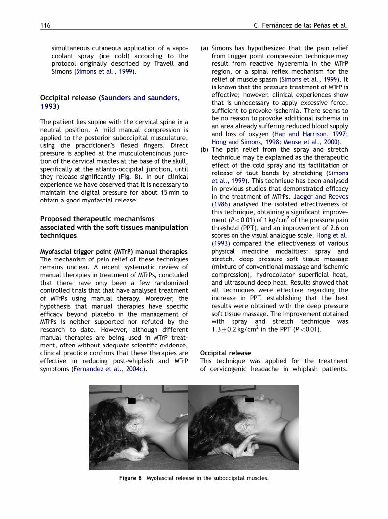
Occipital release (Saunders and saunders,1993)
The patient lies supine with the cervical spine in aneutral position. A mild manual compression isapplied to the posterior suboccipital musculature,using the practitioner’s flexed fingers. Directpressure is applied at the musculotendinous junc-tion of the cervical muscles at the base of the skull,specifically at the atlanto-occipital junction, untilthey release significantly (Fig. 8). In our clinicalexperience we have observed that it is necessary tomaintain the digital pressure for about 15min toobtain a good myofascial release.
Proposed therapeutic mechanismsassociated with the soft tissues manipulationtechniques
Myofascial trigger point (MTrP) manual therapiesThe mechanism of pain relief of these techniquesremains unclear. A recent systematic review ofmanual therapies in treatment of MTrPs, concludedthat there have only been a few randomizedcontrolled trials that that have analysed treatmentof MTrPs using manual therapy. Moreover, thehypothesis that manual therapies have specificefficacy beyond placebo in the management ofMTrPs is neither supported nor refuted by theresearch to date. However, although differentmanual therapies are being used in MTrP treat-ment, often without adequate scientific evidence,clinical practice confirms that these therapies areeffective in reducing post-whiplash and MTrPsymptoms (Fern !andez et al., 2004c).
(a) Simons has hypothesized that the pain relieffrom trigger point compression technique mayresult from reactive hyperemia in the MTrPregion, or a spinal reflex mechanism for therelief of muscle spasm (Simons et al., 1999). Itis known that the pressure treatment of MTrP iseffective; however, clinical experiences showthat is unnecessary to apply excessive force,sufficient to provoke ischemia. There seems tobe no reason to provoke additional ischemia inan area already suffering reduced blood supplyand loss of oxygen (Han and Harrison, 1997;Hong and Simons, 1998; Mense et al., 2000).
(b) The pain relief from the spray and stretchtechnique may be explained as the therapeuticeffect of the cold spray and its facilitation ofrelease of taut bands by stretching (Simonset al., 1999). This technique has been analysedin previous studies that demonstrated efficacyin the treatment of MTrPs. Jaeger and Reeves(1986) analysed the isolated effectiveness ofthis technique, obtaining a significant improve-ment (Po0:01) of 1 kg/cm2 of the pressure painthreshold (PPT), and an improvement of 2.6 onscores on the visual analogue scale. Hong et al.(1993) compared the effectiveness of variousphysical medicine modalities: spray andstretch, deep pressure soft tissue massage(mixture of conventional massage and ischemiccompression), hydrocollator superficial heat,and ultrasound deep heat. Results showed thatall techniques were effective regarding theincrease in PPT, establishing that the bestresults were obtained with the deep pressuresoft tissue massage. The improvement obtainedwith spray and stretch technique was1.370.2 kg/cm2 in the PPT (Po0:01).
Occipital releaseThis technique was applied for the treatmentof cervicogenic headache in whiplash patients.
ARTICLE IN PRESS
Figure 8 Myofascial release in the suboccipital muscles.
116 C. Fern !andez de las Pe *nas et al.

Hanten et al. (1997) reported that this techniquewas no more efficacious than placebo interventionwhen treating MTrPs. However, if the methodologyof this trial is analysed it is clear that the techniquewas not applied in the same way, or with the sameaim, as that in the current protocol. In our clinicalexperience we have observed that it is necessary tomaintain the digital pressure about 15min to obtaina good myofascial release.
The therapeutic rationale for this technique isbased on the existence of a connective tissuebridge between the RCPM muscle, and the dorsalspinal dura at the atlanto-occipital junction (Hacket al., 1995). The mild and continuous pressureover RCPM provokes a myofascial release of thesuboccipital region, potentially offering a benefi-cial effect to the spinal dura at this level. In ourclinical practice we have observed that, after theapplication of this technique, post-whiplash pa-tients commonly report relief of their head andneck symptoms.
Clinical dissertationSpinal joint dysfunction can be defined as atemporary reduction of mobility, in one or moreplanes, of a spinal segment (Triano, 2001). Thisreduction of spinal motion is caused by a hyperto-nus of the deep muscles supplied from the spinalsegment (Denslow, 1944). This hypertonus isthought to be caused by incorrect spinal-cordsetting of the gamma neuron control of intrafusalmuscle fibres. This high ‘‘gamma gain’’ may be thebasis for the reduction of vertebral motion (Korr,1975). The present definition of spinal jointdysfunction implies that muscle shortening isindeed a feature. Moreover, there are differentauthors (Lewit, 1991; Kuan et al., 1997) who havedescribed the existence of a relationship betweenjoint dysfunction and MTrPs. Lewit emphasizes theclinical importance of the treatment of MTrPs, andjoint dysfunction, when both are present (Lewit,1991). This situation implies that all manualtreatment in people suffering from whiplash injuryshould include the treatment of muscular andfascial dysfunction (principally MTrPs), as well asthe treatment of spinal joint dysfunctions.
Conclusions
The manipulative protocol developed by our re-search group has been shown to be effective in themanagement of whiplash injury (Fern !andez et al.,2004a). The biomechanical analysis of a rear-endimpact justifies some of the manipulative techni-
ques: upper cervical manipulation, dorsal manip-ulation, cervicothoracic joint manipulation, andpelvic girdle manipulation. However, lower cervicalspine manipulation seems to not be necessary inthe management of these patients (Fern !andezet al., 2003a). MTrPs in trapezius muscles, sub-occipital muscles, scalene muscles and sternoclei-domastoid muscles, commonly play an importantrole in the treatment of people suffering from post-whiplash symptoms (Fern !andez et al., 2003b).
Acknowledgements
To our deceased friend Alejandro Plaza Fern !andez,for his priceless support in whiplash injuryinvestigation.
To all teachers of the Escuela de Osteopat!ıa deMadrid (EOM) for their continued support, specifi-cally to Francois Ricard and Gin !es Almaz!an.
References
Baker, B.A., 1986. The muscles trigger: evidence of overloadinjury. Journal of Neurological and Orthopaedic Medicine andSurgery 7, 35–43.
Barnsley, L., Lord, S., Bogduk, N., 1998. The pathophysiology ofwhiplash. In: Malanga, G.A. (Ed.), Cervical Flexion–Exten-sion/Whiplash Injuries. Spine: State of the Art Reviews.Hanley and Belfus, Philadelphia, pp. 209–242.
Bogduk, N., 1986. The anatomy and pathophysiology ofwhiplash. Clinical Biomechanics 1, 92–101.
Bogduk, N., Yoganandan, N., 2001. Biomechanic of the cervicalspine Part 3: minor injuries. Clinical Biomechanics 16,267–275.
Brault, J.R., Wheeler, J.B., Siegmund, G.P., et al., 1998. Clinicalresponse of human subjects to rear-end automobile colli-sions. Archives of Physical Medicine and Rehabilitation 79,72–80.
Bronfort, G., Evan, R., Nelson, B., Aker, P.D., Goldsmith, C.H.,Vernon, H., 2001. A randomized controlled clinical trial ofrehabilitative exercise and chiropractic spinal manipulationfor chronic neck pain. Spine 26 (7), 788–799.
Cassidy, J.D., Lopez, A.A., Yong-Hink, K., 1992. The immediateeffect of manipulation versus mobilization on pain and rangeof motion in the cervical spine: a randomized controlledtrial. Journal of Manipulative and Physiological Therapeutics15 (9), 570–575.
Chaitow, L., 2001. Muscle Energy Techniques 2nd Edition.Churchill Livingstone, Edinburgh.
Chaitow, L., 2003. Modern Neuromuscular Techniques 2ndEdition. Churchill Livingstone, Edinburgh.
Denslow, J.S., 1944. An analysis of the variablity of spinal reflexthreshold. Journal of Neurophysiology 7, 207–216.
Drottning, M., Staff, P.H., Sjaastad, O., 2002. Cervicogenicheadache after whiplash injury. Cephalalgia 22 (3), 165–171.
Dvorak, J., Valach, I., Schmid, S.T., 1989. Cervical spine injuriesin Switzerland. Journal of Manual Medicine 16, 7–16.
Fern!andez de las Pe *nas, C., Fern!andez Carnero, J., MiangolarraPage, J.C., 2003a. Vertebral manipulation in whiplashinjury management. A biomechanical justification. 2003
ARTICLE IN PRESS
Manual treatment of post-whiplash injury 117

International Whiplash Trauma Congress, 9–10 October;Denver, USA.
Fern!andez de las Pe*nas, C., Fern!andez Carnero, J., AlonsoBlanco, C., Miangolarra Page, J.C., 2003b. Myofascial PainSyndrome in whiplash injury. A critical review of theliterature. 2003 International Whiplash Trauma Congress,9–10 October; Denver, USA.
Fern!andez de las Pe*nas, C., Fern!andez Carnero, J., Palomequedel Cerro, L., Miangolarra Page, J.C., 2004a. Manipulativetreatment vs conventional physiotherapy treatment inwhiplash injury. A randomized controlled trial. Journal ofWhiplash and Related Disorders 3(2), in press.
Fern!andez de las Pe *nas, C., Fern!andez Carnero, J., PlazaFern!andez, A., Lomas Vega, R., Miangolarra Page, J.C.,2004b. Dorsal manipulation in whiplash injury treatment. Arandomized controlled trial. Journal of Whiplash and RelatedDisorders 3(2), in press.
Fern!andez de las Pe *nas, C., Sohrbeck Campo, M., Fern!andezCarnero, J., Miangolarra Page, J.C., 2004c. Manual therapiesin the myofascial trigger point treatment: a systematicreview. Journal of Bodywork and Movement Therapies, inpress.
Gay, R.E., Levine, R., 2002. The biomechanics of whiplash. In:Malanga, G.A., Nadler, S.F. (Eds.), Whiplash. Hanley andBelfus, Philadelphia, pp. 27–40.
Gross, A.R., Kay, T., Hondrass, M., et al., 2002. Manual therapyfor mechanical neck disorders: a systematic review. ManualTherapy 7 (3), 131–149.
Hack, G.D., Koritzer, R.T., Robinson, W.L., Hallgren, R.C.,Greenman, P.E., 1995. Anatomic relation between the rectuscapitis posterior minor muscle and the dura mater. Spine 20(23), 2484–2486.
Hallgren, R.C., Greenman, P.E., Rechtien, J.J., 1994. Atrophy ofsuboccipital muscles in patients with chronic pain: a pilotstudy. Journal of the American Osteopathic Association 94,1032–1038.
Han, S.C., Harrison, P., 1997. Myofascial pain syndromeand trigger point management. Regional Anesthesia 22,89–101.
Hanten, W.P., Barret, M., Gillespie-Plesko, M., Jump, K.A.,Olson, S.L., 1997. Effects of active head retraction withretraction/extension and occipital release on the pressurepain threshold of cervical and scapular trigger points.Physiotherapy Theory and Practice 13 (4), 285–291.
Hohl, M., 1975. Soft tissue injuries of the neck. Clinics ofOrthopaedic 109, 42–49.
Hong, C.Z., Simons, D.G., 1993. Response to treatment forpectoralis minor. Myofascial Pain Syndrome after whiplash.Journal of Musculoskeletal Pain 1 (1), 89–131.
Hong, C.Z., Simons, D.G., 1998. Pathophysiologic and electro-physiologic mechanism of myofascial trigger points. Archivesof Physical Medicine and Rehabilitation 79, 863–872.
Hong, C.Z., Chen, Y.C., Pon, C.H., Yu, J., 1993. Immediateeffects of various physical medicine modalities on painthreshold of an active myofascial trigger point. Journal ofMusculoskeletal Pain 1 (2), 37–53.
Hou, C.R., Tsai, L.C., Cheng, K.F., et al., 2002. Imme-diate effects of various physical therapeutic modalities oncervical myofascial pain and trigger point sensitivity.Archives of Physical Medicine and Rehabilitation 83 (10),1406–1414.
Jaeger, B., Reeves, J.L., 1986. Quantification of changes inmyofascial trigger point sensitivity with the pressure alg-ometer following passive stretch. Pain 27, 203–210.
Korr, I., 1975. Proprioceptors and somatic dysfunction. Journal ofthe American Osteopathic Association 74, 638–650.
Koelbaek, M.T., Graven-Nielsen, T., Schour, A., Arendt-Nielsen,L., 1999. Generalised muscular hyperalgesia in chronicwhiplash syndrome. Pain 83 (2), 229–234.
Kuan, T.S., Wu, C.T., Chen, S., Chen, J.T., Hong, C.Z., 1997.Manipulation of the cervical spine to release pain andtightness caused by myofascial trigger points. Archives ofPhysical Medicine and Rehabilitation 78 (9), 1042.
Lewit, K., 1991. Manipulative Therapy in Rehabilitation of theLocomotor System 2nd Edition.. Buterworth Heinemann,Oxford.
Lord, S.M., Barnsley, L., Wallis, B.J., McDonald, G.J., Bogduk,N., 1996. Percutaneous radio-frequency neurotomy forchronic cervical zygapophyseal joint pain. New EnglandJournal of Medicine 335, 1721–1726.
Matsushita, T., Sato, T.B., Hirabayashi, K., Fujimara, S.,Asazuma, T., 1994. X-ray study of the human neck motiondue to head inertia loading. Proceedings of the 38th StappCar Crash Conferenc, Fort Lauderdale, FL, pp. 55–64.
Mense, S., Simons, D.G., Jon Russell, I., 2000. Muscle Pain:Understanding its Nature, Diagnosis and Treatment. Lippin-contt Williams & Wilkins, Philadelphia.
Mitchell, F., 1995. The Muscle Energy Manual. Vol. I. Conceptsand Mechanism. The musculoskeletal screen. Cervical Region
Evaluation And Treatment. MET Press, Michigan.
Mitchell, J.A., 2003. Changes in vertebral artery bloodflow following normal rotation of the cervical spine. Journalof Manipulative and Physiological Therapeutics 26 (6),347–351.
Mitchell, B.S., Humphreys, D.C., O’Sullivan, E., 1998. Attach-ments of the ligamentum nuchae to cervical posteriorspinal dura and the lateral part of the occipital bone. Journalof Manipulative and Physiological Therapeutics 21 (3),145–148.
Osterbauer, P.J., Derickson, K.L., Peles, J.D., Deboer, K.F., Fuhr,A.W., Winters, J.M., 1992. Three dimensional head kine-matics and clinical outcome of patients with neck injurytreated with spinal manipulative therapy: a pilot study.Journal of manipulative and Physiological Therapeutics 15(8), 501–511.
Panjabi, M.M., Cholewicki, J., Nibu, K., Grauer, J.N., Babat,K.B., Dvorak, J., 1998a. Mechanism of whiplash injury.Clinical Biomechanics 13, 239–249.
Panjabi, M.M., Nibu, K., Cholewicki, J., 1998b. Whiplash injuriesand the potential for mechanical instability. European SpineJournal 7, 484–492.
Peeters, G.M., Verhagen, A.P., de Bie, R.A., Oostendorp, A.B.,2001. The efficacy of conservative treatment in patients withwhiplash injury. Spine 26, 64–73.
Penning, I., 1992a. Acceleration injury of the cervical spine byhypertranslation of the head. Part I: effects of normaltranslation of the head on the cervical spine motion: aradiological study. European Spine Journal 1, 7–12.
Penning, I., 1992b. Acceleration injury of the cervical spineby hypertranslation of the head. Part II: effects of hyper-translation of the head on the cervical spine motion:discussion of literature data. European Spine Journal 1,13–19.
Saunders, H.D., Saunders, R., 1993. Evaluation, Treatmentand Prevention of Musculoskeletal Disorders. Vol. 1:Spine. Educational Opportunities, Chaska, New York,pp. 246–247.
Schuller, E., Einsenmenger, W., Beier, G., 2000. Whiplash injuryin low speed car accidents. Journal of Musculoskeletal Pain 8,55–67.
ARTICLE IN PRESS
118 C. Fern !andez de las Pe *nas et al.

Shrawan, K., Yogesh, N., Tyler, A., 2002. An electromyographicstudy of low-velocity rear-end impacts. Spine 27 (10),1044–1055.
Simons, D.G., Travell, J., Simons, L.S., 1999. Myofascial Pain andDisfunction. The Trigger Point Manual, Vol. 1, 2nd Edition.Williams & Wilkins, Baltimore.
Spitzer, W.O., Skovron, M.L., Salmi, L.R., et al., 1995. Quebec taskforce on whiplash-associated disorders. Spine 20 (85), 1S–73S.
Taylor, J.R., Taylor, M.M., Twomey, L.T., 1996. Posterior cervicaldura is much thicker than anterior cervical dura (comment).Spine 21, 2300–2301.
Teasell, R.W., Shapiro, A.P., 2001. Whiplash injury and the uppercervical spine. In: Vernon, H. (Ed.), The Cranio-CervicalSyndrome. Mechanism, Assessment and Treatment. Butter-worth-Heinemann, London, pp. 197–206.
Triano, J.J., 2001. Biomechanics of spinal manipulative therapy.The Spine Journal 1, 121–130.
Valera, F., Garoz, S., Lav!ın, M., Pe *na, C., 2003. Algorithm oftreatment for the physiotherapy options of patients withwhiplash. XIV World Congress of Physical Therapy (WCPT),Barcelona, Spain, 7–12 June.
Vernon, H., 1995. Cervicogenic headache. In: Gatterman, M.(Ed.), Foundations of Chiropractic Subluxation. Mosby YearBook, St Luois, pp. 306–316.
Vernon, H.T., Aker, P., Burns, S., Viljakaanen, S., Short, L., 1990.Pressure pain threshold evaluation of the effect of spinalmanipulation in the treatment of chronic neck pain: a pilotstudy. Journal of Manipulative and Physiological Therapeutics13 (1), 13–16.
von Piekartz, H.V., Bryden, L., 2001. Craniofacial Dysfunctionand Pain. Butterworth-Heinemann, London.
Whittingham, W., Nillson, N., 2001. Active range of motion incervical spine increases after spinal manipulation (togglerecoil). Journal of Manipulative and Physiological Therapeu-tics 24, 552–555.
Yoganandan, N., Pintar, F.A., Larson, S.J., Sances, A. (Eds.),1998. Frontiers in Head and Neck Trauma: Clinical andBiomechanical. IOS Press, Amsterdam.
Yoganandan, N., Pintar, F.A., Cusick, J.F., 2002. Biomecha-nical analises of whiplash injuries using an experi-mental model. Accident Analysis and Prevention 34,663–671.
ARTICLE IN PRESS
Manual treatment of post-whiplash injury 119