Magnesium Decreases Inflammatory Cytokine Production… · Magnesium Decreases Inflammatory...
10
of June 21, 2018. This information is current as Immunomodulatory Mechanism Cytokine Production: A Novel Innate Magnesium Decreases Inflammatory Bernstein Kitchen, Nicholas Funderburg, Sam Mesiano and Helene B. Valentin-Torres, Angel A. Luciano, Christina M. Ramirez Jun Sugimoto, Andrea M. Romani, Alice M. ol.1101765 http://www.jimmunol.org/content/early/2012/05/18/jimmun published online 18 May 2012 J Immunol Material Supplementary 5.DC1 http://www.jimmunol.org/content/suppl/2012/05/18/jimmunol.110176 average * 4 weeks from acceptance to publication Fast Publication! • Every submission reviewed by practicing scientists No Triage! • from submission to initial decision Rapid Reviews! 30 days* • Submit online. ? The JI Why Subscription http://jimmunol.org/subscription is online at: The Journal of Immunology Information about subscribing to Permissions http://www.aai.org/About/Publications/JI/copyright.html Submit copyright permission requests at: Email Alerts http://jimmunol.org/alerts Receive free email-alerts when new articles cite this article. Sign up at: Print ISSN: 0022-1767 Online ISSN: 1550-6606. Immunologists, Inc. All rights reserved. Copyright © 2012 by The American Association of 1451 Rockville Pike, Suite 650, Rockville, MD 20852 The American Association of Immunologists, Inc., is published twice each month by The Journal of Immunology by guest on June 21, 2018 http://www.jimmunol.org/ Downloaded from by guest on June 21, 2018 http://www.jimmunol.org/ Downloaded from
Transcript of Magnesium Decreases Inflammatory Cytokine Production… · Magnesium Decreases Inflammatory...

of June 21, 2018.This information is current as
Immunomodulatory MechanismCytokine Production: A Novel Innate Magnesium Decreases Inflammatory
BernsteinKitchen, Nicholas Funderburg, Sam Mesiano and Helene B.Valentin-Torres, Angel A. Luciano, Christina M. Ramirez Jun Sugimoto, Andrea M. Romani, Alice M.
ol.1101765http://www.jimmunol.org/content/early/2012/05/18/jimmun
published online 18 May 2012J Immunol
MaterialSupplementary
5.DC1http://www.jimmunol.org/content/suppl/2012/05/18/jimmunol.110176
average*
4 weeks from acceptance to publicationFast Publication! •
Every submission reviewed by practicing scientistsNo Triage! •
from submission to initial decisionRapid Reviews! 30 days* •
Submit online. ?The JIWhy
Subscriptionhttp://jimmunol.org/subscription
is online at: The Journal of ImmunologyInformation about subscribing to
Permissionshttp://www.aai.org/About/Publications/JI/copyright.htmlSubmit copyright permission requests at:
Email Alertshttp://jimmunol.org/alertsReceive free email-alerts when new articles cite this article. Sign up at:
Print ISSN: 0022-1767 Online ISSN: 1550-6606. Immunologists, Inc. All rights reserved.Copyright © 2012 by The American Association of1451 Rockville Pike, Suite 650, Rockville, MD 20852The American Association of Immunologists, Inc.,
is published twice each month byThe Journal of Immunology
by guest on June 21, 2018http://w
ww
.jimm
unol.org/D
ownloaded from
by guest on June 21, 2018
http://ww
w.jim
munol.org/
Dow
nloaded from

The Journal of Immunology
Magnesium Decreases Inflammatory Cytokine Production: ANovel Innate Immunomodulatory Mechanism
Jun Sugimoto,* Andrea M. Romani,† Alice M. Valentin-Torres,‡ Angel A. Luciano,x
Christina M. Ramirez Kitchen,{ Nicholas Funderburg,‡ Sam Mesiano,* and
Helene B. Bernstein*,‡
MgSO4 exposure before preterm birth is neuroprotective, reducing the risk of cerebral palsy and major motor dysfunction.
Neonatal inflammatory cytokine levels correlate with neurologic outcome, leading us to assess the effect of MgSO4 on cytokine
production in humans. We found reduced maternal TNF-a and IL-6 production following in vivo MgSO4 treatment. Short-term
exposure to a clinically effective MgSO4 concentration in vitro substantially reduced the frequency of neonatal monocytes
producing TNF-a and IL-6 under constitutive and TLR-stimulated conditions, decreasing cytokine gene and protein expression,
without influencing cell viability or phagocytic function. In summary, MgSO4 reduced cytokine production in intrapartum
women, term and preterm neonates, demonstrating effectiveness in those at risk for inflammation-associated adverse perinatal
outcomes. By probing the mechanism of decreased cytokine production, we found that the immunomodulatory effect was
mediated by magnesium and not the sulfate moiety, and it was reversible. Cellular magnesium content increased rapidly upon
MgSO4 exposure, and reduced cytokine production occurred following stimulation with different TLR ligands as well as when
magnesium was added after TLR stimulation, strongly suggesting that magnesium acts intracellularly. Magnesium increased
basal IĸBa levels, and upon TLR stimulation was associated with reduced NF-kB activation and nuclear localization. These
findings establish a new paradigm for innate immunoregulation, whereby magnesium plays a critical regulatory role in NF-kB
activation, cytokine production, and disease pathogenesis. The Journal of Immunology, 2012, 188: 000–000.
Magnesium sulfate is widely used in obstetrics for seizureprophylaxis in preeclampsia and as a tocolytic to arrestpreterm labor. Despite widespread use, the mechanism
by which MgSO4 exerts its action is poorly understood. Retro-spective, clinical studies associated antepartum MgSO4 exposurewith reduced risk of adverse neurologic outcome in prematurenewborns (1, 2), leading to randomized clinical trials (3–7); ad-ditionally, a recent review concluded that antenatal MgSO4 ther-apy significantly reduces the risk of cerebral palsy and substantialgross motor dysfunction (8). These findings raise a critical ques-tion, “How does MgSO4 mediate neuroprotection?”Cerebral palsy is the most common cause of pediatric motor
dysfunction (9). Multiple prospective studies strongly associatecerebral palsy with antepartum and intrapartum inflammation,whereas isolated birth asphyxia accounts for ,10% of the cases(9, 10). These conclusions are supported by animal studies dem-
onstrating that proinflammatory cytokines are neurotoxic, causingCNS damage (11), as well as by epidemiologic research corre-lating increased neonatal serum levels of inflammatory cytokineswith adverse neurologic outcome (12–18). Preterm parturition isassociated with a fetal inflammatory response syndrome definedby increased cord blood IL-6 levels (19), as well as increasedlevels of IL-1, IL-8, RANTES, TNF-a, and other inflammatorycytokines (20). Magnesium sulfate is used both as a tocolytic toarrest preterm labor and for seizure prophylaxis in women withpreeclampsia, a condition sharing features with atherosclerosis,including endothelial dysfunction and systemic inflammation (21,22). Inflammation is also linked to seizure activity. A very recentstudy linked TLR4 signaling to seizure activity; remarkably, sei-zure activity was ameliorated with TLR4 antagonists, supportinga mechanism of inflammation-induced seizure ictogenesis (23).Inflammation plays a central role in the three conditions for
which MgSO4 is used as therapy: 1) to treat preterm labor, 2) toprevent preeclamptic seizures, and 3) to reduce the developmentof cerebral palsy. This led us to hypothesize that MgSO4 exerts itsneuroprotective effect by downregulating inflammatory cytokineproduction in neonates. Following in vivo MgSO4 treatment, weobserved a reduced frequency of monocytes producing TNF-a andIL-6 in women receiving MgSO4 for clinical indications. Expos-ing peripheral and/or cord blood mononuclear cells in vitro toMgSO4 yielded similar results. MgSO4 exposure was accompa-nied by decreased cytokine and IkBa gene expression and di-minished NF-kB activation; moreover, reduced cytokine pro-duction was observed following exposure to different TLRligands, suggesting that magnesium has broad anti-inflammatoryactivity. Taken together, our data establish a new paradigm forinnate immunoregulation whereby magnesium plays a criticalregulatory role in NF-kB activation, cytokine production, anddisease pathogenesis.
*Department of Reproductive Biology, Case Western Reserve University Schoolof Medicine, Cleveland, OH 44106; †Department of Physiology and Biophysics,Case Western Reserve University School of Medicine, Cleveland, OH 44106;‡Department of Molecular Biology and Microbiology, Case Western Reserve Uni-versity School of Medicine, Cleveland, OH 44106; xDivision of Neonatology, De-partment of Pediatrics, College of Medicine, University of South Florida, Tampa,FL 33606; and {Department of Biostatistics, School of Public Health, Universityof California at Los Angeles, Los Angeles, CA 90095
Received for publication June 15, 2011. Accepted for publication April 13, 2012.
H.B.B. received support from American Cancer Society Grant RSG-07-070-01-LIB.
Address correspondence and reprint requests to Dr. Helene B. Bernstein, Case West-ern Reserve University, Wood Building, W210D, 10900 Euclid Avenue, Cleveland,OH 44106-4960. E-mail address: [email protected]
The online version of this article contains supplemental material.
Abbreviations used in this article: CBMC, cord blood mononuclear cell; HAB,human serum from AB donors; ICS, intracellular cytokine staining; MALP, macro-phage-activating lipopeptide; poly(I:C), polyinosinic-polycytidylic acid.
Copyright� 2012 by The American Association of Immunologists, Inc. 0022-1767/12/$16.00
www.jimmunol.org/cgi/doi/10.4049/jimmunol.1101765
Published May 18, 2012, doi:10.4049/jimmunol.1101765 by guest on June 21, 2018
http://ww
w.jim
munol.org/
Dow
nloaded from

Materials and MethodsAbs and reagents
LPS (from Escherichia coli 0111:B4) and brefeldin A were from Sigma-Aldrich (St. Louis, MO). Macrophage-activating lipopeptide (MALP)-2was purchased from Imgenex. Polyinosinic-polycytidylic acid (poly(I:C))was provided by Dr. Aaron Weinberg of Case Western Reserve University.Fluorochrome-labeled Abs and reagents used were: FITC-annexin V, PE-anti-CD14, PerCP-anti-CD3, and allophycocyanin-anti–TNF-a from BDBiosciences, FITC-anti-CD14, allophycocyanin-anti-CD4, PE-anti-CD56,and ECD-anti-CD19 were from Beckman Coulter, and PE-anti–IL-6 wasfrom R&D Systems. Mouse anti-IkBa (L35A5) was obtained from CellSignaling Technology, mouse anti-tubulin (DM1A) was obtained fromSigma-Aldrich, and rabbit anti-TFIID (TBP) (N-12) and NF-kB p65(C-20) were obtained from Santa Cruz Biotechnology.
Cell isolation and culture
Anti-coagulated umbilical cord blood and peripheral blood were collectedunder protocols approved by the University Hospitals Institutional ReviewBoard; all donors provided written informed consent. Mononuclear cellswere isolated by density gradient centrifugation on lymphocyte separationmedium (density, 1.077–1.080 g/ml) (Mediatech). Monocytes were purifiedby positive selection using anti-CD14 magnetic beads (Miltenyi Biotec),and cultures were maintained in RPMI 1640 (HyClone; magnesium con-centration is 1 mg/dl or 0.4 mM) supplemented with 10% heat-inactivatedhuman serum from male AB donors (HAB) (Sigma-Aldrich), 2 mML-glutamine, 100 IU/ml penicillin, and 100 mg/ml streptomycin. Cells weresupplemented with MgSO4 to a final concentration of 60 mg/l or 2.5 mM,a concentration known to be clinically effective. THP-1 cells were ob-tained from the Skowronski Laboratory and maintained in media describedabove, supplemented with 10% FCS. Cyclohexamide (Sigma-Aldrich) wasused at 100 mg/ml to inhibit protein synthesis, whereas 10 mM 6-amino-4-(4-phenoxyphenylethylamino)quinazoline and 80 mM 4-methyl-N1-(3-phenylpropyl)benzene-1,2-diamine (JSH-23) (Calbiochem) were used toinhibit NF-kB activation.
TLR ligand stimulation
Mononuclear cells (1 3 106 cells/ml) were cultured in six-well plates andin some cases stimulated with 50 pg/ml LPS, 1–10 ng/ml MALP-2, or 0.1–1.0 mg/ml poly(I:C) for 6 h. For the dose-response determination, 0–1 mg/ml LPS was used.
Intracellular cytokine staining
Two hours following the addition of TLR ligands, brefeldin Awas added toinhibit cytokine secretion (1 mg/ml; Sigma-Aldrich). Cells were harvestedand blocked with excess HAB (5% HAB in PBS). Cells were stained withFITC-conjugated anti-CD14 Ab. After fixation with 2% paraformaldehyde,cells were permeabilized using 13 Perm/Wash buffer (BD Biosciences),blocked with 5% HAB, followed by staining with intracellular Abs (ICS),allophycocyanin-conjugated anti–TNF-a, and PE-conjugated anti–IL-6,followed by flow cytometric analysis (BD FACSCalibur, FlowJo). Mater-nal whole blood (1 ml) was stimulated by the direct addition of TLRligands and brefeldin A as described above, followed by surface staining,fixation, and RBC lysis using BD FACS Lyse solution and ICS as de-scribed above.
Quantitative PCR
Total RNA was extracted using RNeasy (Qiagen) with QIAshreddersaccording to the manufacturer’s protocol. Single-stranded cDNA wassynthesized using TaqMan reverse transcription reagents (Applied Bio-systems) in a thermal cycler. Quantitative PCR was carried out in an ABIPrism sequence detection system. Cycling conditions were: 95˚C for 10min, followed by 40 cycles of 95˚C for 15 s and 60˚C for 1 min usingPower SYBR Green PCR Master Mix (Applied Biosystems) with relativequantification methods. All reactions yielded a single amplification prod-uct. Primers used for quantitative PCR included: TNF-a, sense, 59-AGT-GACAAGCCTGTAGCCCATGTT-39, anti-sense, 59-GTTATCTCTCAGCTCCACGCCATT-39; IL-6, sense, 59-ACCTGAACCTTCCAAAGATGG-CTG-39, anti-sense, 59-ACTCATCTGCACAGCTCTGG CTT-39; IkBa,sense, 59-AAGTGATCCGCCAGGTGAAG-39, anti-sense, 59-TGCTGC-AGGTTGTTCTGGAA-39; Gus, sense, 59-AGCAGTACCATCTGGGT-CTG-39, anti-sense, 59-TTGGTTGTCTCTGCCGAGTG-39. Values werenormalized to human Gus (b-glucuronidase), expression of this gene wasfound to be stable during stimulation, and the value of unstimulated cells attime 0 was set to 1 and used to calculate the fold change in stimulatedcells. Results are mean values of triplicates.
Phagocytosis assays
To determine the effect of magnesium on monocyte phagocytic function,cord blood mononuclear cells (CBMCs) in the presence or absence ofmagnesium supplementation were exposed to either 0.1 or 0.5 mM FITC-conjugated latex beads or Alexa Fluor 488-conjugated albumin (5 ml/ml)for 2–4 h (a gift from the Canaday Laboratory). Cell identification andsubstrate uptake on a per cell basis was quantitated via flow cytometry toassess fluid phase-type endocytosis and macropinocytosis.
Western blot analysis
Cells were stimulated with LPS for 30 min and lysed in 13 SDS loadingbuffer (62.5 mM Tris-HCl, 2% [w/v] SDS, 10% glycerol, 50 mM DTT,0.01% [w/v] bromophenol blue). Lysates were heated to 95˚C for 5 min,and samples were resolved by SDS-PAGE on 12% Tris-HCl Ready Gels(Bio-Rad, Hercules, CA) and transferred to nitrocellulose membranes(Amersham Biosciences, Piscataway, NJ). Phosphorylated proteins weredetected by using primary monoclonal Abs to p–NF-kB p65 (Ser536) (CellSignaling Technology, Beverly, MA). mAbs to actin (Santa Cruz Bio-technology, Santa Cruz, CA) were used to confirm comparable proteinloading between specimens. Secondary anti-rabbit HRP-conjugated Abswere used to detect primary Abs (Cell Signaling Technology). Followingincubation with HRP-conjugated secondary Abs, proteins were detectedby chemiluminescence (Western Lightning; PerkinElmer Life Sciences,Boston, MA) and were visualized by x-ray film exposure (Denville Sci-entific, Metuchen, NJ).
Nuclear and cytoplasmic proteins were obtained by washing cells twicein ice-cold PBS. Cells were resuspended in cytosolic extract lysis buffer (10mM HEPES-KOH [pH 7.9] , 10 mM KCl, 1 mM EDTA, 1.5mM MgCl2, 1mM DTT, and 1 mM PMSF) containing protease inhibitor mixture (Roche)and incubated on ice for 10 min as described (24). Nuclei were pelleted bycentrifugation for 10 min at 4000 rpm, cytoplasmic extracts were col-lected, and nuclei were washed three times in cytosolic extract lysis buffer.RIPA buffer (20 mM Tris [pH 7.5], 150 mM NaCl, 0.5% Triton X-100,0.5% sodium deoxycholate, 0.1% SDS, 5 mM EDTA) with proteaseinhibitors was added to nuclei, which were sonicated prior to collection ofnuclear protein containing supernatants. Protein concentrations were de-termined using the BCA protein assay kit (Pierce) or the Bradford reagent(Bio-Rad). The NuPAGE system (Invitrogen) was used to resolve andtransfer proteins on a 4–12% Bis-Tris gel. Protein bands on polyvinylidenedifluoride membranes were detected and quantified by Western blottingwith the Odyssey system imager (Li-Cor Biosciences) using IRDye800CW goat anti-mouse IgG and IRDye 800CW goat anti-rabbit IgG.
Magnesium determination
Total cellular Mg2+ content was assessed by atomic absorbance spectro-photometry in a PerkinElmer 3100 (PerkinElmer, Waltham, MA), as re-ported previously (25). Briefly, aliquots of cells (1 3 105) exposed or notto 2.5 mM extracellular Mg2+ were rapidly sedimented through a 0.5-mloil layer (dibutyl phthalate/dioctyl phthalate 2:1 [v/v]) in microfuge tubesat 14,000 rpm for 5 min. The supernatant and oil layer were removed, andthe cell pellets were digested overnight in 0.5 ml 10% HNO3. Followingsedimentation of denatured protein at 14,000 rpm for 5 min in Microfugetubes, the Mg2+ content of the acid extracts was measured by atomic ab-sorbance spectrophotometry calibrated with appropriate standards.
Statistical analysis
Data are expressed and plotted as means 6 standard deviations. A Wil-coxon signed-rank test was used to compare differences between relatedsamples; for poly(I:C) stimulation and maternal blood stimulation, meanswere compared using a Student t test. Statistical significance was definedas p , 0.05 as indicated.
ResultsIn vivo MgSO4 therapy reduces monocyte-mediated cytokineproduction
To determine the in vivo effect of MgSO4 exposure, we assessedcytokine production within heparinized blood samples obtainedfrom women immediately prior to initiating MgSO4 therapy and 6–12 h after beginning MgSO4 therapy for clinical indications. Cy-tokine production was assessed in untouched and LPS-challengedwhole blood from the same donor via ICS. We found that in vivoMgSO4 treatment significantly decreased the frequency of maternalcells producing TNF-a and IL-6 (Fig. 1). The frequency of TNF-
2 MAGNESIUM DECREASES INFLAMMATORY CYTOKINE PRODUCTION
by guest on June 21, 2018http://w
ww
.jimm
unol.org/D
ownloaded from

a–producing cells was reduced by 25% (p = 0.03, n = 7), andrelative TNF-a expression as determined by the median fluores-cence intensity was reduced by 27%. Induction of IL-6 productionwas reduced by ∼20% (p , 0.05, n = 7) as assessed by botha reduced cell frequency and median fluorescence intensity, andmonocytes comprised most of the cytokine-producing cells.Circulating monocytes compromise 10% of mononuclear cells,
playing a key role in systemic inflammation and cytokine pro-duction and differentiating into macrophages when recruited intotissue. We next assessed whether in vitro exposure to MgSO4
influences cytokine production. A dose-response curve measuringthe effect of LPS on TNF-a production was established usingPBMCs. LPS concentrations of 1 mg/ml are typically used forin vitro stimulation assays (26). However, in our assay conditionsan LPS concentration of 50–100 pg/ml (5 3 1025–1024 mg/ml)generated ∼50% of the maximal response, permitting assessmentof the influence of MgSO4 on cytokine production. This LPSconcentration is above the mean plasma LPS concentration (25 pg/ml) within normal, adult nonbacteremic individuals (27). Overall,these results demonstrate that in vivo MgSO4 treatment decreasesthe frequency of cells producing inflammatory cytokines and es-tablishes in vitro conditions for further analysis.
MgSO4 reduces monocyte-mediated IL-6 and TNF-aproduction in neonates
Because neonatal serum cytokine levels are associated with the de-velopment of cerebral palsy, we assessed the influence of MgSO4
exposure on cytokine production within CBMCs. Magnesiumrapidly crosses the placenta, resulting in equivalent maternal andfetal levels, so CBMCs were cultured under standard, physiologic
conditions or exposed to 6 mg/dl MgSO4, a clinically effectivematernal magnesium concentration. Magnesium supplementation
significantly decreased the frequency of LPS-stimulated cord
blood monocytes producing IL-6 and TNF-a (Fig. 2A); the results
of multiple patients are shown in Fig. 2B (p , 0.01, Wilcoxon
signed-rank test). Magnesium supplementation also significantly
decreased IL-6 and TNF-a expression by .60% (p , 0.05), as
measured by median fluorescence intensity; these results were
confirmed by measuring secreted cytokines via ELISA (data not
shown). Cytokine production is much lower in unstimulated cells,
but MgSO4 also significantly reduced the frequency of neonatal
monocytes producing TNF-a and IL-6 under constitutive or
unstimulated conditions (Fig. 2C; p , 0.01 and p , 0.05, re-
spectively; Wilcoxon signed-rank test).To rule out the possibility that decreased cytokine production in
monocytes was secondary to altered cell viability or function, we
quantitated cell count and composition via flow cytometry after
overnight culture under standard conditions or in the presence of
magnesium supplementation. When examining T cell (CD4+ and
CD8+), B cell, NK cell, and monocyte populations, no differences
in cell counts or apoptosis (as assessed via annexin V staining)
were found within individual donors in the presence and absence
of MgSO4 supplementation (data not shown). To evaluate an ad-
ditional aspect of monocyte function, we assessed phagocytosis
using FITC-labeled 0.1 and 0.5 mM latex beads and Alexa Fluor
488-conjugated albumin. No differences in fluid phase-type en-
docytosis or macropinocytosis were observed in the presence
of magnesium supplementation (Supplemental Fig. 1), suggesting
that monocyte function is intact.
FIGURE 1. MgSO4 treatment in vivo and
in vitro significantly decreases baseline and
LPS-stimulated cytokine production. Hepa-
rinized maternal blood samples were ob-
tained immediately prior (Pre Mg) and 6–
12 h after the initiation of clinically indi-
cated parenteral MgSO4 treatment (Mg Tx).
Unmanipulated whole blood was cultured in
the presence or absence of LPS stimulation
for 6 h with brefeldin A followed by ICS.
The percentage of monocytes producing
TNF-a (A) and IL-6 (B) are shown in the
upper right quadrant of each dot plot; rela-
tive decreases in cytokine production for
this patient are shown at the bottom of each
section. Cumulatively, the frequency of
monocytes producing TNF-a was reduced
by 25% (p = 0.03, n = 7), and the frequency
of IL-6–producing monocytes was reduced
by 20% (p , 0.05, n = 7) following in vivo
magnesium treatment. Similar reductions
in cytokine expression were observed, as de-
termined by the median fluorescence inten-
sity within monocytes (p , 0.05).
The Journal of Immunology 3
by guest on June 21, 2018http://w
ww
.jimm
unol.org/D
ownloaded from
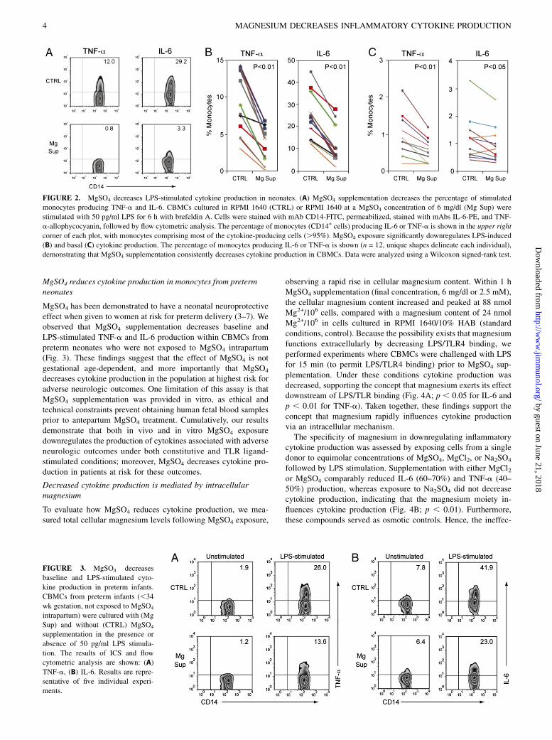
MgSO4 reduces cytokine production in monocytes from pretermneonates
MgSO4 has been demonstrated to have a neonatal neuroprotectiveeffect when given to women at risk for preterm delivery (3–7). Weobserved that MgSO4 supplementation decreases baseline andLPS-stimulated TNF-a and IL-6 production within CBMCs frompreterm neonates who were not exposed to MgSO4 intrapartum(Fig. 3). These findings suggest that the effect of MgSO4 is notgestational age-dependent, and more importantly that MgSO4
decreases cytokine production in the population at highest risk foradverse neurologic outcomes. One limitation of this assay is thatMgSO4 supplementation was provided in vitro, as ethical andtechnical constraints prevent obtaining human fetal blood samplesprior to antepartum MgSO4 treatment. Cumulatively, our resultsdemonstrate that both in vivo and in vitro MgSO4 exposuredownregulates the production of cytokines associated with adverseneurologic outcomes under both constitutive and TLR ligand-stimulated conditions; moreover, MgSO4 decreases cytokine pro-duction in patients at risk for these outcomes.
Decreased cytokine production is mediated by intracellularmagnesium
To evaluate how MgSO4 reduces cytokine production, we mea-sured total cellular magnesium levels following MgSO4 exposure,
observing a rapid rise in cellular magnesium content. Within 1 hMgSO4 supplementation (final concentration, 6 mg/dl or 2.5 mM),the cellular magnesium content increased and peaked at 88 nmolMg2+/106 cells, compared with a magnesium content of 24 nmolMg2+/106 in cells cultured in RPMI 1640/10% HAB (standardconditions, control). Because the possibility exists that magnesiumfunctions extracellularly by decreasing LPS/TLR4 binding, weperformed experiments where CBMCs were challenged with LPSfor 15 min (to permit LPS/TLR4 binding) prior to MgSO4 sup-plementation. Under these conditions cytokine production wasdecreased, supporting the concept that magnesium exerts its effectdownstream of LPS/TLR binding (Fig. 4A; p , 0.05 for IL-6 andp , 0.01 for TNF-a). Taken together, these findings support theconcept that magnesium rapidly influences cytokine productionvia an intracellular mechanism.The specificity of magnesium in downregulating inflammatory
cytokine production was assessed by exposing cells from a singledonor to equimolar concentrations of MgSO4, MgCl2, or Na2SO4
followed by LPS stimulation. Supplementation with either MgCl2or MgSO4 comparably reduced IL-6 (60–70%) and TNF-a (40–50%) production, whereas exposure to Na2SO4 did not decreasecytokine production, indicating that the magnesium moiety in-fluences cytokine production (Fig. 4B; p , 0.01). Furthermore,these compounds served as osmotic controls. Hence, the ineffec-
FIGURE 2. MgSO4 decreases LPS-stimulated cytokine production in neonates. (A) MgSO4 supplementation decreases the percentage of stimulated
monocytes producing TNF-a and IL-6. CBMCs cultured in RPMI 1640 (CTRL) or RPMI 1640 at a MgSO4 concentration of 6 mg/dl (Mg Sup) were
stimulated with 50 pg/ml LPS for 6 h with brefeldin A. Cells were stained with mAb CD14-FITC, permeabilized, stained with mAbs IL-6-PE, and TNF-
a-allophycocyanin, followed by flow cytometric analysis. The percentage of monocytes (CD14+ cells) producing IL-6 or TNF-a is shown in the upper right
corner of each plot, with monocytes comprising most of the cytokine-producing cells (.95%). MgSO4 exposure significantly downregulates LPS-induced
(B) and basal (C) cytokine production. The percentage of monocytes producing IL-6 or TNF-a is shown (n = 12, unique shapes delineate each individual),
demonstrating that MgSO4 supplementation consistently decreases cytokine production in CBMCs. Data were analyzed using a Wilcoxon signed-rank test.
FIGURE 3. MgSO4 decreases
baseline and LPS-stimulated cyto-
kine production in preterm infants.
CBMCs from preterm infants (,34
wk gestation, not exposed to MgSO4
intrapartum) were cultured with (Mg
Sup) and without (CTRL) MgSO4
supplementation in the presence or
absence of 50 pg/ml LPS stimula-
tion. The results of ICS and flow
cytometric analysis are shown: (A)
TNF-a, (B) IL-6. Results are repre-
sentative of five individual experi-
ments.
4 MAGNESIUM DECREASES INFLAMMATORY CYTOKINE PRODUCTION
by guest on June 21, 2018http://w
ww
.jimm
unol.org/D
ownloaded from

tiveness of Na2SO4 at reducing cytokine production rules out anosmotic effect as a possible cause of altered cytokine production.Cumulatively, these data indicate that the immunomodulatoryeffect is mediated by magnesium and not the sulfate moiety of thecompound and that magnesium functions intracellularly.
Magnesium reversibly regulates cytokine production viatranscriptional regulation
To assess whether the effects of magnesium are reversible, cellswere exposed (or not) to MgSO4 for 2 h; cells were then washedand immediately challenged with LPS in the presence of controlor magnesium-supplemented media. The effect of magnesium wasreversible, because exposure prior to LPS challenge had minimalinfluence on the ability of cells to produce IL-6 and TNF-a (Fig.5). These results are consistent with the swift rise and peak in
cellular magnesium concentrations observed following MgSO4
supplementation. By pursuing the mechanism of diminished cy-tokine production, cytokine gene expression within TLR-ligandstimulated cells exposed to MgSO4 was assessed using real-timePCR (Fig. 6). At 2 and 4 h after LPS exposure there was a sta-tistically significant decrease (p , 0.05) in TNF-a and IL-6mRNA levels within cells receiving MgSO4 supplementation.These results indicate that MgSO4 downregulates TNF-a andIL-6 production prior to transcription.
Magnesium decreases cytokine production by reducing NF-kBactivation
The impact of MgSO4 on NF-kB activation was evaluated usingmultiple independent methods. First, we assessed IkBa gene ex-pression, as NF-kB activation leads to increased transcription of
FIGURE 4. MgSO4 decreases cytokine production when added following LPS exposure, and decreased cytokine production is mediated by magnesium.
(A) CBMCs were stimulated with LPS for 15 min prior to MgSO4 exposure (15 min Post LPS) to permit LPS/TLR binding. Inhibition of cytokine
production was observed (at levels similar to those seen when MgSO4 and LPS are added simultaneously; n = 4) and compared with cells from the same
donor not supplemented with magnesium (f). (B) CBMCs were stimulated with LPS alone, in the presence of MgSO4 supplementation (2.5 mM), or with
equimolar concentrations of related salts (MgCl2 or Na2SO4). The percentage change in cytokine production for each salt was calculated based on ICS
(n = 5, *p , 0.01); SEM is shown. Both MgCl2 and MgSO4, decrease TNF-a and IL-6 production compared with untreated cells, whereas Na2SO4 failed
to decrease cytokine production.
FIGURE 5. MgSO4 causes a reversible de-
crease in cytokine production. CBMCs were
exposed or not (CRTL) to magnesium supple-
mentation (Mg Sup) for 2 h. CBMCs were then
washed and immediately challenged with LPS
in the presence of control or magnesium sup-
plemented media. (A) Histogram overlays
show IL-6 and TNF-a production under each
of the four conditions; the condition in which
Mg2+ was supplemented prior to and during
LPS-stimulation is shaded gray. (B) Bar graph
shows the percentage of neonatal monocytes
producing either IL-6 (black) or TNF-a (gray)
in the above histogram under each condition,
illustrating that the effect of magnesium sup-
plementation is reversible. Results are repre-
sentative of three individual experiments.
The Journal of Immunology 5
by guest on June 21, 2018http://w
ww
.jimm
unol.org/D
ownloaded from

its repressor, IkBa. Magnesium supplementation decreased TLR-induced IkBa mRNA levels 3-fold 1 h after stimulation, sug-gesting that NF-kB activation is decreased in the presence of in-creased cellular magnesium levels (Fig. 7A). We next determinedthe impact of MgSO4 on NF-kB p65 phosphorylation. ActivatedNF-kB p65 is phosphorylated at Ser536, regulating activation,nuclear localization, and transcriptional activity. Exposure tomagnesium was correlated with diminished phosphorylated NF-kB p65 levels following TLR stimulation (Fig. 7B), providingfurther evidence that magnesium downregulates TLR-inducedinflammatory cytokine production in an NF-kB–dependent man-ner. We next assessed nuclear NF-kB levels in purified neonatalmonocytes, whereby we demonstrate a 3- to 4-fold increase innuclear NF-kB levels following LPS stimulation (Fig. 7C; the NF-kB p65/TFIID ratio is shown below each lane). In the presence ofmagnesium supplementation, LPS-stimulated nuclear NF-kB levelswere reduced by half, confirming decreased NF-kB activation. Themechanism of magnesium-reduced NF-kB activation leading to de-creased cytokine production was confirmed using the NF-kB specificinhibitors 6-amino-4-(4-phenoxyphenylethylamino)quinazoline and4-methyl-N1-(3-phenylpropyl)benzene-1,2-diamine (JSH-23). Theseinhibitors reduced TNF-a expression by 80 and 50%, respectively,eliminating reduced cytokine production within magnesium-supple-mented cells.
Magnesium decreases TLR-mediated cytokine production byincreasing IkBa levels
Pathogens associated with preterm parturition include group BStreptococcus, Mycoplasma, and Ureaplasma. These clinicallyrelevant perinatal pathogens express molecules interacting withTLR2 (28) leading to NF-kB activation, prompting us to investi-gate whether MgSO4 supplementation also impacts cytokine pro-duction following TLR2 ligand stimulation. Using MALP-2, asynthetic TLR2/6 ligand (29), at a concentration determined toinduce IL-6 production in ∼50% of neonatal monocytes, we foundthat MgSO4 supplementation significantly reduces the percentageof monocytes producing TNF-a and IL-6 following TLR2/6stimulation (Fig. 8A; p , 0.01). This strongly suggests that ourfindings are applicable to pathogens likely to be encountered withinan obstetrical setting. We next investigated whether MgSO4 influ-ences TLR3 signaling. TLR3 recognizes dsRNA and is presentwithin the lysosomal compartment, signaling via an MyD88-
independent pathway. MgSO4 supplementation resulted in di-minished cytokine production following TLR3 signaling (Fig. 8B;p , 0.01), providing additional evidence that intracellular mag-nesium influences cytokine production.Because IkBa is the first signaling molecule in the NF-kB
pathway used by all of the TLRs we assessed (TLRs 2, 3, 4, and6), we measured monocyte IkBa levels. As shown in Fig. 9A,IkBa levels were reduced following TLR stimulation, and mag-nesium supplementation did appear to inhibit IkBa degradation.However, quantitating IkBa by fluorochrome-labeled secondaryAbs and normalizing expression to tubulin levels we observedthat basal IkBa levels were increased ∼25% in magnesium-supplemented cells. Moreover, following LPS stimulation, IkBalevels in magnesium-supplemented cells remained slightly ele-vated for our 1-h-long assessment. Magnesium did not influencebasal IkBa gene expression (data not shown), leading us toquestion how magnesium modulates IkBa levels. We next quan-titated IkBa in unstimulated THP-1 cells, some of which weretreated with cyclohexamide before and during magnesium expo-sure to inhibit protein synthesis. We found that both treated anduntreated cells had increased IkBa levels in the presence ofmagnesium supplementation, with enhanced preservation of IkBalevels in the presence of cyclohexamide (Fig. 9B). These resultssupport the concept that magnesium supplementation increasesconstitutive IkBa levels, leading to reduced NF-ĸB activation andcytokine production.
DiscussionTo our knowledge, this study shows for the first time that in vitroand in vivo exposure to a clinically effective MgSO4 concentration(6 mg/dl) decreases constitutive and TLR-stimulated TNF-a andIL-6 production. Decreased cytokine production is observed inboth adults and neonates and is mediated via increased constitu-tive IĸBa levels and reduced NF-kB activation and nuclear lo-calization. Our results define a novel immunomodulatory func-tion for MgSO4, whereby it regulates NF-kB activation, cytokineproduction, and limits systemic inflammation.By exploring the mechanism of action of MgSO4, we found that
cellular magnesium content rapidly increased following MgSO4
exposure, in accordance with clinical data indicating that MgSO4
rapidly crosses the placenta, resulting in equivalent maternal andfetal concentrations. The anti-inflammatory effect was reversible,
FIGURE 6. MgSO4 decreases IL-6 and TNF-a gene expression following LPS stimulation. CBMCs in the presence (Mg Sup) or absence (CTRL) of
magnesium supplementation were stimulated with LPS; RNA was extracted and reverse transcribed at the time points shown. Analysis of real-time
PCR, showing the relative abundance of mRNAs encoding for IL-6 and TNF-a normalized relative to a stably expressed housekeeping gene (Gus), is
shown. To control for differences in RNA extraction and RT, PCR efficiency samples were run in triplicate; error bars (SEM) are shown. For IL-6 at 1, 2,
and 4 h time points and TNF-a at the 2 and 4 h time points, p , 0.05. Data shown are representative of three individual experiments using different
donors.
6 MAGNESIUM DECREASES INFLAMMATORY CYTOKINE PRODUCTION
by guest on June 21, 2018http://w
ww
.jimm
unol.org/D
ownloaded from

mediated by magnesium and not the sulfate moiety of the com-pound, and reduced cytokine production was unrelated to osmoticchanges. MgSO4 exposure also decreased cytokine and IkBa gene
expression, in addition to reducing phosphorylated NF-kB p65levels and NF-kB nuclear localization following TLR4 stimula-
tion, and decreased cytokine production was abrogated in the
presence of NF-kB inhibitors, proving that MgSO4 downregulates
cytokine production in an NF-kB–dependent manner.Using multiple TLR ligands, we further probed the breadth and
mechanism of magnesium’s action. MgSO4 supplementation re-
duced the percentage of monocytes producing TNF-a and IL-6
following TLR2/6 agonist exposure. Group B Streptococcus,
Mycoplasma, and Ureaplasma express molecules recognized by
TLRs 2 and 6 (28), demonstrating that our findings are applicable
to pathogens prevalent within the clinical obstetrical setting.
MgSO4 also decreased cytokine production following TLR3 li-
gand exposure. TLR3 is expressed intracellularly, signaling via
IKKε/IRF3, a MyD88-independent/Toll/IL-1R domain-containing
adapter inducing IFN-b–dependent pathway. This result, com-
bined with our observations that magnesium decreases cytokine
production when added after LPS exposure and that cellular
magnesium content increases following MgSO4 exposure, per-
suasively indicates that magnesium acts within the cell.NF-kB is a central regulator of inflammation-induced cyto-
kine production and is linked to cancer, diabetes, autoimmune
diseases, and is critical to the development of the adaptive im-
mune response. TLR4 and TLR2/6 activate the classical NF-kB
pathway, whereas TLR3 (TLR4 also has this capacity) utilizes a
MyD88-independent/Toll/IL-1R domain-containing adapter induc-
ing IFN-b–dependent pathway. Molecules shared by both path-
ways include IkBa and NF-kB. By evaluating IkBa in monocytes,
we found that magnesium increases basal IkBa levels by ∼25%without effecting IkBa gene expression. IkBa has a short half life
secondary to its proline, glutamic acid , serine, and threonine
domain, which is thought to be responsible for constitutive pro-
teolytic degradation and protein turnover (30–32). Based on these
findings, IkBa was quantitated in unstimulated cells treated with
an inhibitor of protein synthesis prior to and during magnesium
exposure. Magnesium enhanced preservation of IkBa levels in
the absence of protein synthesis, suggesting that magnesium in-
creases IkBa stability. This finding contrasts with observations in
Hs294T cells whereby constitutive CXCL1 expression was asso-
ciated with a shortened IkBa half life, without changes in IkBa
mRNA levels (33); however, the overall conclusions demonstrat-
ing an inverse correlation between IkBa half life and cytokine
production are analogous. Although these findings do not preclude
the possibility that magnesium influences other mediators within
the TLR signaling cascade, our results strongly suggest that
FIGURE 7. MgSO4 influences NF-kB activation. (A) LPS-induced
IkBa gene expression is decreased in the presence of magnesium sup-
plementation. CBMCs were stimulated with 50 pg/ml LPS, and relative
IkBa gene expression, normalized to a stably expressed housekeeping
gene, was assessed using real-time PCR at various time points. Gray cir-
cles/dashed line delineate magnesium-supplemented samples; black
squares/solid line delineate control samples; error bars indicate the SEM of
triplicate samples. (B) Magnesium exposure decreases phosphorylated NF-
kB p65 (S536) levels following TLR stimulation. CBMCs were stimulated
for 30 min, lysed, and proteins resolved by SDS-PAGE followed by
Western blotting. Proteins were identified with specific rabbit polyclonal
Abs and detected via HRP-conjugated secondary Abs and electrochemi-
luminescence. Detection of b-actin demonstrates comparable protein
loading; magnesium supplementation and LPS exposure are indicated
at the bottom. (C) Magnesium reduces NF-kB p65 nuclear localization.
Neonatal monocytes were stimulated with LPS for 30 min; nuclear extracts
were prepared and analyzed via Western blotting using labeled Abs and an
infrared imaging system (Odyssey; Li-Cor Biosciences). TFIID quantita-
tion was used as a protein loading control and the p65/TFIID ratio, rep-
resenting relative p65 abundance in the nucleus, is shown beneath the blot.
Results are representative of three experiments.
FIGURE 8. MgSO4 decreases cytokine
production induced by other TLR ligands.
CBMCs in the presence (Mg Sup) or ab-
sence (CTRL) of MgSO4 supplementation
were stimulated with (A) 1 ng/ml MALP-2
or (B) 1 mg/ml poly(I:C) and cytokine pro-
duction was assessed via ICS. Magnesium
decreased TNF-a and IL-6 production fol-
lowing either MALP-2 (Wilcoxon signed-
rank test, n = 7) or poly(I:C) (Student t test,
n = 3) stimulation; symbols identify paired
samples from the same individual.
The Journal of Immunology 7
by guest on June 21, 2018http://w
ww
.jimm
unol.org/D
ownloaded from

magnesium supplementation increases IkBa levels, leading toreduced NF-kB activation and cytokine production.These studies were initiated secondary to recent randomized,
controlled clinical trials establishing that antepartum MgSO4
treatment reduces the risk of cerebral palsy and major motordysfunction in preterm infants (3–8). Inflammatory cytokines arefound within periventricular leukomalacia lesions (14, 34, 35);TNF-a and IL-1b exposure induce white matter glial cell death inanimals (11); and epidemiologic studies associate increased neo-natal serum TNF-a, IL-6, IL-8, IL-9, and RANTES levels withadverse neurologic outcomes (12–18). This knowledge led us tohypothesize that MgSO4 exerts its neuroprotective effect bydownregulating inflammatory cytokine production. Our resultssupport our hypothesis and correlate with the findings of clinicaltrials where MgSO4 treatment reduced the risk of cerebral palsyand major motor dysfunction in preterm infants (3–8). Impor-tantly, we confirm the efficacy of MgSO4 at reducing inflamma-tion in preterm neonates, the population at highest risk for thedevelopment of cerebral palsy.MgSO4 has recently been shown to decrease maternal and fetal
inflammation following LPS injection (36), whereas magnesiumdeficiency leads to cardiac dysfunction and inflammation, in-cluding increased TNF-a, IL-6, and IL-1 production in rats (37–39). MgSO4 also reduces inflammation-associated brain injury infetal mice (40), supporting a link between magnesium, inflam-mation, and neurologic injury in rodents. In contrast, previousstudies in humans have not found a correlation between magne-sium levels and secreted cytokines (41, 42). These studies werelimited by small samples sizes, measured serum cytokine levels innonrandomized patients, or exposed diluted blood to a high LPSconcentration. By using intracellular cytokine staining, we ob-served decreased cytokine production at low TLR ligand con-centrations, where not all cells were induced to produce cytokines.In contrast, high TLR ligand concentrations abrogate the magne-sium effect. These findings are consistent with clinical observa-tions demonstrating that MgSO4 is not associated with increased
maternal or neonatal mortality, particularly that secondary to in-fection (8).In current obstetrical practice, MgSO4 is administered for sei-
zure prophylaxis in pregnancies complicated by preeclampsia andas a tocolytic for preterm labor. The cytokines TNF-a and IL-6 arelinked to both preterm birth and preeclampsia, and a recent studylinked TLR4 signaling to seizure activity (23). In vivo MgSO4
exposure decreased inflammatory cytokine production, confirmingclinical significance and leading us to conclude that magnesium’sfunctions include decreasing maternal and neonatal inflammationassociated with preterm labor, preeclampsia, and the developmentof cerebral palsy. MgSO4 is safe and well tolerated, and ourfindings suggest that magnesium could be used therapeuticallyas a broad-spectrum anti-inflammatory agent.Magnesium is the fourth most prevalent cation within the human
body. However, .90% of total body magnesium is intracellular,compartmentalized within organelles, bound to protein, or com-plexed to ATP (43). Extracellular ionized magnesium is readilymeasurable, but intracellular magnesium, which is not measuredclinically and does not correlate with extracellular magnesiumlevels (44), is the biologically relevant form. This limitation in ourability to accurately evaluate magnesium status has been a criticalbarrier to progress in understanding the prevalence and impactof magnesium deficiency. Published work also suggests that the“Western diet” contains inadequate magnesium (45), predisposingindividuals to deficiency that could be exacerbated by pregnancy.Within the fetus magnesium accumulation occurs after 28 wkgestation (46, 47), leading us to speculate that preterm infants aremagnesium deficient. Our observations that MgSO4 exposure in-creased cellular magnesium levels within CBMCs and decreasedcytokine production within preterm neonatal monocytes supportsthis concept. However, additional studies to determine magnesiumlevels at birth and delineate cellular magnesium concentrationslimiting basal inflammation are needed.Demonstrating that magnesium influences human innate im-
mune function challenges current paradigms regarding immuno-regulation and the biologic function of magnesium. Likewise,a very recent study showed that magnesium influx is critical forappropriate TCR-mediated T cell activation (48). Our resultsshowing that MgSO4 decreases cytokine production are both noveland clinically relevant, but not without precedent, as zinc defi-ciency increases systemic inflammation and mortality in a sepsismodel, whereas zinc supplementation decreases the incidenceof age-related macular degeneration (49–51). Zinc mediates itsfunction, in part, by upregulating the zinc-finger protein A20inhibiting TRAF-mediated NF-kB activation (51); we show thatMgSO4 also decreases NF-kB activation. These findings expandour insight regarding micronutrients and molecular processesinfluencing immune function, potentially elucidating the mecha-nism by which MgSO4 mediates neuroprotection. Moreover, be-cause maternal cytokine production is also reduced by MgSO4,our results could have far-reaching implications relevant to a widerange of inflammatory-mediated diseases, including the develop-ment of interventions inhibiting pathologic inflammation whileleaving the immune system capable of responding appropriately.
AcknowledgmentsWe thank Method Duchon for critical reading of this manuscript, Joseph
DiDonato for advice regarding IĸBa studies, and members of the Skow-
ronski, Karn, Canaday, and Carlin Laboratories for reagents and help at
various stages of this project.
DisclosuresThe authors have no financial conflicts of interest.
FIGURE 9. MgSO4 increases IkBa levels. (A) Neonatal monocytes
were stimulated with 50 pg/ml LPS, and IkBa levels, normalized to tu-
bulin, a stably expressed housekeeping protein, were assessed at the in-
dicated time points. Numbers below the blot indicate the IkBa/tubulin
ratio, as calculated via infrared imaging, representing normalized IkBa
levels. (B) Cyclohexamide (CHX) treatment maintains increased IkBa
levels. THP-1 cells were treated with CHX, and after 1 h magnesium was
added for an additional 2 h in the absence of stimulation. Cell lysates were
analyzed by Western blotting, and IkBa levels under each condition are
shown. Results are representative of three experiments.
8 MAGNESIUM DECREASES INFLAMMATORY CYTOKINE PRODUCTION
by guest on June 21, 2018http://w
ww
.jimm
unol.org/D
ownloaded from

References1. Kuban, K. C., A. Leviton, M. Pagano, T. Fenton, R. Strassfeld, and M. Wolff.
1992. Maternal toxemia is associated with reduced incidence of germinal matrixhemorrhage in premature babies. J. Child Neurol. 7: 70–76.
2. Schendel, D. E., C. J. Berg, M. Yeargin-Allsopp, C. A. Boyle, and P. Decoufle.1996. Prenatal magnesium sulfate exposure and the risk for cerebral palsy ormental retardation among very low-birth-weight children aged 3 to 5 years.JAMA 276: 1805–1810.
3. Magpie Trial Follow-Up Study Collaborative Group. 2007. The Magpie Trial:a randomised trial comparing magnesium sulphate with placebo for pre-eclampsia. Outcome for children at 18 months. BJOG 114: 289–299.
4. Crowther, C. A., J. E. Hiller, L. W. Doyle, and R. R. Haslam; AustralasianCollaborative Trial of Magnesium Sulphate (ACTOMg SO4) CollaborativeGroup. 2003. Effect of magnesium sulfate given for neuroprotection beforepreterm birth: a randomized controlled trial. JAMA 290: 2669–2676.
5. Marret, S., L. Marpeau, V. Zupan-Simunek, D. Eurin, C. Leveque, M. F. Hellot,and J. Benichou; PREMAG trial group. 2007. Magnesium sulphate given beforevery-preterm birth to protect infant brain: the randomised controlled PREMAGtrial*. BJOG 114: 310–318.
6. Mittendorf, R., J. Dambrosia, P. G. Pryde, K. S. Lee, J. G. Gianopoulos,R. E. Besinger, and P. G. Tomich. 2002. Association between the use of antenatalmagnesium sulfate in preterm labor and adverse health outcomes in infants. Am.J. Obstet. Gynecol. 186: 1111–1118.
7. Rouse, D. J., D. G. Hirtz, E. Thom, M. W. Varner, C. Y. Spong, B. M. Mercer,J. D. Iams, R. J. Wapner, Y. Sorokin, J. M. Alexander, et al; Eunice KennedyShriver NICHD Maternal-Fetal Medicine Units Network. 2008. A randomized,controlled trial of magnesium sulfate for the prevention of cerebral palsy.N. Engl. J. Med. 359: 895–905.
8. Doyle, L. W., C. A. Crowther, P. Middleton, S. Marret, and D. Rouse. 2009.Magnesium sulphate for women at risk of preterm birth for neuroprotection ofthe fetus. Cochrane Database Syst. Rev. (1):CD004661.
9. Yeargin-Allsopp, M., K. Van Naarden Braun, N. S. Doernberg, R. E. Benedict,R. S. Kirby, and M. S. Durkin. 2008. Prevalence of cerebral palsy in 8-year-oldchildren in three areas of the United States in 2002: a multisite collaboration.Pediatrics 121: 547–554.
10. Girard, S., H. Kadhim, M. Roy, K. Lavoie, M.-E. Brochu, A. Larouche, andG. Sebire. 2009. Role of perinatal inflammation in cerebral palsy. Pediatr.Neurol. 40: 168–174.
11. Sherwin, C., and R. Fern. 2005. Acute lipopolysaccharide-mediated injury in neonatalwhite matter glia: role of TNF-a, IL-1b, and calcium. J. Immunol. 175: 155–161.
12. Dammann, O., and A. Leviton. 1997. Maternal intrauterine infection, cytokines,and brain damage in the preterm newborn. Pediatr. Res. 42: 1–8.
13. Nelson, K. B., J. M. Dambrosia, J. K. Grether, and T. M. Phillips. 1998. Neonatalcytokines and coagulation factors in children with cerebral palsy. Ann. Neurol.44: 665–675.
14. Rezaie, P., and A. Dean. 2002. Periventricular leukomalacia, inflammation andwhite matter lesions within the developing nervous system. Neuropathology 22:106–132.
15. Romero, R., F. Gotsch, B. Pineles, and J. P. Kusanovic. 2007. Inflammation inpregnancy: its roles in reproductive physiology, obstetrical complications, andfetal injury. Nutr. Rev. 65: S194–S202.
16. Shalak, L. F., and J.M. Perlman. 2002. Infectionmarkers and early signs of neonatalencephalopathy in the term infant.Ment. Retard. Dev. Disabil. Res. Rev. 8: 14–19.
17. Yoon, B. H., R. Romero, C. J. Kim, J. N. Koo, G. Choe, H. C. Syn, and J. G. Chi.1997. High expression of tumor necrosis factor-a and interleukin-6 in periven-tricular leukomalacia. Am. J. Obstet. Gynecol. 177: 406–411.
18. Yoon, B. H., R. Romero, S. H. Yang, J. K. Jun, I. O. Kim, J. H. Choi, andH. C. Syn. 1996. Interleukin-6 concentrations in umbilical cord plasma are el-evated in neonates with white matter lesions associated with periventricularleukomalacia. Am. J. Obstet. Gynecol. 174: 1433–1440.
19. Gomez, R., R. Romero, F. Ghezzi, B. H. Yoon, M. Mazor, and S. M. Berry. 1998.The fetal inflammatory response syndrome. Am. J. Obstet. Gynecol. 179: 194–202.
20. Romero, R., J. Espinoza, L. F. Goncalves, J. P. Kusanovic, L. Friel, andS. Hassan. 2007. The role of inflammation and infection in preterm birth. Semin.Reprod. Med. 25: 21–39.
21. Luppi, P., and J. A. Deloia. 2006. Monocytes of preeclamptic women sponta-neously synthesize pro-inflammatory cytokines. Clin. Immunol. 118: 268–275.
22. Schiessl, B. 2007. Inflammatory response in preeclampsia. Mol. Aspects Med.28: 210–219.
23. Maroso, M., S. Balosso, T. Ravizza, J. Liu, E. Aronica, A. M. Iyer, C. Rossetti,M. Molteni, M. Casalgrandi, A. A. Manfredi, et al. 2010. Toll-like receptor 4 andhigh-mobility group box-1 are involved in ictogenesis and can be targeted toreduce seizures. Nat. Med. 16: 413–419.
24. Kim, Y. K., U. Mbonye, J. Hokello, and J. Karn. 2011. T-cell receptor signalingenhances transcriptional elongation from latent HIV proviruses by activatingP-TEFb through an ERK-dependent pathway. J. Mol. Biol. 410: 896–916.
25. Romani, A., C. Marfella, and A. Scarpa. 1993. Regulation of magnesium uptake andrelease in the heart and in isolated ventricular myocytes. Circ. Res. 72: 1139–1148.
26. Damsgaard, C. T., L. Lauritzen, P. C. Calder, T. M. Kjaer, and H. Frøkiaer. 2009.Whole-blood culture is a valid low-cost method to measure monocytic cytokines:
a comparison of cytokine production in cultures of human whole-blood,mononuclear cells and monocytes. J. Immunol. Methods 340: 95–101.
27. Jiang, W., M. M. Lederman, P. Hunt, S. F. Sieg, K. Haley, B. Rodriguez,A. Landay, J. Martin, E. Sinclair, A. I. Asher, et al. 2009. Plasma levels ofbacterial DNA correlate with immune activation and the magnitude of immunerestoration in persons with antiretroviral-treated HIV infection. J. Infect. Dis.199: 1177–1185.
28. Han, Y. W., T. Shen, P. Chung, I. A. Buhimschi, and C. S. Buhimschi. 2009.Uncultivated bacteria as etiologic agents of intra-amniotic inflammation leadingto preterm birth. J. Clin. Microbiol. 47: 38–47.
29. Burger-Kentischer, A., I. S. Abele, D. Finkelmeier, K.-H. Wiesmuller, andS. Rupp. 2010. A new cell-based innate immune receptor assay for the exami-nation of receptor activity, ligand specificity, signalling pathways and the de-tection of pyrogens. J. Immunol. Methods 358: 93–103.
30. DiDonato, J. A., F. Mercurio, and M. Karin. 1995. Phosphorylation of IkBaprecedes but is not sufficient for its dissociation from NF-kB.Mol. Cell. Biol. 15:1302–1311.
31. Rechsteiner, M., and S. W. Rogers. 1996. PEST sequences and regulation byproteolysis. Trends Biochem. Sci. 21: 267–271.
32. Rice, N. R., and M. K. Ernst. 1993. In vivo control of NF-kB activation by IkBa.EMBO J. 12: 4685–4695.
33. Luan, J., R. Shattuck-Brandt, H. Haghnegahdar, J. D. Owen, R. Strieter, M. Burdick,C. Nirodi, D. Beauchamp, K. N. Johnson, and A. Richmond. 1997. Mechanism andbiological significance of constitutive expression of MGSA/GRO chemokines inmalignant melanoma tumor progression. J. Leukoc. Biol. 62: 588–597.
34. Duggan, P. J., E. F. Maalouf, T. L. Watts, M. H. Sullivan, S. J. Counsell,J. Allsop, L. Al-Nakib, M. A. Rutherford, M. Battin, I. Roberts, andA. D. Edwards. 2001. Intrauterine T-cell activation and increased proin-flammatory cytokine concentrations in preterm infants with cerebral lesions.Lancet 358: 1699–1700.
35. Kadhim, H., B. Tabarki, C. De Prez, and G. Sebire. 2003. Cytokine immuno-reactivity in cortical and subcortical neurons in periventricular leukomalacia: arecytokines implicated in neuronal dysfunction in cerebral palsy? Acta Neuro-pathol. 105: 209–216.
36. Tam Tam, H. B., O. Dowling, X. Xue, D. Lewis, B. Rochelson, and C. N. Metz.2011. Magnesium sulfate ameliorates maternal and fetal inflammation in a ratmodel of maternal infection. Am. J. Obstet. Gynecol. 204: 364.e1-8.
37. Malpuech-Brugere, C., W. Nowacki, E. Rock, E. Gueux, A. Mazur, andY. Rayssiguier. 1999. Enhanced tumor necrosis factor-alpha production follow-ing endotoxin challenge in rats is an early event during magnesium deficiency.Biochim. Biophys. Acta 1453: 35–40.
38. Shogi, T., H. Oono, M. Nakagawa, A. Miyamoto, S. Ishiguro, and A. Nishio.2002. Effects of a low extracellular magnesium concentration and endotoxin onIL-1b and TNF-a release from, and mRNA levels in, isolated rat alveolarmacrophages. Magnes. Res. 15: 147–152.
39. Weglicki, W. B., T. M. Phillips, A. M. Freedman, M. M. Cassidy, andB. F. Dickens. 1992. Magnesium-deficiency elevates circulating levels of in-flammatory cytokines and endothelin. Mol. Cell. Biochem. 110: 169–173.
40. Burd, I., K. Breen, A. Friedman, J. Chai, and M. A. Elovitz. 2010. Magnesiumsulfate reduces inflammation-associated brain injury in fetal mice. Am. J. Obstet.Gynecol. 202: 292.e1-9.
41. Nowacki, W., C. Malpuech-Brugere, E. Rock, and Y. Rayssiguier. 2009. High-magnesium concentration and cytokine production in human whole blood model.Magnes. Res. 22: 93–96.
42. Mezad, D., M. Hallak, M. Huleihel, L. Gortzak-Uzan, A. Smolin, and M. Mazor.2002. Intravenous magnesium sulphate effect on maternal serum and amnioticfluid cytokines levels in preterm labour patients. Magnes. Res. 15: 247–252.
43. Romani, A. 2007. Regulation of magnesium homeostasis and transport inmammalian cells. Arch. Biochem. Biophys. 458: 90–102.
44. Franz, K. B. 2004. A functional biological marker is needed for diagnosingmagnesium deficiency. J. Am. Coll. Nutr. 23: 738S–741S.
45. Shils, M. E. 1999. Magnesium. In Modern Nutrition in Health and Disease.M. E. Shils, J. A. Olson, M. Shike, and A. C. Ross, eds. Lippincott Williams &Wilkins, New York, p. 169–192.
46. Caddell, J. L. 1996. A review of evidence for a role of magnesium and possiblycopper deficiency in necrotizing enterocolitis. Magnes. Res. 9: 55–66.
47. CIBA-Geigy. 1981. Geigy Scientific Tables. CIBA-Geigy, Basel, Switzerland.48. Li, F.-Y., B. Chaigne-Delalande, C. Kanellopoulou, J. C. Davis, H. F. Matthews,
D. C. Douek, J. I. Cohen, G. Uzel, H. C. Su, and M. J. Lenardo. 2011. Secondmessenger role for Mg2+ revealed by human T-cell immunodeficiency. Nature475: 471–476.
49. Bao, S., M. J. Liu, B. Lee, B. Besecker, J. P. Lai, D. C. Guttridge, andD. L. Knoell. 2010. Zinc modulates the innate immune response in vivo topolymicrobial sepsis through regulation of NF-kB. Am. J. Physiol. Lung Cell.Mol. Physiol. 298: L744–L754.
50. Knoell, D. L., M. W. Julian, S. Bao, B. Besecker, J. E. Macre, G. D. Leikauf,R. A. DiSilvestro, and E. D. Crouser. 2009. Zinc deficiency increases organdamage and mortality in a murine model of polymicrobial sepsis. Crit. CareMed. 37: 1380–1388.
51. Prasad, A. S. 2009. Zinc: role in immunity, oxidative stress and chronic in-flammation. Curr. Opin. Clin. Nutr. Metab. Care 12: 646–652.
The Journal of Immunology 9
by guest on June 21, 2018http://w
ww
.jimm
unol.org/D
ownloaded from