Maggot Therapy: The Science and Implication for...
6
Advance Access Publication 5 May 2006 eCAM 2006;3(2)223–227 doi:10.1093/ecam/nel021 Review Maggot Therapy: The Science and Implication for CAM Part I—History and Bacterial Resistance Yamni Nigam 1 , Alyson Bexfield 1,2 , Stephen Thomas 3 and Norman Arthur Ratcliffe 2 1 School of Health Science, University of Wales Swansea, Singleton Park, Swansea SA2 8PP, UK, 2 Department of Biological Sciences, University of Wales Swansea, Singleton Park, Swansea SA2 8PP, UK and 3 Biosurgical Research Unit (SMTL), Princess of Wales Hospital, Coity Road, Bridgend CF31 1RQ, UK It is now a universally acknowledged fact that maggot therapy can be used successfully to treat chronic, long-standing, infected wounds, which have previously failed to respond to conventional treatment. Such wounds are typically characterized by the presence of necrotic tissue, underlying infection and poor healing. Maggot therapy employs the use of freshly emerged, sterile larvae of the common green- bottle fly, Phaenicia (Lucilia) sericata, and is a form of artificially induced myiasis in a controlled clin- ical situation. In this review article, we will discuss the role of maggots and their preparation for clinical use. Maggot therapy has the following three core beneficial effects on a wound: debridement, disinfec- tion and enhanced healing. In part I we explore our current understanding of the mechanisms underlying these effects. Keywords: Maggot debridement therapy – MRSA – antimicrobial – Lucilia sericata – wounds Introduction I—The Rise and Fall of Maggot Therapy Numerous clinical reports have been published that describe the outstanding effects of maggot therapy, most notably on debridement, cleansing, disinfection and healing of indolent wounds, many of which have previously failed to respond to conventional treatment (1–11). Current day maggot therapy, with its multi-action approach to wound cleansing and healing, is highly successful. Records of maggots in wounds, however, and the recogni- tion of improvement in the wound state as a consequence of infestation, date back to the 16th century (12). In 1829, Baron Dominic Larrey, Napoleon’s battlefield surgeon, described how men had arrived at his field hospital with healing maggot-infested wounds (13). The wounds were sustained in battle, but, owing to the presence of maggots, were not infected and showed accelerated healing. Such positive accounts were made by many surgeons who followed, but it was William Baer, Professor of Orthopaedic Surgery at the John Hopkins School of Medicine in Maryland, USA, who is believed to be the founder of modern maggot therapy (14). It was Baer who pioneered the use of sterile maggots as a reputable method of wound therapy, following observations he made about the value of maggots in traumatic wounds on the battlefield in France during World War 1. Such was the success of Baer’s work that by the mid-1930s almost 1000 North American surgeons employed maggot therapy (15) and by the end of the decade it was in use in over 300 hospitals in the US and Canada. However, by 1940, a new era was dawn- ing. This era which saw the introduction and widespread use of antibiotics following the mass production of penicillin (16). So despite the obvious success of maggot therapy, by the mid-1940s it had practically disappeared from use. In Part 1 of this review, we introduce the stages involved in the wound healing process, the advantages in the use of maggots for the cleaning (debridement) of infected wounds, and the possible mechanisms underlying the debridement of wounds by mag- gots. The antimicrobial activity of maggots to treat methicillin- resistant Staphylococcus aureus (MRSA)-infected wounds For reprints and all correspondence: Yamni Nigam, University of Wales Swansea, Singleton Park, Swansea SA2 8PP, UK. E-mail: [email protected] Ó The Author (2006). Published by Oxford University Press. All rights reserved. The online version of this article has been published under an open access model. Users are entitled to use, reproduce, disseminate, or display the open access version of this article for non-commercial purposes provided that: the original authorship is properly and fully attributed; the Journal and Oxford University Press are attributed as the original place of publication with the correct citation details given; if an article is subsequently reproduced or disseminated not in its entirety but only in part or as a derivative work this must be clearly indicated. For commercial re-use, please contact [email protected]
Transcript of Maggot Therapy: The Science and Implication for...
Advance Access Publication 5 May 2006 eCAM 2006;3(2)223–227
doi:10.1093/ecam/nel021
Review
Maggot Therapy: The Science and Implication for CAMPart I—History and Bacterial Resistance
Yamni Nigam1, Alyson Bexfield1,2, Stephen Thomas3 and Norman Arthur Ratcliffe2
1School of Health Science, University of Wales Swansea, Singleton Park, Swansea SA2 8PP, UK,2Department of Biological Sciences, University of Wales Swansea, Singleton Park, Swansea SA2 8PP, UK and3Biosurgical Research Unit (SMTL), Princess of Wales Hospital, Coity Road, Bridgend CF31 1RQ, UK
It is now a universally acknowledged fact that maggot therapy can be used successfully to treat chronic,
long-standing, infected wounds, which have previously failed to respond to conventional treatment.
Such wounds are typically characterized by the presence of necrotic tissue, underlying infection and
poor healing. Maggot therapy employs the use of freshly emerged, sterile larvae of the common green-
bottle fly, Phaenicia (Lucilia) sericata, and is a form of artificially induced myiasis in a controlled clin-
ical situation. In this review article, we will discuss the role of maggots and their preparation for clinical
use. Maggot therapy has the following three core beneficial effects on a wound: debridement, disinfec-
tion and enhanced healing. In part I we explore our current understanding of the mechanisms underlying
these effects.
Keywords: Maggot debridement therapy – MRSA – antimicrobial – Lucilia sericata – wounds
Introduction I—The Rise and Fall ofMaggot Therapy
Numerous clinical reports have been published that describe
the outstanding effects of maggot therapy, most notably on
debridement, cleansing, disinfection and healing of indolent
wounds, many of which have previously failed to respond to
conventional treatment (1–11). Current day maggot therapy,
with its multi-action approach to wound cleansing and healing,
is highly successful.
Records of maggots in wounds, however, and the recogni-
tion of improvement in the wound state as a consequence of
infestation, date back to the 16th century (12). In 1829, Baron
Dominic Larrey, Napoleon’s battlefield surgeon, described
how men had arrived at his field hospital with healing
maggot-infested wounds (13). The wounds were sustained in
battle, but, owing to the presence of maggots, were not infected
and showed accelerated healing. Such positive accounts were
made by many surgeons who followed, but it was William
Baer, Professor of Orthopaedic Surgery at the John Hopkins
School of Medicine in Maryland, USA, who is believed to be
the founder of modern maggot therapy (14).
It was Baer who pioneered the use of sterile maggots as a
reputable method of wound therapy, following observations
he made about the value of maggots in traumatic wounds on
the battlefield in France during World War 1. Such was the
success of Baer’s work that by the mid-1930s almost 1000
North American surgeons employed maggot therapy (15) and
by the end of the decade it was in use in over 300 hospitals
in the US and Canada. However, by 1940, a new era was dawn-
ing. This era which saw the introduction and widespread use of
antibiotics following the mass production of penicillin (16). So
despite the obvious success of maggot therapy, by the
mid-1940s it had practically disappeared from use. In Part 1
of this review, we introduce the stages involved in the wound
healing process, the advantages in the use of maggots for the
cleaning (debridement) of infected wounds, and the possible
mechanisms underlying the debridement of wounds by mag-
gots. The antimicrobial activity of maggots to treat methicillin-
resistant Staphylococcus aureus (MRSA)-infected wounds
For reprints and all correspondence: Yamni Nigam, University of WalesSwansea, Singleton Park, Swansea SA2 8PP, UK. E-mail:[email protected]
� The Author (2006). Published by Oxford University Press. All rights reserved.
The online version of this article has been published under an open access model. Users are entitled to use, reproduce, disseminate, or display the open accessversion of this article for non-commercial purposes provided that: the original authorship is properly and fully attributed; the Journal and Oxford UniversityPress are attributed as the original place of publication with the correct citation details given; if an article is subsequently reproduced or disseminated not in itsentirety but only in part or as a derivative work this must be clearly indicated. For commercial re-use, please contact [email protected]

is also described and details of the nature of the potent
antibacterial activity of maggot secretions are then considered
in Part II of this review.
Review of Wounds and Events during Healing
A wound is a breach in the skin, which may allow the entry of
microorganisms, possibly leading to infection. Wound tissue
provides the rich environment necessary for the proliferation
of microbes. It is characterized by hypoxia, necrosis and often
an accompanying impaired immune response owing to subop-
timal delivery of immune effector molecules through damaged
blood vessels (17). This compromised, necrotic, sloughy tissue
provides a warm, moist and nutritive environment, perfect for
replication of colonizing bacteria. Bacterial species which
were previously harmless commensals of the human body,
most commonly on the skin, may become pathogenic in a
wound environment (18). In order for a wound to heal, it
must progress through the following four main stages of the
healing process (19): (i) the inflammatory phase where hemo-
stasis occurs and numerous inflammatory mediators are
released. Leukocytes migrate into the wound, and the bacterial
burden of the wound is decreased. (ii) The destructive phase
which sees the phagocytosis of necrotic tissue and killing of
ingested microbes and foreign particles. Numerous growth fac-
tors are released during this phase. (iii) The proliferative phase
involves the formation of new capillary loops and granulation
tissue (angiogenesis), fibroplasia and the synthesis of new
matrix and collagen. (iv) The maturation phase occurs when
wound collagen is remodeled and reorganized. The wound
contracts and epithelialization occurs. These events often hap-
pens during wound healing using adaptogens (20) (natural herb
products that increase the body’s resistance to physical, chem-
ical or biological stresses). There is considerable overlap
between the various stages, and the entire healing process
can take months to complete, with full maturation often not
achieved until a year after the wound was initiated.
Types of Wounds
Wounds can be broadly divided into two types, acute and
chronic, which exhibit significant differences in the healing
process. An acute wound is one which is usually instigated
by a sudden, solitary insult, such as a traumatic injury. Such
wounds generally proceed through the healing process in an
orderly manner. In contrast, a chronic wound, such as a leg
ulcer, is usually owing to an underlying pathological process,
such as diabetes or vascular insufficiency, which produces a
repeated and prolonged insult to the tissue, resulting in severe
damage. The chronic wound does not normally progress
through the healing process, often remaining in the inflammat-
ory, infected phase and causing much discomfort and distress
to the patient. Although maggots can be used for any kind of
purulent, sloughy wound on the skin, independent of the
underlying disease or the location on the body (21), it is in
the cleansing and healing of such chronic wounds that maggot
therapy becomes an invaluable tool.
Maggot Therapy: Selection of the Flies
Many dipteran species are capable of infesting living verte-
brate hosts (a condition termed myiasis). Maggot therapy is
essentially artificially induced myiasis, performed in a con-
trolled environment by experienced medical practitioners.
Myiasis-causing flies may be grouped into two categories as
follows: obligate and facultative parasites. Obligate parasites
require the ingestion of living tissue in order to complete their
lifecycles (22). Larvae of obligate parasites can cause severe
damage to healthy tissue and are therefore unsuitable for use
in maggot therapy. Facultative parasites are able to parasitize
living hosts if conditions are favorable, but more commonly
develop on carrion and therefore have greater potential for
therapeutic use.
Selection of a suitable fly species for use in maggot therapy
is of paramount importance, determining both the safety and
success of the treatment. It is imperative to select a species
that feeds almost exclusively on necrotic tissue. William
Baer chose the larvae of Phaenicia sericata, the common
green-bottle, as the most appropriate species for this applica-
tion and this is the species still used by practitioners today.
Phaenicia larvae are facultative parasites, unable to ingest or
significantly damage healthy human tissue (2). Infestations
of living hosts by Phaenicia do, however, occur, most com-
monly in sheep to induce an often fatal condition known as
sheep strike. Exactly why Phaenicia attack the healthy tissue
of sheep and appear unable to do the same to human tissue is
as yet unknown.
Female Flies, Eggs, Larvae andPreparation for Clinical Use
In the wild, adult female Phaenicia lay a large number of eggs
(2000–3000) over the course of a few weeks, a necessity as
relatively few will survive to adults. The eggs are laid in
clusters directly onto the chosen food source, upon which the
emerging larvae will feed. Larval development requires a
moist environment to prevent desiccation, so larvae are gener-
ally found in nutritious, damp places such as decaying animal
corpses or moist, necrotic wounds (22). Eggs hatch within
18–24 h, depending on optimal conditions, into first instar
larvae (maggots), �1–2 mm in length, which immediately
and actively begin to feed. It is this vigorous feeding activity,
which is beneficial to an infected or necrotic wound. Maggots
feed by the extracorporeal secretion of a wide spectrum of
proteolytic enzymes that liquefy the host tissue (23–26). This
semi-digested liquid material is then ingested as a source
of nutrients. The maturing first instar larvae continue to feed
for �4–5 days, molting twice as they increase in size to
�8–10 mm, at which point they stop feeding and leave the
wound or corpse to search for a dry place in the ground where
they pupate (25). Following metamorphosis, an adult fly
emerges from the pupa. In preparation for clinical use, flies
typically oviposit onto porcine liver, and the eggs are separated
and chemically sterilized. Resultant (L1) larvae are sterile
224 Maggot therapy: Part I
upon emergence from the egg and undergo rigorous testing to
ensure their microbiological status (3). Larvae are then main-
tained under aseptic conditions prior to wound application.
Debridement (Wound Clearing)
Maggot therapy has the following three core beneficial effects
on a wound: debridement, disinfection and enhanced healing.
Debridement is the removal of cellular debris and non-viable
necrotic tissue from the wound bed. This is a first, essential
step before healing can commence. Removal of necrotic tissue
abolishes many of the associated bacteria and also reduces
wound odor. The removal of necrotic tissue, which acts as a
microbial substrate, may also reduce the risk of infection. Dur-
ing the inflammatory stage of wound healing host leucocytes
play an important role in debridement of wound sites, degrad-
ing damaged extracellular matrix (ECM) components through
the release of proteases. The injury is initially filled with a pro-
visional wound matrix consisting predominantly of fibrin and
fibronectin. Key proteases are involved in ECM degradation
(see below). These are released from neutrophils, macro-
phages, fibroblasts, epithelial and endothelial cells. As healing
proceeds, and new ECM constituents such as collagen, elastin
and proteoglycans are synthesized, damaged ECM is removed
by these proteases (27).
Chronic Wounds
Chronic wounds do not proceed through the normal healing
process and are typically characterized by prolonged inflam-
mation, inhibition of cell proliferation (28,29), incomplete
ECM remodeling and a failure to epithelialize (30). Over
expression and inefficient debridement of temporary ECM
components, e.g. fibronectin and fibrin, contribute to the fail-
ure of chronic wounds to heal. The entire environment of a
chronic wound must be rebalanced for wound repair to proceed
to completion, an undertaking which is unlikely to occur
without extraneous intervention and one of the explanations
as to why chronic wounds may persist for many years.
There are a number of existing methods for the debridement
of chronic wounds as described by Schultz et al. (27). These
include surgical and sharp debridement (using scalpel or
scissors to remove debris and necrotic tissue), mechanical
debridement using methods such as wet-to-dry dressings,
wound irrigation and whirlpool techniques, enzymatic debri-
dement using the application of exogenous enzymes, and
autolytic debridement using hydrogels and hydrocolloids.
Each of these techniques has associated disadvantages such
as extended treatment times, pain and mechanical damage to
underlying healthy tissue.
Maggot Debridement Therapy
The alternative is maggot therapy. Maggots debride wounds
quickly and effectively, without damage to viable tissue.
Maggots are photophobic and will naturally move into the
deep crevices that may be beyond the reach of a surgeon’s
scalpel. Reports have been published marveling at the benefits
of maggot debridement therapy (MDT) in all sorts of wounds,
including abscesses, burns, gangrenous wounds, arterial and
venous ulcers, osteomyelitis, diabetic foot ulcers and pressure
sores (7,9,31–33). One such study compared MDT with con-
servative debridement therapy for the treatment of pressure
sores (34). Here, 80% of maggot-treated wounds (n ¼ 43)
were completely debrided, while only 48% of conventionally
treated wounds (n ¼ 49) were completely debrided. Also, by
using maggots, total wound surface area decreased, whereas
during conventional debridement therapy, the total wound
area had increased (p ¼ 0.001) (34). The report concluded
that maggot therapy was a more effective and efficient way
of debriding chronic pressure sores than the conventional
treatments prescribed.
Mechanisms of MDT
How exactly maggots remove devitalized, necrotic tissue from
the wound is currently actively being investigated. Research
into the debridement mechanisms underlying maggot therapy
has revealed that maggots secrete a rich soup of digestive
enzymes while feeding, including carboxypeptidases A and B
(35), leucine aminopeptidase (35), collagenase (23,36) and
serine proteases (trypsin-like and chymotrypsin-like enzymes)
(35,37). Recently, workers in Nottingham, UK, demonstrated
in vitro a range of enzymes secreted by P. sericata larvae
(26). Four proteolytic enzymes, comprising two serine pro-
teases, a metalloproteinase and an aspartyl proteinase, were
detected, with molecular weights ranging from 20 to 40 kDa,
with activity across a wide pH range. A chymotrypsin-like
serine proteinase exhibited excellent degradation of ECM
components laminin, fibronectin, and collagen types I and III
(26), and may therefore play a significant role in the digestion
of wound matrix and effective debridement.
The mechanical action of numerous wriggling maggots in a
necrotic debris-filled wound has also been suggested in aiding
wound debridement. Maggots possess a pair of mandibles
(hooks) which assist with locomotion and attachment to tissue.
This probing and maceration of wound tissue with maggot
mouthhooks may enhance debridement (38), but these hooks
are used during feeding to disrupt membranes and thus facilit-
ate the penetration of proteolytic enzymes (3). Together, this
mechanical action and the secretion of powerful, proteolytic
enzymes may be the secret of efficient tissue debridement.
Disinfection (Introduction toAntibiotic Activity)
For wounds to heal, and progress through stages of destruc-
tion and proliferation onto maturation, infection needs to be
eliminated. The majority of wounds are polymicrobial, hosting
a range of both anaerobic and aerobic bacteria (18,39).
Antimicrobial treatment of clinically infected and non-healing
wounds, should, therefore, encompass broad-spectrum anti-
eCAM 2006;3(2) 225
microbials in order to cleanse the wound effectively. The
application of maggots to an infected wound results in
the rapid elimination of such infecting microorganisms
(2,6,40,41). The most frequently isolated pathogen from acute
and chronic wounds is Staphylococcus aureus. S. aureus is
carried innocuously by �30% of the general population (42)
[40–70% of hospital staff (43,44)], usually on the moist skin
in the nose, axillae (armpits) and perineum (groin), but can
become pathogenic when able to enter damaged skin. S. aureus
has caused great concern owing to its ability to acquire
resistance to a range of antimicrobials.
Penicillin Methicillin Resistance and MRSA
In 1948, 4 years after the widespread introduction of penicillin,
over 50% of nosocomial S. aureus were penicillin-resistant
(45) owing to the production of penicillinase (b-lactamase),
an enzyme which inactivates b-lactam antibiotics (46).
Currently, the majority (80–90%) of S. aureus are penicillin-
resistant. In 1960, a structural modification of penicillin saw
the synthetic production of methicillin, which was active
against penicillin-resistant strains of S. aureus. The launch
of methicillin, however, failed to control the proliferation of
resistant strains of bacteria and the first clinical isolate of
MRSA was reported in 1961 (47). Since then, MRSA has con-
tinued to disseminate rapidly, causing serious hospital and
community infections all over the world, with global increases
in both the numbers of infected patients and mortality. The
recent isolation of vancomycin-resistant strains of S. aureus
(VRSA) in Japan (48) severely reduces the repetoire of drugs
available to treat infections caused by resistant strains of
S. aureus.
In the literature, there is an ever increasing trend supporting
the clinical use of maggots for treating wounds infected with
MRSA (6,40,49,50). This support, initially anecdotal, was
strengthened by case studies and most recently, strong laborat-
ory evidence indicates that maggots do possess the ability to
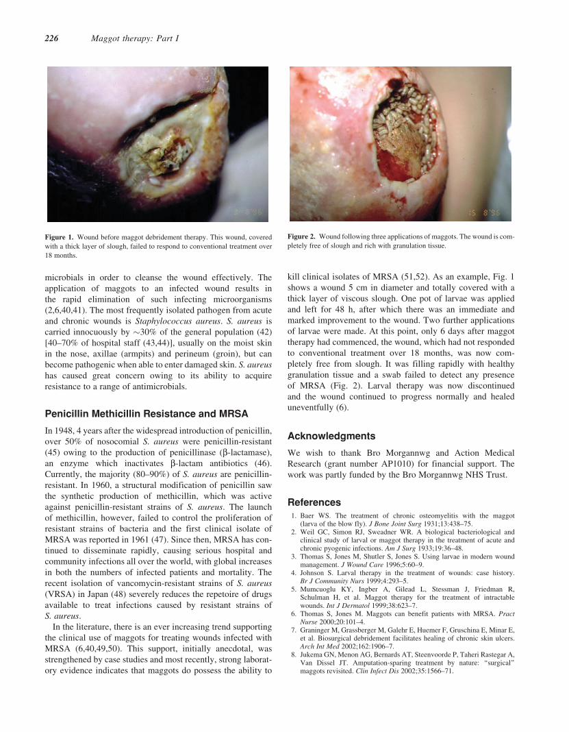
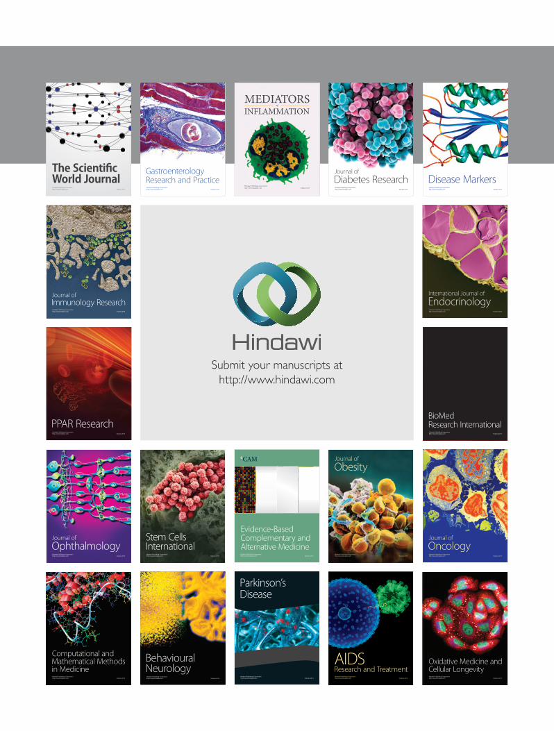
kill clinical isolates of MRSA (51,52). As an example, Fig. 1
shows a wound 5 cm in diameter and totally covered with a
thick layer of viscous slough. One pot of larvae was applied
and left for 48 h, after which there was an immediate and
marked improvement to the wound. Two further applications
of larvae were made. At this point, only 6 days after maggot
therapy had commenced, the wound, which had not responded
to conventional treatment over 18 months, was now com-
pletely free from slough. It was filling rapidly with healthy
granulation tissue and a swab failed to detect any presence
of MRSA (Fig. 2). Larval therapy was now discontinued
and the wound continued to progress normally and healed
uneventfully (6).
Acknowledgments
We wish to thank Bro Morgannwg and Action Medical
Research (grant number AP1010) for financial support. The
work was partly funded by the Bro Morgannwg NHS Trust.
References1. Baer WS. The treatment of chronic osteomyelitis with the maggot
(larva of the blow fly). J Bone Joint Surg 1931;13:438–75.2. Weil GC, Simon RJ, Sweadner WR. A biological bacteriological and
clinical study of larval or maggot therapy in the treatment of acute andchronic pyogenic infections. Am J Surg 1933;19:36–48.
3. Thomas S, Jones M, Shutler S, Jones S. Using larvae in modern woundmanagement. J Wound Care 1996;5:60–9.
4. Johnson S. Larval therapy in the treatment of wounds: case history.Br J Community Nurs 1999;4:293–5.
5. Mumcuoglu KY, Ingber A, Gilead L, Stessman J, Friedman R,Schulman H, et al. Maggot therapy for the treatment of intractablewounds. Int J Dermatol 1999;38:623–7.
6. Thomas S, Jones M. Maggots can benefit patients with MRSA. PractNurse 2000;20:101–4.
7. Graninger M, Grassberger M, Galehr E, Huemer F, Gruschina E, Minar E,et al. Biosurgical debridement facilitates healing of chronic skin ulcers.Arch Int Med 2002;162:1906–7.
8. Jukema GN, Menon AG, Bernards AT, Steenvoorde P, Taheri Rastegar A,Van Dissel JT. Amputation-sparing treatment by nature: ‘‘surgical’’maggots revisited. Clin Infect Dis 2002;35:1566–71.
Figure 2. Wound following three applications of maggots. The wound is com-
pletely free of slough and rich with granulation tissue.Figure 1. Wound before maggot debridement therapy. This wound, covered
with a thick layer of slough, failed to respond to conventional treatment over
18 months.
226 Maggot therapy: Part I

9. Wollina U, Liebold K, Schmidt W-D, Hartmann M, Fassler D. Biosurgerysupports granulation and debridement in chronic wounds—clinicaldata and remittance spectroscopy measurement. Int J Dermatol 2002;41:635–9.
10. Sherman RA. Maggot therapy for treating diabetic foot ulcersunresponsive to conventional therapy. Diabetes Care 2003;26:446–51.
11. Sherman RA, Shimoda KJ. Presurgical maggot debridement of softtissue wounds is associated with decreased rates of postoperativeinfection. Clin Infect Dis 2004;39:1067–70.
12. Goldstein H. Maggots in the treatment of wound and bone infections.J Bone Joint Surg 1931;13:476–8.
13. Larrey DJ. Observations on Wounds and Their Complications byErysipelas Gangrene and Tetanus etc51–52 Paris: Clin. Chir. Transl.EF Rivinus, 1932. p. 34. Philadelphia: Key Mielke & Biddle.
14. Sherman RA, Hall MJR, Thomas S. Medicinal maggots: an ancientremedy for some contemporary afflictions. Annu Rev Entomol 2000;45:55–81.
15. RobinsonW. Stimulation of healing in non-healing wounds by allantoin inmaggot secretions and of wide biological distribution. J Bone Joint Surg1935;17:267–71.
16. Chain E, Florey HW, Gardner AD, Heatley HG, JenningMA, Orr-Ewing J,et al. Penicillin as a chemotherapeutic agent. Lancet 1940;2:226–8.
17. Waldrop J, Doughty D. In: Bryant R (ed). Wound-healing Physiology.Acute and Chronic Wounds. Nursing Management. London: Mosby Inc.,2000, 17–39.
18. Bowler P. The anaerobic and aerobic microbiology of wounds: a review.Wounds 1998;10:170–8.
19. Torrance C. The physiology of wound healing. Wound care in accidentand emergency supplement. Nursing 1985;2:1–3.
20. Olalde JA, Margarici M, Amendola F, Del Castillo O. The systemictheory of livivng systems. Part IV: Systemic medicine—the praxis.Evid Based Complement Alternat Med 2005;2:429–39.
21. Mumcuoglu KY. Clinical applications for maggots in wound care.Am J Derm 2001;2:219–27.
22. Erzinclioglu Z. The biology of blowflies. Naturalist’s Handbook 23:Blowflies Slough. England: Richmond Publishing Co. Ltd, 1996.
23. Ziffren SE, Heist HE, May SC, Womack NA. The secretion of collagenaseby maggots and its implication. Ann Surg 1953;138:932–4.
24. Terra WR, Ferreira C. Insect digestive enzymes: properties com-partmentalization and function. Comp Biochem Physiol 1994;109B:1–62.
25. Sherman RA. Maggot debridement in modern medicine. Infect Med1998;15:651–6.
26. Chambers L, Woodrow S, Brown AP, Harris PD, Philips D, Hall M, et al.Degradation of extracellular matrix componets by defined proteinasesfrom the greenbottle larva Lucilia sericata used for the clinicaldebridement of non-healing wounds. Br J Dermatol 2003;148:14–23.
27. Schultz GS, Sibbald RG, Falanga V, Ayello EA, Dowsett C, Harding K,et al. Wound bed preparation: a systematic approach to wound manage-ment. Wound Repair Regen 2003;11:1–28.
28. Bucalo B, Eaglstein WH, Falanga V. Inhibition of cell proliferation bychronic wound fluid. Wound Repair Regen 1993;1:181–6.
29. Agren MS, Steenfos HH, Dabelsteen S, Hansen JB, Dabelsteen E. Prolif-eration and mitogenic response to PDGF-BB of fibroblasts isolated fromchronic leg ulcers is ulcer-dependent. J Invest Dermatol 1999;112:463–9.
30. Tarnuzzer RW, Shultz GS. Biochemical analysis of acute and chronicwound environments. Wound Repair Regen 1996;4:321–5.
31. Sherman RA, Tran JM-T, Sullivan R. Maggot therapy for venous stasisulcers. Arch Dermatol 1996;132:254–6.
32. Mumcuoglu KY, Lipo M, Ioffe-Uspensky I, Miller J, Galun R. Maggottherapy for gangrene and osteomyelitis. Harefuah 1997;132:323–5.
33. Namias N, Varela JE, Varas RP, Quintana O, Ward CG. Biodebridement:a case report of maggot therapy for limb salvage after forth degree burns.J Burn Care Rehabil 2000;21:254–7.
34. Sherman RA. Maggot versus conservative debridement therapy for thetreatment of pressure ulcers. Wound Repair Regen 2002;10:208–14.
35. Vistnes L, Lee R, Ksander A. Proteolytic activity of blowfly larvaesecretions in experimental burns. Surgery 1981;90:835–41.
36. Hobson RP. Studies on the nutrition of blow-fly larvae. I. Structure andfunction of the alimentary tract. J Exp Biol 1931;8:110–23.
37. Casu RE, Pearson RD, Jarmey JM, Cadogan LC, Riding GA, Tellam RL.Extretory/secretory chymotrypsin from Lucilia cuprina: purificationenzymatic specificity and amino avid sequence deduced from mRNA.Insect Mol Biol 1994;3:201–11.
38. Barnard DR. Skeletal-muscular mechanisms of the larva of Luciliasericata (Meigen) in relation to feeding habit. Pan-Pac Entomol1977;53:223–9.
39. Bowler P, Davies BJ. The microbiology of acute and chronic wounds.Wounds 1999;11:72–8.
40. Courtenay M. The use of larval therapy in wound management in the UK.J Wound Care 1999;8:177–9.
41. Hinshaw J. Larval therapy: a review of clinical human andveterinary studies. Available at: www.worldwidewounds.com/2000/oct/janet-hinshaw/larval-therapy-human-and-veterinary.html.
42. Casewell MW, Hill RL. The carrier state: methicillin-resistantStaphylococcus aureus. J Antimicrob Chemother 1986;18(Suppl A):1–12.
43. Williams RE. Healthy carriage of Staphylococcus aureus: its prevalenceand importance. Bacteriol Rev 1963;27:56–71.
44. Parnaby RM, O’Dwyer G, Monsey HA, Shafi MS. Carriage ofStaphylococcus aureus in the elderly. J Hosp Infect 1995;33:201–6.
45. Barber M, Rozwadowska-Dowzenko M. Infection by penicillin-resistantstaphylococci. Lancet 1948;ii:641–4.
46. Livermore DM. Antibiotic resistance in staphylococci. Int J AntimicrobAgents 2000;16:S3–10.
47. Jevons MP. Celbenin-resistant staphylococci. Br Med J 1961;1:124–5.48. Hiramatsu K, Hanaki H, Ino T, Yabuta K, Oguzi T, Tenover FC.
Methicillin-resistant Staphylococcus aureus clinical strain with reducedvancomycin susceptibility. J Antimicrob Chemother 1997;40:135–6.
49. Dissemond J, Kopperman M, Esser S, Schultewolter T, Goos M,Wagner SN. Treatment of methicillin-resistant Staphyloccus of a chronicleg ulcer. Hautarzt 2002;53:608–12.
50. Beasley WD, Hirst G. Making a meal of MRSA—the role of biosurgery inhospital-acquired infection. J Hosp Infect 2004;56:6–9.
51. Thomas S, Andrews A, Hay P, Bourgoise S. The anti-microbial activityof maggot secretions: results of a preliminary study. J Tissue Viability1999;9:127–32.
52. Bexfield A, Nigam Y, Thomas S, Ratcliffe NA. Detection and partialcharacterisation of two antibacterial factors from the excretions/secretionsof the medicinal maggot Lucilia sericata and their activity againstmethicillin-resistant Staphylococcus aureus (MRSA). Microbes Infect2004;6:1297–304.
Received September 13, 2005; accepted March 23, 2006
eCAM 2006;3(2) 227
Submit your manuscripts athttp://www.hindawi.com
Stem CellsInternational
Hindawi Publishing Corporationhttp://www.hindawi.com Volume 2014
Hindawi Publishing Corporationhttp://www.hindawi.com Volume 2014
MEDIATORSINFLAMMATION
of
Hindawi Publishing Corporationhttp://www.hindawi.com Volume 2014
Behavioural Neurology
EndocrinologyInternational Journal of
Hindawi Publishing Corporationhttp://www.hindawi.com Volume 2014
Hindawi Publishing Corporationhttp://www.hindawi.com Volume 2014
Disease Markers
Hindawi Publishing Corporationhttp://www.hindawi.com Volume 2014
BioMed Research International
OncologyJournal of
Hindawi Publishing Corporationhttp://www.hindawi.com Volume 2014
Hindawi Publishing Corporationhttp://www.hindawi.com Volume 2014
Oxidative Medicine and Cellular Longevity
Hindawi Publishing Corporationhttp://www.hindawi.com Volume 2014
PPAR Research
The Scientific World JournalHindawi Publishing Corporation http://www.hindawi.com Volume 2014
Immunology ResearchHindawi Publishing Corporationhttp://www.hindawi.com Volume 2014
Journal of
ObesityJournal of
Hindawi Publishing Corporationhttp://www.hindawi.com Volume 2014
Hindawi Publishing Corporationhttp://www.hindawi.com Volume 2014
Computational and Mathematical Methods in Medicine
OphthalmologyJournal of
Hindawi Publishing Corporationhttp://www.hindawi.com Volume 2014
Diabetes ResearchJournal of
Hindawi Publishing Corporationhttp://www.hindawi.com Volume 2014
Hindawi Publishing Corporationhttp://www.hindawi.com Volume 2014
Research and TreatmentAIDS
Hindawi Publishing Corporationhttp://www.hindawi.com Volume 2014
Gastroenterology Research and Practice
Hindawi Publishing Corporationhttp://www.hindawi.com Volume 2014
Parkinson’s Disease
Evidence-Based Complementary and Alternative Medicine
Volume 2014Hindawi Publishing Corporationhttp://www.hindawi.com