M13 BACTERIOPHAGE NANOMATERIALS FOR REGERATIVE...
38
Chapter 8 M13 BACTERIOPHAGE NANOMATERIALS FOR REGERATIVE MEDICINE So Young Yoo * BIO-IT Foundry Technology Institute, Pusan National University, Busan 609-735, Republic of Korea; Research Institute for Convergence of Biomedical Science and Technology, Pusan National University Yangsan Hospital, Yangsan 626-770, Republic of Korea * Email: [email protected]
Transcript of M13 BACTERIOPHAGE NANOMATERIALS FOR REGERATIVE...
Chapter
8
M13 BACTERIOPHAGE NANOMATERIALS FOR REGERATIVE MEDICINE
So Young Yoo*
BIO-IT Foundry Technology Institute, Pusan National University, Busan 609-735, Republic of Korea; Research Institute for Convergence of Biomedical Science and Technology, Pusan National University Yangsan Hospital, Yangsan 626-770, Republic of Korea *Email: [email protected]
Chapter 8
Contents 8.1. INTRODUCTION ........................................................................................................................................ 187
8.1.1. The M13 bacteriophage........................................................................................................... 188 8.1.2. Phage structure ........................................................................................................................... 188
8.2. PHAGE ENGINEERING ........................................................................................................................... 190 8.2.1. Genetic engineering of phages ............................................................................................. 190
8.2.1.1. pIII minor coat, or pVIII protein engineering .............................................. 191 8.2.1.2. pVI, pVII or pIX minor coat protein engineering ........................................ 192 8.2.1.3. NN type engineering ............................................................................................... 192
8.2.2. Directed evolution of phages ................................................................................................ 192 8.2.2.1. Phage display to select functional peptide sequences ............................. 193 8.2.2.2. Phage display to identify protein interactions ............................................ 193
8.2.3. Chemical engineering of phages .......................................................................................... 194 8.2.4. Self-assembly of phages .......................................................................................................... 196 8.2.5. Fabrication of the M13 bacteriophage self-assembly (M13SA) building
block ............................................................................................................................................... 197 8.2.6. Application of (M13SA) as an artificial extracellular matrix (ECM) .................... 198
8.3. TISSUE ENGINEERING ........................................................................................................................... 199 8.3.1. Architecture for tissue engineering materials (physical cues) .............................. 200 8.3.2. Receptor-ligand interactions for tissue engineering materials
(chemical cues) .......................................................................................................................... 201 8.3.3. Current technologies for tissue engineering materials ............................................. 202
8.4. PHAGES FOR TISSUE REGENERATION .......................................................................................... 204 8.4.1. Chemical cue control by engineered phages .................................................................. 205 8.4.2. Physical cue control by engineered phages ................................................................... 206 8.4.3. Multifunctional phage materials ......................................................................................... 207 8.4.4. Immune study of phage materials ...................................................................................... 209 8.4.5. Mechanical and degradation properties of phage materials .................................. 210 8.4.6. Gene delivery systems ............................................................................................................. 211 8.4.7. Diagnosis and therapeutic applications ........................................................................... 211 8.4.8. Tissue engineering and regenerative medicine applications ................................. 212
8.5. SUMMARY AND FUTURE PERSPECTIVES ..................................................................................... 213
ACKNOWLEDGEMENTS ................................................................................................................................. 214
REFERENCES ...................................................................................................................................................... 215
186
8.1. INTRODUCTION Recent advances in nanotechnology have enabled us to see, measure, and control nano-scaled objects at the desired level. This technology has allowed us to envision how to fabricate and regenerate tissues and organs by smart tissue scaffolding, and has facilitated the development of highly efficient clean and green energy conversion devices. Although we are now in the stage of utilizing various methodologies to manipulate atoms and molecules to get new functional materials, the design of novel materials with well-defined structures that perform particular functions is still challenging in materials science [1]. Many material researchers have focused their efforts on developing new materials through endless reiteration of rational design and performance characterization processes. However, nature may have already solved such material design issues through the process of evolution. Thus, mimicking natural functional materials may be crucial to developing the desired functional materials. Some examples includes glass sponges (optical fibers) [2], brittle stars (optical lens array) [3], diatoms (sophisticated periodic structures), abalone shells (fracture resistant materials) [4], bones (support structures for vertebrates) [5], and cells (exquisite self-replicating biomachines) [6-8]. Most bio-organisms have specific functions and self-templated hierarchical structures that are difficult to mimic. Most unique characteristics are dependent on genetic information, which works as a molecular machine to control many specific functions by translating the desired proteins and precisely programmed processes in biosystems. Protein-based “bottom-up” synthesis of nanoscale functional materials and devices is thus one of the most promising areas in the newly emerging field of nanobiomaterials [9-12]. Identifying active basic buildings blocks from biological examples is crucial in biomaterial design because of the complicated nature of biological materials. Meanwhile, the genetic engineering of phage viruses has recently provided various opportunities for building novel bio-nanomaterials by integrating various disciplines, including biology, chemistry, physics, materials science, and electric engineering. By mimicking the evolutionary process in nature, phages can be used for identifying protein (or peptide) sequences that can specifically recognize desired (target) materials at the molecular level [6,8]. These recognition elements can be used as a basis to design unprecedented materials by synthesizing inorganic nanomaterials and developing sensory materials. Additionally, due to their well-defined shape, viral particles can be used to self-assemble various ordered structures which can create novel functional nanomaterials for various applications, including energy generation [13,14], biosensors [15,16], semiconductors, and tissue regenerating materials.
187

Chapter 8
In this chapter, I would like to introduce the unique features of phage viral particles and the recent accomplishments in phage-based materials as a tool to fabricate functional nanomaterials and potential future applications in regenerative medicine.
8.1.1. The M13 bacteriophage Bacteriophages (phages) are prokaryotic viruses that can infect bacterial host cells. The name “bacteriophage” can be translated to “bacteria eater” in Greek. Like its name, once the phage infects the bacterial host, the virus exploits the host biosynthetic machinery to produce many identical copies of the viral particle itself. Phages are also one of the most common organisms on earth. Phages are composed of protein capsids surrounding outer surfaces of the viral particles (shells) and genomic materials (DNA or RNA) inside of the protein shells (Figure 1). There are many different types of phages depending on their genomic material, life cycle, and shape. Genomic materials can be either DNA or RNA in a single stranded or double stranded form. Depending on the replication process, the phage can be lysogenic or lytic. Lysogenic phages infect host cells and inject their genomic materials. The genetic materials then overtake host cell metabolism to reproduce the identical genetic material and corresponding proteins. These products are delivered to the host cell membrane and the new phages are packaged and released. Therefore, lysogenic phages do not break the host cell wall. However, lytic phages invade the host cell and replicate inside of the host cells. Right after replication, the phages destroy the host cell wall and the newly amplified phages can infect other host cells. There are many different shapes of phages, such as linear (M13, Fd, F1, Ff) or spherical (MS2). Some phages possess very sophisticated shapes. For example, the T4 phage possesses an icosahedral head and a long tail connected by a cylindrical body. Although the shape, composition, and life cycles are different, phages can replicate exact copies of themselves because they possess the phenotype and genotype in the same body. Because of this property, phages are excellent candidates to develop nanomaterials (Figure 1). Thanks to commercially available genetic tool kits, the M13 phage has been extensively used for the further development of evolvable nanoscale materials for information mining, the synthesis of new materials, and the self-assembly of various nanostructures.
8.1.2. Phage structure The M13 phage is a bacterial virus comprised of a single stranded DNA encapsulated by various major and minor coat proteins. It has a long rod filament shape that is approximately 880 nm in length and 6.6 nm in width (Figure 1A) [17,18]. The viral capsid is composed of 2,700 copies of helically arranged major coat protein, pVIII, and 5–7 copies of minor coat proteins, pIII, pVI, pIX and pVII, located at either of its ends [17,18]. The M13 phage can infect and propagate only within bacteria displaying F-pili, such as Escherichia
188

M13 bacteriophage nanomaterials for regerative medicine
coli (E. coli) [19]. It is a non-lytic bacterial virus, meaning that it does not break the bacterial cell membrane upon exit, but instead is secreted through a protein pore channel in the bacterial membrane [17,20,21]. Bacterial host growth is slowed down due to increased metabolic demands of phage production, but continues after infection [22]. These qualities allow for easy mass amplification of the bacteriophage in bacterial culture. For the last two decades, the chemical or biochemical landscape of the phage structure was greatly expanded through genetic engineering of the phage [23-26] and site-specific bio-orthogonal organic synthesis approaches [27-30].
Figure 1. Schematic diagram of various distinct structures of various phages. (A) Long
rod structure of the M13 bacteriophage with genomic schematic diagrams to show each protein expressed on the M13 phage surface. (B) Sophisticated structure of the T4 bacteriophage with an icosahedral head and long tail connected through the cylindrical
body. (C) Spherical structure of the MS2 bacteriophage.
189
Chapter 8
8.2. PHAGE ENGINEERING Many foreign DNA or synthetic DNA sequences can be integrated into the phage genome and expressed at various sites of the phage body through genetic engineering [26,31]. Various non-natural amino acids have been expressed on the phage surface using amber codon tRNA approaches [32,33]. In addition, site-specific chemical reactions have been developed and enable further modification of the phage surface with various chemicals such as fluorescent dyes or chromophores for various applications, including biochemical imaging and energy harvesting applications [28-30].
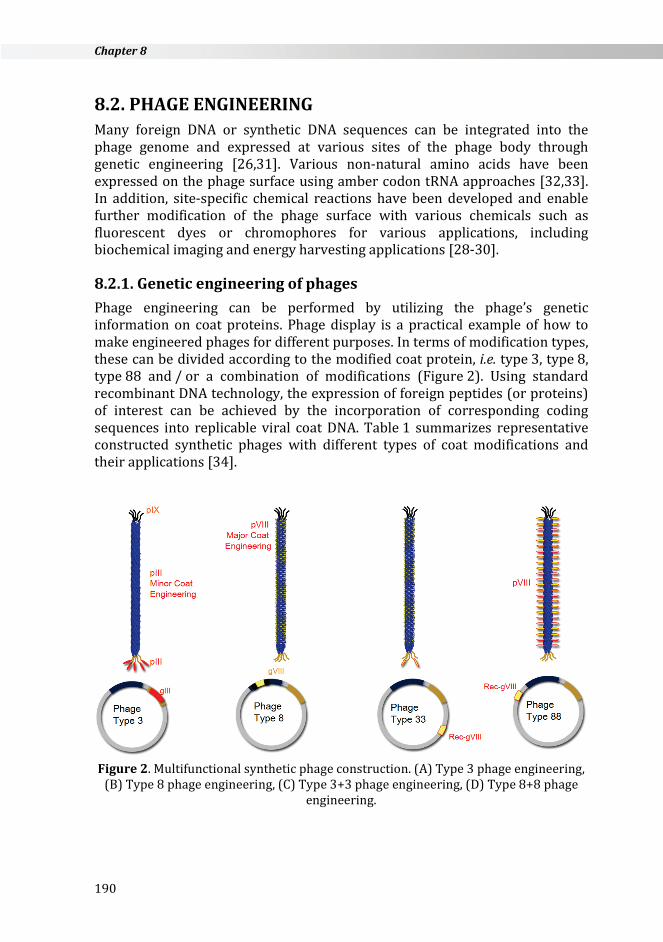
8.2.1. Genetic engineering of phages Phage engineering can be performed by utilizing the phage’s genetic information on coat proteins. Phage display is a practical example of how to make engineered phages for different purposes. In terms of modification types, these can be divided according to the modified coat protein, i.e. type 3, type 8, type 88 and / or a combination of modifications (Figure 2). Using standard recombinant DNA technology, the expression of foreign peptides (or proteins) of interest can be achieved by the incorporation of corresponding coding sequences into replicable viral coat DNA. Table 1 summarizes representative constructed synthetic phages with different types of coat modifications and their applications [34].
Figure 2. Multifunctional synthetic phage construction. (A) Type 3 phage engineering,
(B) Type 8 phage engineering, (C) Type 3+3 phage engineering, (D) Type 8+8 phage engineering.
190
M13 bacteriophage nanomaterials for regerative medicine
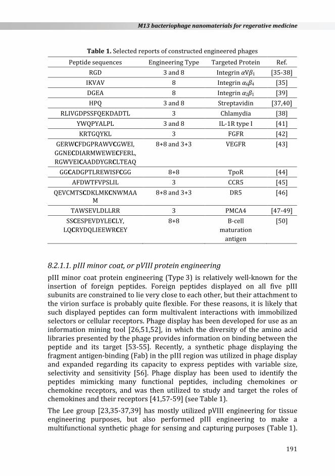
Table 1. Selected reports of constructed engineered phages
Peptide sequences Engineering Type Targeted Protein Ref. RGD 3 and 8 Integrin αVβ1 [35-38]
IKVAV 8 Integrin α6β4 [35] DGEA 8 Integrin α2β1 [39] HPQ 3 and 8 Streptavidin [37,40]
RLIVGDPSSFQEKDADTL 3 Chlamydia [38] YWQPYALPL 3 and 8 IL-1R type I [41] KRTGQYKL 3 FGFR [42]
GERWCFDGPRAWVCGWEI, GGNECDIARMWEWECFERL, RGWVEICAADDYGRCLTEAQ
8+8 and 3+3 VEGFR [43]
GGCADGPTLREWISFCGG 8+8 TpoR [44] AFDWTFVPSLIL 3 CCR5 [45]
QEVCMTSCDKLMKCNWMAAM
8+8 and 3+3 DR5 [46]
TAWSEVLDLLRR 3 PMCA4 [47-49] SSCESPEVDYLECLY,
LQCRYDQLIEEWRCEY 8+8 B-cell
maturation antigen
[50]
8.2.1.1. pIII minor coat, or pVIII protein engineering pIII minor coat protein engineering (Type 3) is relatively well-known for the insertion of foreign peptides. Foreign peptides displayed on all five pIII subunits are constrained to lie very close to each other, but their attachment to the virion surface is probably quite flexible. For these reasons, it is likely that such displayed peptides can form multivalent interactions with immobilized selectors or cellular receptors. Phage display has been developed for use as an information mining tool [26,51,52], in which the diversity of the amino acid libraries presented by the phage provides information on binding between the peptide and its target [53-55]. Recently, a synthetic phage displaying the fragment antigen-binding (Fab) in the pIII region was utilized in phage display and expanded regarding its capacity to express peptides with variable size, selectivity and sensitivity [56]. Phage display has been used to identify the peptides mimicking many functional peptides, including chemokines or chemokine receptors, and was then utilized to study and target the roles of chemokines and their receptors [41,57-59] (see Table 1). The Lee group [23,35-37,39] has mostly utilized pVIII engineering for tissue engineering purposes, but also performed pIII engineering to make a multifunctional synthetic phage for sensing and capturing purposes (Table 1).
191
Chapter 8
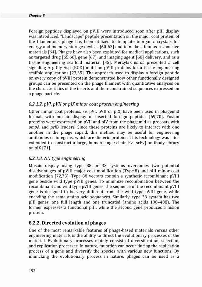
Foreign peptides displayed on pVIII were introduced soon after pIII display was introduced. “Landscape” peptide presentation on the major coat protein of the filamentous phage has been utilized to template inorganic crystals for energy and memory storage devices [60-63] and to make stimulus-responsive materials [64]. Phages have also been exploited for medical applications, such as targeted drug [65,66], gene [67], and imaging agent [68] delivery, and as a tissue engineering scaffold material [35]. Merzylak et al. presented a cell signaling Arg-Gly-Asp (RGD) motif on pVIII proteins for a tissue engineering scaffold applications [23,35]. The approach used to display a foreign peptide on every copy of pVIII protein demonstrated how other functionally designed groups can be presented on the phage filament with quantitative analyses on the characteristics of the inserts and their constrained sequences expressed on a phage particle.
8.2.1.2. pVI, pVII or pIX minor coat protein engineering Other minor coat proteins, i.e. pVI, pVII or pIX, have been used in phagemid format, with mosaic display of inserted foreign peptides [69,70]. Fusion proteins were expressed on pVII and pIV from the phagemid as procoats with ompA and pelB leaders. Since these proteins are likely to interact with one another in the phage capsid, this method may be useful for engineering antibodies or integrins, which are dimeric proteins. This technology was later extended to construct a large, human single-chain Fv (scFv) antibody library on pIX [71].
8.2.1.3. NN type engineering Mosaic display using type 88 or 33 systems overcomes two potential disadvantages of pVIII major coat modification (Type 8) and pIII minor coat modification [72,73]. Type 88 vectors contain a synthetic recombinant pVIII gene beside wild type pVIII genes. To minimize recombination between the recombinant and wild type pVIII genes, the sequence of the recombinant pVIII gene is designed to be very different from the wild type pVIII gene, while encoding the same amino acid sequences. Similarly, type 33 system has two pIII genes, one full length and one truncated (amino acids 198–408). The former expresses a functional pIII, while the second gene produces a fusion protein.
8.2.2. Directed evolution of phages One of the most remarkable features of phage-based materials versus other engineering materials is the ability to direct the evolutionary processes of the material. Evolutionary processes mainly consist of diversification, selection, and replication processes. In nature, mutation can occur during the replication process of a gene and diversify the species with various new functions. By mimicking the evolutionary process in nature, phages can be used as a
192
M13 bacteriophage nanomaterials for regerative medicine
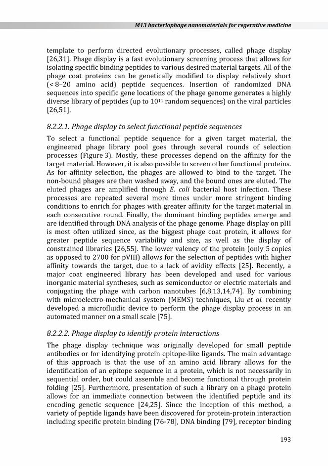
template to perform directed evolutionary processes, called phage display [26,31]. Phage display is a fast evolutionary screening process that allows for isolating specific binding peptides to various desired material targets. All of the phage coat proteins can be genetically modified to display relatively short (< 8–20 amino acid) peptide sequences. Insertion of randomized DNA sequences into specific gene locations of the phage genome generates a highly diverse library of peptides (up to 1011 random sequences) on the viral particles [26,51].
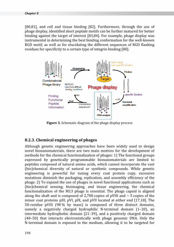
8.2.2.1. Phage display to select functional peptide sequences To select a functional peptide sequence for a given target material, the engineered phage library pool goes through several rounds of selection processes (Figure 3). Mostly, these processes depend on the affinity for the target material. However, it is also possible to screen other functional proteins. As for affinity selection, the phages are allowed to bind to the target. The non-bound phages are then washed away, and the bound ones are eluted. The eluted phages are amplified through E. coli bacterial host infection. These processes are repeated several more times under more stringent binding conditions to enrich for phages with greater affinity for the target material in each consecutive round. Finally, the dominant binding peptides emerge and are identified through DNA analysis of the phage genome. Phage display on pIII is most often utilized since, as the biggest phage coat protein, it allows for greater peptide sequence variability and size, as well as the display of constrained libraries [26,55]. The lower valency of the protein (only 5 copies as opposed to 2700 for pVIII) allows for the selection of peptides with higher affinity towards the target, due to a lack of avidity effects [25]. Recently, a major coat engineered library has been developed and used for various inorganic material syntheses, such as semiconductor or electric materials and conjugating the phage with carbon nanotubes [6,8,13,14,74]. By combining with microelectro-mechanical system (MEMS) techniques, Liu et al. recently developed a microfluidic device to perform the phage display process in an automated manner on a small scale [75].
8.2.2.2. Phage display to identify protein interactions The phage display technique was originally developed for small peptide antibodies or for identifying protein epitope-like ligands. The main advantage of this approach is that the use of an amino acid library allows for the identification of an epitope sequence in a protein, which is not necessarily in sequential order, but could assemble and become functional through protein folding [25]. Furthermore, presentation of such a library on a phage protein allows for an immediate connection between the identified peptide and its encoding genetic sequence [24,25]. Since the inception of this method, a variety of peptide ligands have been discovered for protein-protein interaction including specific protein binding [76-78], DNA binding [79], receptor binding
193
Chapter 8
[80,81], and cell and tissue binding [82]. Furthermore, through the use of phage display, identified short peptide motifs can be further matured for better binding against the target of interest [83,84]. For example, phage display was instrumental in determining the best binding conformation for the well-known RGD motif, as well as for elucidating the different sequences of RGD flanking residues for specificity to a certain type of integrin binding [80].
Figure 3. Schematic diagram of the phage display process
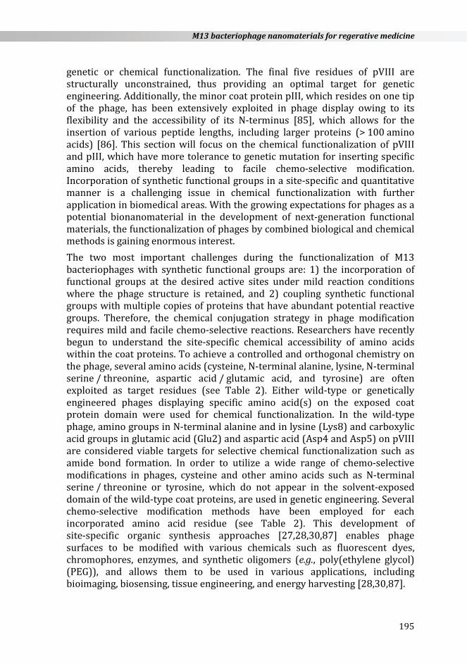
8.2.3. Chemical engineering of phages Although genetic engineering approaches have been widely used to design novel bionanomaterials, there are two main motives for the development of methods for the chemical functionalization of phages: 1) The functional groups expressed by genetically programmable bionanomaterials are limited to peptides composed of natural amino acids, which cannot incorporate the vast (bio)chemical diversity of natural or synthetic compounds. While genetic engineering is powerful for tuning every coat protein copy, excessive mutations diminish the packaging, replication, and assembly efficiency of the phage. 2) To expand the use of phages in novel functional applications such as (bio)chemical sensing, bioimaging, and tissue engineering, the chemical functionalization of the M13 phage is essential. The phage capsid is aligned along the shaft and is composed of 2,700 copies of pVIII and ~ 5 copies of the minor coat proteins pIII, pVI, pIX, and pVII located at either end [17,18]. The 50-residue pVIII (98 % by mass) is composed of three distinct domains, namely a negatively charged hydrophilic N-terminal domain (1–20), an intermediate hydrophobic domain [21–39), and a positively charged domain (40–50) that interacts electrostatically with phage genomic DNA. Only the N-terminal domain is exposed to the medium, allowing it to be targeted for
194
M13 bacteriophage nanomaterials for regerative medicine
genetic or chemical functionalization. The final five residues of pVIII are structurally unconstrained, thus providing an optimal target for genetic engineering. Additionally, the minor coat protein pIII, which resides on one tip of the phage, has been extensively exploited in phage display owing to its flexibility and the accessibility of its N-terminus [85], which allows for the insertion of various peptide lengths, including larger proteins (> 100 amino acids) [86]. This section will focus on the chemical functionalization of pVIII and pIII, which have more tolerance to genetic mutation for inserting specific amino acids, thereby leading to facile chemo-selective modification. Incorporation of synthetic functional groups in a site-specific and quantitative manner is a challenging issue in chemical functionalization with further application in biomedical areas. With the growing expectations for phages as a potential bionanomaterial in the development of next-generation functional materials, the functionalization of phages by combined biological and chemical methods is gaining enormous interest. The two most important challenges during the functionalization of M13 bacteriophages with synthetic functional groups are: 1) the incorporation of functional groups at the desired active sites under mild reaction conditions where the phage structure is retained, and 2) coupling synthetic functional groups with multiple copies of proteins that have abundant potential reactive groups. Therefore, the chemical conjugation strategy in phage modification requires mild and facile chemo-selective reactions. Researchers have recently begun to understand the site-specific chemical accessibility of amino acids within the coat proteins. To achieve a controlled and orthogonal chemistry on the phage, several amino acids (cysteine, N-terminal alanine, lysine, N-terminal serine / threonine, aspartic acid / glutamic acid, and tyrosine) are often exploited as target residues (see Table 2). Either wild-type or genetically engineered phages displaying specific amino acid(s) on the exposed coat protein domain were used for chemical functionalization. In the wild-type phage, amino groups in N-terminal alanine and in lysine (Lys8) and carboxylic acid groups in glutamic acid (Glu2) and aspartic acid (Asp4 and Asp5) on pVIII are considered viable targets for selective chemical functionalization such as amide bond formation. In order to utilize a wide range of chemo-selective modifications in phages, cysteine and other amino acids such as N-terminal serine / threonine or tyrosine, which do not appear in the solvent-exposed domain of the wild-type coat proteins, are used in genetic engineering. Several chemo-selective modification methods have been employed for each incorporated amino acid residue (see Table 2). This development of site-specific organic synthesis approaches [27,28,30,87] enables phage surfaces to be modified with various chemicals such as fluorescent dyes, chromophores, enzymes, and synthetic oligomers (e.g., poly(ethylene glycol) (PEG)), and allows them to be used in various applications, including bioimaging, biosensing, tissue engineering, and energy harvesting [28,30,87].
195
Chapter 8
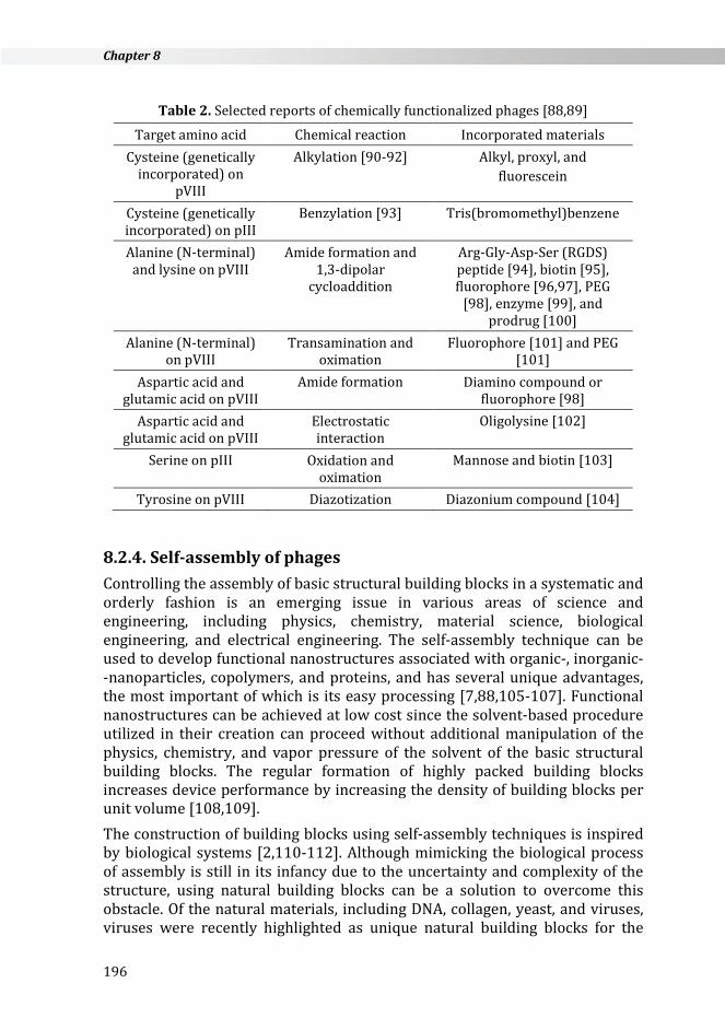
Table 2. Selected reports of chemically functionalized phages [88,89]
Target amino acid Chemical reaction Incorporated materials Cysteine (genetically
incorporated) on pVIII
Alkylation [90-92] Alkyl, proxyl, and fluorescein
Cysteine (genetically incorporated) on pIII
Benzylation [93] Tris(bromomethyl)benzene
Alanine (N-terminal) and lysine on pVIII
Amide formation and 1,3-dipolar
cycloaddition
Arg-Gly-Asp-Ser (RGDS) peptide [94], biotin [95], fluorophore [96,97], PEG
[98], enzyme [99], and prodrug [100]
Alanine (N-terminal) on pVIII
Transamination and oximation
Fluorophore [101] and PEG [101]
Aspartic acid and glutamic acid on pVIII
Amide formation Diamino compound or fluorophore [98]
Aspartic acid and glutamic acid on pVIII
Electrostatic interaction
Oligolysine [102]
Serine on pIII Oxidation and oximation
Mannose and biotin [103]
Tyrosine on pVIII Diazotization Diazonium compound [104]
8.2.4. Self-assembly of phages Controlling the assembly of basic structural building blocks in a systematic and orderly fashion is an emerging issue in various areas of science and engineering, including physics, chemistry, material science, biological engineering, and electrical engineering. The self-assembly technique can be used to develop functional nanostructures associated with organic-, inorganic- -nanoparticles, copolymers, and proteins, and has several unique advantages, the most important of which is its easy processing [7,88,105-107]. Functional nanostructures can be achieved at low cost since the solvent-based procedure utilized in their creation can proceed without additional manipulation of the physics, chemistry, and vapor pressure of the solvent of the basic structural building blocks. The regular formation of highly packed building blocks increases device performance by increasing the density of building blocks per unit volume [108,109]. The construction of building blocks using self-assembly techniques is inspired by biological systems [2,110-112]. Although mimicking the biological process of assembly is still in its infancy due to the uncertainty and complexity of the structure, using natural building blocks can be a solution to overcome this obstacle. Of the natural materials, including DNA, collagen, yeast, and viruses, viruses were recently highlighted as unique natural building blocks for the
196
M13 bacteriophage nanomaterials for regerative medicine
self-assembly process [113-116]. Furthermore, the M13 bacteriophage has attracted significant attention in the field because of its easy growth and handling properties. The use of biological materials as templates enables non-toxic synthesis at low cost. Phages are virus nanofibers approximately 880 nm in length and 6.6 nm in diameter and are safe for use in humans. They can be easily modified genetically and chemically to provide specific functions. Phages can also be used as a template to reveal the homogeneous distribution and percolated network structure of inorganic nanostructures under ambient conditions [13,89,117-118]. Inexpensive and environmentally friendly synthesis is possible through M13 bacteriophage engineering.
8.2.5. Fabrication of the M13 bacteriophage self-assembly (M13SA) building block
The traditional preparation of M13SA is based on the layer-by-layer (LBL) assembly technique. By using linear poly(ethylene imine) and poly(acrylic acid), this controlled molecular interaction leads to a regular orientation of the bacteriophage [119-121]. Recently, novel techniques for 2-D and three- -dimensional (3-D) assembly of the M13 bacteriophage were introduced. 2-D structure fabrication: Low permeability has been a significant issue in 2-D fabrication because of the thickness of traditional membranes in the active area. Current research efforts are therefore primarily focused on making thin membranes that maintain separation efficiency with high permeability [122-125]. Interconnected basic structural building blocks can provide structural integrity and large-scale production with excellent permeability [126,127]. However, the range of the pore size distribution due to the non-uniform integration of the building blocks limits the application of this system; using the M13 bacteriophage could be a solution to this problem. Lee et al. generated a unidirectionally aligned 2-D M13 bacteriophage structure on a graphene oxide (GO) surface using a simple fabrication method [128]. The genetically programmed pIII protein placed at the end of the M13 bacteriophage had specific binding interactions with the carboxylate-functionalized edges of the GO. The relatively neutral body of the virus had a weaker connection with the GO surface, which was less chemically active than the end of the virus with the GO edge. The genetically modified pIII protein made strong salt-bridge type interactions with the GO edge via hydrogen bonding. The pIII protein, displayed with a disulfide bond-constrained peptide, made a strong cyclic structure with the edge of the GO and the aligned virus in the same direction owing to the energetic affinity of peptide geometry [129]. In contrast, the pVIII protein on the virus body demonstrated slight electrostatic repulsion due to the inherent negative charge. The fabricated M13 virus on the GO surface could be unidirectionally aligned by the external shear force of a water stream. This technique, which orients the direction of M13 by the dipping and sweeping
197
Chapter 8
procedure, was previously reported [130]. Actual alignment of the M13 virus was observed using transmission electron microscopy (TEM). Through this approach, it is anticipated that highly orientated 2-D viral structures can be produced on both a large scale and at low cost, while maintaining high performance [129]. 3-D structure fabrication: While 2-D phage formation is important for the application of thin structures such as ultrathin membranes, the 3-D structure of phage is also essential for engineering batteries [131], piezoelectric generators [132], and photovoltaics [133]. Chen et al. reported the generation of a polyaniline (PANI) and single walled carbon nanotube (SWNT) composite 3-D structure using the M13 bacteriophage as a template [134]. PANI is an excellent conductive polymer and exhibits enhanced performance when mixed with SWNT. PANI and SWNT composites have been studied extensively through different assembly methods such as colloidal mixtures [135], electrostatic deposition [136], electropolymerization [137,138], radical polymerization in solution [139], and direct polymerization on SWNT supports [140-143]. However, poor dispersion and aggregation of SWNTs in the composites has limited the progress of these studies. Chemical modification of SWNTs [139,141] by adding surfactants [140,143] or binders [137,144] was applied to overcome this hurdle. Although the fabrication of composites was successful in this approach, heavy loading of SWNTs to achieve efficiency created another obstacle for further applications due to the high associated costs. A phage was introduced as a template to generate a better PANI/SWNT composite because of its efficient dispersion ability. It was genetically engineered to bind SWNTs along the length of the bacteriophage, thereby allowing SWNTs to be clustered without aggregation. The M13/SWNTs composite was cross-linked to form a hydrogel type scaffold. The continuous 3-D porous M13/SWNTs scaffold was successfully combined with PANI using a direct polymerization method. This result represents one of the best cost- -effective solutions, since the aqueous-based synthetic process allows for reduced cost and easy large-scale production.
8.2.6. Application of (M13SA) as an artificial extracellular matrix (ECM)
Wang et al. reported that specific fibronectin peptides (RGD and PHSRN) displayed on the bacteriophage matrix with unique topology serve as an ECM for differentiating mesenchymal stem cells (MSC) into osteoblasts [145]. The phage-based film can provide a unique rigid / groove nanostructure, and the ECM topology had a significant influence on cell behavior such as cell differentiation by cell shape elongation [146-148]. This demonstrates that the rigid / groove structure formed through self-assembly of the M13 bacteriophage significantly induced the elongation and parallel alignment of rat MSC. In addition, insertion of RGD and PHSRN peptides into the M13 bacteriophage increased cell adhesion and viability [145]. The simultaneous
198
M13 bacteriophage nanomaterials for regerative medicine
functionality of ECM topology and peptides via the M13 bacteriophage could be a novel strategy in stem cell research. Wang et al. also suggested a 3-D structure of M13 bacteriophage-based ECM for vascularized osteogenesis of MSC [149]. It is known that new bone formation is promoted by proper angiogenesis [150]. Thus, to induce bone tissue angiogenesis in an artificial ECM, RGD peptides that contain the blood vessel regeneration factor, αv integrin, were introduced through the M13 bacteriophage. RGD peptides are highly unstable when mixed physically or chemically with ECM or medium. Therefore, an RGD-displayed virus matrix could be a possible solution for maintaining RGD peptides in the ECM for a long period in order to achieve successful bone regeneration in vivo. To test this, an RGD-displayed virus-based matrix was implanted into a rat radial bone defect. The vascular endothelial growth factor (VEGF) and wild type M13 were used as positive and negative controls, respectively, for comparison. Quantification of the bone volume of the RGD-displayed virus-based matrix sample showed comparable results in comparison to the normal bone tissue sample. The number of blood vessels formed was approximately 50 % greater than the positive control. The RGD-displayed virus-based matrix induced the formation of vascularized bone without VEGF [149]. Based on these results, the application of various peptide-displayed bacteriophages in nanomedicine and regenerative medicine is highly anticipated.
8.3. TISSUE ENGINEERING The field of tissue engineering strives to fulfill a need to regenerate, repair, or replace biological tissue damaged due to injury or disease. The regulatory approval process for medical devices, and especially combination therapies that encompass products containing both biomaterials and cells, is staggeringly long. Therefore, most of the commercial products currently available are based on some of the initial efforts in tissue engineering that focused on cellular therapies, such as biomaterial skin substitutes and bioactive bone filler materials [151]. As the market and clinical penetration of tissue engineering-based technology is still extremely low, while the basic science knowledge of materials and molecular biology, as well as the know-how of technology transfer strategies for this field is rapidly growing, the creative space for improvement and innovation is vast. The blueprint design considerations of tissue engineers are based on the cellular microenvironment in vivo. There the cells are in close contact with the other cells, as well as ECM. The structure and composition of the ECM is highly dependent on the cell and tissue type it contains, but is generally an interconnected network of proteins, carbohydrates, and proteoglycans (highly branched combinations of the other two). The ECM is continuously synthesized and remodeled by the cells that it surrounds. The ECM scaffold is fibrillar in nature, and contains proteins with
199

Chapter 8
diameters ranging from 5–300 nm [152]. This matrix provides physical support to cells, but also along with neighboring cells often provides topographical cues for cell polarization or migration. Furthermore, the components of the ECM, as well as the ligands and receptors displayed on other cells, serve to provide chemical signaling to cells. The signals for adhesion, migration, proliferation, and differentiation are provided by differential exposure of integrin binding sites, as well as growth factors and cytokines. These signaling motifs can either be presented directly upon contact, or be hidden and only exposed upon matrix remodeling, or in the context of tissue injury [153-155]. Furthermore, the matrix components can bind soluble growth factor molecules from the physiological milieu and present them to cells as needed, as seen with the laminin-bound heparan sulfate proteoglycan that binds and presents bFGF upon demand to neural cells [153,154]. Such a complicated and dynamic environment is nearly impossible to capture with current technologies. A highly simplified cellular environment is the goal of engineered materials that strive to preserve only the most important physiological functionalities.
8.3.1. Architecture for tissue engineering materials (physical cues) In imitating tissue-specific architectures, many tissue engineering efforts have been directed towards making a biomimetic nanofibrous environment conducive to cell growth. Nanoscale features are much smaller than the size of a cell, and so allow cells to experience a more physiological 3-D environment and retain their normal shape. Materials that are in the microscale range are often bigger than the size of a cell, and present a 2-D surface for cell binding, which forces the cells to spread and flatten, therefore changing their natural morphology [156]. Nanoscale features more closely resemble the dimensions of receptors and extracellular proteins, and provide a greatly increased surface area for ligand immobilization and cell attachment. Filopodia, i.e. cell sensory units, have previously been observed to interact with islands as small as 10 nm in height [157]. Additionally, cells grown on substrates with nanoscale topographical features have been shown to upregulate a different set of genes than on substrates with microscale features [158], explaining differences in cell behavior, including increased proliferation and differentiation seen on nanoscale topographies [158,159]. Many tissues such as nerves, cornea, muscles, blood vessels and cartilage, and cell processes such as migration, contractility, and polarization are influenced by the underlying topography of their environments. Cell alignment, elongation, and migration have long been noted to occur in a preferential direction if grown on a material with a directionally oriented topography such as fibers, ridges, or steps [159-163]. This is in part is due to the structural organization and assembly of the cytoskeleton that can converts the mechanical input stimuli of the surrounding substrate into the chemical response output of regulatory cell signaling pathways [164]. Cell alignment and elongation depend on a probability that a
200
M13 bacteriophage nanomaterials for regerative medicine
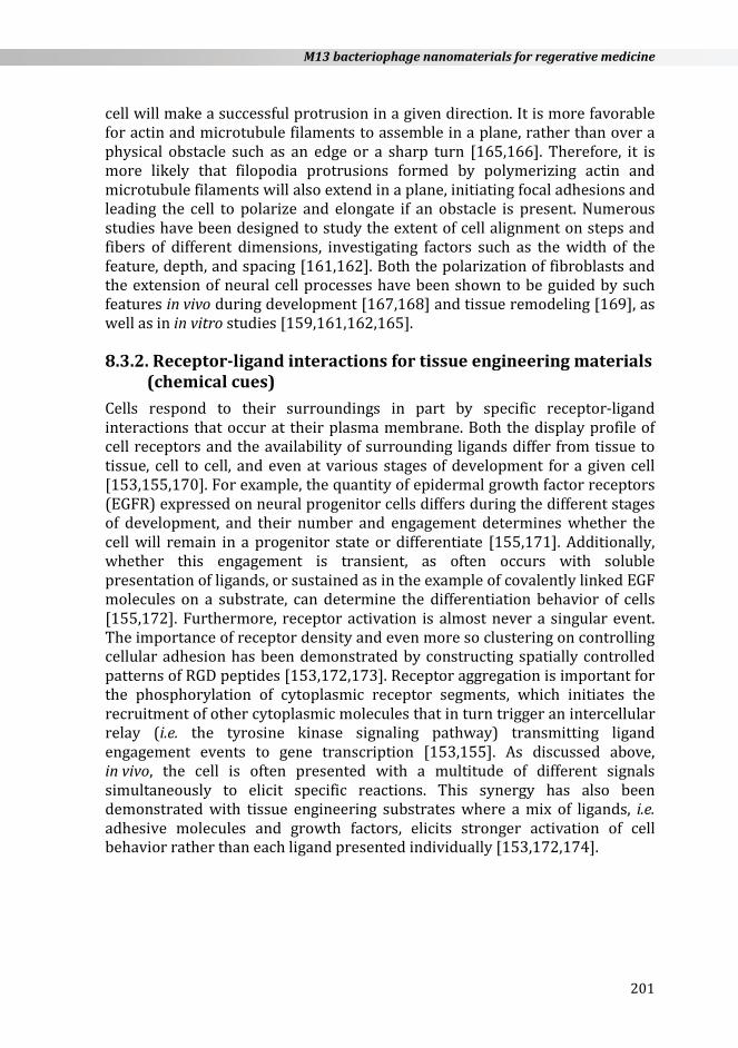
cell will make a successful protrusion in a given direction. It is more favorable for actin and microtubule filaments to assemble in a plane, rather than over a physical obstacle such as an edge or a sharp turn [165,166]. Therefore, it is more likely that filopodia protrusions formed by polymerizing actin and microtubule filaments will also extend in a plane, initiating focal adhesions and leading the cell to polarize and elongate if an obstacle is present. Numerous studies have been designed to study the extent of cell alignment on steps and fibers of different dimensions, investigating factors such as the width of the feature, depth, and spacing [161,162]. Both the polarization of fibroblasts and the extension of neural cell processes have been shown to be guided by such features in vivo during development [167,168] and tissue remodeling [169], as well as in in vitro studies [159,161,162,165].
8.3.2. Receptor-ligand interactions for tissue engineering materials (chemical cues)
Cells respond to their surroundings in part by specific receptor-ligand interactions that occur at their plasma membrane. Both the display profile of cell receptors and the availability of surrounding ligands differ from tissue to tissue, cell to cell, and even at various stages of development for a given cell [153,155,170]. For example, the quantity of epidermal growth factor receptors (EGFR) expressed on neural progenitor cells differs during the different stages of development, and their number and engagement determines whether the cell will remain in a progenitor state or differentiate [155,171]. Additionally, whether this engagement is transient, as often occurs with soluble presentation of ligands, or sustained as in the example of covalently linked EGF molecules on a substrate, can determine the differentiation behavior of cells [155,172]. Furthermore, receptor activation is almost never a singular event. The importance of receptor density and even more so clustering on controlling cellular adhesion has been demonstrated by constructing spatially controlled patterns of RGD peptides [153,172,173]. Receptor aggregation is important for the phosphorylation of cytoplasmic receptor segments, which initiates the recruitment of other cytoplasmic molecules that in turn trigger an intercellular relay (i.e. the tyrosine kinase signaling pathway) transmitting ligand engagement events to gene transcription [153,155]. As discussed above, in vivo, the cell is often presented with a multitude of different signals simultaneously to elicit specific reactions. This synergy has also been demonstrated with tissue engineering substrates where a mix of ligands, i.e. adhesive molecules and growth factors, elicits stronger activation of cell behavior rather than each ligand presented individually [153,172,174].
201
Chapter 8
8.3.3. Current technologies for tissue engineering materials Several technologies have been utilized to create materials incorporating the biomimetic features described above to study cell responses and the utility of these materials as tissue engineering matrices. Nanotextured surface topographies have been created with a variety of lithography-based techniques. Lithographic fabrication methods such as nanoimprint lithography, e-beam lithography, microcontact printing, and embossing are based on the concept of transferring a pattern to a surface [175,176]. This is accomplished by using a mask or masking materials (such as in colloidal lithography) to shield a pattern, and then applying energy, either UV or electrons, to selectively strengthen or etch the exposed parts of the sensitive substrate material. Very precisely ordered patterns maybe produced, and a resolution of 5 nm has been achieved with techniques such as nanoimprint lithography [161,162,177]. Even though such microfabrication methods provide incredible techniques to study cell behavior in vitro, they are difficult to transfer to large scale in vivo tissue engineering applications due to the high costs of production, small area of patterning, and difficulties in translating this method to three dimensions [162]. To create a more biomimetic 3-D scaffold for cell growth, both fibrous and gel materials have been implemented. Nanofibrous networks have been made utilizing techniques such as electrospinning, peptide self-assembly and polymer phase separation. Electrospinning methodology uses a high strength electrical field to spin fibers from melted polymer solution droplets and deposit them on a substrate. The various parameters of the process, including polymer composition, field strength, and the distance between the spinneret and the substrate can be adjusted to produce fibers of various diameters. Furthermore, an electrically grounded rotating drum can be used as a collector to achieve orientation alignment of the deposited fibers [159,175,176,178]. A variety of biodegradable synthetic polymers (poly(L-lactide) (PLLA), poly(glycolic acid) (PGA), and polycaprolactone (PCL)) and biopolymers (collagen, fibrin, and silk) have been successfully spun into fibrous mats and used as cell substrates. The advantages of electrospinning methodology include the ease of production, biomimetic nanofibrous structure, and the wide applicability to a variety of materials. Some of the disadvantages include difficulty in fiber reproducibility from study to study due to many adjustment parameters, as well as difficulties in producing porous 3-D materials [178,179]. Phase separation is another method to create a polymeric nanofibrous scaffolds. Phase separation is based on multi-component systems becoming unstable and separating into multi-phase materials under certain thermodynamic conditions [180]. The phase that contains the majority of the solvent is removed, leaving a porous nanofibrous polymer foam [178]. Both synthetic (PLLA or polyurethane) and bio polymers (collagen) have been used to create such scaffolds [176,178,181]. The process is low cost and does not
202
M13 bacteriophage nanomaterials for regerative medicine
require specialized technology. The morphology of this material closely resembles the structure of the native ECM, with nanofiber dimensions controlled to tens of nanometers. The porosity of the material can be controlled with varying parameters such as the choice of solvent, the concentration of polymer solutions, and the incorporation of beads. Disadvantages include the time-consuming process involving several steps such as raw material dissolution, gelation, solvent extraction, freezing, and drying. Also, the orientation of the fibers within the created matrix cannot be controlled with the current phase separation techniques [176,178,179]. Specially designed self-assembling peptide amphiphilic materials have been shown to form nanofibrous materials. The mechanism of self-assembly is based on the design of the peptide unit and depends on the ionic concentration of the solvent, and the hydrophobic-hydrophilic interactions of these units with the solvent, which drive the packing of these amphiphilic molecules into β-sheet type materials [12] or nanofibers [11]. This method has been shown to form highly hydrated and porous hydrogels that can either be used in vitro as a cell culture substrate, or can be injected and formed in vivo to facilitate tissue repair [11,12]. Additionally, amphiphilic peptide units have been previously designed to carry physiological cell receptor ligands such as RGD or IKVAV. Fibers resulting after the assembly of these units present a very high density of peptides, and have been shown to influence cell differentiation behavior [11]. Some of the disadvantages of this approach include the high cost of the self-assembling units obtained via peptide synthesis. Furthermore, the orientation of these materials is hard to manage, and the resulting structures are often susceptible to uncontrolled degradation by enzymes [179]. Hydrogels are a class of highly hydrated polymer materials that are used for tissue engineering scaffolds. They can be composed of hydrophilic synthetic (PLLA or PGA) or natural (collagen, fibrin, or hyaluronic acid) polymer chains. The biological and mechanical properties of hydrogel materials can be controlled by varying the composition of the gel, and the degree as well as the method of its crosslinking [153,182]. As hydrophilic materials, the gels are not inherently adhesive to cells; however, they can be readily decorated with adhesive peptide groups via crosslinking, or by the incorporation of peptide sequences within the polymer chain [153,182,183]. Additionally, incorporation of enzyme substrates can allow for the control of gel degradation and sequestering active peptide groups. Another attractive property of these materials as tissue engineering scaffolds is their ability to be injected to the injury site and gel in vivo, if this polymerization process is, for example, temperature dependent [153,182]. A disadvantage of this material is the lack of orientation control in the hydrogel structure [179]. As a monodisperse population of filamentous particles, concentrated phage solutions can be aligned through both self-assembly to form liquid-crystalline like structures, and via the application of an external force for longer
203

Chapter 8
macroscale orientation. Furthermore, the major and minor coat proteins located on the phage shell can be genetically and chemically modified to create a controlled, spatially dense presentation of biologically active molecules. Due to these reasons, we will investigate the use of the M13 filamentous phage as a macromolecular building block for the creation of directionally oriented and biologically functionalized tissue regeneration scaffolds in the next section.
8.4. PHAGES FOR TISSUE REGENERATION Tissue engineering scaffolding materials are ultimately designed to imitate ECM, the fibrous protein network that houses cells in vivo. This network provides cells with physical support and guidance through the specific topographical and chemical presentation of various adhesive sites and growth factors. Therefore, in order to control cellular behaviors such as adhesion, proliferation, and differentiation within man-made scaffolds, their surface functionalization with bioactive molecules is highly desirable [172,184,185]. Furthermore, control over the density of such bioactive groups [11,174,186] and their geometric patterning [186-188] has been shown to be important in the ability of biomaterials to modulate such behaviors. The majority of current fabrication methods rely on chemical processing to functionalize biomaterials. With this method, the final density of bioactive groups presented on the surface is ultimately dictated by the bulk solution concentration [172,185,189]. The local binding properties of the material surface, such as charge, or the availability of reactive groups or receptors dictate the final spacing of the bioactive groups. Most techniques that allow for a very precise micro and nanoscale chemical patterning of a substrate are lithography based (i.e. dip-pen lithography) and are hard to replicate in large scale or in 3-D scaffold materials [184,187]. Recently developed nanofabrication techniques, such as peptide self-assembly, electro-spinning, and polymer phase-separation come closer to mimicking the natural ECM topographically. However, the controlled presentation of single or multiple functional groups is still lacking [11,174,184]. Viruses are some of the best characterized structurally organized large molecules. Their nanoscale size and the inherent monodispersity of their shape and surface chemistry are better than can be achieved with most synthetic nanoparticles to date [190]. Both genetic and chemical pathways have been used to modify either single or multiple virus capsid proteins with functional groups [67,186,190-192]. Moreover, novel binding ligands can be found through evolutionary phage display screening methods [26,52,54]. Such functionalized virus particles have been demonstrated to selectively bind both inorganic and organic particles. Additionally, the templation of virus particles has been utilized for electronic and magnetic materials [193,194], as well as a variety of medical applications [65,66,68]. The M13 bacteriophage is a
204
M13 bacteriophage nanomaterials for regerative medicine
filamentous bacterial virus. It has a defined long-rod shape at 880 nm long and 6.7 nm in diameter, with precisely positioned major and minor capsid proteins. These coat proteins can be genetically engineered to express short peptide groups [115,193]. M13 has been previously genetically engineered phage to display cell-adhesive peptides such as RGD and IKVAV on every copy of its pVIII protein [195]. Furthermore, such modified filamentous phages for the construction of aligned 2-D and 3-D materials that are able to support and control the polarization of cells such as fibroblasts and neural progenitor cells was demonstrated [195]. Chimeric displays of binding groups on M13 phage have been demonstrated previously for drug delivery [67], enzyme-linked immunosorbent assay (ELISA) [196], and electronic [193] applications. Additional engineering of the M13 phage to express biotin-like His-Pro-Gln (HPQ) motifs on their capsid proteins will allow for a functional expansion of potential scaffold interactions with the cells, as it will be able to present a variety of immobilized avidin conjugated growth factors and cytokines. The unique biochemical and structural features of genetically engineered phages can be also used in the context of tissue engineering / regeneration in order to control cellular growth or differentiation.
8.4.1. Chemical cue control by engineered phages Merzlyak et al., for example, have explored the use of genetically modified M13 phages as a novel building block for neural cell engineering materials to make functional biomaterials for tissue regeneration by chemical cue control [35]. This was accomplished by engineering the phage to display specific cell signaling motifs, and then assembling the viral particles into a macroscopic scaffolding material. Many peptide expression systems have previously been demonstrated on the various capsid proteins of the phage through the creation of peptide libraries [26,72]. However, as a biological particle for peptide display, phages possess the inherent limitation of having to be successfully expressed and assembled within the E. coli bacterial host, which restricts the type and number of peptides that can be displayed [18,197-199]. These researchers developed a novel cloning approach for the display of an integrin-binding RGD motif on every copy of the pVIII major coat protein [35]. The researchers constructed the phage using a partial library, in which an engineered octamer insert for pVIII included a constrained RGD group that was surrounded by flanking degenerate residues. This allowed for the expression of inserts that retained the desired function of the RGD motif, and yet were biologically compatible with E. coli during the intricate phage replication process. After the construction of engineered phages that stably displayed either RGD or IKVAV peptide groups on every copy of the pVIII protein, they constructed aligned 2-D and 3-D scaffolding materials containing phages and tested their applicability for tissue engineering. The biocompatibility of the engineered phage materials was tested by growing NIH-3T3 fibroblasts and neural progenitor cells on phage films and in phage-
205

Chapter 8
-containing media [35,200]. Both cell types showed normal morphology and proliferation when in direct contact with phage materials. Neural progenitor cells either retained their progenitor state or differentiated towards the neural cell phenotype depending on the medium conditions. It was then demonstrated that 3-D phage materials could support the proliferation and differentiation of neural progenitor cells. Both RGD and IKVAV phage matrices facilitated colony formation of neural progenitor cells, which sustained over 85 % viability during the 7 day observation period. In comparison to RGE and wild type phage controls, RGD and IKVAV phages resulted in enhanced binding and spreading of neural progenitor cells with high specificity. Finally, by simple extrusion or spinning of the phage solution, the researchers constructed aligned 3-D phage fiber matrices with embedded neural progenitor cells. The resulting phage fibers encouraged neural cell differentiation and directed cell growth in parallel to the long axis of the fibers [35]. Chung et al. showed that mechanical shearing of the phage solution on a glass substrate resulted in 2-D directionally oriented films. These oriented films were shown to direct the alignment and morphology of fibroblasts, osteoblasts, and neural cells [200].
8.4.2. Physical cue control by engineered phages Studies on the chemical cues and physical cues provided by synthetic phages were performed with RGD and Asp-Gly-Glu-Ala (DGEA) peptides on engineered phage films and fibers. Yoo et al. demonstrated the early osteogenic differentiation of mouse preosteoblasts by using a collagen-derived DGEA peptide on nanofibrous phage tissue matrices [39]. They constructed a major coat protein engineered with DGEA, Asp-Gly-Asp-Ala (DGDA) or Glu-Gly-Glu-Ala (EGEA) peptides. By genetic engineering of the phages, they constructed nanofiber-like phages having 2700 copies of the target peptide from the inserted genes with 2 and 2.7 nm spacing laterally and axially, respectively. By constructing the phage-based tissue matrix system, they could investigate the specific effect of biochemical cues, which can be tuned precisely at the single amino acid level with little changes to other physical and chemical properties. They characterized the chemical cue or physical cue effects of DGEA and of RGD peptides on the synthetic M13 phage backbone by applying MC3T3 preosteoblast cells on fabricated phage 2-D film and 3-D fibers. They observed pronounced outgrowth of preosteoblasts on DGEA phage matrices, and the cells spread very well throughout the samples on the DGEA phage matrices. Cells on DGDA, EGEA or RGE phages, which were different by one single amino acid from DGEA or RGD phages, showed that the responses were DGEA peptide-specific, demonstrating that synthetic phage-based chemical cues can be controlled by genetic engineering. A competition assay with the peptide corresponding to the engineered phage confirmed that the peptide- -specific chemical cues were controlled by the synthetic phage. The DGEA peptide-specific outgrown morphology of preosteoblasts on the 2-D cultures
206

M13 bacteriophage nanomaterials for regerative medicine
phage matrices were also observed in 3-D cultures. In addition, the DGEA-specific morphological responses of preosteoblast cells were linked to early osteogenic differentiation by DGEA peptides. Virus structure can give more effective and efficient physical cues. The self-assembly capabilities of phages with patterning techniques can enhance phage-specific biochemical and physical cues. Yoo et al. developed a facile method of patterning genetically engineered M13 bacteriophage by employing microcontact printing methods to provide human fibroblast cells with specific biochemical and physical cues [36]. They demonstrated that nanofibrous structures, along with the biochemical signals presented by the phage microstructures, were critical to guiding cellular growth and morphology. The enhanced cellular morphological responses to RGD phage topology, rather than the RGD peptide itself, showed that the phage nanofibrous structure contributes to controlling physical cues. Especially rod-like viruses such as M13 and tobacco mosaic virus (TMV) can control their physical cues and mechanical cues, even based on concentration alone. Lin et al. reported on the formation of diverse patterns resulting from drying a solution of rod-like TMV particles in a glass capillary tube [201]. The concentration of TMV, the salt concentration in the aqueous solution, and the surface properties of the capillary tube interior were used as three key factors to govern such combined self-assembly behavior. The formation of hierarchical structures, which can be used for guiding directional cellular growth, was determined by the preferred orientation of TMV at the air-liquid interface as well as the pinning-depinning process. By controlling these key factors, they could generate surface roughness together with a patterned structure, which was then used for rat aortic smooth muscle cell (SMC) culture to direct the orientation of cells. These researchers could generate either stress-induced SMC alignment or 2-D patterns by utilizing the TMV patterns.
8.4.3. Multifunctional phage materials The physiological cellular environment presents a variety of cell signaling motifs simultaneously, including adhesive sites, growth factors, and cytokines that influence cell behavior [153,172,202,203]. Similarly, engineering materials incorporating several signaling motifs simultaneously have shown this synergy to be more effective than a single motif alone [153,172,174,204]. For example, a study by Dr. Jeffrey Hubbell’s group demonstrated that the incorporation of several functional peptide groups derived from laminin into a fibrin matrix at the same time resulted in a synergistic effect on cell differentiation. The cell neurites extended further into the combination peptide matrix then was predicted by just an additive effect from each peptide [174]. Immobilization of growth factor molecules to the matrix surface, instead of their untethered encapsulation within it, can decrease the uncontrolled release of these molecules, as well as their internalization and metabolism by cells, and therefore provide cells with a more directed and sustained signal,
207
Chapter 8
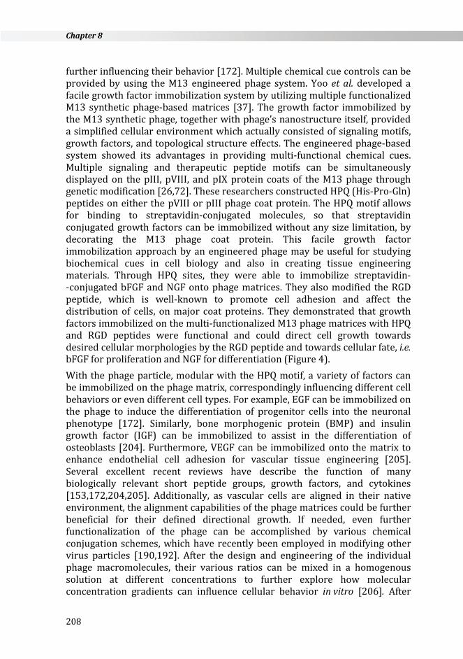
further influencing their behavior [172]. Multiple chemical cue controls can be provided by using the M13 engineered phage system. Yoo et al. developed a facile growth factor immobilization system by utilizing multiple functionalized M13 synthetic phage-based matrices [37]. The growth factor immobilized by the M13 synthetic phage, together with phage’s nanostructure itself, provided a simplified cellular environment which actually consisted of signaling motifs, growth factors, and topological structure effects. The engineered phage-based system showed its advantages in providing multi-functional chemical cues. Multiple signaling and therapeutic peptide motifs can be simultaneously displayed on the pIII, pVIII, and pIX protein coats of the M13 phage through genetic modification [26,72]. These researchers constructed HPQ (His-Pro-Gln) peptides on either the pVIII or pIII phage coat protein. The HPQ motif allows for binding to streptavidin-conjugated molecules, so that streptavidin conjugated growth factors can be immobilized without any size limitation, by decorating the M13 phage coat protein. This facile growth factor immobilization approach by an engineered phage may be useful for studying biochemical cues in cell biology and also in creating tissue engineering materials. Through HPQ sites, they were able to immobilize streptavidin- -conjugated bFGF and NGF onto phage matrices. They also modified the RGD peptide, which is well-known to promote cell adhesion and affect the distribution of cells, on major coat proteins. They demonstrated that growth factors immobilized on the multi-functionalized M13 phage matrices with HPQ and RGD peptides were functional and could direct cell growth towards desired cellular morphologies by the RGD peptide and towards cellular fate, i.e. bFGF for proliferation and NGF for differentiation (Figure 4). With the phage particle, modular with the HPQ motif, a variety of factors can be immobilized on the phage matrix, correspondingly influencing different cell behaviors or even different cell types. For example, EGF can be immobilized on the phage to induce the differentiation of progenitor cells into the neuronal phenotype [172]. Similarly, bone morphogenic protein (BMP) and insulin growth factor (IGF) can be immobilized to assist in the differentiation of osteoblasts [204]. Furthermore, VEGF can be immobilized onto the matrix to enhance endothelial cell adhesion for vascular tissue engineering [205]. Several excellent recent reviews have describe the function of many biologically relevant short peptide groups, growth factors, and cytokines [153,172,204,205]. Additionally, as vascular cells are aligned in their native environment, the alignment capabilities of the phage matrices could be further beneficial for their defined directional growth. If needed, even further functionalization of the phage can be accomplished by various chemical conjugation schemes, which have recently been employed in modifying other virus particles [190,192]. After the design and engineering of the individual phage macromolecules, their various ratios can be mixed in a homogenous solution at different concentrations to further explore how molecular concentration gradients can influence cellular behavior in vitro [206]. After
208
M13 bacteriophage nanomaterials for regerative medicine
such systematic analysis, the design parameters that work best can be incorporated into a final mix solution to be tested on in vivo systems.
Figure 4. Multifunctional phage-based tissue engineering materials. Neural progenitor cells cultured on top of synthetic phages responded to the growth factor immobilized by HPQ phages via streptavidin. The physical and chemical cues provided by synthetic
phages could control cellular behaviors [37].
8.4.4. Immune study of phage materials As the phage material we discussed is ultimately designed for in vivo applications, synthetic phage-based future works will explore both the in vitro and in vivo immunogenic response to phage matrices. We hypothesize that the phage matrix as a foreign protein mass will be recognized as a “non-self” material via the complement system [207]. In the immune privileged environment of the central nervous system, microglia, i.e. the specialized immune cells of the brain, will likely mediate the immune response [68,208]. Previous studies have seen no inflammation-related damage at phage targeted tissue sites [68]. However, if a greater concentration of the phage activates
209
Chapter 8
microglia, their recruitment to the site of injury may actually facilitate nerve tissue regeneration by enhancing the clearance of cellular and ECM debris in the glial scar, and expression of the growth factors, and the expression of native extracellular proteins such as laminin [209]. To explore a similar mechanism of action, there is currently a Phase II clinical trial study to test the efficacy of injecting macrophages into the site of spinal injury on stimulating regeneration [187]. In vitro immunogenic studies will be conducted to assess the potential of phage materials to induce an immunogenic inflammatory reaction. Similar to a study conducted by Ainslie et al. testing the inflammation reaction of the material nPTFE [210], a panel measuring the level of immunostimulatory or inhibitory cytokines can be performed on the supernatant from macrophages grown on phage substrates. Tissue culture polystyrene can serve as a negative control, and macrophages stimulated by lipopolysaccharides as a positive control. The levels of cytokines present can be assessed for their immunostimulatory and immunoinhibitory activity. If very high levels of immunostimulating molecules such as IL-1 or TNF-α are observed, the phage may be modified to express complement inhibiting peptides [211,212]. Furthermore, as was done in a study by Silva et al., in vivo studies can be performed by injecting the phage solution into the spinal cord area of rat subjects [11]. Following the injection, the behavior of the animals can be evaluated for changes. After culling the animal, the injection site can be evaluated using histological studies to assess tissue inflammation and fibrosis. A previous study that targeted an engineered phage solution to a β-amyloid plaques in the brain did not observe any adverse tissue reactions in the histological analysis [68].
8.4.5. Mechanical and degradation properties of phage materials Control of the mechanical and degradation properties of biomaterials are important for tissue engineering applications. In an optimal engineering scenario, the material that is intended to replace or repair a tissue will remain at the site of injury until it is remodeled by cells and replaced by the naturally produced ECM [153,182]. Previous work with hydrogels has demonstrated that both the concentration of the polymer macromolecule units and the degree of their cross-linking can be used to tune the mechanical properties and the rate of degradation of these materials [153,182]. The Lee group encapsulated the phage materials in an agarose gel to keep them stable in the media solution over the course of the experiment [35,39]. A future project that can further improve upon phage scaffolds is to increase their stability in aqueous media environments. Preliminary work conducted on crosslinking chemically biotinylated phages with streptavidin showed much improved stability of the phage fibers, which remained in solution for over a week without degradation [95].
210
M13 bacteriophage nanomaterials for regerative medicine
8.4.6. Gene delivery systems Drug delivery and tissue regeneration materials are often very closely related in both function and architecture. In fact, there is one perspective in the scientific community that tissue engineering scaffolds are just a delivery system of cells into the body [185]. Additionally, the line between the two areas becomes blurred when controlled growth factor or cytokine release is incorporated into the matrix to influence either the contained or the surrounding cells [172,182,185,204]. By the streptavidin crosslinking methods described above, small therapeutic drug molecules may be incorporated into the matrix. Furthermore, the link to the phage can be engineered to be dependent on enzymatic cleavage [153,213], so that the delivered molecules are released only when they are sequestered by cell activity. Therapeutic genetic material to induce tissue regeneration can be incorporated into the phage DNA and carried within the phage capsule for specific delivery to cells via receptor uptake [67]. As the M13 phage is non-lytic, they will be continuously produced by bacteria without causing bacterial wall rupture or the resulting debris. By designing peptide expression on the phage capsid, phages can be more locally targeted to cell receptors (i.e. via RGD or another ligand). Phage display technology has allowed for the identification of novel homing peptides that target unknown cell surface proteins. The targeting peptides can be incorporated into bacteriophage coat proteins through the genetic engineering techniques described previously [67]. These include peptides (RGD, glioma-binding peptide) [214,215], HER2 receptor targeting antibody [216], growth factors (EGF or FGF2) [217-219] and the penton base of adenovirus [220]. Similar to drug delivery, nucleic acid materials are now being incorporated into scaffolding materials for delivery to cells. Furthermore, it has been shown that DNA materials that are tethered to the matrix, rather than just encapsulated, are more effectively transferred to cells [221]. Phage particles engineered as described above to contain the genetic load for cell delivery, as well as specific cell targeting peptides, can be cross- -linked with streptavidin units to produce stable tissue engineering scaffolds. As these scaffolds are taken up and degraded by endocytosis [222], the phage could release its gene cargo and further induce cell behavior.
8.4.7. Diagnosis and therapeutic applications Thanks to phage display technology, we have considerable useful peptide information, which can be developed further for imaging and the diagnosis of certain diseases, such as cancer [223]. Such applications can be directly assayed by Phage-Chips [17]. Yoo et al. demonstrated multifunctional phage matrices that were optically readable for cell proliferation and morphology. The self-assembled nanofibrous network structure enhanced surface plasmon resonance (SPR) monitoring signals, and biochemical cues displayed by phage surfaces controlled cellular proliferation and morphology at the same time. For therapeutic applications, antibody phage display has been developed and
211
Chapter 8
tested for clinical approval [224]. Another application study of utilizing M13 engineered phage properties was also introduced by adopting different useful virus parts. Hajitou et al. constructed a hybrid phage with two genes from the phage and the nucleus integrating gene from AAV, called inverted terminal repeats (ITR). Additionally, these phages were engineered to express an integrin binding peptide on minor coat proteins. Therefore, the RGD peptide induced internalization of the phage through integrin-mediated endocytosis and the ITR led to improved transgene expression, which was linked to the function of the delivered gene in the cytoplasm. The resulting AAV/phage system provided superior tumor transduction over the phage alone. Topical delivery of this therapeutic synthetic phage material onto localized disease areas with specific integrating functions might reduce the risk of the side effects and enhance the efficiency of drug delivery.
8.4.8. Tissue engineering and regenerative medicine applications The aim of tissue engineering is to create desired artificial tissues or organ scaffold structures. Consequently, studies in this field require combining biomaterials and cells to closely mimic in vivo tissue environments. Natural tissue microenvironments are composed of networks of various nanofibrous proteins, such as collagen or fibronectin. Therefore, as discussed above, the M13 phage can be the predominant viral type utilized for this purpose because of its ability to display peptides on its coat proteins in various manners, its ability to replicate in large quantities, and its ability to self-assemble into nanofilaments. The multifunctional merits of M13 phage engineering can be further synergized with chemical modification or by conjugating growth factors. As a result of these traits, many studies have investigated using these phages as a platform to enhance the chemical, physical, and mechanical properties of biomimetic matrices (Figure 5). 2-D phage films with nanogroove topography using a facile shearing method or a layer-by-layer method were formed for neural progenitor cell regeneration and differentiation (left top in the Figure 5) [35,95]. In combination with signaling peptides on phage coats, physical properties, such as phage alignment and hierarchical ordering of phages, can help to control cell behavior (left bottom in the Figure 5) [36]. These phages displaying DGEA, a bone-specific peptide, have also been used for the engineering of hard tissues, such as bone (right top in the Figure 5) [39]. Multiple protein coat engineered M13 phages expressing HPQ, a biotin- -like peptide, on minor coat proteins and RGD on major and minor coat proteins promoted neural progenitor cell proliferation or differentiation according to the streptavidin-conjugated growth factors, which were specific for neural cells (right bottom in the Figure 5) [37]. Many of the uses of phages in tissue engineering applications are similar to those previously discussed. Although the lack of patentability and the food and drug administration (FDA) guidelines, as well as safety concerns, are still challenging, the success of previous studies using M13 phages for tissue
212

M13 bacteriophage nanomaterials for regerative medicine
engineering / regeneration research drives optimism that further clinical applications will emerge from this field. In vivo animal studies can help to further characterize the efficacy and safety of phage-based tissue engineering matrices.
Figure 5. Phage based tissue engineering for soft and hard tissues. M13 phage coat
proteins can be engineered for various tissue engineering purposes.
8.5. SUMMARY AND FUTURE PERSPECTIVES In this chapter, the use of M13 bacteriophages (phages) has been explored as a novel building block together with providing specific functions for tissue engineering / regeneration materials. Prior to using M13 as a biomimetic tissue engineering scaffold material, the phage is decorated with cell signaling motifs. An incredible diversity of peptide expression has been previously demonstrated on the various capsid proteins of the phage through the creation of peptide libraries [26,51,52]. A novel cloning approach to display an integrin binding RGD motif on every copy of pVIII was introduced to decorate the major coat protein of the M13 phage. Merzlyak et al. did this by using a partial library method, where an engineered octamer insert for pVIII included a constrained RGD group surrounded by a degenerate residue library. This allowed the expression of full inserts that retained the desired RGD motif yet were favorably compatible with all the protein interactions inherent in the phage replication process within E. coli. Furthermore, they systematically analyzed
213
Chapter 8
the allowed amino acid sequence space for pVIII inserts by making constrained libraries with chemically variable residues (positive, negative, and hydrophobic) [35]. This approach can be useful for engineering phage particles with a very dense uniform display of short signal peptide motifs that may be beneficial for tissue engineering materials [11,35,174,183]. After demonstrating the phage as a filament particle able to form aligned scaffolds that are both conducive and instructive to cell growth, further phage design improvements can be made by making it multifunctional. The phage was then engineered to incorporate an adhesive peptide motif (RGD) on pVIII, and a constrained biotin-like HPQ motif on the pIII protein [37,67,225]. There are limits in the ability of the phage to display a multivalency of protein molecules based on the size and sequence of the insert [26], and it cannot be altered via genetic means to present functional carbohydrate molecules. By exploiting the binding affinity of the biotin-streptavidin bond, with an engineered biotin-like HPQ group, we imparted modular functionality to the phage building block [37]. Any growth factor, cytokine, or otherwise therapeutic molecule conjugated to avidin will be able to bind to our engineered HPQ phage and further functionalize the matrix [37,226]. In summary, we have introduced the utilization of M13 bacteriophage as a functional building block for tissue engineering matrices that can guide the adhesion, polarization, and alignment behavior of cells and promote tissue regeneration. We have also presented a number of avenues that can be used to expand this area of research further to immune / chemokine study and use phages for highly functional and useful biomaterials with potential applications in diagnosis and therapy.
ACKNOWLEDGEMENTS This work was supported by the Basic Science Research Program through the National Research Foundation of Korea (NRF) funded by the Ministry of Science, ICT and Future Planning and the Korean Government (2013R1A1A3008484) and by the Korean Government (NRF- -2014S1A2A2027641).
214
M13 bacteriophage nanomaterials for regerative medicine
REFERENCES 1. A. Merzlyak, S.-W. Lee. Curr. Opin. Chem. Biol. 10 (2006) 246. 2. V.C. Sundar, A.D. Yablon, J.L. Grazul, M. Ilan, J. Aizenberg. Nature 424 (2003)
899. 3. J. Aizenberg, J.C. Weaver, M.S. Thanawala, V.C. Sundar, D.E. Morse, P. Fratzl.
Science 309 (2005) 275. 4. J.N. Cha, G.D. Stucky, D.E. Morse, T.J. Deming. Nature 403 (2000) 289. 5. S. Weiner, W. Traub, H.D. Wagner. J. Struct. Biol. 126 (1999) 241. 6. S.R. Whaley, D.S. English, E.L. Hu, P.F. Barbara, A.M. Belcher. Nature 405
(2000) 665. 7. S. Zhang. Nat. Biotechnol. 21 (2003) 1171. 8. S.-W. Lee, C. Mao, C.E. Flynn, A.M. Belcher. Science 296 (2002) 892. 9. J.M. Nam, C.S. Thaxton, C.A. Mirkin. Science 301 (2003) 1884.
10. Y. Huang, X. Duan, Y. Cui, L.J. Lauhon, K.H. Kim, C.M. Lieber. Science 294 (2001) 1313.
11. G.A. Silva, C. Czeisler, K.L. Niece, E. Beniash, D.A. Harrington, J.A. Kessler, S.I. Stupp. Science 303 (2004) 1352.
12. S. Zhang. Adv. Cancer Res. 99 (2008) 335. 13. Y.J. Lee, H. Yi, W.J. Kim, K. Kang, D.S. Yun, M.S. Strano, G. Ceder, A.M. Belcher.
Science 324(2009) 1051. 14. K.T. Nam, D.-W. Kim, P.J. Yoo, C.-Y. Chiang, N.L. Meethong, P.T. Hammond,
Y.-M. Chiang, A.M. Belcher. Science 312 (2006) 885. 15. M. Cerruti, J. Jaworski, D. Raorane, C. Zueger, J. Varadarajan, C. Carraro,
S.-W. Lee, R. Maboudian, A. Majumdar. Anal. Chem. 81(2009) 4192. 16. J.W. Jaworski, D. Raorane, J.H. Huh, A. Majumdar, S.W. Lee. Langmuir 24 (2008)
4938. 17. S.Y. Yoo, J.-W. Oh, S.-W. Lee. Langmuir 28 (2012) 2166. 18. D.J. Rodi, S. Mandova, L. Makowski, in Phage Display in Biotechnology and Drug
Discovery, S.S. Sidhu (Ed.), CRC Press Taylor & Francis Group, Boca Raton, FL, 2005, p. 748.
19. A. Jacobson. J Virol 10 (1972) 835. 20. N. Opalka, R. Beckmann, N. Boisset, M.N. Simon, M. Russel, S.A. Darst.
J. Mol. Biol. 325 (2003) 461. 21. M. Russel, N.A. Linderoth, A. Sali. Gene 192 (1997) 23. 22. B. Hohn, H. Lechner, D.A. Marvin. J. Mol. Biol. 56 (1971) 143. 23. A. Merzlyak, S.-W. Lee. Bioconjug. Chem. 20 (2009) 2300. 24. S.F. Parmley, G.P. Smith. Gene 73 (1988) 305. 25. J.K. Scott, G.P. Smith. Science 249 (1990) 386. 26. G.P. Smith, V.A. Petrenko. Chem. Rev. 97 (1997) 391. 27. Z.M. Carrico, D.W. Romanini, R.A. Mehl, M.B. Francis. Chem. Commun. (2008)
1205. 28. R.A. Miller, A.D. Presley, M.B. Francis. J. Am. Chem. Soc. 129 (2007) 3104. 29. S. Nicholas, C. Zacharywirt, F. Matthewwofx. Angew. Chem. Int. Ed. 48 (2009)
9498. 30. T.L. Schlick, Z. Ding, E.W. Kovacs, M.B. Francis. J. Am. Chem. Soc. 127 (2005)
3718. 31. G.P. Smith. Science 228 (1985) 1315.
215
Chapter 8
32. C.C. Liu, A.V. Mack, M.L. Tsao, J.H. Mills, H.S. Lee, H. Choe, M. Farzan, P.G. Schultz, V.V. Smider. Proc. Natl. Acad. Sci. U. S. A. 105 (2008) 17688.
33. T. Feng, M.L. Tsao, P.G. Schultz. J. Am. Chem. Soc. 126 (2004) 15962. 34. S.Y. Yoo, A. Merzlyak, S.-W. Lee. Mediat. Inflamm. 2014 (2014) 11. 35. A. Merzlyak, S. Indrakanti, S.-W. Lee. Nano Lett. 9 (2009) 846. 36. S.Y. Yoo, W.-J. Chung, T.H. Kim, M. Le, S.-W. Lee. Soft. Matter. 7 (2011) 363. 37. S.Y. Yoo, A. Merzlyak, S.-W. Lee. Soft. Matter. 7 (2011) 1660. 38. S.R. Bhattarai, S.Y. Yoo, S.-W. Lee, D. Dean. Biomaterials 33 (2012) 5166. 39. S.Y. Yoo, M. Kobayashi, P.P. Lee, S.-W. Lee. Biomacromolecules 12 (2011) 987. 40. S.Y. Yoo, J.-W. Oh, S.-W. Lee. Langmuir 28 (2012) 2166. 41. S.D Yanofsky, D.N. Baldwin, J.H. Butler, F.R. Holden, J.W. Jacobs,
P. Balasubramanian, J.P. Chinn, S.E. Cwirla, E. Peters-Bhatt, E.A. Whitehorn, E.H. Tate, A. Akeson, T.L. Bowlin, W.J. Dower, R.W. Barrett. Proc. Natl. Acad. Sci. 93 (1996) 7381.
42. A. Yayon, D. Aviezer, M. Safran, J.L. Gross, Y. Heldman, S. Cabilly, D. Givol, E. Katchalski-Katzir. Proc. Natl. Acad. Sci. 90 (1993) 10643.
43. W.J. Fairbrother, H.W. Christinger, A.G. Cochran, G. Fuh, C.J. Keenan, C. Quan, S.K. Shriver, J.Y. Tom, J.A. Wells, B.C. Cunningham. Biochemistry 37 (1998) 17754.
44. N.C. Wrighton, F.X. Farrell, R. Chang, A.K. Kashyap, F.P. Barbone, L.S. Mulcahy, D.L. Johnson, R.W. Barrett, L.K. Jolliffe, W.J. Dower. Science 273 (1996) 458.
45. F.-Y. Wang, T.-Y. Zhang, J.-X. Luo, G.-A. He, Q.-L. Gu, F. Xiao. Biosci. Biotech. Biochem. 70 (2006) 2035.
46. B. Li, S.J. Russell, D.M. Compaan, K. Totpal, S.A. Marsters, A. Ashkenazi, A.G. Cochran, S.G. Hymowitz, S.S. Sidhu. J. Mol. Biol. 361 (2006) 522.
47. J. Pande, M.M. Szewczyk, A.K. Grover. Biotechnol. Adv. 28 (2010) 849. 48. J. Pande, K.K. Mallhi, A.K. Grover. Eur. J. Pharmacol. 508 (2005) 1. 49. J. Pande, K.K. Mallhi, A. Sawh, M.M. Szewczyk, F. Simpson, A.K. Grover.
Am. J. Physiol. Cell Physiol. 290 (2006) C1341. 50. N.C. Gordon, S. Lien, J. Johnson, H.J. Wallweber, T. Tran, B. Currell, M. Mathieu,
C. Quan, M.A. Starovasnik, S.G. Hymowitz, R.F. Kelley. J. Mol. Biol. 396 (2010) 166.
51. V.A. Petrenko, G.P. Smith, X. Gong, T. Quinn. Protein Eng. 9 (1996) 797. 52. V.A. Petrenko, G.P. Smith, M.M. Mazooji, T. Quinn. Protein Eng. 15 (2002) 943. 53. G.P. Smith. Filament. Virol. 167 (1988) 156. 54. G.A. Kuzmicheva, P.K. Jayanna, I.B. Sorokulova, V.A. Petrenko.
Protein Eng. Des. Sel. 22 (2009) 9. 55. D.J. Rodi, A.S. Soares, L. Makowski. J. Mol. Biol. 322 (2002) 1039. 56. M.L. Scalley-Kim, B.W. Hess, R.L. Kelly, A.R. Krostag, K.H. Lustig, J.S. Marken,
P.J. Ovendale, A.R. Posey, P.J. Smolak, J.D. Taylor, C.L. Wood, D.L. Bienvenue, P. Probst, R.A. Salmon, D.S. Allison, T.M. Foy, C.J. Raport. PLoS ONE 7 (2012) 1.
57. K. Even-Desrumeaux, D. Nevoltris, M.N. Lavaut, K. Alim, J.P. Borg, S. Audebert, B. Kerfelec, D. Baty, P. Chames. Mol. Cell. Proteomics 13 (2013) 653.
58. H.-M. Zheng, Y. Jiang, J.-R. Wang, X.-L. Gong, B.-Y. Guo. Inflamm. Res. 60 (2011) 759.
59. C. Blanchetot, D. Verzijl, A. Mujić-Delić, L. Bosch, L. Rem, R. Leurs, C.T. Verrips, M. Saunders, H. de Haard, M.J. Smit. J. Biol. Chem. 288 (2013) 25173.
60. S.W. Lee, B.M. Wood, A.M. Belcher. Langmuir 19 (2003) 1592. 61. A. Merzlyak, S.W. Lee. Curr. Opin. Chem. Biol. 10 (2006) 246.
216

M13 bacteriophage nanomaterials for regerative medicine
62. R.R. Naik, S.J. Stringer, G. Agarwal, S.E. Jones, M.O. Stone. Nature Mater. 1 (2002) 169.
63. K.T. Nam, D.W. Kim, P.J. Yoo, C.Y. Chiang, N. Meethong, P.T. Hammond, Y.M. Chiang, A.M. Belcher. Science 312 (2006) 885.
64. H. Bermudez, A.P. Hathorne. Faraday Discuss. 139 (2008) 327. 65. T.J. Dickerson, K.D. Janda. AAPS J 7 (2005) E579. 66. D.N. Krag, G.S. Shukla, G.P. Shen, S. Pero, T. Ashikaga, S. Fuller, D.L. Weaver,
S. Burdette-Radoux, C. Thomas. Cancer Res. 66 (2006) 8925. 67. A. Hajitou, M. Trepel, C.E. Lilley, S. Soghomonyan, M.M. Alauddin, F.C. Marini
3rd, B.H. Restel, M.G. Ozawa, C.A. Moya, R. Rangel, Y. Sun, K. Zaoui, M. Schmidt, C. von Kalle, M.D. Weitzman, J.G. Gelovani, R. Pasqualini, W. Arap. Cell 125 (2006) 385.
68. D. Frenkel, B. Solomon. Proc. Natl. Acad. Sci. U S A 99 (2002) 5675. 69. L.S. Jespers, J.H. Messens, A. De Keyser, D. Eeckhout, I. Van den Brande,
Y.G. Gansemans, M.J. Lauwereys, G.P. Vlasuk, P.E. Stanssens. Nat. Biotech. 13 (1995) 378.
70. S.M. Changshou Gao, C.-H. L. Lo, P. Wirsching, R.A. Lerner, K.D. Janda. Proc. Natl. Acad. Sci. U S A 96 (1999) 6025.
71. C. Gao, S. Mao, G. Kaufmann, P. Wirsching, R.A. Lerner, K.D. Janda. Proc. Natl. Acad. Sci. 99 (2002) 12612.
72. V. Petrenko, G. Smith, in Phage Display In Biotechnology and Drug Discovery, CRC Press, 2005, p. 63.
73. D.S. Choi, H.-E. Jin, S.Y. Yoo, S.-W. Lee. Bioconjug. Chem. 25 (2014) 216. 74. S.W. Lee, A.M. Belcher, Nano Lett. 4 (2004) 387. 75. Y.L. Liu, J.D. Adams, K. Turner, F.V. Cochran, S.S. Gambhir, H.T. Soh. Lab Chip 9
3604 (2009) 1033. 76. B. Cao, C. Mao. Biomacromolecules 10 (2009) 555. 77. Y. Chen, C. Wiesmann, G. Fuh, B. Li, H.W. Christinger, P. McKay, A.M. de Vos,
H.B. Lowman. J. Mol. Biol. 293 (1999) 865. 78. J.J. Devlin, L.C. Panganiban, P.E. Devlin. Science 249 (1990) 404. 79. X. Cheng, B.K. Kay, R.L. Juliano. Gene 171 (1996) 1. 80. E. Ruoslahti. Annu. Rev. Cell Dev. Biol. 12 (1996) 697. 81. R.J. Giordano, M. Vila, J.L. Ahdenranta, R. Apasqualini, W. Arap. Nat. Med. 7
(2001) 1249. 82. R. Pasqualini, E. Ruoslahti. Nature 380 (1996) 364. 83. R. Brissette, N.I. Goldstein, in Methods in Molecular Biology, P. Fisher (Ed.),
Humana Press, Inc, Totowa, NJ, 2007, p. 203. 84. R. Li, R.H. Hoess, J.S. Bennett, W.F. DeGrado. Protein Eng. 16 (2003) 65. 85. J. Lubkowski, F. Hennecke, A. Pluckthun, A. Wlodawer. Nat. Struct. Mol. Biol. 5
(1998) 140. 86. S.S. Sidhu, G.A. Weiss, J.A. Wells. J. Mol. Biol. 296 (2000) 487. 87. S. Nicholas, C. Zachary, F. Matthew. Angew. Chem. Int. Ed. 48 (2009) 9498. 88. G. Whitesides, J. Mathias, C. Seto. Science 254 (1991) 1312. 89. W.-J. Chung, D.-Y. Lee, S.Y. Yoo. Int. J. Nanomedicine 9 (2014) 5825. 90. R.B. Spruijt, C.J.A.M. Wolfs, J.W.G. Verver, M.A. Hemminga. Biochemistry 35
(1996) 10383. 91. M.A. Hemminga, W.L. Vos, P.V. Nazarov, R.B.M. Koehorst, C.J.A.M. Wolfs,
R.B. Spruijt, D. Stopar. Eur. Biophys. J. 39 (2010) 541.
217
Chapter 8
92. A.R. Khan, K.A. Williams, J.M. Boggs, C.M. Deber. Biochemistry 34 (1995) 12388.
93. C. Heinis, T. Rutherford, S. Freund, G. Winter. Nat. Chem. Biol. 5 (2009) 502. 94. J. Rong, L.A. Lee, K. Li, B. Harp, C.M. Mello, Z. Niu, Q. Wang. Chem. Commun. 41
(2008) 5185. 95. W.-J. Chung, A. Merzlyak, S.-W. Lee. Soft. Matter. 6 (2010) 4454. 96. K. Li, Y. Chen, S. Li, H.G. Nguyen, Z. Niu, S. You, C.M. Mello, X. Lu, Q. Wang.
Bioconjug. Chem. 21 (2010) 1369. 97. K.A. Kelly, P. Waterman, R. Weissleder. Neoplasia 8 (2006) 1011. 98. Z. Zhang, J. Buitenhuis, A. Cukkemane, M. Brocker, M. Bott, J.K.G. Dhont
Langmuir 26 (2010) 10593. 99. M. Adhikari, S. Dhamane, A.E. Hagström, G. Garvey, W.H. Chen, K. Kourentzi,
U. Strych, R.C. Willson. Analyst. 138 (2013) 5584. 100. I. Yacoby, M. Shamis, H. Bar, D. Shabat, I. Benhar.
Antimicrob. Agents Chemother. 50 (2006) 2087. 101. Z.M. Carrico, M.E. Farkas, Y. Zhou, S.C. Hsiao, J.D. Marks, H. Chokhawala,
D.S. Clark, M.B. Francis. ACS Nano 6 (2012) 6675. 102. J.A. Lamboy, P.Y. Tam, L.S. Lee, P.J. Jackson, S.K. Avrantinis, H.J. Lee, R.M. Corn,
G.A. Weiss. ChemBioChem. 9 (2008) 2846. 103. S. Ng, M.R. Jafari, W.L. Matochko, R. Derda. ACS Chem. Biol. 7 (2012), 1482. 104. M. Murugesan, G. Abbineni, S.L. Nimmo, B. Cao, C. Mao. Sci. Rep. 3 (2013) 1820. 105. G.M. Whitesides, B. Grzybowski. Science 295 (2002) 2418. 106. R.M. Capito, H.S. Azevedo, Y.S. Velichko, A. Mata, S.I. Stupp. Science 319 (2008)
1812. 107. J.-S. Moon, W.-G. Kim, C. Kim, G.-T. Park, J. Heo, S.Y. Yoo, J.-W. Oh.
Mini-Rev. Org. Chem. 12 (2015) 271. 108. S. Giménez, T. Lana-Villarreal, R. Gómez, S. Agouram, V. Muñoz-Sanjosé,
I. Mora-Seró. J. Appl. Phys. 108 (2010) 064310. 109. V.M. Karpan, P.A. Khomyakov, A.A. Starikov, G. Giovannetti, M. Zwierzycki,
M. Talanana, G. Brocks, J. van den Brink, P.J. Kelly. Phys. Rev. B 78 (2008) 195419.
110. H.D. Espinosa, A.L. Juster, F.J. Latourte, O.Y. Loh, D. Gregoire, P.D. Zavattieri. Nat. Commun. 2 (2011) 173.
111. J. Aizenberg, J.C. Weaver, M.S. Thanawala, V.C. Sundar, D.E. Morse, P. Fratzl. Science 309 (2005) 275.
112. H. Budzikiewicz. Bacterial Mini Rev. Org. Chem. 3 (2006) 93. 113. N.S. Malvankar, M. Vargas, K.P. Nevin, A.E. Franks, C. Leang, B.C. Kim, K. Inoue,
T. Mester, S.F. Covalla, J.P. Johnson, V.M. Rotello, M.T. Tuominen, D.R. Lovley. Nat. Nano 6 (2011) 573.
114. H. Yan, S.H. Park, G. Finkelstein, J.H. Reif, T.H. LaBean. Science 301 (2003) 1882.
115. C. Mao, D.J. Solis, B.D. Reiss, S.T. Kottmann, R.Y. Sweeney, A. Hayhurst, G. Georgiou, B. Iverson, A.M. Belcher. Science 303 (2004) 213.
116. S.W. Lee, C.B. Mao, C.E. Flynn, A.M. Belcher. Science 296 (2002) 892. 117. Y. Lee, J. Kim, D.S. Yun, Y.S. Nam, Y. Shao-Horn, A.M. Belcher.
Energy Environ. Sci. 5 (2012) 8328. 118. X. Dang, H. Yi, M.H. Ham, J. Qi, D.S. Yun, R. Ladewski, M.S. Strano,
P.T. Hammond, A.M. Belcher. Nat. Nano 6 (2011) 377. 119. Z. Tang, Y. Wang, P. Podsiadlo, N.A. Kotov. Adv. Mater. 18 (2006) 3203.
218

M13 bacteriophage nanomaterials for regerative medicine
120. C. Jiang, V.V. Tsukruk. Adv. Mater. 18 (2006) 829. 121. A. Javey, Nam, R.S. Friedman, H. Yan, C.M. Lieber. Nano Lett. 7 (2007) 773. 122. X. Peng, J. Jin, Y. Nakamura, T. Ohno, I. Ichinose. Nat. Nano 4 (2009) 353. 123. S. Karan, S. Samitsu, X. Peng, K. Kurashima, I. Ichinose. Science 335 (2012) 444. 124. C.C. Striemer, T.R. Gaborski, J.L. McGrath, P.M. Fauchet. Nature 445 (2007)
749. 125. J. He, X.-M. Lin, H. Chan, L. Vuković, P. Král, H.M. Jaeger. Nano Lett. 11 (2011)
2430. 126. H.-W. Liang, L. Wang, P.-Y. Chen, H.-T. Lin, L.-F. Chen, D. He, S.-H. Yu.
Adv. Mater.22 (2010) 4691. 127. X.W. Zhang, T. Zhang, J. Ng, D.D. Sun, Adv. Funct. Mater. 19 (2009) 3731. 128. Y.M. Lee, B. Jung, Y.H. Kim, A.R. Park, S. Han, W.S. Choe, P.J. Yoo. Adv. Mater. 26
(2014) 3899. 129. J. Kim, L.J. Cote, F. Kim, W. Yuan, K.R. Shull, J. Huang. J. Am. Chem. Soc. 132
(2010) 8180. 130. T. Sawada, T. Serizawa. J. Mater. Chem. B 1 (2013) 149. 131. X. Chen, K. Gerasopoulos, J. Guo, A. Brown, C. Wang, R. Ghodssi, J.N. Culver.
ACS Nano 4 (2010) 5366. 132. B.Y. Lee, J. Zhang, C. Zueger, W.J. Chung, S.Y. Yoo, E. Wang, J. Meyer, R. Ramesh,
S.W. Lee. Nat. Nanotechnol. 7 (2012) 351. 133. P.Y. Chen, X. Dang, M.T. Klug, J. Qi, N.M. Dorval Courchesne, F.J. Burpo, N. Fang,
P.T. Hammond, A.M. Belcher. ACS Nano 7 (2013) 6563. 134. P.-Y. Chen, M. Nasim Hyder, D. Mackanic, N.-M. Dorval Courchesne, J. Qi,
M.T. Klug, A.M. Belcher, P.T. Hammond. Adv. Mater. 26 (2014) 5069. 135. Q. Liu, M.H. Nayfeh, S.-T. Yau. J. Power Sources 195 (2010) 7480. 136. J.H. Hur, K.H. Im, S.W. Kim, U.J Kim, J.H. Lee, S.Y. Hwang, J.J. Song, S.J. Kim,
S.W. Hwang, N.Y. Park. J. Mater. Chem. A 1 (2013) 14460. 137. V. Gupta, N. Miura. Electrochim. Acta 52 (2006) 1721. 138. Y.-K. Zhou, B.-L. He, W.-J. Zhou, H.-L. Li. J. Electrochem. Soc. 151 (2004) A1052. 139. T. Abdiryim, A. Ubul, R. Jamal, A. Rahman. Materials 5 (2012) 1219. 140. J. Ge, G. Cheng, L. Chen. Nanoscale 3 (2011) 3084. 141. J. Liu, J. Sun, L. Gao. J. Phys. Chem. C 114 (2010), 19614. 142. Z.Q. Niu, P.S. Luan, Q. Shao, H. Dong, J.Z. Li, J. Chen, D. Zhao, L. Cai, W.Y. Zhou,
X.D. Chen, S.S. Xie Energy Environ. Sci.5 (2012) 8726. 143. K. Wang, P. Zhao, X. Zhou, H. Wu, Z. Wei. J. Mater. Chem. 21 (2011) 16373. 144. V. Gupta, N. Miura. J. Power Sources 157 (2006) 616. 145. K.A. Kilian, B. Bugarija, B.T. Lahn, M. Mrksich. P. Natl. Acad. Sci. 107 (2010)
4872. 146. M.J.P. Biggs, R.G. Richards, N. Gadegaard, R.J. McMurray, S. Affrossman,
C.D.W. Wilkinson, R.O.C. Oreffo, M.J. Dalby. J. Biomed. Mater. Res. Part A 91A (2009) 195.
147. M.J.P. Biggs, R.G. Richards, S. McFarlane, C.D.W. Wilkinson, R.O.C. Oreffo, M.J. Dalby. J. R. Soc. Interface 5 (2008) 1231.
148. A.J. Engler, S. Sen, H.L. Sweeney, D.E. Discher. Cell 126 (2006) 677. 149. J. Wang, M. Yang, Y. Zhu, L. Wang, A.P. Tomsia, C. Mao. Adv. Mater. 26 (2014)
4961. 150. J. Street, M. Bao, L. deGuzman, S. Bunting, F.V. Peale Jr, N. Ferrara, H. Steinmetz,
J. Hoeffel, J.L. Cleland, A. Daugherty, N. van Bruggen, H.P. Redmond, R.A. Carano, E.H. Filvaroff. Proc. Natl. Acad. Sci. U. S. A. 99 (2002) 9656.
219
Chapter 8
151. M.J. Lysaght, A. Jaklenec, E. Deweerd. Tissue Eng. Part A 14 (2008) 305. 152. R.G. Flemming, C.J. Murphy, G.A. Abrams, S.L. Goodman, P.F. Nealey.
Biomaterials 20 (1999) 573. 153. J.F. Pollock, K. Healy, in Strategies in Regenerative Medicine, M. Santin (Ed.),
Springer New York, 2009, p. 97. 154. R. Raghow. FASEB J 8 (1994) 823. 155. T. Zigova, E.Y. Snyder, P.R. Sanberg (Eds.), Neural Stem Cells for Brain and
Spinal Cord Repair, Humana Press, Totowa, NJ, 2003. 156. M.M. Stevens, J.H. George. Science 310 (2005) 1135. 157. M.J. Dalby, M.O. Riehle, H. Johnstone, S. Affrossman, A.S. Curtis. Cell. Biol. Int. 28
(2004) 229. 158. M.J. Dalby, N. Gadegaard, R. Tare, A. Andar, M.O. Riehle, P. Herzyk,
C.D.W. Wilkinson, R.O.C. Oreffo. Nat. Mater. 6 (2007) 997. 159. F. Yang, R. Murugan, S. Wang, S. Ramakrishna. Biomaterials 26 (2005) 2603. 160. D. Ceballos, X. Navarro, N. Dubey, G. Wendelschafer-Crabb, W.R. Kennedy,
R.T. Tranquillo. Exp. Neurol. 158 (1999) 290. 161. P. Clark, P. Connolly, A.S. Curtis, J.A. Dow, C.D. Wilkinson. J. Cell. Sci. 99 (Pt 1)
(1991) 73. 162. A.S. Crouch, D. Miller, K.J. Luebke, W. Hu. Biomaterials 30 (2009) 1560. 163. P. Weiss. J. Exp. Zool. 100 (1945) 353. 164. M.J. Dalby. Med. Eng. Phys. 27 (2005) 730. 165. N. Dubey, P.C. Letourneau, R.T. Tranquillo. Exp. Neurol. 158 (1999) 338. 166. G.A. Dunn, J.P. Heath. Exp. Cell. Res. 101 (1976) 1. 167. A.B.J. Alberts, J. Lewis, M. Raff, K. Roberts, P. Walter (Eds.), Molecular Biology of
the Cell, Garland Science, New York and London, 4th Ed., 2002. 168. H.G. Hentschel, A. van Ooyen. Proc. Biol. Sci. 266 (1999) 2231. 169. M.E. Fini. Prog. Retin. Eye Res. 18 (1999) 529. 170. R. Milner, I.L. Campbell. J. Neurosci. Res. 69 (2002) 286. 171. R.C. Burrows, D. Wancio, P. Levitt, L. Lillien. Neuron 19 (1997) 251. 172. Y. Ito. Soft. Matter. 4 (2008) 46. 173. G. Maheshwari, G. Brown, D.A. Lauffenburger, A. Wells, L.G. Griffith.
J. Cell. Sci. 113 (Pt 10) (2000) 1677. 174. J.C. Schense, J. Bloch, P. Aebischer, J.A. Hubbell. Nat. Biotechnol. 18 (2000) 415. 175. E. Engel, A. Michiardi, M. Navarro, D. Lacroix, J.A. Planell. Trends Biotechnol. 26
(2008) 39. 176. J.J. Norman, T.A. Desai. Ann. Biomed. Eng. 34 (2006) 89. 177. A. Curtis, C. Wilkinson. Biomaterials 18 (1997) 1573. 178. P.X. Ma. Adv. Drug. Deliv. Rev. 60 (2008) 184. 179. M. Goldberg, R. Langer, X. Jia. J. Biomater. Sci. Polym. Ed. 18 (2007) 241. 180. P.X. Ma, in Encyclopedia of Polymer Science and Technology, J.I. Kroschwitz
(Ed.), JohnWiley & Sons Inc, Hoboken, NJ, 2005, vol. 12, p. 261. 181. J. Guan, K.L. Fujimoto, M.S. Sacks, W.R. Wagner. Biomaterials 26 (2005) 3961. 182. J. L. Drury, D. J. Mooney, Biomaterials 24 (2003), 4337. 183. K. Saha, E.F. Irwin, J. Kozhukh, D.V. Schaffer, K.E. Healy. J. Biomed. Mater. Res. A
81 (2007) 240. 184. E. Engel, A. Michiardi, M. Navarro, D. Lacroix, J. A. Planell. Trends Biotechnol. 26
(2008) 39. 185. M. Goldberg, R. Langer, X.Q. Jia. J. Biomat. Sci. Polym. E 18 (2007) 241.
220
M13 bacteriophage nanomaterials for regerative medicine
186. J.H. Huang, S.V. Gräter, F. Corbellini, S. Rinck-Jahnke, E. Bock, R. Kemkemer, H. Kessler, J.D. Ding, J.P. Spatz. Nano Lett. 9 (2009) 1111.
187. M. Arnold, M. Schwieder, J. Blümmel, E.A. Cavalcanti-Adam, M. López-Garcia, H. Kessler, B. Geiger, J.P. Spatz. Soft. Matter. 5 (2009) 72.
188. N. Gomez, S.C. Chen, C.E. Schmidt, J. R. Soc. Interface 4 (2007) 223. 189. J.M. Goddard, J.H. Hotchkiss. Prog. Polym. Sci. 32 (2007) 698. 190. E. Strable, M.G. Finn, in Viruses and Nanotechnology, M. Manchester,
N.F. Steinmetz (Eds.), Springer-Verlag, Berlin, 2009, vol. 327, pp. 1–21. 191. S.S. Gupta, J. Kuzelka, P. Singh, W.G. Lewis, M. Manchester, M.G. Finn.
Bioconjug. Chem. 16 (2005) 1572. 192. E.W. Kovacs, J.M. Hooker, D.W. Romanini, P.G. Holder, K.E. Berry, M.B. Francis.
Bioconjug. Chem. 18 (2007) 1140. 193. Y. Huang, C.-Y. Chiang, S.K. Lee, Y. Gao, E.L. Hu, J.D. Yoreo, A.M. Belcher.
Nano Lett. 5 (2005) 1429. 194. S.K. Lee, D.S. Yun, A.M. Belcher. Biomacromolecules 7 (2006) 14. 195. A. Merzlyak, S. Indrakanti, S.W. Lee. Nano Lett. 9 (2009) 846. 196. J. Light, R.A. Lerner. Bioorg. Med. Chem. Lett. 2 (1992) 1073. 197. G. Iannolo, O. Minenkova, R. Petruzzelli, G. Cesareni. J. Mol. Biol. 248 (1995)
835. 198. G.A. Kuzmicheva, P.K. Jayanna, I.B. Sorokulova, V.A. Petrenko.
Protein Eng. Des. Sel. 22 (2009) 9. 199. L. Makowski. Gene 128 (1993) 5. 200. W.-J. Chung, A. Merzlyak, S.Y. Yoo, S.-W. Lee. Langmuir 26(2010) 9885. 201. Y. Lin, E. Balizan, L.A. Lee, Z. Niu, Q. Wang. Angew. Chem. Int. Ed. 49 (2010) 868. 202. B. Grimpe, J. Silver. Prog. Brain. Res. 137 (2002) 333. 203. C. H. Streuli, N. Akhtar. Biochem. J. 418 (2009) 491. 204. T. Boontheekul, D.J. Mooney. Curr. Opin. Biotechnol. 14 (2003) 559. 205. A. de Mel, G. Jell, M.M. Stevens, A.M. Seifalian. Biomacromolecules 9 (2008)
2969. 206. L.G. Griffith, M.A. Swartz. Nat. Rev. Mol. Cell. Biol. 7 (2006) 211. 207. K. Elward, P. Gasque. Mol. Immunol. 40 (2003) 85. 208. G.W. Kreutzberg. Trends Neurosci. 19 (1996) 312. 209. W.J. Streit, Microglia in the Regenerating and Degenerating Central Nervous
System, W.J. Streit (Ed.), Springer, New York, 2002, p. 315. 210. K.M. Ainslie, E.M. Bachelder, S. Borkar, A.S. Zahr, A. Sen, J.V. Badding,
M.V. Pishko. Langmuir 23 (2007) 747. 211. R.J. Johnson, S.A. Maves, in USPTO C. Baxter Healthcare (Ed.), Innate Biotech
Inc., USA, 2004. 212. A. Kapil, B. Kundu, S.K. Khare. Protein Pept. Lett. 4 (1997) 405. 213. H. Bar, I. Yacoby, I. Benhar. BMC Biotechnol. 8 (2008) 14. 214. S.L. Hart, A.M. Knight, R.P. Harbottle, A. Mistry, H.D. Hunger, D.F. Cutler,
R. Williamson, C. Coutelle. J.Biol. Chem. 269 (1994) 12468. 215. J.D. Mount, T.I. Samoylova, N.E. Morrison, N.R. Cox, H.J. Baker, V.A. Petrenko.
Gene 341(2004) 59. 216. M.-A. Poul, J.D. Marks. J. Mol. Biol. 288 (1999) 203. 217. M.A. Bur, K. Jensen-Pergakes, A.M. Gonzalez, P. Ravey, A. Baird, D. Larocca.
Cancer Res. 62 (2002) 977. 218. D. Larocca, A. Witte, W. Johnso, G.F. Pierce, A. Baird. Hum. Gene Ther. 9 (1998)
2393.
221

Chapter 8
219. Y. Seow, M.J. Wood. Mol. Ther. 17 (2009) 767. 220. S. Piersanti, G. Cherubini, Y. Martina, B. Salone, Da. Avitabile, F. Grosso,
E. Cundari, G. Di Zenzo, I. Saggio. J. Mol. Med. 82 (2004) 467. 221. T. Segura, L.D. Shea. Bioconjug. Chem. 13 (2002) 621. 222. V.V. Ivanenkov, F. Felici, A.G. Menon. Biochim. Biophys. Acta – Molecular Cell
Research 1448 (1999) 450. 223. S.L. Deutscher. Chem. Rev. 110 (2010) 3196. 224. T.M. Holger Thie, T. Schirrmann, M. Hust, S. Dubel. Curr. Pharm. Biotechnol. 9
(2008) 439. 225. L. B. Giebel, R.T. Cass, D.L. Milligan, D.C. Young, R. Arze, C.R. Johnson.
Biochemistry 34 (1995) 15430. 226. S. Y. Yoo, S. M. Kwon. Mediat. Inflamm. 2013 (2013) 11.
222