Bacteriophage - microinmuno.qb.fcen.uba.ar
21
Chapter 7 Bacteriophage Bacteriophage or phage for short are viruses that infect only bacteria. In contrast to cells that grow from an increase in the number of their components and reproduce by division, viruses are assembled from pre-made components. Viruses are nucleic acid molecules surrounded by a protective coating. They are not capable of generat- ing energy and reproduce inside of cells. The nucleic acid inside the coating, called the phage genome in a bacteriophage, encodes most of the gene products needed for making more phage. The phage genome can be made of either double- or single-stranded DNA or RNA, depending on the bacteriophage in question. The genome can be circular or linear. The protective coating or capsid surrounding the phage genome is composed of phage-encoded proteins. Many important discoveries have been made using phage as model systems. From the discovery that a nonsense codon stopped protein synthesis to the first develop- mental switch to be understood at the molecular level, phage have proven to be very useful. In this chapter, we will look at phage development using T4, l (lambda), P1, and M13 as examples. Each of these phage infect E. coli. We will examine specific dis- coveries using these phage or specific properties of the phage that have made them particularly useful to biologists. The structure of phage All phage have a chromosome encased in a capsid that is composed of phage-encoded proteins. For many phage types, the capsid is attached to a tail structure that is also made from phage-encoded proteins. T4 and P1 contain a linear double-stranded DNA genome enclosed in a capsid and attached to a tail (Fig. 7.1a). The T4 genome is 172 kb, while P1 is a smaller phage with a genome of 90 kb. The T4 capsid is an elongated icosahedron. T4 has a very elaborate tail structure including a collar at the base of the head and a rigid tail core surrounded by a contractile sheath. The core and sheath are attached to a hexagonal base plate. Also attached to the tail plate are tail pins and six kinked tail fibers. P1 also has an icosahedral capsid, a tail with a contractile sheath, a base plate, and tail fibers. l contains a linear double-stranded DNA genome of 48.5 kb, a capsid, and a tail (Fig. 7.1b). The finished capsid is again shaped like an icosahedron whereas the tail is a thin flexible tube that ends in a small conical part and a single tail fiber. M13 contains a circular single-stranded DNA genome of 6407 nucleotides sur- rounded by five phage-encoded proteins (Fig. 7.1c). The M13 chromosome is coated FYI 7.1 Discovery of phage Phage were first described in 1915 by Frederick Twort and 1917 by Felix d’Herelle. Both men discovered phage when the bacteria they were working with lysed. The agent responsible for the lysis was transferable from culture to culture, invisible by light microscopy, and would go through the smallest filter they had. d’Herelle coined the term “bacteriophage”, signifying an entity that eats bacteria.
-
Upload
vuongnguyet -
Category
Documents
-
view
236 -
download
1
Transcript of Bacteriophage - microinmuno.qb.fcen.uba.ar

Chapter 7
Bacteriophage
Bacteriophage or phage for short are viruses that infect only bacteria. In contrastto cells that grow from an increase in the number of their components and reproduceby division, viruses are assembled from pre-made components. Viruses are nucleicacid molecules surrounded by a protective coating. They are not capable of generat-ing energy and reproduce inside of cells. The nucleic acid inside the coating, called the phage genome in a bacteriophage, encodes most of the gene products neededfor making more phage. The phage genome can be made of either double- or single-stranded DNA or RNA, depending on the bacteriophage in question. Thegenome can be circular or linear. The protective coating or capsid surrounding thephage genome is composed of phage-encoded proteins.
Many important discoveries have been made using phage as model systems. Fromthe discovery that a nonsense codon stopped protein synthesis to the first develop-mental switch to be understood at the molecular level, phage have proven to be veryuseful. In this chapter, we will look at phage development using T4, l (lambda), P1,and M13 as examples. Each of these phage infect E. coli. We will examine specific dis-coveries using these phage or specific properties of the phage that have made themparticularly useful to biologists.
The structure of phage
All phage have a chromosome encased in a capsid that is composed of phage-encodedproteins. For many phage types, the capsid is attached to a tail structure that is alsomade from phage-encoded proteins. T4 and P1 contain a linear double-stranded DNAgenome enclosed in a capsid and attached to a tail (Fig. 7.1a). The T4 genome is 172kb, while P1 is a smaller phage with a genome of 90kb. The T4 capsid is an elongatedicosahedron. T4 has a very elaborate tail structure including a collar at the base of thehead and a rigid tail core surrounded by a contractile sheath. The core and sheath areattached to a hexagonal base plate. Also attached to the tail plate are tail pins and sixkinked tail fibers. P1 also has an icosahedral capsid, a tail with a contractile sheath, abase plate, and tail fibers. l contains a linear double-stranded DNA genome of 48.5kb,a capsid, and a tail (Fig. 7.1b). The finished capsid is again shaped like an icosahedronwhereas the tail is a thin flexible tube that ends in a small conical part and a single tailfiber. M13 contains a circular single-stranded DNA genome of 6407 nucleotides sur-rounded by five phage-encoded proteins (Fig. 7.1c). The M13 chromosome is coated
FYI 7.1
Discovery of phage
Phage were first described in1915 by Frederick Twort and1917 by Felix d’Herelle. Bothmen discovered phage whenthe bacteria they were workingwith lysed. The agentresponsible for the lysis wastransferable from culture toculture, invisible by lightmicroscopy, and would gothrough the smallest filter theyhad. d’Herelle coined the term“bacteriophage”, signifying anentity that eats bacteria.
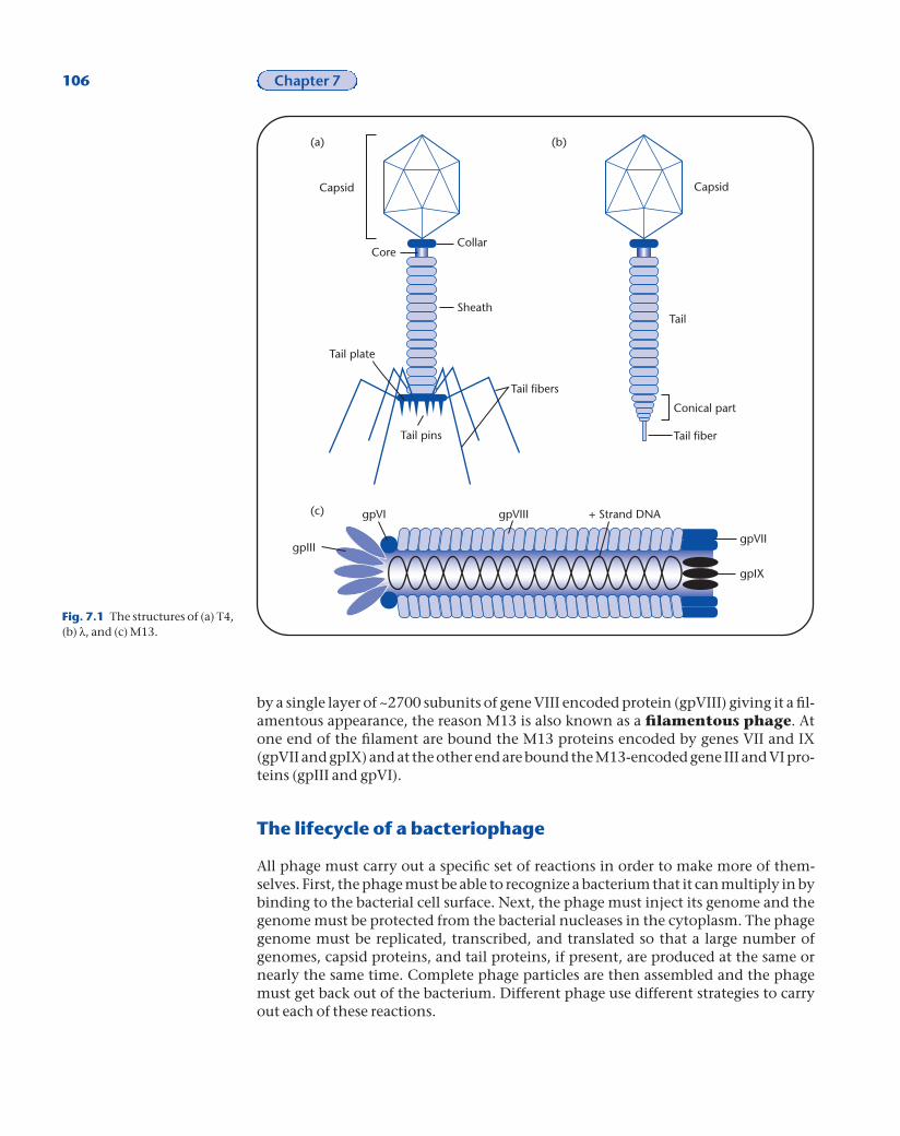
by a single layer of ~2700 subunits of gene VIII encoded protein (gpVIII) giving it a fil-amentous appearance, the reason M13 is also known as a filamentous phage. Atone end of the filament are bound the M13 proteins encoded by genes VII and IX(gpVII and gpIX) and at the other end are bound the M13-encoded gene III and VI pro-teins (gpIII and gpVI).
The lifecycle of a bacteriophage
All phage must carry out a specific set of reactions in order to make more of them-selves. First, the phage must be able to recognize a bacterium that it can multiply in bybinding to the bacterial cell surface. Next, the phage must inject its genome and thegenome must be protected from the bacterial nucleases in the cytoplasm. The phagegenome must be replicated, transcribed, and translated so that a large number ofgenomes, capsid proteins, and tail proteins, if present, are produced at the same ornearly the same time. Complete phage particles are then assembled and the phagemust get back out of the bacterium. Different phage use different strategies to carryout each of these reactions.
106 Chapter 7
Capsid Capsid
Tail
Conical part
Tail fiber
CollarCore
Sheath
Tail fibers
Tail pins
Tail plate
+ Strand DNA
gpVII
gpIX
gpIII
gpVI gpVIII
(a) (b)
(c)
Fig. 7.1 The structures of (a) T4,(b) l, and (c) M13.
Phage are very choosy as to what bacteria they infect. This is referred to as the hostrange of the phage. For example, l only infects certain E. coli, whereas Spo1 phage infect only Bacillus subtilis. Several phage types may infect a single bacterial species. E. coli can be infected by l, M13, P1, T4, and Mu phages, to name a few.
The number of phage that can be released from one bacterium after infection andgrowth by one phage is known as the burst size. Every phage has a characteristicburst size. Different phage also take different amounts of time to go through onegrowth cycle. We know when a phage has successfully reproduced when we are ableto detect plaques or circular areas with little or no bacterial growth on an agar platecovered with a thin layer of bacteria.
Once bound to the cell, the phage must get its genome into the cytoplasm. The rateof phage DNA transport can be very rapid. It is different for different phages but canreach values as high as 3000 base pairs per second. In contrast, two other methods forgetting DNA from the outside of the cell to the cytoplasm (conjugation, Chapter 10and transformation, Chapter 11) transfer the DNA at a rate of approximately 100bases per second. In many cases the details of how a phage genome gets into the cyto-plasm are not known. From the information we do have, it is clear that not just onemechanism is used.
Lytic–Lysogenic options
The process of a phage infecting a bacterium and producing progeny is referred to as alytic infection. Some phage, like T4, are only capable of lytic growth. Some phage arealso capable of maintaining their chromosome in a stable, silent state within the bac-teria. This is called lysogeny. Phage that are capable of both a lytic and lysogenicpathway are called temperate phage. P1 and l are temperate phage. M13 is unusu-al because phage continually exit from a bacterium without killing it. For this reason,M13 is not considered to have a true lysogenic state and is not a temperate phage.When the bacterium contains a silent phage chromosome, it is referred to as a lyso-gen. The incorporated phage genome is referred to as a prophage.
The l lifecycle
l adsorption
Phage identify a host bacterium by binding or adsorbing to a specific structure on thesurface of the cell. Many different cell surface structures can be used as binding sites.The basics of adsorption are that a specific structure on the surface of the phage inter-acts with a specific structure on the surface of the bacterium. l binds to an outer mem-brane protein called LamB via a protein that resides at the tip of the l tail called the Jprotein. LamB normally functions in the binding and uptake of the sugars maltoseand maltodextrin.
l DNA injection
Initially, l binds to LamB and the binding is reversible. This step requires only the ltail and the LamB protein. Next, the bound phage undergoes a change and the bind-ing to LamB becomes irreversible. The nature of the change is unknown but it requires
Bacteriophage 107

that a phage head be attached to the phage tail. Next the l DNA is ejected from thephage and taken up by the bacterium. The DNA in the phage head is very tightlypacked. If the condensed state of the phage DNA is stabilized, ejection of the DNAdoes not occur. The addition of small positively charged molecules such as putrescineto the phage counteracts the negatively charged DNA and stabilizes the DNA in thephage head. This implies that the tight packing of the DNA is used to help eject theDNA from the phage particle. When l DNA is put into the capsid, one end known asthe left end is inserted first. When the l DNA comes out of the phage head, the rightend exits first. Unlike phage T4 (see below), no change in the l tail structure is seen
108 Chapter 7
5'
Head/tail genes
att
Recombination
Immunity
DNA replication
Lysis
3'
Single-stranded overhang
3'
5'
(a)
(b)
Head genes
Recombination
Immunity
DNA replication
Lysis
Tail genes
attcos
Cut cos site
Cut cos siteFig. 7.2 The structure of the lDNA in the phage capsid (a) andafter circularization in thecytoplasm (b). The DNAcircularizes via the cos site.
when the DNA is ejected. In addition to LamB, l also uses an inner membrane proteincalled PstM to gain entry to the cytoplasm. How the l DNA physically traverses thepeptidoglycan and periplasm and gets through PtsM is not known.
Protecting the l genome in the bacterial cytoplasm
What protection the phage genome needs in the cytoplasm depends on the physicalstate of the injected nucleic acid. l contains a linear double-stranded DNA moleculein its capsid. In the bacterial cytoplasm, dsDNA molecules are subject to degradationby exonucleases that need a free end to digest the DNA. The first event that happensto newly injected l DNA is that the DNA circularizes to prevent it from being degraded.
l has a specific site on its DNA, termed the cos site, which it uses to circularize theDNA (Fig. 7.2). The cos site is a 22bp sequence that is cut asymmetrically when the lDNA is packaged (see below). The cut cos site has a 12bp overhang. There is one cut cossite at the left end of the l genome and another cut cos site at the right end of the lgenome (Fig. 7.2a). When the l DNA is injected into the cytoplasm, the cut cos sites ateither of the linear l genome anneal (Fig. 7.2b). A host enzyme, DNA ligase, seals thenicks at either end of the cos site generating a covalently closed, circular l genome.The host encoded enzyme, DNA gyrase, supercoils the l molecule.
What happens to the l genome after it is stabilized?
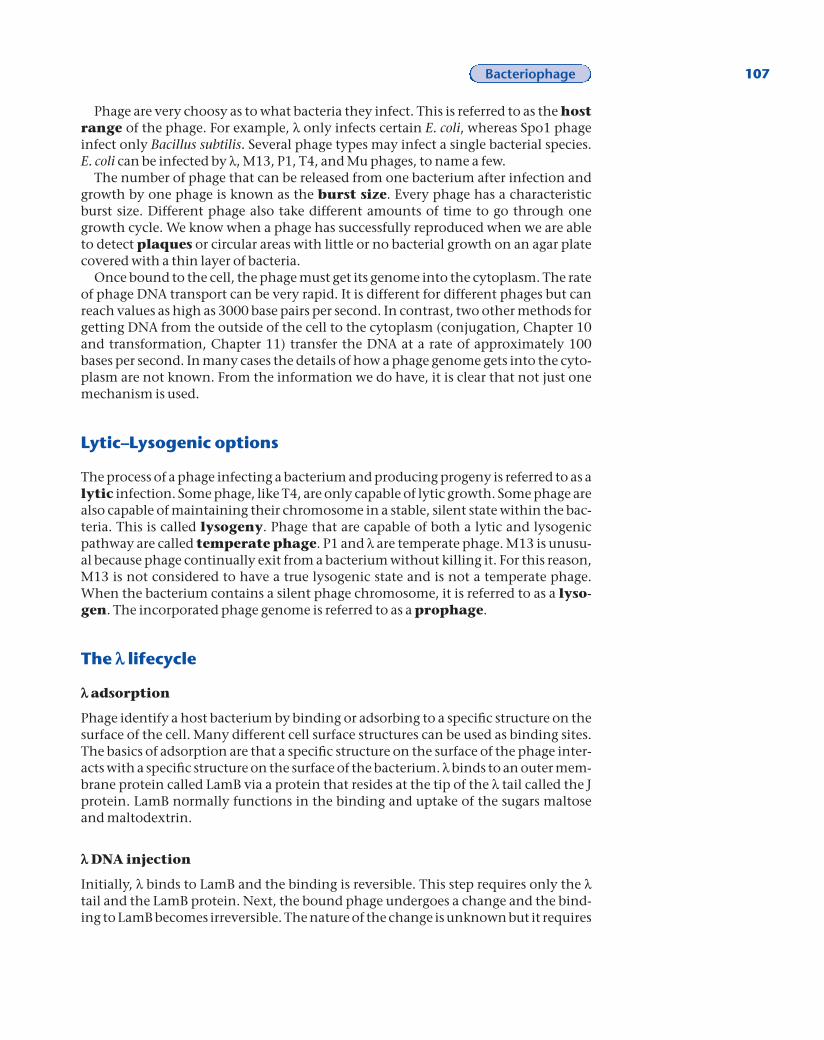
The l genome contains six major promoters known as PLfor promoter leftward, PR for promoter rightward, PRE forpromoter for repressor establishment, PRM for promoterfor repressor maintenance, PI for promoter for integra-tion, and PR¢ for secondary rightward promoter (Fig. 7.3).After the genome is circularized and supercoiled, tran-scription begins from PL and PR. A series of genes known asearly genes are transcribed and translated. These geneproducts are the initial proteins needed for further phagedevelopment. E. coli RNA polymerase interacts with PL togive rise to a short mRNA transcript that is translated intothe N protein (Fig. 7.4a). E. coli RNA polymerase interactswith PR to give rise to a short mRNA transcript that istranslated into the Cro protein (Fig. 7.4a).
The N protein is able to extend transcription when RNApolymerase encounters a sequence in the DNA that tells itto stop. For this reason, N is called an anti-terminationprotein. N allows RNA polymerase to transcribe throughthe tL and tR1 termination signals resulting in the synthe-sis of longer mRNA transcripts (Fig. 7.4b). The longertranscripts from PR encode the O, P, and CII proteins, anda small amount of another anti-terminator, the Q protein.From PL, CIII, the recombination proteins Gam and Red and a small amount of Xisand Int are made.
The N protein anti-terminates by binding to RNA polymerase after a specific basepair sequence, located upstream of the transcriptional termination site, has been
Bacteriophage 109
Fig. 7.3 The location of the sixmajor promoters on the lgenome and the direction inwhich they specify mRNAproduction.
att
Recombination
IntXis
PICIII N CI Cro
CII
DNA replicationQ
Lysis
cos
Head genesTail genes
PL PR
PRM PRE
PR'
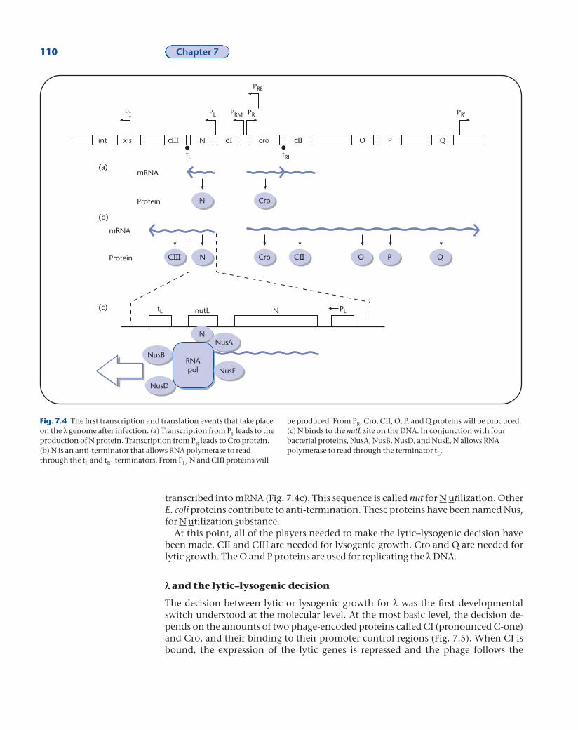
transcribed into mRNA (Fig. 7.4c). This sequence is called nut for N utilization. OtherE. coli proteins contribute to anti-termination. These proteins have been named Nus,for N utilization substance.
At this point, all of the players needed to make the lytic–lysogenic decision havebeen made. CII and CIII are needed for lysogenic growth. Cro and Q are needed forlytic growth. The O and P proteins are used for replicating the l DNA.
l and the lytic–lysogenic decision
The decision between lytic or lysogenic growth for l was the first developmentalswitch understood at the molecular level. At the most basic level, the decision de-pends on the amounts of two phage-encoded proteins called CI (pronounced C-one)and Cro, and their binding to their promoter control regions (Fig. 7.5). When CI isbound, the expression of the lytic genes is repressed and the phage follows the
110 Chapter 7
xisint N cro O P Q
PL PRM PR
PRE
PR'
tL
tL
tRI
N Cro
N Cro O P Q
Protein
mRNA
Protein
mRNA
nutL N PL
N
NusD
NusBRNApol
NusA
NusE
(a)
(b)
(c)
Fig. 7.4 The first transcription and translation events that take placeon the l genome after infection. (a) Transcription from PL leads to theproduction of N protein. Transcription from PR leads to Cro protein.(b) N is an anti-terminator that allows RNA polymerase to readthrough the tL and tR1 terminators. From PL, N and CIII proteins will
be produced. From PR, Cro, CII, O, P, and Q proteins will be produced.(c) N binds to the nutL site on the DNA. In conjunction with fourbacterial proteins, NusA, NusB, NusD, and NusE, N allows RNApolymerase to read through the terminator tL.

lysogenic pathway (Fig. 7.5a). For this reason, CI is also known as CI repressor or l repressor. The expression and binding of Cro leads to lytic development.
Cro is made from PR and CI is made from either PRE or PRM. Both Cro and CI bind tothe same DNA sequences called operators (Fig. 7.5b). l contains two operators thatbind Cro and CI. One, called OR, overlaps the PRM and PR promoters. The other, called
Bacteriophage 111
cro
PRM PR
OR1OR2OR3
N cro
PL
OL
PRM PR
OR
cro
PRM PR
Cro
PRM PR
Cro Cro
Lysogeny
Lytic growth
or
(a)
(b)
(c)
(d)
(e)
N cro
Cro
Lysogeny Lytic growth
Fig. 7.5 CI and Cro are the proteins responsible for the two developmental fates of l. (a) CI leads tolysogeny and Cro leads to lytic growth. (b) Both CI and Cro bind to two operator regions, OR and OL. ORoverlaps with both PR and PRM. OL overlaps with PL. (c) OR is required for the switch betweendevelopmental pathways. It is composed of three 17 base pair sequences called OR1, OR2, and OR3. Theyare similar in sequence but not identical. (d) CI binds to OR1 first then OR2. It will bind to OR3 but only atvery high concentrations. When CI binds to OR, it represses transcription from PR and activates it fromPRM. CI binding to OR is actually required for PRM to be activated. CI binding leads to lysogeny. (e) Cro alsobinds to OR1, OR2, and OR3 but in the opposite order from CI. Cro binds to OR3 first then OR2 and at highconcentrations OR1. Cro binding to OR3 inhibits PRM and leads to lysogeny.

112 Chapter 7
Fig. 7.6 The CII protein is themajor player in the switchbetween lytic and lysogenicgrowth. CII is unstable andrapidly degraded by the host-encoded HflA protease. InactiveCII leads to lytic growth. CII canbe protected by the phage-encoded CIII protein. Active CIIleads to lysogenic growth.
OL, is behind the PL promoter. OR is a major player in the lytic–lysogenic decision,while OL is not part of the decision.
OR is composed of three 17 base pair sequences called OR1, OR2, and OR3 (Fig. 7.5c).CI repressor binds to OR1 10 times better than it binds to OR2 or OR3. At increasing con-centrations of CI, it will bind to OR2 and eventually to OR3. When CI is bound to OR, itstimulates the PRM promoter and the production of CI repressor and inhibits the PRpromoter and the production of Cro, leading to lysogeny (Fig. 7.5d). Cro also binds toOR1, OR2, and OR3 but in the reverse order from CI repressor. Cro binds to OR3 first, thenOR2, and finally at high concentrations to OR1. When Cro is bound to OR, it inhibitsthe PRM promoter and the production of CI, leading to lytic growth (Fig. 7.5e). This isthe basis for either lytic or lysogenic growth.
How does the phage switch between these two developmental pathways? Themajor protein involved in the switch is another phage-encoded protein called CII(pronounced C-two, Fig. 7.6). CII activates the PRE and PI promoters. This leads to theproduction of repressor and the Integrase protein, which is also needed for lysogeny(Fig. 7.6b). The gene for CII (cII) resides just to the right of the cro gene. When l infectsa cell, transcription automatically begins from PL and PR using host proteins. Tran-scription from PR leads to production of both the Cro and CII proteins. If CII is activeit will lead to production of CI and Integrase and lysogeny. If CII is inactive then Crowill repress PRM, preventing expression of CI and leading to lytic growth.
The CII protein is inherently unstable. Several factors influence this feature of theprotein. CII is degraded by the bacterial-encoded HflA protease. When cells are actively growing in nutrient-rich conditions, the amount of HflA in the cell is high,leading to degradation of CII and lytic growth. When cell are growing slowly, HflAlevels are low, leading to stabilization of CII, production of CI, and lysogeny. In thismanner, CII is used to monitor the health of the cell and impact the lytic–lysogenicdecision accordingly. It is thought that l wants to produce more phage when cells arehealthy, nutrients are plentiful, and the prospect of completing phage development
is good. Lysogeny is a betterbet when cells are growingpoorly. CII is also stabilizedby a phage-encoded proteincalled CIII. CIII is producedfrom PL by infecting phage.
The l lysogenicpathway
If CII prevails, CI will beproduced, initially from thePRE promoter and eventu-ally from the PRM promoter.CI activates PRM ensuringthat a continuous supply ofCI is made. CI also activatesthe PI promoter, leading tothe production of the Inte-grase protein. The recombi-nation of l DNA into thechromosome occurs at a
CII
Int CI
CII
LysogenyLytic growth
ActiveInactive
Degradation by HfLA Protection by CIII
CI Cro CIIInt Xis
PI PRE
++
(a)
(b)
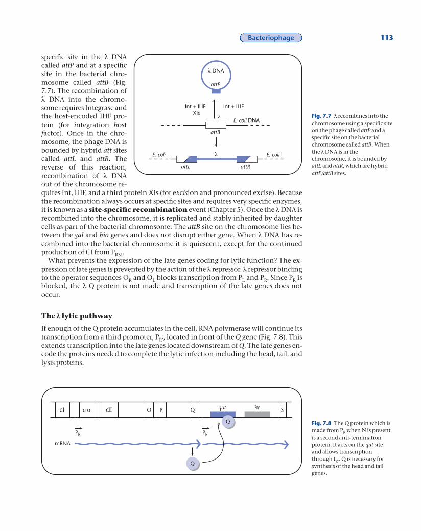
specific site in the l DNAcalled attP and at a specificsite in the bacterial chro-mosome called attB (Fig.7.7). The recombination ofl DNA into the chromo-some requires Integrase andthe host-encoded IHF pro-tein (for integration hostfactor). Once in the chro-mosome, the phage DNA isbounded by hybrid att sitescalled attL and attR. The reverse of this reaction, recombination of l DNAout of the chromosome re-quires Int, IHF, and a third protein Xis (for excision and pronounced excise). Becausethe recombination always occurs at specific sites and requires very specific enzymes,it is known as a site-specific recombination event (Chapter 5). Once the l DNA isrecombined into the chromosome, it is replicated and stably inherited by daughtercells as part of the bacterial chromosome. The attB site on the chromosome lies be-tween the gal and bio genes and does not disrupt either gene. When l DNA has re-combined into the bacterial chromosome it is quiescent, except for the continuedproduction of CI from PRM.
What prevents the expression of the late genes coding for lytic function? The ex-pression of late genes is prevented by the action of the l repressor. l repressor bindingto the operator sequences OR and OL blocks transcription from PL and PR. Since PR isblocked, the l Q protein is not made and transcription of the late genes does notoccur.
The l lytic pathway
If enough of the Q protein accumulates in the cell, RNA polymerase will continue itstranscription from a third promoter, PR¢, located in front of the Q gene (Fig. 7.8). Thisextends transcription into the late genes located downstream of Q. The late genes en-code the proteins needed to complete the lytic infection including the head, tail, andlysis proteins.
Bacteriophage 113
Fig. 7.7 l recombines into thechromosome using a specific siteon the phage called attP and aspecific site on the bacterialchromosome called attB. Whenthe l DNA is in thechromosome, it is bounded byattL and attR, which are hybridattP/attB sites.
Int + IHFInt + IHFXis
l DNA
attP
E. coli DNA
attB
attL attR
E. coli E. colil
cro O P Q Squt tR'
PR PR'
Q
Q
mRNA
Fig. 7.8 The Q protein which ismade from PR when N is presentis a second anti-terminationprotein. It acts on the qut siteand allows transcriptionthrough tR¢. Q is necessary forsynthesis of the head and tailgenes.

DNA replication during the l lytic pathway
After the infecting l DNA has been converted to a double-stranded circular molecule,it replicates from a specific origin using both the phage-encoded O and P proteins andbacterial-encoded proteins. Replication proceeds bidirectionally, much like the E. colichromosome. This form of replication produces molecules that look like the Greekletter theta and is called theta replication (Fig. 7.9a). Later in lytic development, lswitches to a second mode of replication called rolling circle replication.
Rolling circle replication of l DNA commences when an endonuclease, en-coded by l exo, cuts one strand of the covalently closed circular double-stranded DNAmolecule (Fig. 7.9b). The cut strand is called the plus strand. The 5¢ end of the cut plusstrand is peeled away from the intact minus strand. DNA polymerase adds deoxyri-bonucleotides to the free 3¢ OH of the cut plus strand using the intact circular minusstrand as the template. This produces new plus strands through a process of continu-ally elongating the original plus strand. The new plus strands are used as a template tosynthesize new minus strands. Rolling circle replication produces long DNA mole-cules containing multiple phage genomes called concatomers.
Making l phage
The structure of the fin-ished capsid is determinedby the physical characteris-tics of the structural pro-teins that they are madefrom and the phage andhost proteins used for as-sembly. Assembly of thecapsids requires at least 10 phage-encoded proteinsand two host-encoded pro-teins. The final capsid ismade up of eight proteins,E, D, B, W, FII, B*, X1, andX2. Initially, B, C, and Nu3(all phage proteins) form asmall, ill-defined initiatorstructure (Fig. 7.10a). Thisstructure is a substrate forthe host-encoded GroELand GroES proteins. GroELand GroES act on proteinsor protein complexes andhelp remodel them. Themajor coat protein, E, isadded to this structure toform an immature phagehead (Fig. 7.10b). The immature phage head isconverted to the mature
114 Chapter 7
Fig. 7.9 l has two modes ofDNA replication: thetareplication (a) and rolling circlereplication (b). Theta replicationoccurs early in infection androlling circle replication occurslate in infection. Rolling circlereplication producesconcatomers for packaging intophage heads.
Theta replication
Rolling circle replication
Theta structure
3'5'
3'
5'
ñ+
3'
3'5'
5'cos
cos
cos
Newly synthesized DNA
ori
O P
(a)
(b)
Bacteriophage 115
BCNu3
Convert B B*
Fuse E + C Digest to X1, X2
GroELGroES
Terminase
cos cos
D
W + F
Tails
Infectivephage
(a) (b)
(c)
(d)
(e)
(f)
E
Nu3
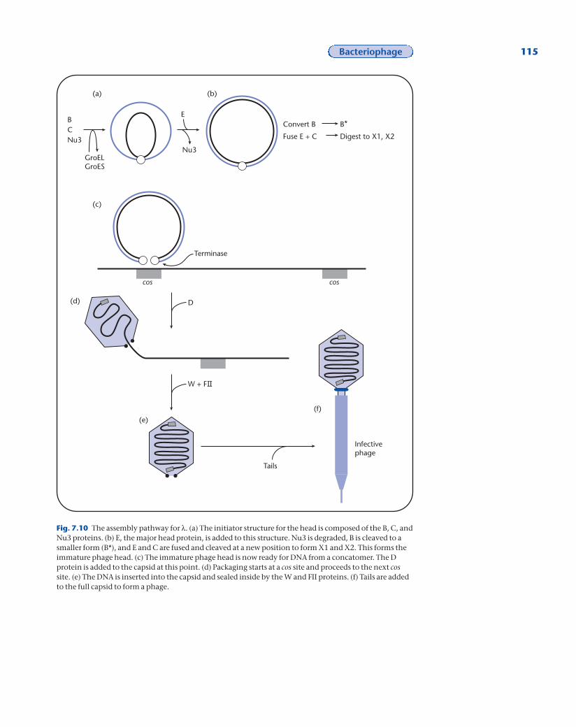
Fig. 7.10 The assembly pathway for l. (a) The initiator structure for the head is composed of the B, C, andNu3 proteins. (b) E, the major head protein, is added to this structure. Nu3 is degraded, B is cleaved to asmaller form (B*), and E and C are fused and cleaved at a new position to form X1 and X2. This forms theimmature phage head. (c) The immature phage head is now ready for DNA from a concatomer. The Dprotein is added to the capsid at this point. (d) Packaging starts at a cos site and proceeds to the next cossite. (e) The DNA is inserted into the capsid and sealed inside by the W and FII proteins. (f) Tails are addedto the full capsid to form a phage.

phage head by the degradation of Nu3, the cleavage of B to B*, and the fusion of C pro-tein and some E protein followed by the cleavage of the fused protein into two newproteins, X1 and X2 (Fig. 7.10c).
The mature phage head is now ready for DNA. As the DNA is inserted into the phagehead, it expands and the D protein is added to the surface of the capsid. l DNA cannotbe packaged from a monomer of l DNA but only from concatomers usually producedby rolling circle replication (Fig. 7.10c). The DNA is cut at one cos site by a l encodedenzyme and put into the phage head. Terminase binds to a cos site and to a phagehead, cuts the cos site, and inserts that end of the l DNA into the phage head (Fig.7.10d). Terminase cuts the cos site asymmetrically, leaving the 12 base pair overhang.The terminase enzyme then tracks along the concatomer of l DNA until it reaches asecond cos site. As terminase tracks, the DNA is inserted into the phage head. When asecond cos site is reached, terminase cuts the DNA and the last bit of DNA is insertedinto the phage head (Fig. 7.10e).
This phage head with newly inserted DNA is unstable and not able to join to phagetails. The W and FII proteins are added to the base of the full head (Fig. 7.10e). Thisboth stabilizes the DNA-containing head and builds the connector to which the tailwill bind. Tails add spontaneously to this structure (Fig. 7.10f).
Tails are constructed from 12 gene products. Like the capsids, the tails are formedfrom an ill-defined initiator complex. This complex requires the J, I, L, K, H, G, and Mphage proteins. They are added to the complex in the order listed beginning with theJ protein. For this reason, it is thought that tails are built from the tip that recognizesthe bacterium towards the end that binds to the phage head. Once the initiator struc-ture is formed, the major tail protein, V, is added. The H protein is used as a measuringstick and determines how long the tail will be. Once the tails reaches the correctlength, the U protein is added to prevent further growth and the H protein is cleaved.The Z protein is added last and is required to make an infectious phage. A tail withoutZ will bind to a full phage head but the resulting particle is not infectious.
The l phage packaging system packages DNA molecules on the basis of the cossites rather than on the basis of the length of the DNA molecule. Varying lengths ofDNA molecules, within set limits, can be packaged as long as the molecule contains a cos site at both ends. If the distance between the two cos sites is less than ~37kb, the resulting phage particle will be unstable. When the DNA is inside the capsid, it exerts pressure on the capsid. Likewise the capsid exerts an inward force on the DNA. If there is not enough DNA inside the capsid, it will implode from the inwardforce of the capsid. If the distance between the two cos sites is too far (~52kb), then thecapsid will be filled before the second cos is reached. The tail cannot be added becausethe DNA hanging out of the capsid is in the way and no infectious phage particle isproduced.
Getting out of the cell —the l S and R proteins
The l R and S proteins are required for l to release progeny phage into the environ-ment. The R protein is an endolysin that degrades the peptidoglycan cell wall and allows the phage to escape from the cell. The S protein forms a hole in the inner membrane to allow the endolysin to gain access to the cell wall. After the hole isformed, approximately 100 intact l phage particles are released into the environ-ment. The entire lytic cycle lasts ~35 minutes.
116 Chapter 7

Induction of l by the SOS System
When a l lysogen is treated with ultraviolet light (UV),~35 minutes later the cells lyse and release phage. Whatdoes the UV do to the cell? UV damages the DNA and trig-gers a cellular response called the SOS response to dealwith this damage (Fig. 7.11). The RecA protein, which isnormally used for homologous recombination, is activat-ed and becomes a special kind of protease. The activatedRecA interacts with LexA, leading to cleavage of LexA.This leads to the activation of a number of genes whoseproducts repair the DNA damage in the cell. l has tappedinto this system through the CI protein. CI repressor caninteract with activated RecA, leading to the cleavage of CI.This leads to expression of the phage lytic genes andphage production. The rational for this response is that ldoes not want to risk staying in a cell that has DNA dam-age and may not survive.
Superinfection
If a cell is a l lysogen, another l phage that infects is notable to undergo lytic development and produce phage.The incoming phage can inject its DNA, however, the DNA is immediately shut downand no transcription or translation of the l initiates. l lysogens are immune to infec-tion by another l phage particle, which is called superinfection. Superinfection isblocked because the lysogen is continuously producing CI repressor. The lysogen actually produces more repressor than it needs to shut down one phage. This extra repressor binds to the superinfecting phage DNA at OL and OR and prevents transcription from PL and PR.
Restriction and modification of DNA
A simple experiment with l leads to the discovery of how bacteria tell their own DNAfrom foreign DNA. l is capable of making plaques on two different types of E. coli, E.coli K12 and E. coli C. If l is grown on E. coli K12, it will form plaques on E. coli K12 orE. coli C with equal efficiency. If l is grown on E. coli C, it will form plaques on E. coli Cbut if it is plated on E. coli K12, only a few phage will form plaques. The efficiency offorming plaques or efficiency of plating (EOP) is decreased by 10,000-fold. This isknown as restriction. If the E. coli C grown phage that did plaque on E. coli K12 arereplated on E. coli K12, the EOP is 1. This is known as modification. The few phagethat survive the replating on E. coli K12 have been modified so that they can effici-ently plate on E. coli K12.
While this originated as a curiosity of phage growth, it has proven to be essential formany molecular techniques. Further investigation showed that the protein responsi-ble for restriction, a restriction enzyme or restriction endonuclease, actuallyrecognizes a specific DNA sequence and cleaves the DNA on both strands. The cut ordigested DNA is sensitive to nucleases that degrade DNA. The modification part of thesystem is a protein that specifically modifies the DNA sequence recognized by the re-striction enzyme and prevents the DNA from being digested. E. coli K12 has a restric-
Bacteriophage 117
Fig. 7.11 The SOS responseinduces l. UV treatment of cells(a) damages the DNA and leavesstretches of single-stranded DNA(b). The single-stranded DNAactivates RecA (c). ActivatedRecA interacts with CI, leadingto cleavage of CI and inductionof the l lysogen (d). ActivatedRecA also interacts with LexAand leads to LexA inactivation(e). LexA inactivation leads toexpression of a number of genes,including some DNA repairenzymes.
UV
RecA
LexA
CleavedLexA
ActivatedRecA
CI
Cleaved CI
Lytic growth Activate DNArepair genes
(a)
(b)
(c)
(d) (e)
tion/modification system and E. coli C does not. This explains the original observa-tion with l growth. If a bacterium carries the restriction enzyme, it must also carry themodification enzyme so that the bacterial chromosome is not digested and degraded.The restriction/modification system allows a bacterium to tell DNA from its ownspecies from foreign DNA. Many different bacteria contain restriction/modificationsystems that recognize different DNA sequences. The restriction enzymes are purifiedand used in vitro to cleave DNA at specific DNA sequences, depending on the recog-nition sequence of the enzyme in question. Restriction enzymes are used to cleaveand clone DNA fragments as described in Chapter 14.
The lifecycle of M13
M13 adsorption and injection
M13 adsorbs to the tip of the F pilus, a hair-like structure on the surface of some bac-teria. It can only infect bacteria that carry an F or F-like conjugative plasmid that en-codes the proteins that make up the F pilus (see Chapter 10). For the filamentousphage, it is known that infection is initiated by the binding of gpIII to the tip of the Fpilus. GpIII then interacts with the inner membrane protein TolA. Two additionalfacts about gpIII suggest a mechanism for phage DNA entry. GpIII contains aminoacid sequences that are fusogenic or promote localized fusion of two membranes andgpIII is capable of forming pores in membranes that are large enough for DNA to gothrough. If each of these properties of gpIII are important for phage entry, then thephage could bind to the F pilus, promote fusion of the membranes, and use gpIII toform holes in the membrane to gain entry into the cytoplasm.
Protection of the M13 genome
The M13 DNA that ends up in the cytoplasm is a circular single-stranded DNA mole-cule. The strand present in phage particle is known as the plus or + strand. After entryinto the cytoplasm, the + strand DNA is immediately coated with an E. coli single-stranded DNA binding protein known as SSB. The SSB coating protects the DNA fromdegradation.
M13 DNA replication
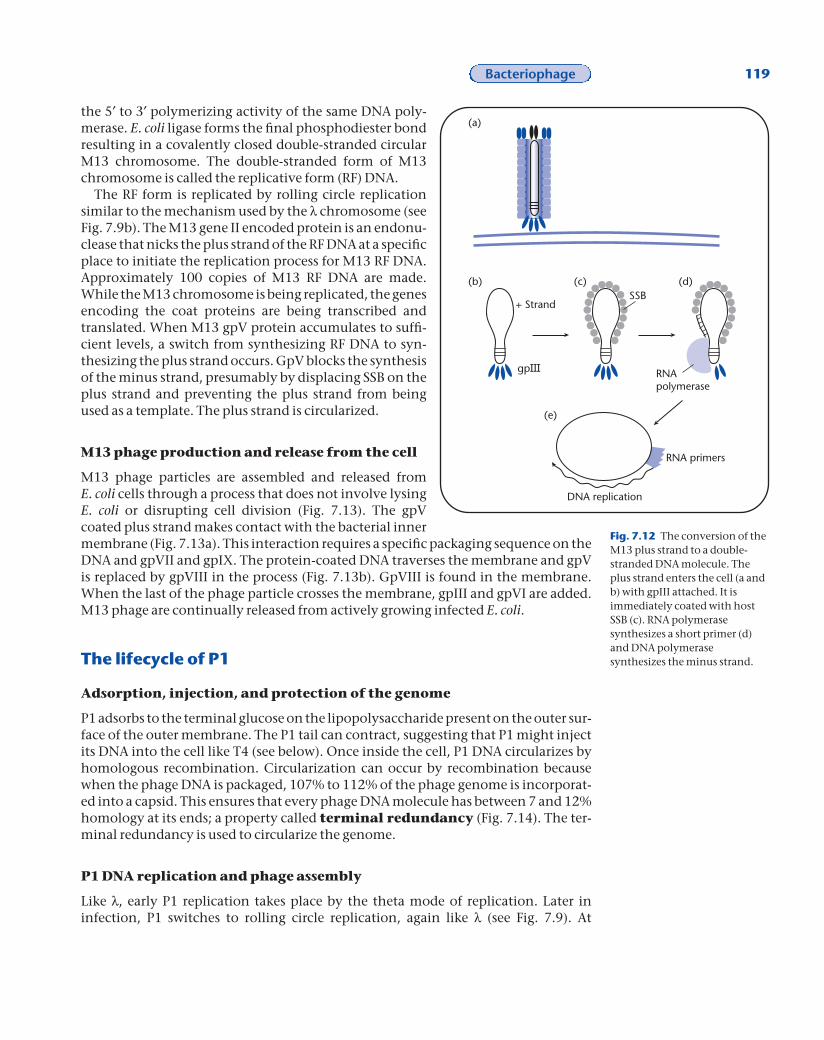
The M13 plus strand is converted to a double-stranded molecule immediately uponentry into E. coli (Fig. 7.12). Synthesis of the complementary strand is carried out en-tirely by E. coli’s DNA synthesis machinery. The complementary strand is called theminus or —strand. Only the minus strand is used as the template for mRNA synthesisand ultimately it is the template for the translation of the encoded M13 gene prod-ucts. The SSB that coats the plus strand upon entry of the DNA into the E. coli cyto-plasm fails to bind to ~60 nucleotides of the molecule (Fig 7.12c). These nucleotidesform a hairpin loop that is protected from nuclease degradation. M13 gpIII from thephage is found associated with the hairpin loop. The hairpin loop is recognized by E.coli RNA polymerase as a DNA replication origin and is used to initiate transcription ofa short RNA primer (Fig. 7.12d). The RNA primer is extended by E. coli DNA poly-merase III to create the minus strand (Fig. 7.12e). The RNA primer is eventually re-moved by the exonuclease activities of E. coli DNA polymerase I. The gap is filled in by
118 Chapter 7
FYI 7.2
Pathogenicity inVibrio cholera
Cholera is caused by thebacterium, Vibrio cholera.Many of the genes that makethis bacteria pathogenic ordisease causing are part of aprophage located in the V.cholera chromosome. Thisprophage bears strikingresemblance to M13 and otherfilamentous phage. It ispossible that the transmissionof these pathogenic genes is assimple as the phage movingfrom one bacterial species toanother sensitive bacterialspecies.
Bacteriophage 119
the 5¢ to 3¢ polymerizing activity of the same DNA poly-merase. E. coli ligase forms the final phosphodiester bondresulting in a covalently closed double-stranded circularM13 chromosome. The double-stranded form of M13chromosome is called the replicative form (RF) DNA.
The RF form is replicated by rolling circle replicationsimilar to the mechanism used by the l chromosome (seeFig. 7.9b). The M13 gene II encoded protein is an endonu-clease that nicks the plus strand of the RF DNA at a specificplace to initiate the replication process for M13 RF DNA.Approximately 100 copies of M13 RF DNA are made.While the M13 chromosome is being replicated, the genesencoding the coat proteins are being transcribed andtranslated. When M13 gpV protein accumulates to suffi-cient levels, a switch from synthesizing RF DNA to syn-thesizing the plus strand occurs. GpV blocks the synthesisof the minus strand, presumably by displacing SSB on theplus strand and preventing the plus strand from beingused as a template. The plus strand is circularized.
M13 phage production and release from the cell
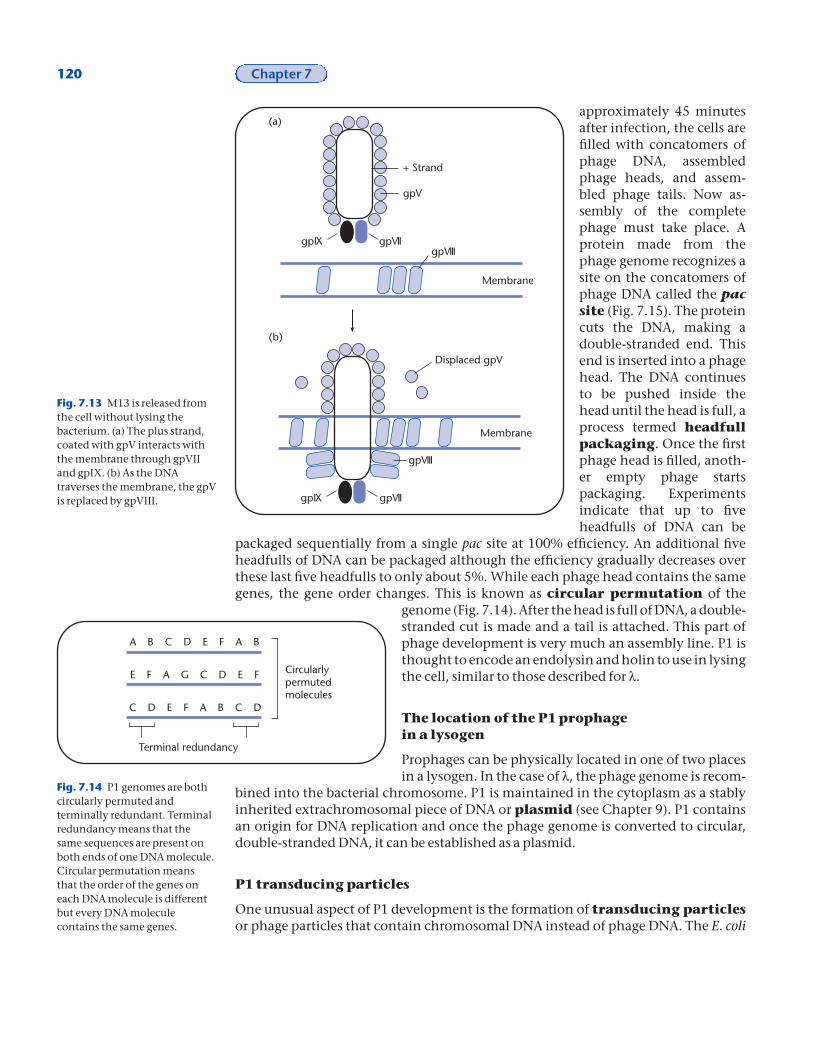
M13 phage particles are assembled and released from E. coli cells through a process that does not involve lysingE. coli or disrupting cell division (Fig. 7.13). The gpV coated plus strand makes contact with the bacterial innermembrane (Fig. 7.13a). This interaction requires a specific packaging sequence on theDNA and gpVII and gpIX. The protein-coated DNA traverses the membrane and gpVis replaced by gpVIII in the process (Fig. 7.13b). GpVIII is found in the membrane.When the last of the phage particle crosses the membrane, gpIII and gpVI are added.M13 phage are continually released from actively growing infected E. coli.
The lifecycle of P1
Adsorption, injection, and protection of the genome
P1 adsorbs to the terminal glucose on the lipopolysaccharide present on the outer sur-face of the outer membrane. The P1 tail can contract, suggesting that P1 might injectits DNA into the cell like T4 (see below). Once inside the cell, P1 DNA circularizes byhomologous recombination. Circularization can occur by recombination becausewhen the phage DNA is packaged, 107% to 112% of the phage genome is incorporat-ed into a capsid. This ensures that every phage DNA molecule has between 7 and 12%homology at its ends; a property called terminal redundancy (Fig. 7.14). The ter-minal redundancy is used to circularize the genome.
P1 DNA replication and phage assembly
Like l, early P1 replication takes place by the theta mode of replication. Later in infection, P1 switches to rolling circle replication, again like l (see Fig. 7.9). At
Fig. 7.12 The conversion of theM13 plus strand to a double-stranded DNA molecule. Theplus strand enters the cell (a andb) with gpIII attached. It isimmediately coated with hostSSB (c). RNA polymerasesynthesizes a short primer (d)and DNA polymerasesynthesizes the minus strand.
(a)
(b) (c) (d)
(e)
+ Strand
gp
SSB
RNApolymerase
RNA primers
DNA replication
120 Chapter 7
Fig. 7.14 P1 genomes are bothcircularly permuted andterminally redundant. Terminalredundancy means that thesame sequences are present onboth ends of one DNA molecule.Circular permutation meansthat the order of the genes oneach DNA molecule is differentbut every DNA moleculecontains the same genes.
approximately 45 minutesafter infection, the cells arefilled with concatomers ofphage DNA, assembledphage heads, and assem-bled phage tails. Now as-sembly of the completephage must take place. Aprotein made from thephage genome recognizes asite on the concatomers ofphage DNA called the pacsite (Fig. 7.15). The proteincuts the DNA, making adouble-stranded end. Thisend is inserted into a phagehead. The DNA continuesto be pushed inside thehead until the head is full, aprocess termed headfullpackaging. Once the firstphage head is filled, anoth-er empty phage starts packaging. Experiments indicate that up to fiveheadfulls of DNA can be
packaged sequentially from a single pac site at 100% efficiency. An additional fiveheadfulls of DNA can be packaged although the efficiency gradually decreases overthese last five headfulls to only about 5%. While each phage head contains the samegenes, the gene order changes. This is known as circular permutation of the
genome (Fig. 7.14). After the head is full of DNA, a double-stranded cut is made and a tail is attached. This part ofphage development is very much an assembly line. P1 isthought to encode an endolysin and holin to use in lysingthe cell, similar to those described for l.
The location of the P1 prophage in a lysogen
Prophages can be physically located in one of two placesin a lysogen. In the case of l, the phage genome is recom-
bined into the bacterial chromosome. P1 is maintained in the cytoplasm as a stablyinherited extrachromosomal piece of DNA or plasmid (see Chapter 9). P1 containsan origin for DNA replication and once the phage genome is converted to circular,double-stranded DNA, it can be established as a plasmid.
P1 transducing particles
One unusual aspect of P1 development is the formation of transducing particlesor phage particles that contain chromosomal DNA instead of phage DNA. The E. coli
Fig. 7.13 M13 is released fromthe cell without lysing thebacterium. (a) The plus strand,coated with gpV interacts withthe membrane through gpVIIand gpIX. (b) As the DNAtraverses the membrane, the gpVis replaced by gpVIII.
+ Strand
gpVII
gpV
gpIXgpVIII
Membrane
(a)
gpVIIgpIX
gpVIII
Membrane
(b)
Displaced gpV
Circularlypermutedmolecules
A B C D E F A B
E F A G C D E F
C D E F A B C D
Terminal redundancy

Bacteriophage 121
chromosome contains many pseudopac sites or sites that can be used to initiatepackaging of host chromosomal DNA into maturing phage. These pseudopac sites areused much less frequently than the phage pac sites but they are used. The resultingphage carry random pieces of the chromosome in place of phage genomes. The ability to package any piece of chromosomal DNA instead of phage DNA makes P1 ageneralized transducing phage. Transducing particles are used to move pieces of host chromosomal DNA from one strain to another for the purposes described in Chapter 8.
The lifecycle of T4
T4 adsorption and injection
For T4, the phage binds to the lipopolysaccharide. The tips of the tail fibers contactthe cell first (Fig. 7.16). Once the phage has bound to the cell, the base plate rearrangescreating a hole in the base plate. The outer sheath contracts and the internal tube goesthrough the outer membrane, peptidoglycan, and periplasm and comes close to thecytoplasmic membrane. The DNA is injected and crosses the cytoplasmic membranein about 30 seconds. Not all phage that have the structure of T4 inject their DNA thisway. Some phage such as T7, have tails that cannot contract. The T7 genome is only 40kb but takes 9 to 12 minutes to cross into the cytoplasm. For T7, a small portion of
pac pac pac
pacpac
Concatomers
(a)
(b)
(c) (d)
Fig. 7.15 P1 packages DNAfrom a pac site (a) and packagesbetween 7 and 12% more thanone P1 genome, until the phagehead is full (b and c). Once thephage head is full, apreassembled head is added (d).

122 Chapter 7
genome (about 8%) crosses both membranes, the peptidoglycan and periplasm, andenters the cytoplasm. After a 4-minute lag during which two proteins encoded by this piece of DNA are synthesized, the rest of the phage DNA enters the cytoplasm. Binding of these two phage proteins to the DNA is thought to pull theDNA into the cytoplasm.
Once T4 DNA is in the cell cytoplasm, it specifies a highly organized and coordin-ated program of gene expression. A group of genes with similar promoters, called theearly genes, are transcribed and translated by host enzymes. One early gene enco-ded protein activates a second set of promoters for the middle genes. A differentearly gene encoded protein shuts off synthesis of the early genes. One product of middle transcription is required to activate the late genes. The early genes encodethe proteins needed for DNA synthesis and late genes encode the proteins needed tobuild the capsid and tail structures. Many phage stage the expression of their genes in this temporal fashion to ensure proper construction of the phage particles.
The T4 genome does not contain cytosine residues. All of the cytosines are modifiedby methylation. Several of the early genes encode proteins that degrade the cytosine-containing host DNA. The phage DNA is protected from degradation. The T4genome, like P1 is both circularly permuted and terminally redundant. T4 has about3% terminal redundancy. Unlike P1, T4 does not appear to use this terminal redun-dancy to circularize upon infection. T4 begins replication immediately after the earlygene products are made. T4 replicates as a linear molecule and uses replication and re-combination to both replicate the entire genome and to make concatomers to pack-age into phage heads (Fig. 7.17). Like other phage, capsids, tails, and concatomers ofphage DNA are premade and assembled into infectious phage particles late in phagedevelopment.
(a)
(b)
(c)
(d)
DNA
Fig. 7.16 Injection of T4 DNAinto the cell. (a) T4 “looks” for asusceptible bacterium with itstail fibers. (b) The tail fibersrecognize the membrane first. (c)The tail spikes interact with themembrane. (d) The tail sheathcontacts, driving the internal tailtube through the outermembrane, peptidoglycan, andto the inner membrane wherethe DNA is released.

Bacteriophage 123
T4 rII mutations and the nature of the genetic code
The study of two genes in T4 has contributed significantly to our understanding of thegenetic code and the nature of the gene. In the late 1950s and 1960s the understand-ing of the nature of the gene was in its infancy. The prevailing thought was that thegene was the smallest genetic unit and it was inherited as a unit. The chemical natureof DNA had just been described by Watson and Crick. The relationship between DNAand the gene was not understood. Seymour Benzer used the T4 rII locus and geneticlogic to describe several key features of the gene.
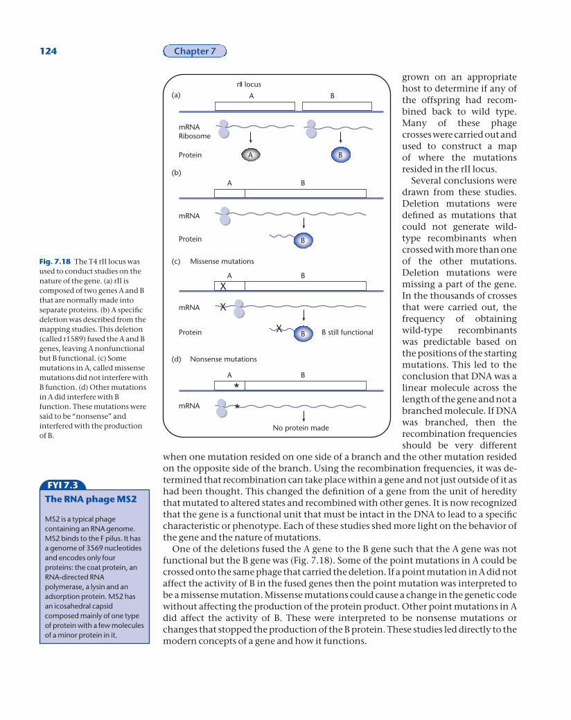
rII encodes two proteins, A and B. Several thousand point mutations and deletionswere isolated in these two genes. These mutations were put to good use. A phage car-rying one mutation and a phage carrying a second mutation were mixed together and
B C D E F A B
A B C D E F A B
A B C D E F A B
A B C D E F A
C D E F A B
A B C D E F A B
A B C D E F A
A B C D E F
BB
A B C D E F A B C D E F A
A B C D E F A B C D E F A B
E F A B
A B C D E F A
B C D
A B C
A B C
D E F A B
D E F A B
3'3'5'
5'
3'
5'
5'
A B C
C
B
BA
A
(a)
(b)
and
DNA replication
Strand invasion
DNA replication
Free ssDNA
Free ssDNA
(c)
(d)
Fig. 7.17 T4 replicates its DNA using both replication and recombination. (a) Linear T4 DNA moleculesare injected into the cytoplasm of the host. (b) DNA replication begins at an origin and proceedsbidirectionally to the ends. However, because of DNA polymerase’s requirement for a primer, a piece ofthe DNA at one end of the molecule cannot be replicated and remains single stranded. (c) This piece ofsingle-stranded DNA can invade duplexed DNA at any place where it has homology, like the initialreaction in recombination. (d) DNA replication of this molecule will lead to concatomers of the phagegenome. Depending on where strand invasion takes place, branched molecules can also be formed. T4packages its DNA out of the concatomers. The displaced single strands are free to strand invade theconcatomer structures.
124 Chapter 7
grown on an appropriatehost to determine if any ofthe offspring had recom-bined back to wild type.Many of these phage crosses were carried out andused to construct a map of where the mutationsresided in the rII locus.
Several conclusions weredrawn from these studies.Deletion mutations weredefined as mutations thatcould not generate wild-type recombinants whencrossed with more than oneof the other mutations.Deletion mutations weremissing a part of the gene.In the thousands of crossesthat were carried out, thefrequency of obtainingwild-type recombinantswas predictable based onthe positions of the startingmutations. This led to theconclusion that DNA was alinear molecule across thelength of the gene and not abranched molecule. If DNAwas branched, then the recombination frequenciesshould be very different
when one mutation resided on one side of a branch and the other mutation residedon the opposite side of the branch. Using the recombination frequencies, it was de-termined that recombination can take place within a gene and not just outside of it ashad been thought. This changed the definition of a gene from the unit of hereditythat mutated to altered states and recombined with other genes. It is now recognizedthat the gene is a functional unit that must be intact in the DNA to lead to a specificcharacteristic or phenotype. Each of these studies shed more light on the behavior ofthe gene and the nature of mutations.
One of the deletions fused the A gene to the B gene such that the A gene was notfunctional but the B gene was (Fig. 7.18). Some of the point mutations in A could becrossed onto the same phage that carried the deletion. If a point mutation in A did notaffect the activity of B in the fused genes then the point mutation was interpreted tobe a missense mutation. Missense mutations could cause a change in the genetic codewithout affecting the production of the protein product. Other point mutations in Adid affect the activity of B. These were interpreted to be nonsense mutations orchanges that stopped the production of the B protein. These studies led directly to themodern concepts of a gene and how it functions.
Fig. 7.18 The T4 rII locus wasused to conduct studies on thenature of the gene. (a) rII iscomposed of two genes A and Bthat are normally made intoseparate proteins. (b) A specificdeletion was described from themapping studies. This deletion(called r1589) fused the A and Bgenes, leaving A nonfunctionalbut B functional. (c) Somemutations in A, called missensemutations did not interfere withB function. (d) Other mutationsin A did interfere with Bfunction. These mutations weresaid to be “nonsense” andinterfered with the productionof B.
FYI 7.3
The RNA phage MS2
MS2 is a typical phagecontaining an RNA genome.MS2 binds to the F pilus. It hasa genome of 3569 nucleotidesand encodes only fourproteins: the coat protein, anRNA-directed RNApolymerase, a lysin and anadsorption protein. MS2 hasan icosahedral capsidcomposed mainly of one typeof protein with a few moleculesof a minor protein in it.
A B
B
X B
*
*
A B
X
X
A B
A B
A B
(a)
(b)
(c)
(d)
mRNA
mRNA
mRNA
mRNA
Ribosome
Protein
Protein
Protein
Nonsense mutations
Missense mutations
B still functional
No protein made
rII locus

Bacteriophage 125
Further reading
Campbell, A.M. 1996. Bacteriophages. In Escherichia coli and Salmonella typhimurium: Cellular and MolecularBiology, 2nd edn., eds. F.C. Neidhardt, R. Curtiss III, J.L. Ingraham, E.C.C. Lin, K.B. Low, B. Hagasanik,W.S. Rexnikoff, M. Riley, M. Schaechter, and H.E. Umbarger, pp. 2325–38. Washington, DC: ASM Press.
Ptashne, M. 1993. A Genetic Switch. 2nd edn. Cambridge, MA: Blackwell Scientific.Young, R., Wang, I.-N., and Roof, W. 2002. Phage will out: Strategies of host cell lysis. Trends in Microbiology,
8: 120–8.Zaman, G., Smetsers, A., Kaan, A., Schoenmaters, J., and Konings, R. 1991. Regulation of expression of the
genome of bacteriophage M13. Gene V protein regulated translation of the mRNAs encoded by genes I,II, V and X. Biochimica et Biophysica Acta, 1089: 183–92.
simplicity of phage have made them favorite modelsystems to study many biological processes. While itmay appear that phage carry out some processesusing baroque mechanisms, it usually turns out thatother biological systems share these mechanisms. Forexample, the unusual mechanism used to replicate T4DNA is also used to help maintain bacterial andeukaryotic chromosomes.
Summary
Bacteriophage are a very diverse group of viruses.Their genomes can be made from either DNA or RNA.They can be linear or circular, single stranded ordouble stranded. Phage have evolved many differentways to carry out the limited number of steps in aphage infection. All phage must recognize the correctbacterium to infect, get their genome inside the cell,replicate the genome, transcribe and translate thegenome, and assemble phage particles. The relative
Stu
dy
qu
esti
on
s 1 What processes must be carried out by all phage to produce progeny?2 What is the phenotype of a l mutant containing a defective cI gene?3 Which regulatory proteins and promoters are crucial in l’s decision-making process? Which regulatory proteins and promoters are crucial in l’slytic pathway? Which regulatory proteins and promoters are crucial in l’slysogenic pathway? Describe the roles for all identified participants.4 A new phage from local sewage was recently isolated that infects laboratorystrains of E. coli. How would you determine if this new phage is a temperate orlytic phage using simple genetic tests?5 Contrast and compare the lytic pathway for l and M13 phage. What dothey do that is similar? What do they do that is different?6 Contrast and compare rolling circle replication and theta mode replication.What components of the machinery are similar? What components of themachinery are different? When would one type of mechanism be preferableto the other type? Why?7 How does T4 gets its DNA from the phage head into the cytoplasm?8 How do restriction/modification systems function?9 How do different phage protect their DNA in the cell cytoplasm?10 Why is M13 not considered a temperate phage?


![BACTERIOPHAGE-RESISTANT AND BACTERIOPHAGE-SENSITIVE ...halsmith/phagemutantsubmitted_2.pdf · BACTERIOPHAGE-RESISTANT AND BACTERIOPHAGE-SENSITIVE BACTERIA IN A CHEMOSTAT ... [22],](https://static.fdocuments.in/doc/165x107/5b3839687f8b9a5a518d2ce1/bacteriophage-resistant-and-bacteriophage-sensitive-halsmithphagemutantsubmitted2pdf.jpg)