Lumbar Spinal Conditions
42
STUDENT OUTCOMES 1. Locate and explain the functional significance of the bony and soft-tissue structures of the lumbar spine. 2. Describe the motion capabilities of the lumbar spine. 3. Identify the factors that contribute to mechanical loading on the spine. 4. Describe specific strategies in activities of daily living to reduce spinal stress in the lumbar region. 5. Identify anatomical variations that can predispose individuals to lumbar spine injuries. 6. Explain the measures used to prevent injury to the lumbar spinal region. 7. Describe the common injuries and conditions of the lumbar spine and low back area in physically active individuals. 8. Describe a thorough assessment of the lumbar spine. 9. Identify rehabilitative exercises for the lumbar region. 306 C H A P T E R Lumbar Spinal Conditions 11
Transcript of Lumbar Spinal Conditions
STUDENT OUTCOMES1. Locate and explain the functional significance of the bony and soft-tissue structures of the
lumbar spine.
2. Describe the motion capabilities of the lumbar spine.
3. Identify the factors that contribute to mechanical loading on the spine.
4. Describe specific strategies in activities of daily living to reduce spinal stress in the lumbarregion.
5. Identify anatomical variations that can predispose individuals to lumbar spine injuries.
6. Explain the measures used to prevent injury to the lumbar spinal region.
7. Describe the common injuries and conditions of the lumbar spine and low back area inphysically active individuals.
8. Describe a thorough assessment of the lumbar spine.
9. Identify rehabilitative exercises for the lumbar region.
306
C H A P T E R
Lumbar Spinal Conditions11
ANDERc11.qxd 11/16/07 3:53 PM Page 306
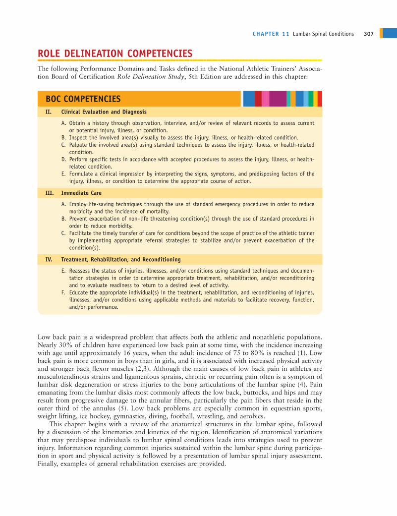
ROLE DELINEATION COMPETENCIESThe following Performance Domains and Tasks defined in the National Athletic Trainers’ Associa-tion Board of Certification Role Delineation Study, 5th Edition are addressed in this chapter:
Low back pain is a widespread problem that affects both the athletic and nonathletic populations.Nearly 30% of children have experienced low back pain at some time, with the incidence increasingwith age until approximately 16 years, when the adult incidence of 75 to 80% is reached (1). Lowback pain is more common in boys than in girls, and it is associated with increased physical activityand stronger back flexor muscles (2,3). Although the main causes of low back pain in athletes aremusculotendinous strains and ligamentous sprains, chronic or recurring pain often is a symptom oflumbar disk degeneration or stress injuries to the bony articulations of the lumbar spine (4). Painemanating from the lumbar disks most commonly affects the low back, buttocks, and hips and mayresult from progressive damage to the annular fibers, particularly the pain fibers that reside in theouter third of the annulus (5). Low back problems are especially common in equestrian sports,weight lifting, ice hockey, gymnastics, diving, football, wrestling, and aerobics.
This chapter begins with a review of the anatomical structures in the lumbar spine, followedby a discussion of the kinematics and kinetics of the region. Identification of anatomical variationsthat may predispose individuals to lumbar spinal conditions leads into strategies used to preventinjury. Information regarding common injuries sustained within the lumbar spine during participa-tion in sport and physical activity is followed by a presentation of lumbar spinal injury assessment.Finally, examples of general rehabilitation exercises are provided.
CHAPTER 11 Lumbar Spinal Conditions 307
BOC COMPETENCIESII. Clinical Evaluation and Diagnosis
A. Obtain a history through observation, interview, and/or review of relevant records to assess currentor potential injury, illness, or condition.
B. Inspect the involved area(s) visually to assess the injury, illness, or health-related condition.C. Palpate the involved area(s) using standard techniques to assess the injury, illness, or health-related
condition.D. Perform specific tests in accordance with accepted procedures to assess the injury, illness, or health-
related condition.E. Formulate a clinical impression by interpreting the signs, symptoms, and predisposing factors of the
injury, illness, or condition to determine the appropriate course of action.
III. Immediate Care
A. Employ life-saving techniques through the use of standard emergency procedures in order to reducemorbidity and the incidence of mortality.
B. Prevent exacerbation of non–life threatening condition(s) through the use of standard procedures inorder to reduce morbidity.
C. Facilitate the timely transfer of care for conditions beyond the scope of practice of the athletic trainerby implementing appropriate referral strategies to stabilize and/or prevent exacerbation of the condition(s).
IV. Treatment, Rehabilitation, and Reconditioning
E. Reassess the status of injuries, illnesses, and/or conditions using standard techniques and documen-tation strategies in order to determine appropriate treatment, rehabilitation, and/or reconditioningand to evaluate readiness to return to a desired level of activity.
F. Educate the appropriate individual(s) in the treatment, rehabilitation, and reconditioning of injuries,illnesses, and/or conditions using applicable methods and materials to facilitate recovery, function,and/or performance.
ANDERc11.qxd 11/16/07 3:53 PM Page 307
ANATOMY OF THE LUMBAR SPINEAs mentioned in Chapter 10,[AU/ED: Please confirm chapter x-ref.] the lumbar and sacral regionsof the spine are anatomically and functionally unique. Normal lumbar curvature is concave, andsacral curvature is convex, from the posterior perspective.
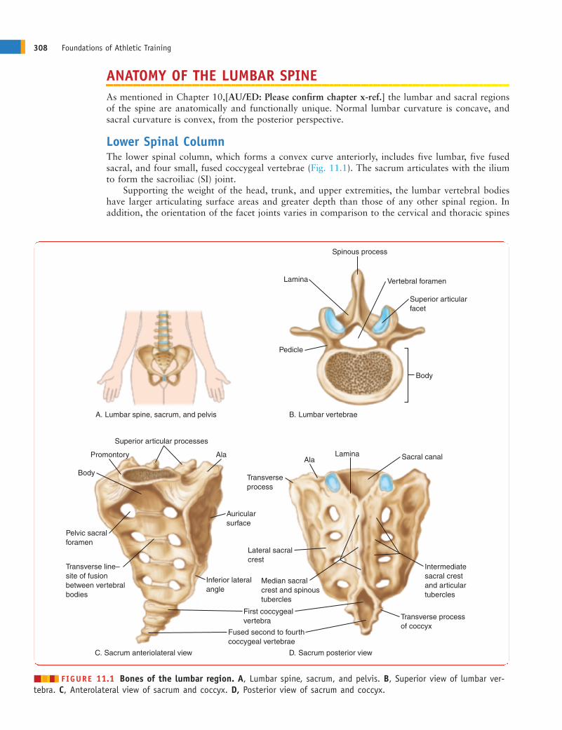
Lower Spinal ColumnThe lower spinal column, which forms a convex curve anteriorly, includes five lumbar, five fusedsacral, and four small, fused coccygeal vertebrae (Fig. 11.1). The sacrum articulates with the iliumto form the sacroiliac (SI) joint.
Supporting the weight of the head, trunk, and upper extremities, the lumbar vertebral bodieshave larger articulating surface areas and greater depth than those of any other spinal region. Inaddition, the orientation of the facet joints varies in comparison to the cervical and thoracic spines
308 Foundations of Athletic Training
Spinous process
Lamina
Lamina
Vertebral foramen
Superior articularfacet
Body
Pedicle
Body
Transverse line–site of fusion between vertebralbodies
Pelvic sacralforamen
Promontory AlaAla
Transverseprocess
Lateral sacral crest
Median sacral crest and spinoustubercles
Auricularsurface
Inferior lateral angle
Transverse processof coccyx
Intermediate sacral crestand articular tubercles
Sacral canal
First coccygealvertebra
Fused second to fourthcoccygeal vertebrae
Superior articular processes
Lumbar vertebrae
Sacrum anteriolateral view Sacrum posterior view
Lumbar spine, sacrum, and pelvis B.
C. D.
A.
������ FIGURE 11.1 Bones of the lumbar region. A, Lumbar spine, sacrum, and pelvis. B, Superior view of lumbar ver-tebra. C, Anterolateral view of sacrum and coccyx. D, Posterior view of sacrum and coccyx.
ANDERc11.qxd 11/16/07 3:53 PM Page 308
(see Fig. 10.4). Information concerning the general structures of the spinal vertebrae and interver-tebral disks is presented in Chapter 10.
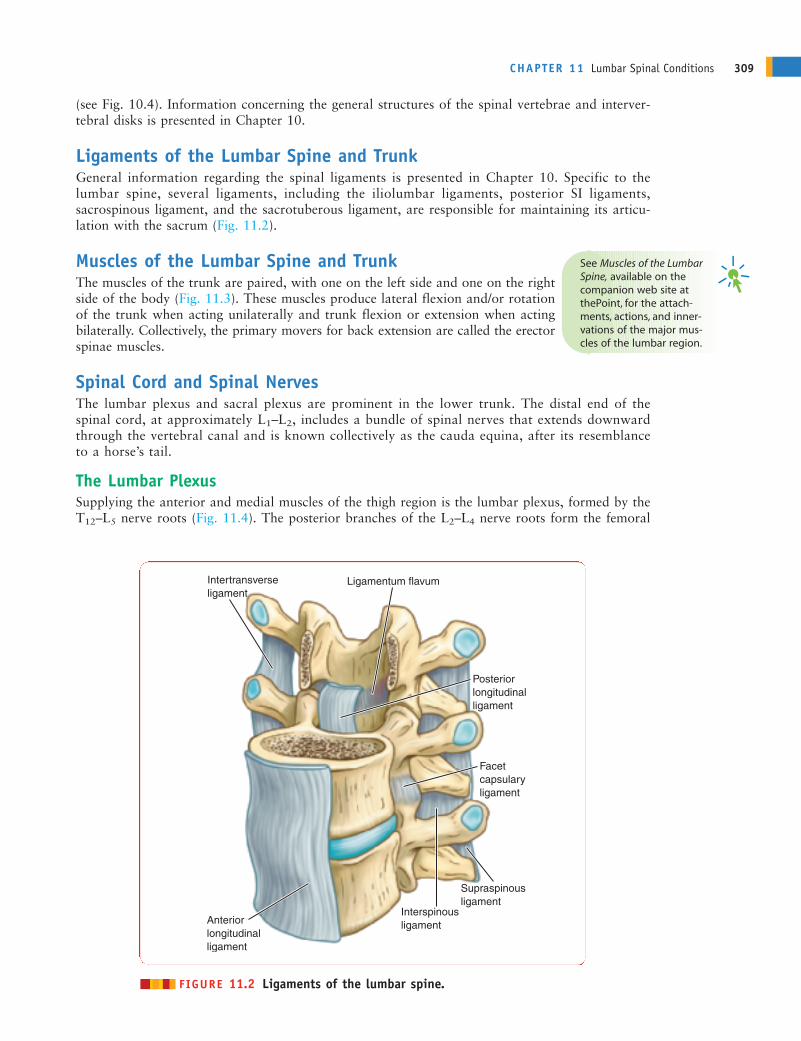
Ligaments of the Lumbar Spine and TrunkGeneral information regarding the spinal ligaments is presented in Chapter 10. Specific to thelumbar spine, several ligaments, including the iliolumbar ligaments, posterior SI ligaments,sacrospinous ligament, and the sacrotuberous ligament, are responsible for maintaining its articu-lation with the sacrum (Fig. 11.2).
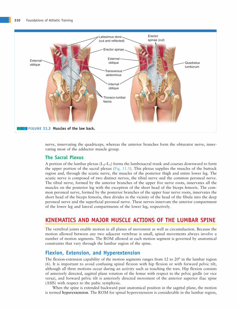
Muscles of the Lumbar Spine and TrunkThe muscles of the trunk are paired, with one on the left side and one on the rightside of the body (Fig. 11.3). These muscles produce lateral flexion and/or rotationof the trunk when acting unilaterally and trunk flexion or extension when actingbilaterally. Collectively, the primary movers for back extension are called the erectorspinae muscles.
Spinal Cord and Spinal NervesThe lumbar plexus and sacral plexus are prominent in the lower trunk. The distal end of thespinal cord, at approximately L1–L2, includes a bundle of spinal nerves that extends downwardthrough the vertebral canal and is known collectively as the cauda equina, after its resemblanceto a horse’s tail.
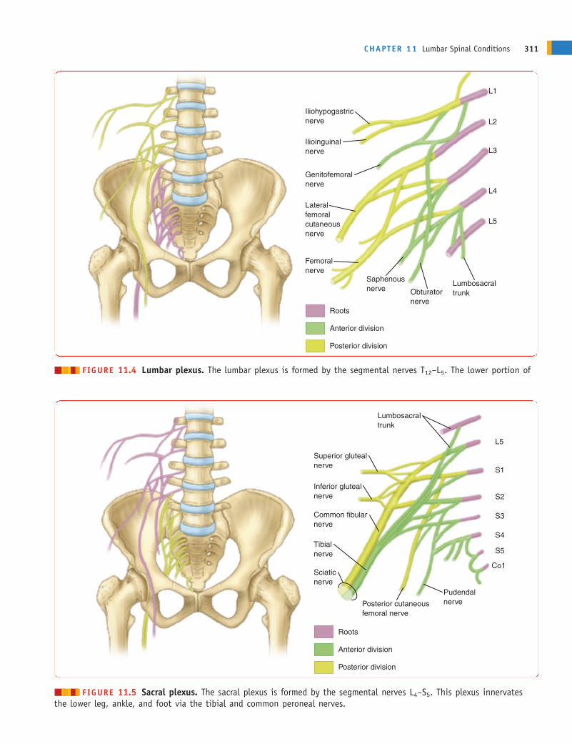
The Lumbar PlexusSupplying the anterior and medial muscles of the thigh region is the lumbar plexus, formed by theT12–L5 nerve roots (Fig. 11.4). The posterior branches of the L2–L4 nerve roots form the femoral
CHAPTER 11 Lumbar Spinal Conditions 309
Ligamentum flavum
Facetcapsularyligament
Interspinousligament
Supraspinousligament
Anteriorlongitudinalligament
Posteriorlongitudinalligament
Intertransverseligament
������ FIGURE 11.2 Ligaments of the lumbar spine.
See Muscles of the LumbarSpine, available on thecompanion web site atthePoint, for the attach-ments, actions, and inner-vations of the major mus-cles of the lumbar region.
ANDERc11.qxd 11/16/07 3:53 PM Page 309
nerve, innervating the quadriceps, whereas the anterior branches form the obturator nerve, inner-vating most of the adductor muscle group.
The Sacral PlexusA portion of the lumbar plexus (L4–L5) forms the lumbosacral trunk and courses downward to formthe upper portion of the sacral plexus (Fig. 11.5). This plexus supplies the muscles of the buttockregion and, through the sciatic nerve, the muscles of the posterior thigh and entire lower leg. Thesciatic nerve is composed of two distinct nerves, the tibial nerve and the common peroneal nerve.The tibial nerve, formed by the anterior branches of the upper five nerve roots, innervates all themuscles on the posterior leg with the exception of the short head of the biceps femoris. The com-mon peroneal nerve, formed by the posterior branches of the upper four nerve roots, innervates theshort head of the biceps femoris, then divides in the vicinity of the head of the fibula into the deepperoneal nerve and the superficial peroneal nerve. These nerves innervate the anterior compartmentof the lower leg and lateral compartments of the lower leg, respectively.
KINEMATICS AND MAJOR MUSCLE ACTIONS OF THE LUMBAR SPINEThe vertebral joints enable motion in all planes of movement as well as circumduction. Because themotion allowed between any two adjacent vertebrae is small, spinal movements always involve anumber of motion segments. The ROM allowed at each motion segment is governed by anatomicalconstraints that vary through the lumbar region of the spine.
Flexion, Extension, and HyperextensionThe flexion-extension capability of the motion segments ranges from 12 to 20° in the lumbar region(6). It is important to avoid confusing spinal flexion with hip flexion or with forward pelvic tilt,although all three motions occur during an activity such as touching the toes. Hip flexion consistsof anteriorly directed, sagittal plane rotation of the femur with respect to the pelvic girdle (or viceversa), and forward pelvic tilt is anteriorly directed movement of the anterior superior iliac spine(ASIS) with respect to the pubic symphysis.
When the spine is extended backward past anatomical position in the sagittal plane, the motionis termed hyperextension. The ROM for spinal hyperextension is considerable in the lumbar region,
310 Foundations of Athletic Training
External oblique
Transversusabdominus
Thoraco-lumbarfascia
Internaloblique
Quadratuslumborum
Erector spinae (cut)
Externaloblique
Erector spinae
Latissimus dorsi(cut and reflected)
������ FIGURE 11.3 Muscles of the low back.
ANDERc11.qxd 11/16/07 3:53 PM Page 310
CHAPTER 11 Lumbar Spinal Conditions 311
Iliohypogastricnerve
Ilioinguinalnerve
Genitofemoralnerve
Femoralnerve
Roots
Anterior division
Posterior division
Lateralfemoralcutaneousnerve
Saphenousnerve Obturator
nerve
Lumbosacraltrunk
L1
L2
L3
L4
L5
������ FIGURE 11.4 Lumbar plexus. The lumbar plexus is formed by the segmental nerves T12–L5. The lower portion of
Superior glutealnerve
Inferior glutealnerve
Tibialnerve
Sciaticnerve
Roots
Anterior division
Posterior division
Common fibularnerve
Posterior cutaneousfemoral nerve
Pudendalnerve
Lumbosacraltrunk
L5
S1
S2
S3
S4
S5
Co1
������ FIGURE 11.5 Sacral plexus. The sacral plexus is formed by the segmental nerves L4–S5. This plexus innervatesthe lower leg, ankle, and foot via the tibial and common peroneal nerves.
ANDERc11.qxd 11/16/07 3:53 PM Page 311

ranging as high as 21° at L5–S1. The cumulative ROM for hyperextension is 54° in the lumbarregion. Lumbar hyperextension is required in many sport skills, including several swimming strokes,the high jump and pole vault, wrestling, and numerous gymnastic skills. Repeated, extreme lumbarhyperextension is associated with increased risk of spondylolysis, a stress fracture of the pars inter-articularis region of the spine (7).
Lateral Flexion and RotationMovement of the spine away from anatomical position in a lateral direction in the frontal plane istermed lateral flexion. In the lumbar region, the cumulative ROM for lateral flexion is approxi-mately 24°. Spinal rotation capability is small in the lumbar region, with only approximately 2° ofmotion allowed because of the interlocking of the articular processes there. The lumbosacral jointpermits approximately 5° of rotation.
KINETICS OF THE LUMBAR SPINEAs discussed in Chapter 10, forces acting on the spine include body weight, tension in the spinal liga-ments and paraspinal muscles, intra-abdominal pressure, and any applied external loads. When the
body is in an upright position, the major form of loading on the spineis axial, and the lumbar spine supports the weight of the body segmentsabove it. Although most of the axial compression load on the spine isborne by the vertebral bodies and disks, the facet joints, when the spineis in hyperextension, may bear as much as approximately 30% of theload. Under significant compressive loading, such as during a heavylifting task, increases in intra-abdominal pressure occur that mayhelp to stiffen the trunk to prevent the spine from buckling (8).
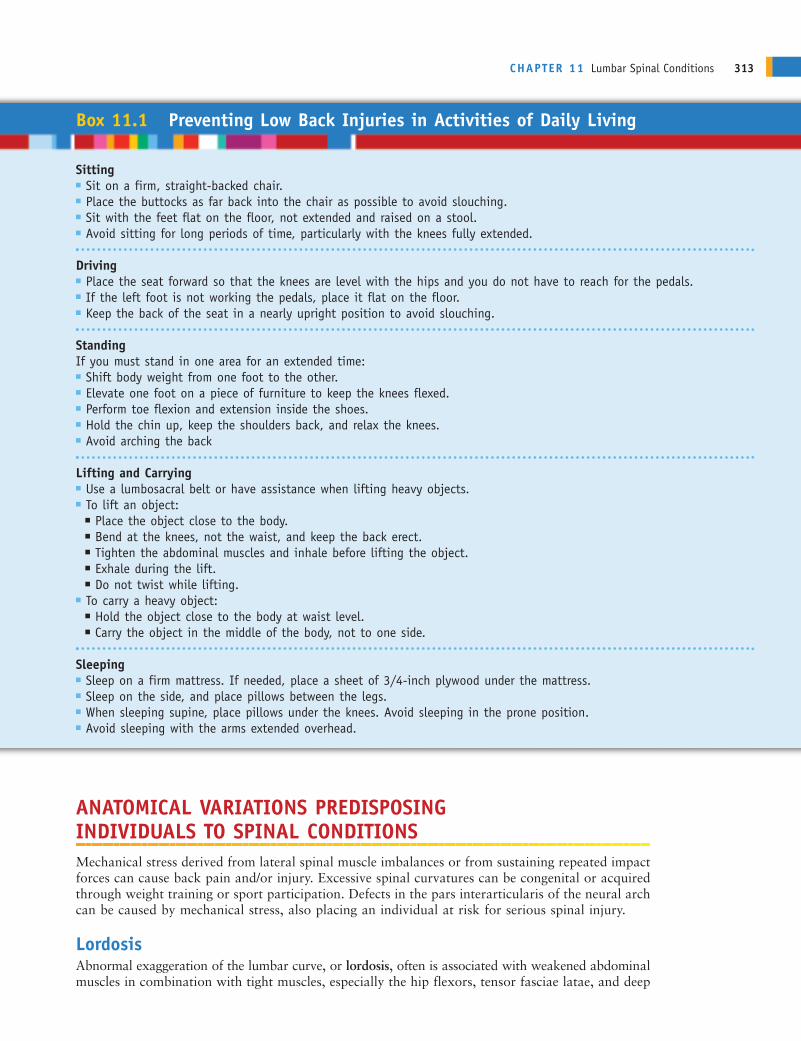
Effect of Body PositionOne factor that can dramatically affect the load on the lumbar spineis body position. When the body is in an upright position, the line ofgravity passes anterior to the spinal column (Fig. 11.6). As a result,the spine is under a constant, forward bending moment. As the trunkis progressively flexed, the line of gravity shifts farther away from thespine. The farther the line of gravity from the spine, the larger themoment arm for body weight and the greater the bending momentgenerated. To maintain body position, this moment must be counter-acted by tension in the back muscles. The more tension that is requiredto maintain body position, the greater the compression load on thespine. Lifting with the trunk being erect minimizes the tension require-ment for the lumbar muscles, because the moment arm for bodyweight is minimized. For the same reason, holding the load as closeto the trunk as possible during lifting and carrying minimizes theload on the back. In comparison to the load that is present duringupright standing, compression on the lumbar spine increases withsitting; increases more with spinal flexion; and increases still furtherwith a slouched sitting position. Box 11.1 lists guidelines for pre-venting lumbar spinal stress during performance of daily activities.
Effect of Movement SpeedAnother factor that affects loading of the lumbar spine is bodymovement speed. Executing a lift in a very rapid, jerking fashiondramatically increases compression and shear forces on the lumbarspine as well as tension in the paraspinal muscles (9). This is one rea-son why isotonic resistance training exercises should be performedin a slow, controlled fashion.
312 Foundations of Athletic Training
Line of gravity passesanterior to spinal column
������ FIGURE 11.6 Line of gravity. The lineof gravity for the head and trunk passes anteriorto the spinal column during upright standing.The moment arm for head/trunk weight at anygiven vertebral joint is the perpendicular dis-tance between the line of gravity and thespinal column.
ANDERc11.qxd 11/16/07 3:53 PM Page 312
ANATOMICAL VARIATIONS PREDISPOSING INDIVIDUALS TO SPINAL CONDITIONSMechanical stress derived from lateral spinal muscle imbalances or from sustaining repeated impactforces can cause back pain and/or injury. Excessive spinal curvatures can be congenital or acquiredthrough weight training or sport participation. Defects in the pars interarticularis of the neural archcan be caused by mechanical stress, also placing an individual at risk for serious spinal injury.
LordosisAbnormal exaggeration of the lumbar curve, or lordosis, often is associated with weakened abdominalmuscles in combination with tight muscles, especially the hip flexors, tensor fasciae latae, and deep
CHAPTER 11 Lumbar Spinal Conditions 313
Box 11.1 Preventing Low Back Injuries in Activities of Daily Living
Sitting� Sit on a firm, straight-backed chair.� Place the buttocks as far back into the chair as possible to avoid slouching.� Sit with the feet flat on the floor, not extended and raised on a stool.� Avoid sitting for long periods of time, particularly with the knees fully extended.
Driving� Place the seat forward so that the knees are level with the hips and you do not have to reach for the pedals.� If the left foot is not working the pedals, place it flat on the floor.� Keep the back of the seat in a nearly upright position to avoid slouching.
StandingIf you must stand in one area for an extended time:� Shift body weight from one foot to the other.� Elevate one foot on a piece of furniture to keep the knees flexed.� Perform toe flexion and extension inside the shoes.� Hold the chin up, keep the shoulders back, and relax the knees.� Avoid arching the back
Lifting and Carrying� Use a lumbosacral belt or have assistance when lifting heavy objects.� To lift an object:
� Place the object close to the body.� Bend at the knees, not the waist, and keep the back erect.� Tighten the abdominal muscles and inhale before lifting the object.� Exhale during the lift.� Do not twist while lifting.
� To carry a heavy object:� Hold the object close to the body at waist level.� Carry the object in the middle of the body, not to one side.
Sleeping� Sleep on a firm mattress. If needed, place a sheet of 3/4-inch plywood under the mattress.� Sleep on the side, and place pillows between the legs.� When sleeping supine, place pillows under the knees. Avoid sleeping in the prone position.� Avoid sleeping with the arms extended overhead.
ANDERc11.qxd 11/16/07 3:53 PM Page 313
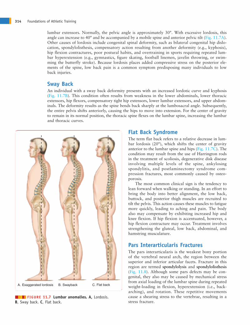
lumbar extensors. Normally, the pelvic angle is approximately 30°. With excessive lordosis, thisangle can increase to 40° and be accompanied by a mobile spine and anterior pelvic tilt (Fig. 11.7A).Other causes of lordosis include congenital spinal deformity, such as bilateral congenital hip dislo-cation, spondylolisthesis, compensatory action resulting from another deformity (e.g., kyphosis),hip flexion contractures, poor postural habits, and overtraining in sports requiring repeated lum-bar hyperextension (e.g., gymnastics, figure skating, football linemen, javelin throwing, or swim-ming the butterfly stroke). Because lordosis places added compressive stress on the posterior ele-ments of the spine, low back pain is a common symptom predisposing many individuals to lowback injuries.
Sway BackAn individual with a sway back deformity presents with an increased lordotic curve and kyphosis(Fig. 11.7B). This condition often results from weakness in the lower abdominals, lower thoracicextensors, hip flexors, compensatory tight hip extensors, lower lumbar extensors, and upper abdom-inals. The deformity results as the spine bends back sharply at the lumbosacral angle. Subsequently,the entire pelvis shifts anteriorly, causing the hips to move into extension. For the center of gravityto remain in its normal position, the thoracic spine flexes on the lumbar spine, increasing the lumbarand thoracic curves.
Flat Back SyndromeThe term flat back refers to a relative decrease in lum-bar lordosis (20°), which shifts the center of gravityanterior to the lumbar spine and hips (Fig. 11.7C). Thecondition may result from the use of Harrington rodsin the treatment of scoliosis, degenerative disk diseaseinvolving multiple levels of the spine, ankylosingspondylitis, and postlaminectomy syndrome com-pression fractures, most commonly caused by osteo-porosis.
The most common clinical sign is the tendency tolean forward when walking or standing. In an effort tobring the body into better alignment, the low back,buttock, and posterior thigh muscles are recruited totilt the pelvis. This action causes these muscles to fatiguemore quickly, leading to aching and pain. The bodyalso may compensate by exhibiting increased hip andknee flexion. If hip flexion is accentuated, however, ahip flexion contracture may occur. Treatment involvesstrengthening the gluteal, low back, abdominal, andhamstring musculature.
Pars Interarticularis FracturesThe pars interarticularis is the weakest bony portionof the vertebral neural arch, the region between thesuperior and inferior articular facets. Fracture in thisregion are termed spondylolysis and spondylolisthesis(Fig. 11.8). Although some pars defects may be con-genital, they also may be caused by mechanical stressfrom axial loading of the lumbar spine during repeatedweight-loading in flexion, hyperextension (i.e., back-arching), and rotation. These repetitive movementscause a shearing stress to the vertebrae, resulting in astress fracture.
314 Foundations of Athletic Training
Exaggerated lordosis Swayback Flat backA. C.B.
������ FIGURE 11.7 Lumbar anomalies. A, Lordosis. B, Sway back. C, Flat back.
ANDERc11.qxd 11/16/07 3:53 PM Page 314
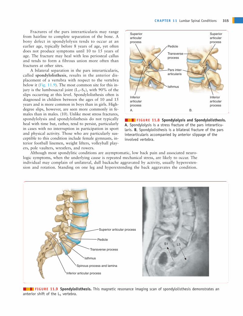
Fractures of the pars interarticularis may rangefrom hairline to complete separation of the bone. Abony defect in spondylolysis tends to occur at anearlier age, typically before 8 years of age, yet oftendoes not produce symptoms until 10 to 15 years ofage. The fracture may heal with less periosteal callusand tends to form a fibrous union more often thanfractures at other sites.
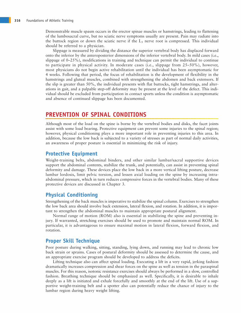
A bilateral separation in the pars interarticularis,called spondylolisthesis, results in the anterior dis-placement of a vertebra with respect to the vertebrabelow it (Fig. 11.9). The most common site for this in-jury is the lumbosacral joint (L5–S1), with 90% of theslips occurring at this level. Spondylolisthesis often isdiagnosed in children between the ages of 10 and 15years and is more common in boys than in girls. High-degree slips, however, are seen more commonly in fe-males than in males. (10). Unlike most stress fractures,spondylolysis and spondylolisthesis do not typicallyheal with time but, rather, tend to persist, particularlyin cases with no interruption in participation in sportand physical activity. Those who are particularly sus-ceptible to this condition include female gymnasts, in-terior football linemen, weight lifters, volleyball play-ers, pole vaulters, wrestlers, and rowers.
Although most spondylitic conditions are asymptomatic, low back pain and associated neuro-logic symptoms, when the underlying cause is repeated mechanical stress, are likely to occur. Theindividual may complain of unilateral, dull backache aggravated by activity, usually hyperexten-sion and rotation. Standing on one leg and hyperextending the back aggravates the condition.
CHAPTER 11 Lumbar Spinal Conditions 315
Superior articularprocess
Pedicle
Transverseprocess
Pars inter-articularis
Isthmus
Inferior articularprocess
Superior articularprocess
Inferior articularprocess
A. B.
������ FIGURE 11.8 Spondylolysis and Spondylolisthesis.A, Spondylolysis is a stress fracture of the pars interarticu-laris. B, Spondylolisthesis is a bilateral fracture of the parsinterarticularis accompanied by anterior slippage of the involved vertebra.
Superior articular process
L5S1Pedicle
Transverse process
Isthmus
Spinous process and lamina
Inferior articular process
������ FIGURE 11.9 Spondylolisthesis. This magnetic resonance imaging scan of spondylolisthesis demonstrates an anterior shift of the L5 vertebra.
ANDERc11.qxd 11/16/07 3:53 PM Page 315
316 Foundations of Athletic Training
Demonstrable muscle spasm occurs in the erector spinae muscles or hamstrings, leading to flatteningof the lumbosacral curve, but no sciatic nerve symptoms usually are present. Pain may radiate intothe buttock region or down the sciatic nerve if the L5 nerve root is compressed. This individualshould be referred to a physician.
Slippage is measured by dividing the distance the superior vertebral body has displaced forwardonto the inferior by the anteroposterior dimensions of the inferior vertebral body. In mild cases (i.e.,slippage of 0–25%), modifications in training and technique can permit the individual to continueto participate in physical activity. In moderate cases (i.e., slippage from 25–50%), however,most physicians do not begin active rehabilitation until the individual has been asymptomatic for4 weeks. Following that period, the focus of rehabilitation is the development of flexibility in thehamstrings and gluteal muscles, combined with strengthening the abdomen and back extensors. Ifthe slip is greater than 50%, the individual presents with flat buttocks, tight hamstrings, and alter-ations in gait, and a palpable step-off deformity may be present at the level of the defect. This indi-vidual should be excluded from participation in contact sports unless the condition is asymptomaticand absence of continued slippage has been documented.
PREVENTION OF SPINAL CONDITIONSAlthough most of the load on the spine is borne by the vertebral bodies and disks, the facet jointsassist with some load bearing. Protective equipment can prevent some injuries to the spinal region;however, physical conditioning plays a more important role in preventing injuries to this area. Inaddition, because the low back is subjected to a variety of stresses as part of normal daily activities,an awareness of proper posture is essential in minimizing the risk of injury.
Protective EquipmentWeight-training belts, abdominal binders, and other similar lumbar/sacral supportive devicessupport the abdominal contents, stabilize the trunk, and potentially, can assist in preventing spinaldeformity and damage. These devices place the low back in a more vertical lifting posture, decreaselumbar lordosis, limit pelvic torsion, and lessen axial loading on the spine by increasing intra-abdominal pressure, which in turn reduces compressive forces in the vertebral bodies. Many of theseprotective devices are discussed in Chapter 3.
Physical ConditioningStrengthening of the back muscles is imperative to stabilize the spinal column. Exercises to strengthenthe low back area should involve back extension, lateral flexion, and rotation. In addition, it is impor-tant to strengthen the abdominal muscles to maintain appropriate postural alignment.
Normal range of motion (ROM) also is essential in stabilizing the spine and preventing in-jury. If warranted, stretching exercises should be used to promote and maintain normal ROM. Inparticular, it is advantageous to ensure maximal motion in lateral flexion, forward flexion, androtation.
Proper Skill TechniquePoor posture during walking, sitting, standing, lying down, and running may lead to chronic lowback strain or sprains. Cases of postural deformity should be assessed to determine the cause, andan appropriate exercise program should be developed to address the deficits.
Lifting technique also can affect spinal loading. Executing a lift in a very rapid, jerking fashiondramatically increases compression and shear forces on the spine as well as tension in the paraspinalmuscles. For this reason, isotonic resistance exercises should always be performed in a slow, controlledfashion. Breathing technique should be emphasized as well. Specifically, it is desirable to inhaledeeply as a lift is initiated and exhale forcefully and smoothly at the end of the lift. Use of a sup-portive weight-training belt and a spotter also can potentially reduce the chance of injury to thelumbar region during heavy weight lifting.
ANDERc11.qxd 11/16/07 3:53 PM Page 316
CONDITIONS OF THE LUMBAR SPINEWhat criteria should be used to determine whether an individual who reports low back painshould be referred to a physician?
The lumbar spine must support the weight of the head, trunk, and arms as well as any load heldin the hands. In addition, the two lower lumbar motion segments (i.e., L4–L5 and L5–S1) provide alarge ROM in flexion-extension. As such, it is not surprising that mechanical abuse often results inepisodes of low back pain or that the lower lumbar disks are injured more frequently than anyothers in the spine.
Lumbar Contusions, Strains, and SprainsAn estimated 75 to 80% of the population experiences low back pain stemming from mechanicalinjury to muscles, ligaments, or connective tissue (Box 11.2). Although low back pain typicallystrikes adults, nearly 30% of children experience low back pain up to the age of 16 years (1).Several known pathologies may cause low back pain, but reduced spinal flexibility, repeated stress,and activities that require maximal extension of the lumbar spine are most associated with chroniclow back pain.
Muscle strains may result from a sudden extension action with trunk rotation on an overtaxed,unprepared, or underdeveloped spine. Chronic strains may stem from improper posture, excessivelumbar lordosis, flat back, or scoliosis.
Signs and SymptomsPain and discomfort can range from diffuse to localized. Pain does not radiate into the buttocks orposterior thigh, and no signs of neural involvement, such as muscle weakness, sensory changes, orreflex inhibition, are seen. If a muscle strain is present, pain will increase with passive flexion andwith active or resisted extension.
ManagementAcute protocol is followed to control pain and hemorrhage. Following cold treatment, passivestretching of the low back can help to relieve muscle spasm. A corset-type brace can be worn tocompress the area (see Fig. 3.5). Following the acute stage, a graduated stretching and strengthen-ing program can be initiated. In moderate to severe cases, the individual should be referred to aphysician. Prescription muscle relaxants or nonsteroidal anti-inflammatory drugs (NSAIDs) may beappropriate.
Low Back Pain in RunnersMany runners develop muscle tightness in the hip flexors and hamstrings. Tight hip flexors tend toproduce a forward body lean, which leads to anterior pelvic tilt and hyperlordosis of the lumbarspine. Because the lumbar muscles develop tension to counteract the forward bending moment ofthe entire trunk when the trunk is in flexion, these muscles are particularly susceptible to strain.Coupled with tight hamstrings, a shorter stride often emerges.
CHAPTER 11 Lumbar Spinal Conditions 317
Box 11.2 Causes of Low Back Pain
� Muscle strains and sprains � Spinal infections (e.g., tuberculosis)� Sciatica � Neoplastic tumor (i.e., primary or metastatic)� Protruded or herniated disk � Ankylosing spondylitis (arthritis of the spine)� Pathologic fracture � Benign space-occupying lesions� Disk space infections � Abdominal aortic aneurysm
ANDERc11.qxd 11/16/07 3:53 PM Page 317
318 Foundations of Athletic Training
Signs and SymptomsSymptoms include localized pain that increases with active and resisted back extension, but radiatingpain and neurologic deficits are not present. Anterior pelvic tilt and hyperlordosis of the lumbarspine also may be present.
ManagementTreatment focuses on avoiding excessive flexion activities and a sedentary posture (Box 11.3). Flex-ion causes the mobile nucleus pulposus to shift posteriorly and press against the annulus fibrosusat its thinnest, least-buttressed place. In most cases, this just leads to pain, but in others, it may leadto a herniated disk. In addition, physical activity is necessary to pump fluid through the spinal disksto keep them properly hydrated; by interfering with this process, immobility can prolong pain.
Ice, NSAIDs, muscle relaxants, transcutaneous electrical nerve stimulation (TENS), and electricalmuscle stimulation may be used to reduce pain and inflammation. Lumbar stabilization exercisescan be combined with extension exercises, progressive activity, and early mobilization. Aerobic exer-cise, such as walking, swimming, or biking, should be included in all programs. If symptoms do notimprove within a week, the individual should be referred to a physician to rule out a more seriousunderlying condition. In an effort to decrease the incidence of low back pain, training techniquesshould allow adequate progression of distance and intensity and include extensive flexibility exercisesfor the hip and thigh region.
Myofascial PainMyofascial pain is referred pain that emanates from a myofascial trigger point, a hypersensitive, lo-calized nodule within a taut band of muscle tissue and its surrounding fascia. When compressed orpalpated, pain is produced in a predictable distribution of referred pain. In the lumbar area, thepiriformis muscle and quadratus lumborum are common trigger point sites associated with extendedsitting, standing, running and walking activities. The piriformis in particular can impact the sciaticnerve as it courses through, above, or below the muscle on its path into the posterior leg. Individualswho slip unexpectedly and catch themselves also can irritate the trigger points.
Signs and SymptomsAggravation of the piriformis can lead to referred pain in the SI area, posterior hip, and upper two-thirds of the posterior thigh. Aching and deep pain increases with activity or with prolonged sittingwith the hip adducted, flexed, and internally rotated. If the sciatic nerve is impinged, pain andpossible changes in sensation may extend into the leg.
Box 11.3 Reducing Low Back Pain in Runners
� Wear properly fitted shoes that control heel motion and provide maximum shock absorption.� Increase flexibility at the hip, knee, ankle plantar flexors, and trunk extensors.� Increase strength in the abdominal and trunk extensor muscles.� Avoid excessive body weight.� Warm up before and after running.� Run with an upright stance rather than with a forward lean.� Avoid excessive side-to-side sway.� Run on even terrain, and limit hill work. Avoid running on concrete.� Avoid overstriding to increase speed, because this increases leg shock.� Gradually increase distance, intensity, and duration. Do not increase any parameter more than 10% in 1 week.� If orthotics are worn and pain persists, check for wear and rigidity.� Consider alternatives to running, such as cycling, rowing, or swimming.
ANDERc11.qxd 11/16/07 3:53 PM Page 318
Referred pain from the quadratus lumborum often gives a false sign of a disk syndrome andoften is overlooked as a source of low back pain. The superficial fibers can refer a sharp, aching painto the low back, iliac crest, or greater trochanter or can extend it to the abdominal wall. The deep fibersmay refer pain to the SI joint or lower buttock region. Pain increases during lateral bending towardthe involved side, while standing for long periods of time, and during coughing or sneezing.
ManagementTrigger point treatment involves stretching the involved muscle back to its normal resting lengthas a way to relieve the irritation that led to the initial pain. The patient should be placed in acomfortable position on the uninvolved side if the piriformis is involved or prone if the quadratuslumborum is involved. Three potential techniques can be used. One technique involves applicationof pressure slowly and progressively over the trigger point. Pressure is maintained until the tender-ness is gone. Another technique involves ice massage applied over the length of the muscle andthen over the referred pain pattern. The ice is applied in longitudinal, parallel strokes in only onedirection while a passive stretch is applied progressively to the involved muscle. The third techniqueinvolves a deep-stroking massage over the length of the muscle, moving in a distal to proximal di-rection. As the massage continues, the taut band should relax, the tender nodules soften, and thepain ease.
Facet Joint PathologyThroughout the longitudinal axis of the spine, three distinct anatomical columns can be defined atany spinal motion segment—namely, the anterior, middle, and posterior columns. The posteriorcolumn contains the pars interarticularis, facet joints, and spinous processes and is supported bythe ligamentum flavum and interspinous ligaments. The facet joint is a synovial joint richly inner-vated via the medial branch of the posterior primary rami of at least two adjacent spinal nerves.The facet joint capsules act as passive restraints against excessive lumbar rotation and flexion andserve as a protective mechanism for the intervertebral disk.
Lumbar facet pathology may involve subluxation or dislocation of the facet, facet joint syn-drome (i.e., inflammation), or degeneration of the facet itself (i.e., arthritis). The exact pathophys-iology is unclear. Theories include possible mechanical irritation of the nearby nerve root, chemicalirritation arising from the inflammatory process (e.g., capsular and synovial inflammation), menis-coid entrapment, synovial impingement, joint subluxation, chondromalacia facette, mechanical in-jury to the joint’s capsule, and restriction to normal articular motion from soft or articular causes.
Signs and SymptomsSigns and symptoms can include nonspecific low back, hip, and buttock pain with a deep and achyquality. The pain may radiate into the posterior thigh, but it does not radiate below the knee. Somepatients describe their pain as being worse in the morning, aggravated by rest and hyperextension,and relieved by repeated motion. Flattening of lumbar lordosis may be visible. Point tenderness maybe elicited to a unilateral or bilateral paravertebral area. Pain often is exacerbated by trunk rotation,stretching into full extension, lateral bending toward the involved side, and with torsion. Sensoryalternations usually are absent unless the nerve root is secondarily involved.
Limited flexibility of the pelvic musculature can directly impact the mechanics of the lumbosacralspine. If facet joint pathology is present, an abnormal pelvic tilt and rotation of the hip secondaryto tight hamstrings, hip rotators, and quadratus may be evident. Typically, manual muscle testingis normal; however, a subtle weakness in the erector spinae and hamstring muscles may contributeto pelvic tilt abnormalities. This subtle weakness may be appreciated with trunk, pelvic, and lowerextremity extension asymmetry. If facet hypertrophy narrows the neural foramen, causing nerve rootimpingement, a straight leg raising test may elicit a positive response. Typically, this maneuver isnormal.
ManagementA definitive diagnosis is made using radiographs or magnetic resonance imaging or by a physicianinjecting the facet with an anesthetic and noting any change in the symptoms. Initial treatment
CHAPTER 11 Lumbar Spinal Conditions 319
ANDERc11.qxd 11/16/07 3:53 PM Page 319
should focus on education, relative rest, pain relief, and maintenance of positions that provide comfort,exercises, and some modalities. Therapeutic exercises should include instruction regarding properposture and body mechanics in activities of daily living that protect the injured joints, reduce symp-toms, and prevent further injury. Positions that cause pain should be avoided. Modalities such assuperficial heat and cryotherapy may help to relax the muscles and reduce pain. In addition, med-ications such as NSAIDs can be advantageous. Spinal manipulation and mobilization also can beused to reduce pain. Once the painful symptoms are controlled during the acute phase of treatment,stretching and strengthening exercises of the lumbar spine and associated muscles can be initiated.
SciaticaSciatica, an inflammatory condition of the sciatic nerve, is classified in terms of four levels of sever-ity, each with its own management strategy (Box 11.4). The condition can be caused by a herniateddisk, annular tear, myogenic or muscle-related disease, spinal stenosis, facet joint arthropathy, orcompression of the nerve between the piriformis muscle.
Signs and SymptomsIf related to a herniated disk, radiating leg pain is greater than back pain and increases with sittingand leaning forward, coughing, sneezing, and straining. Pain is reproduced during an ipsilateralstraight leg raising test (see Fig. 11.18).
In an annular tear, back pain is more prevalent and is exacerbated with straight leg raising.Morning pain and muscular stiffness that worsens if chilled or when the weather changes (arthritic-like symptoms) are characteristic of myogenic or muscle-related disease. Pain typically radiates intothe buttock and thigh region.
If lumbar spinal stenosis is present, back and leg pain develop after the individ-ual walks a limited distance and concomitantly increase as the distance increases.Pain is not reproduced with a straight leg raising test, but it can be reproduced withprolonged spine extension, which is relieved with spine flexion. If a facet joint isinvolved, pain is localized over the joint on spinal extension and is exacerbatedwith ipsilateral lateral flexion. If the sciatic nerve is compressed by the piriformismuscle, pain increases during internal rotation of the thigh.
ManagementReferral to a physician is necessary to check for a potentially serious underlying condition. Undernormal circumstances, bed rest usually is not indicated, although side-lying with the knees flexedmay relieve symptoms. Lifting, bending, twisting, and prolonged sitting and standing aggravate thecondition and, therefore, should be avoided. When asymptomatic, abdominal and extensor musclestrengthening exercises can begin, with gradual return to activity. If symptoms resume, however,activity should cease, and the individual should be referred back to the physician. Occasionally,
320 Foundations of Athletic Training
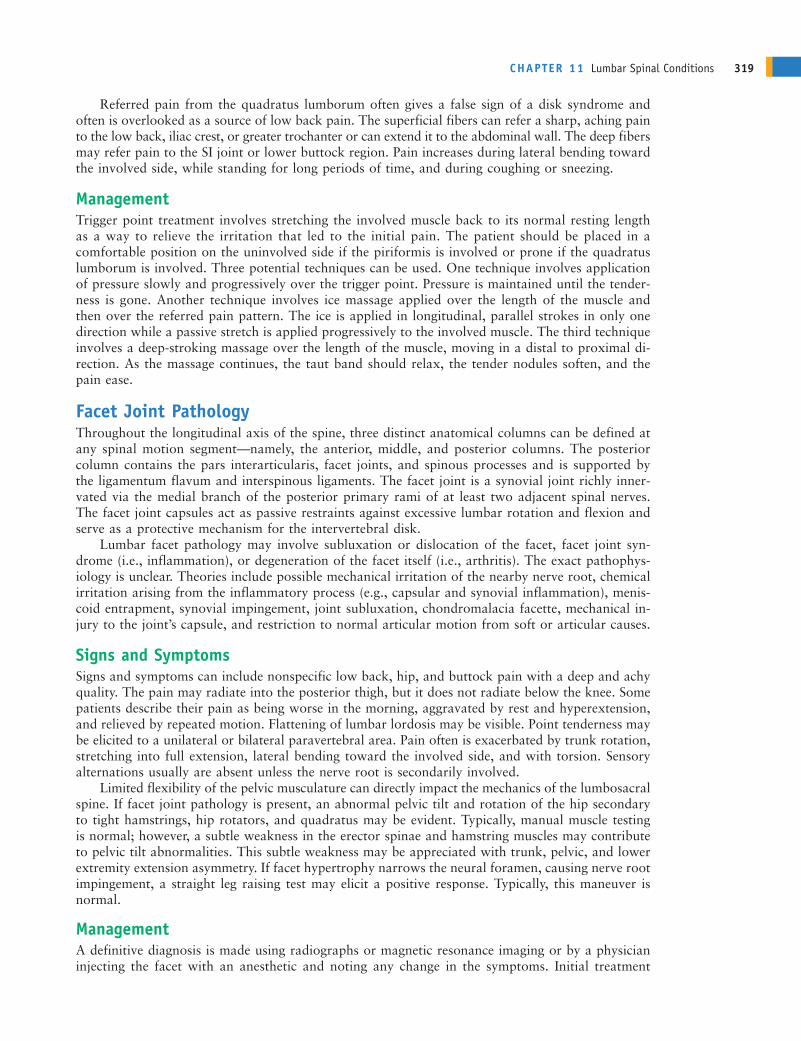
Box 11.4 Classification and Management of Sciatica
� Sciatica only: No sensory or muscle weakness. Modify activity appropriately, and develop rehabilitation and preventionprogram. Any increased pain requires immediate re-evaluation.
� Sciatica with soft signs: Some sensory changes, mild or no reflex change, normal muscle strength, and normal boweland bladder function. Remove from sport participation for 6 to 12 weeks.
� Sciatica with hard signs: Sensory and reflex changes, and muscle weakness caused by repeated, chronic, or acute con-dition. Normal bowel and bladder function. Remove from participation for 12 to 24 weeks.
� Sciatica with severe signs: Sensory and reflex changes, muscle weakness, and altered bladder function. Consider imme-diate surgical decompression.
See Signs and Symptoms ofSciatic, available on thecompanion website at thePoint, for the commonsigns and symptoms thataccompany the various etiologies of sciatica.
ANDERc11.qxd 11/16/07 3:53 PM Page 320
extended rest is needed for symptoms to resolve totally,and if a significant disk protrusion is present, surgerymay be indicated.
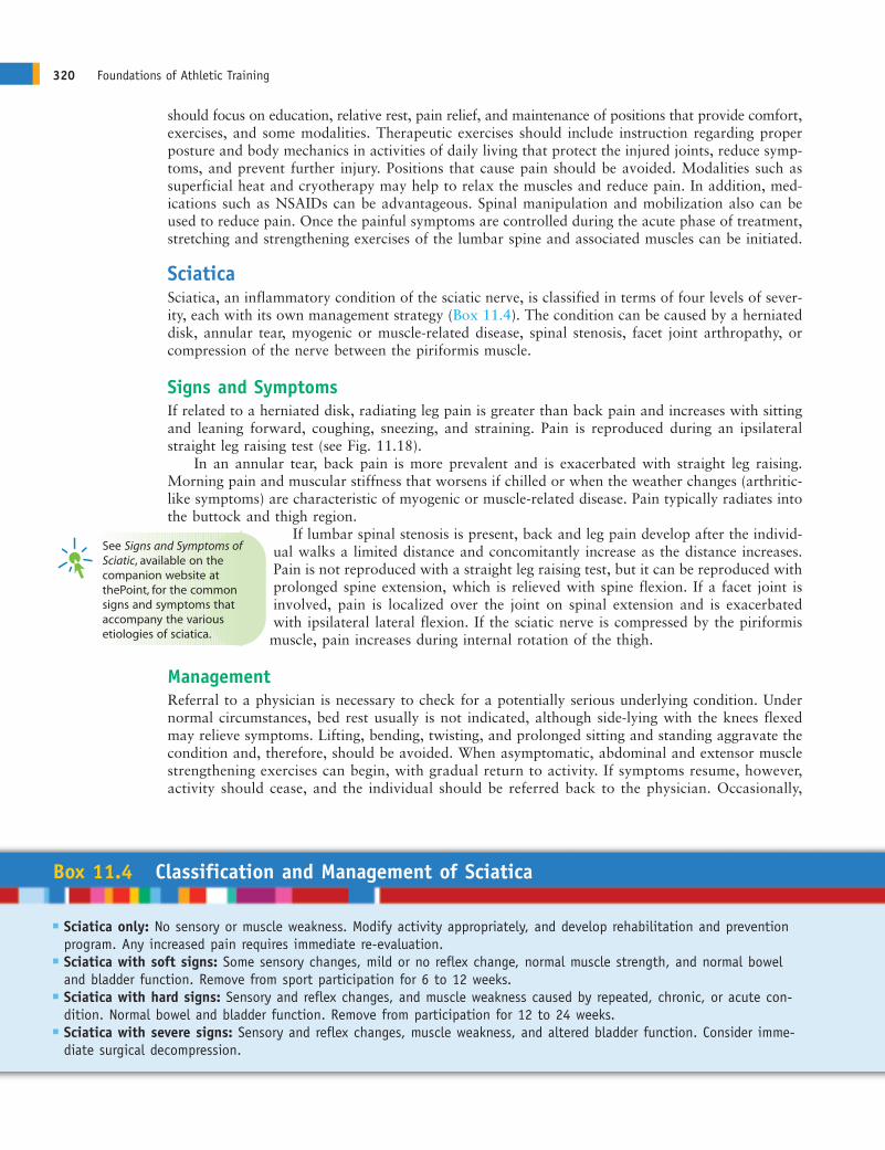
Conditions of the Lumbar DiskProlonged mechanical loading of the spine can lead tomicroruptures in the annulus fibrosus, resulting in de-generation of the disk (Fig. 11.10). Bulging or protrudeddisks refer to some eccentric accumulation of the nucleuswith slight deformity of the annulus. When the eccentricnucleus produces a definite deformity as it works its waythrough the fibers of the annulus, it is called a prolapseddisk. It is called an extruded disk when the materialmoves into the spinal canal, where it runs the risk ofimpinging on adjacent nerve roots. Finally, with a se-questrated disk, the nuclear material has separated fromthe disk itself and, potentially, can migrate. The mostcommonly herniated disks are the lower two lumbardisks at L4-L5 and L5-S1, followed by the two lowercervical disks. Most ruptures move in a posterior orposterolateral direction as a result of torsion and com-pression, not just compression.
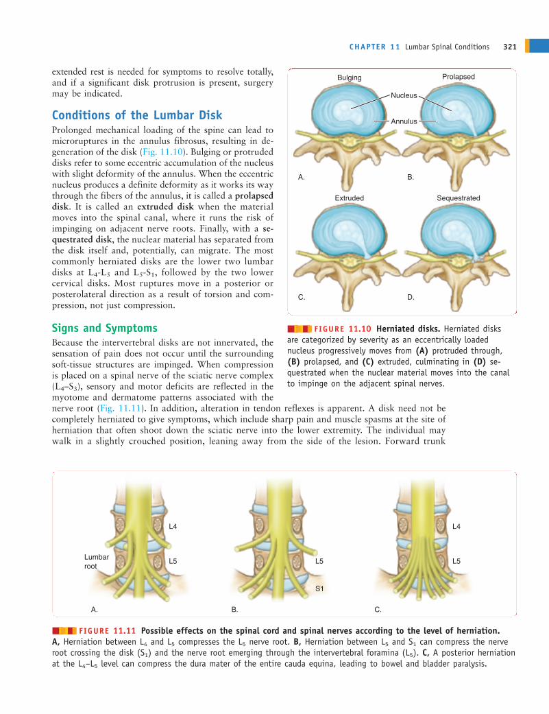
Signs and SymptomsBecause the intervertebral disks are not innervated, thesensation of pain does not occur until the surroundingsoft-tissue structures are impinged. When compressionis placed on a spinal nerve of the sciatic nerve complex(L4–S3), sensory and motor deficits are reflected in themyotome and dermatome patterns associated with thenerve root (Fig. 11.11). In addition, alteration in tendon reflexes is apparent. A disk need not becompletely herniated to give symptoms, which include sharp pain and muscle spasms at the site ofherniation that often shoot down the sciatic nerve into the lower extremity. The individual maywalk in a slightly crouched position, leaning away from the side of the lesion. Forward trunk
CHAPTER 11 Lumbar Spinal Conditions 321
Nucleus
Bulging Prolapsed
Extruded Sequestrated
Annulus
A. B.
C. D.
������ FIGURE 11.10 Herniated disks. Herniated disks are categorized by severity as an eccentrically loaded nucleus progressively moves from (A) protruded through,(B) prolapsed, and (C) extruded, culminating in (D) se-questrated when the nuclear material moves into the canalto impinge on the adjacent spinal nerves.
Lumbar root
L5 L5
S1
L4
L5
L4
A. C.B.
������ FIGURE 11.11 Possible effects on the spinal cord and spinal nerves according to the level of herniation. A, Herniation between L4 and L5 compresses the L5 nerve root. B, Herniation between L5 and S1 can compress the nerveroot crossing the disk (S1) and the nerve root emerging through the intervertebral foramina (L5). C, A posterior herniationat the L4–L5 level can compress the dura mater of the entire cauda equina, leading to bowel and bladder paralysis.
ANDERc11.qxd 11/16/07 3:53 PM Page 321
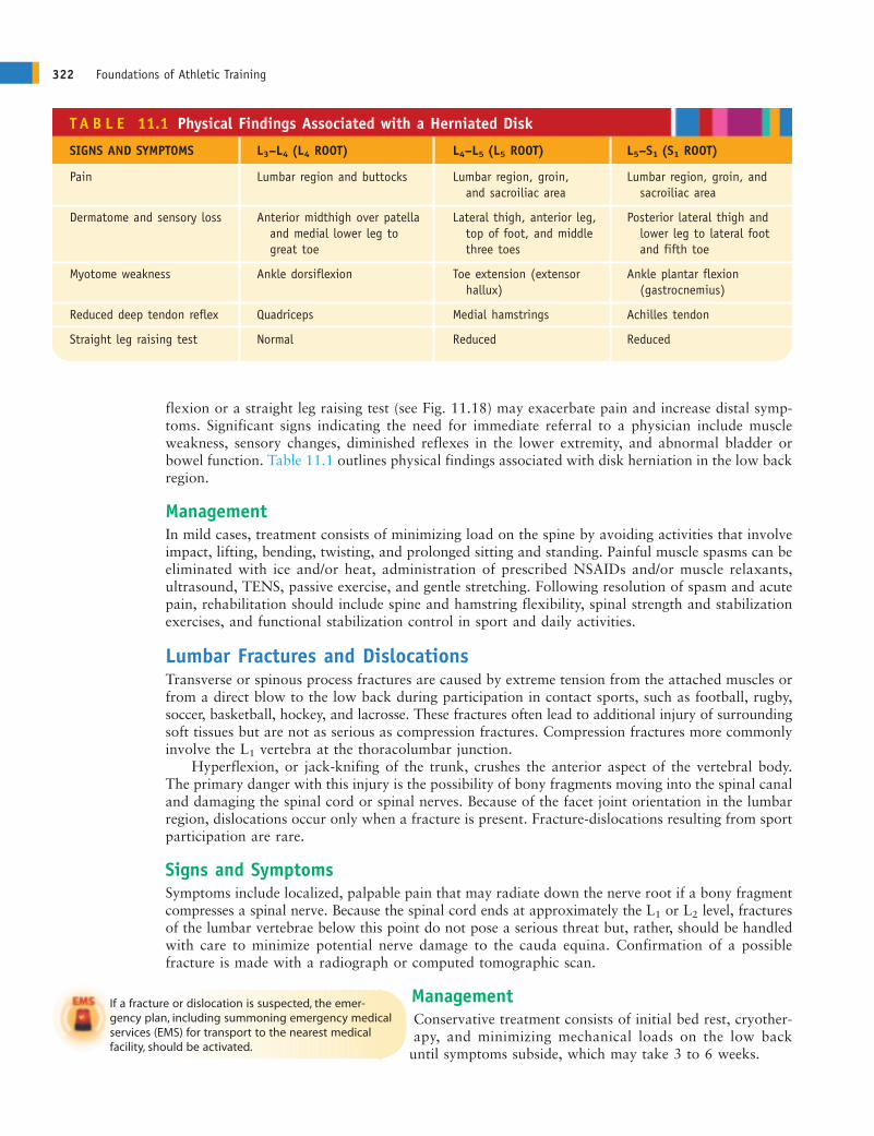
flexion or a straight leg raising test (see Fig. 11.18) may exacerbate pain and increase distal symp-toms. Significant signs indicating the need for immediate referral to a physician include muscleweakness, sensory changes, diminished reflexes in the lower extremity, and abnormal bladder orbowel function. Table 11.1 outlines physical findings associated with disk herniation in the low backregion.
ManagementIn mild cases, treatment consists of minimizing load on the spine by avoiding activities that involveimpact, lifting, bending, twisting, and prolonged sitting and standing. Painful muscle spasms can beeliminated with ice and/or heat, administration of prescribed NSAIDs and/or muscle relaxants,ultrasound, TENS, passive exercise, and gentle stretching. Following resolution of spasm and acutepain, rehabilitation should include spine and hamstring flexibility, spinal strength and stabilizationexercises, and functional stabilization control in sport and daily activities.
Lumbar Fractures and DislocationsTransverse or spinous process fractures are caused by extreme tension from the attached muscles orfrom a direct blow to the low back during participation in contact sports, such as football, rugby,soccer, basketball, hockey, and lacrosse. These fractures often lead to additional injury of surroundingsoft tissues but are not as serious as compression fractures. Compression fractures more commonlyinvolve the L1 vertebra at the thoracolumbar junction.
Hyperflexion, or jack-knifing of the trunk, crushes the anterior aspect of the vertebral body.The primary danger with this injury is the possibility of bony fragments moving into the spinal canaland damaging the spinal cord or spinal nerves. Because of the facet joint orientation in the lumbarregion, dislocations occur only when a fracture is present. Fracture-dislocations resulting from sportparticipation are rare.
Signs and SymptomsSymptoms include localized, palpable pain that may radiate down the nerve root if a bony fragmentcompresses a spinal nerve. Because the spinal cord ends at approximately the L1 or L2 level, fracturesof the lumbar vertebrae below this point do not pose a serious threat but, rather, should be handledwith care to minimize potential nerve damage to the cauda equina. Confirmation of a possiblefracture is made with a radiograph or computed tomographic scan.
ManagementConservative treatment consists of initial bed rest, cryother-apy, and minimizing mechanical loads on the low back
until symptoms subside, which may take 3 to 6 weeks.
322 Foundations of Athletic Training
T A B L E 11.1 Physical Findings Associated with a Herniated Disk
SIGNS AND SYMPTOMS L3–L4 (L4 ROOT) L4–L5 (L5 ROOT) L5–S1 (S1 ROOT)
Pain Lumbar region and buttocks Lumbar region, groin, Lumbar region, groin, and and sacroiliac area sacroiliac area
Dermatome and sensory loss Anterior midthigh over patella Lateral thigh, anterior leg, Posterior lateral thigh and and medial lower leg to top of foot, and middle lower leg to lateral foot great toe three toes and fifth toe
Myotome weakness Ankle dorsiflexion Toe extension (extensor Ankle plantar flexion hallux) (gastrocnemius)
Reduced deep tendon reflex Quadriceps Medial hamstrings Achilles tendon
Straight leg raising test Normal Reduced Reduced
If a fracture or dislocation is suspected, the emer-gency plan, including summoning emergency medicalservices (EMS) for transport to the nearest medical facility, should be activated.
ANDERc11.qxd 11/16/07 3:53 PM Page 322
If assessment of a low back injury reveals signs of nerve root involvement (i.e., sensory ormotor deficits and diminished reflexes) or disk injury, physician referral is warranted.
SACRUM AND COCCYX CONDITIONSA 40-year-old man initiated a training program to improve his cardiovascular fitness.His workout for the past month has consisted of running on a treadmill. Over the past
week, he began to develop pain in the sacral region during his workout. The pain has now becomeso persistent and chronic that it hurts to sit for an extended period of time. What injury maybe present, and what recommendations can be made to this person relative to caring for theinjury?
Because the sacrum and coccyx are essentially immobile, the potential for mechanical injury to theseregions is dramatically reduced. In many cases, injuries result from direct blows and stress on theSI joint.
Sacroiliac Joint SprainSprains of the SI joint may result from a single traumatic episode that involves bending and/ortwisting, repetitive stress from lifting, a fall on the buttocks, excessive side-to-side or up-and-downmotion during running and jogging, running on uneven terrain, suddenly slipping or stumbling for-ward, or wearing new shoes or orthoses. The injury may irritate or stretch the sacrotuberous orsacrospinous ligament, or it may lead to an anterior or posterior rotation of one side of the pelvisrelative to the other. Hypermobility results from rotation of the pelvis. During healing, the joint onthe injured side may become hypermobile, allowing the joint to subluxate in either an anterior- orposterior-rotated position.
Signs and SymptomsSymptoms may involve unilateral, dull pain in the sacral area that extends into the buttock and pos-terior thigh. On observation, the ASIS or posterior superior iliac spine (PSIS) may appear to beasymmetrical when compared bilaterally. A leg-length discrepancy may be present, but musclespasm is not often seen. Standing on one leg and climbing stairs may increase the pain. Forwardbending reveals a block to normal movement, with the PSIS on the injured side moving sooner thanon the uninjured side. Lateral flexion toward the injured side increases pain, as do straight leg raisesbeyond 45°.
ManagementTreatment for SI sprains includes cryotherapy, prescribed NSAIDs, and gentle stretching to allevi-ate stiffness. As the conditions improves, flexibility, pelvic stabilization exercises, mobilization ofthe affected joint, and strengthening exercises for the low back should be initiated.
Coccygeal ConditionsDirect blows to the region can produce contusions and fractures of the coccyx. Pain resulting froma fracture may last for several months. Prolonged or chronic pain in the region also may result fromirritation of the coccygeal nerve plexus. This condition is termed coccygodynia. Treatment for coc-cygeal pain includes analgesics, use of padding for protection, and a ring seat to alleviate compres-sion during sitting.
This individual has probably irritated the SI joint from repeated stress while running on thetreadmill. This individual should ice the region to control inflammation and pain and should
stretch the low back and buttock region. A detailed assessment (including a gait analysis whilerunning) should be performed to determine the potential cause of the injury so that a properrehabilitation program and, subsequently, an appropriate cardiovascular conditioning regimen canbe developed.
CHAPTER 11 Lumbar Spinal Conditions 323
ANDERc11.qxd 11/16/07 3:53 PM Page 323
ASSESSMENT OF SPINAL CONDITIONSA 17-year-old cheerleader reports to the athletic training room complaining of aching painduring trunk flexion and aggravated with resisted hyperextension that produces sharp,
shooting pains into the low back and down the posterior leg. How should the assessment of thisinjury progress to determine the extent and severity of injury?
Injury assessment of the lumbar spine is difficult and complex. In the event of an acute injury withpossible nerve involvement, immobilization and immediate transportation to the nearest medicalfacility is warranted, regardless of whether a total assessment is completed. Box 11.5 identifiesseveral “Red Flags” that warrant immobilization and immediate referral to a physician.
When the individual walks into the examination room and complains of low back pain, it isrelatively safe to assume that a serious spinal injury is not present. Most of the examination willinvolve differentiating symptoms, including distinguishing the presence of radicular symptoms into
the leg from a space-occupying lesion or herniated disk, from other conditions, suchas a strain, sprain, or facet problem more likely to cause localized low back pain.Even after a detailed and methodical assessment, a definitive determination of thesource of pain may not be obvious. As such, referral to a physician for advancedtesting and assessment may be necessary.The assessments that follows focuses on a lumbar assessment for a conscious indi-
vidual. Specific information related to an acute injury is included where appropriate.
HISTORYThe injury assessment of the cheerleader should begin with a history. What questions needto be asked to identify the cause and extent of injury?
A history of the injury should include information regarding the primary complaint, mechanism ofinjury, characteristics of the symptoms, disability resulting from the injury, previous injuries to the
area, and family history that may have some bearing on this specific condition. Incases of lumbar spinal injury, questions should be asked about the location ofpain (i.e., localized or radiating), type of pain (i.e., dull, aching, sharp, burning,or radiating), presence of sensory changes (i.e., numbness, tingling, or absence ofsensation), and possible muscle weakness or paralysis. It also is important to deter-mine the length of time the problem has been present. Acute back pain usually lasts3 to 4 days. Subacute back pain lasts up to 12 weeks, however, and chronic back
pain can extend longer than 3 months.
324 Foundations of Athletic Training
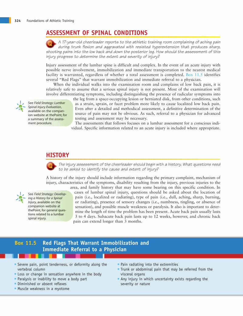
Box 11.5 Red Flags That Warrant Immobilization and Immediate Referral to a Physician
� Severe pain, point tenderness, or deformity along the � Pain radiating into the extremitiesvertebral column � Trunk or abdominal pain that may be referred from the
� Loss or change in sensation anywhere in the body visceral organs� Paralysis or inability to move a body part � Any injury in which uncertainty exists regarding the � Diminished or absent reflexes severity or nature� Muscle weakness in a myotome
See Field Strategy: LumbarSpinal Injury Evaluation,available on the compan-ion website at thePoint, fora summary of the assess-ment procedure.
See Field Strategy: Develop-ing a History for a Spinal Injury, available on thecompanion website at thePoint, for general ques-tions related to a lumbarspinal injury.
ANDERc11.qxd 11/16/07 3:53 PM Page 324
The cheerleader should be asked questions that would assist in determining the cause andextent of injury. Questions should address the primary complaint (i.e., what, when, and how
questions), mechanism of injury, location of pain (i.e., localized or radiating), type of pain (i.e., dull,aching, sharp, burning, or radiating), presence of sensory changes (i.e., numbness, tingling, orabsence of sensation) and possible muscle weakness, unusual sensations (i.e., sound or feelings),onset of symptoms, related medical history, and past injuries/treatment.
OBSERVATION AND INSPECTIONThe 17-year-old cheerleader has been participating in cheerleading for 5 years. The primarycomplaint is an aching pain during trunk flexion and aggravated with resisted hyperex-
tension that produces sharp shooting pains into the low back and down the posterior aspect ofthe right leg. The cheerleader reports the condition has been present for 2 weeks, and shecannot recall a traumatic episode that may have caused the condition. Would it be appropriateto do a scan examination to rule out other painful areas, and what specific factors should beobserved to identify the injury?
The observation component of an assessment should be initiated as soon as the examiner sees thepatient. Body language can signal pain, disability, and muscle weakness. It is important to note theindividual’s willingness or ability to move, general posture, ease in motion, and general attitude.Clothing and protective equipment may prevent visual observation of abnormalities in the spinalalignment. As such, the individual should be suitably dressed so that the back is as exposed as pos-sible; for girls and women, a bra, halter top, or swimsuit can be worn. Observation should beginwith a postural assessment, progress through a scan examination and gait analysis, and end withan inspection of the injury site.
PosturePosture assessment can detect congenital or functional problems that may contribute to the injury.A patient with acute back pain usually exhibits some degree of antalgic (painful) posturing. Thismay present as a decrease in lumbar lordosis with a concomitant lateral shift or scoliosis becauseof muscle spasm. The individual should be observed from an anterior, lateral, and posterior view,paying particular attention to pelvic tilt. It is important to determine if the pelvis is maintained ina neutral position (i.e., normal lordotic curve with ASIS slightly lower than the PSIS).
The head and neck posture should be observed as well. In particular, it is important to determine:
� If the nose is in alignment with the manubrium, sternum, and umbilicus
� If any abnormal spinal curvatures are present
� If the shoulders and clavicles are level (although the dominant side may be slightly lower)
� If the individual leans to one side and, if so, if it appears to be caused by muscle spasm, scolio-sis, or leg length discrepancy
� If the height and position of the ASIS, iliac crests, patella, and malleoli are the same bilaterally
The individual should be instructed to lean forward and touch the toes while keeping the kneesstraight (Adam’s position). The vertebrae and contour of the back should be observed (see Fig.10.18). The presence of a hump or raised scapula on one side (convex side of curve) and of a hollow(concave side of curve) on the other indicates scoliosis. From the side, the presence offlexed posturing at the hips from tight hip flexors should be noted; this results in acompensatory increase in the lumbar and cervical lordoses. It also is appropriateto determine if the trunk is rotated so that one shoulder is forward. Finally, thepresence of prominent ribs on one side should be noted.
From a posterior view, the level of the shoulders, inferior angles of the scapula,and waist angles should be assessed. The PSIS should be level. The gluteal foldsand knee joints should be at the same height. The presence of any dark areas of skin
CHAPTER 11 Lumbar Spinal Conditions 325
See Field Strategy: PosturalAssessment of the Low BackRegion, available on thecompanion website at thePoint, for specific pos-tural factors to observe inthe head and spinal region.
ANDERc11.qxd 11/16/07 3:53 PM Page 325
pigmentation, such as café-au-lait spots, should be noted,because this could indicate a possible collagen disease orabnormal growth of neural tissues (neurofibromatosis).The lower lumbar spine and sacrum should be observedfor tufts of hair (Faun’s beard), indicating possible spinabifida occulta.
Gait AssessmentThe individual should be asked to walk several yardswhile the examiner observes normal body movement.The examiner should stand behind, in front of, and tothe side of the individual to observe from all angles.Subtle posture abnormalities, such as kyphosis, scolio-sis, lordosis, or pelvic tilt, should be noted. A low backinjury may produce a forward lean, a lean to one side,or a noticeable limp.
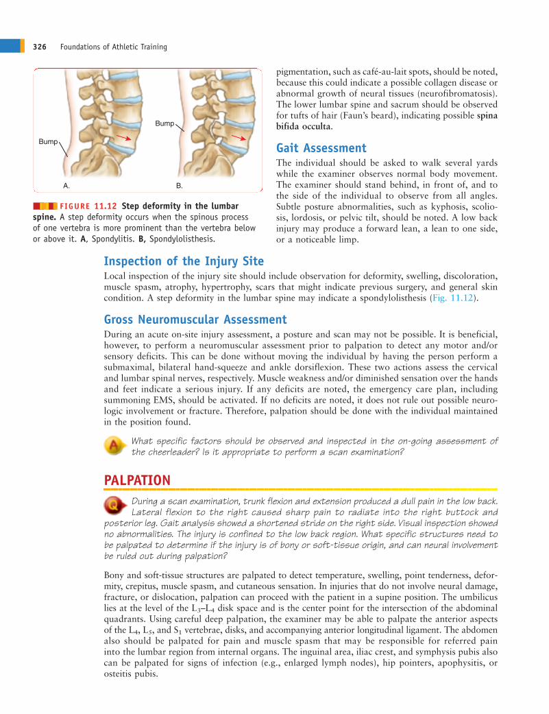
Inspection of the Injury SiteLocal inspection of the injury site should include observation for deformity, swelling, discoloration,muscle spasm, atrophy, hypertrophy, scars that might indicate previous surgery, and general skincondition. A step deformity in the lumbar spine may indicate a spondylolisthesis (Fig. 11.12).
Gross Neuromuscular AssessmentDuring an acute on-site injury assessment, a posture and scan may not be possible. It is beneficial,however, to perform a neuromuscular assessment prior to palpation to detect any motor and/orsensory deficits. This can be done without moving the individual by having the person perform asubmaximal, bilateral hand-squeeze and ankle dorsiflexion. These two actions assess the cervicaland lumbar spinal nerves, respectively. Muscle weakness and/or diminished sensation over the handsand feet indicate a serious injury. If any deficits are noted, the emergency care plan, includingsummoning EMS, should be activated. If no deficits are noted, it does not rule out possible neuro-logic involvement or fracture. Therefore, palpation should be done with the individual maintainedin the position found.
What specific factors should be observed and inspected in the on-going assessment ofthe cheerleader? Is it appropriate to perform a scan examination?
PALPATIONDuring a scan examination, trunk flexion and extension produced a dull pain in the low back.Lateral flexion to the right caused sharp pain to radiate into the right buttock and
posterior leg. Gait analysis showed a shortened stride on the right side. Visual inspection showedno abnormalities. The injury is confined to the low back region. What specific structures need tobe palpated to determine if the injury is of bony or soft-tissue origin, and can neural involvementbe ruled out during palpation?
Bony and soft-tissue structures are palpated to detect temperature, swelling, point tenderness, defor-mity, crepitus, muscle spasm, and cutaneous sensation. In injuries that do not involve neural damage,fracture, or dislocation, palpation can proceed with the patient in a supine position. The umbilicuslies at the level of the L3–L4 disk space and is the center point for the intersection of the abdominalquadrants. Using careful deep palpation, the examiner may be able to palpate the anterior aspectsof the L4, L5, and S1 vertebrae, disks, and accompanying anterior longitudinal ligament. The abdomenalso should be palpated for pain and muscle spasm that may be responsible for referred paininto the lumbar region from internal organs. The inguinal area, iliac crest, and symphysis pubis alsocan be palpated for signs of infection (e.g., enlarged lymph nodes), hip pointers, apophysitis, orosteitis pubis.
326 Foundations of Athletic Training
Bump
Bump
A. B.
������ FIGURE 11.12 Step deformity in the lumbarspine. A step deformity occurs when the spinous process of one vertebra is more prominent than the vertebra belowor above it. A, Spondylitis. B, Spondylolisthesis.
ANDERc11.qxd 11/16/07 3:53 PM Page 326
When moving into the prone position, a pillow or blanket should be placed under the hip regionto tilt the pelvis back and relax the lumbar curvature. Muscle spasm in the lower erector spinae,lower trapezius, serratus posterior, quadratus lumborum, latissimus dorsi, or gluteus maximus canindicate dysfunction of the thoracic or lumbar spine. The following surface landmarks in the lumbarregion can facilitate palpation:
� L4—top of iliac crest� L5—demarcated by bilateral dimples� S2—level of PSIS
In palpating the spinous processes of the lumbar spine, particular attention should be noted at theL4, L5, and S1 level. A visible or palpable dip or protrusion can indicate spondylolisthesis. If thefingers are moved laterally 2 to 3 cm (0.8–1.2 inches), the facet joints can be palpated for signs ofpathology. Because of their depth, it may be difficult to palpate the joints; however, spasm in the over-lying paraspinal muscles can be palpated. The spinous processes of the sacrum also can be palpated.Because no interposing soft-tissue spaces are between them, they may be harder to distinguish. TheS2 spinous process is at the level of a line drawn between the two PSIS (“posterior dimples”). Inmoving to the PSIS, the examiner can palpate the iliac crest for signs of injury and then palpate thegluteal muscles for pain, spasm, or possible nodules. Having the patient flex the hip at 90° allowseasier palpation of the ischial tuberosity, greater trochanter, and sciatic nerve, which is located mid-way between the ischial tuberosity and greater trochanter. Finally, the piriformis muscle should bepalpated deep to the gluteal muscles for pathology. The following structures should be palpated:
Anterior Aspect
1. Umbilicus and abdominal area. Note any abnormal tenderness or masses indicating internalpathology that is referring pain to the spinal region.
2. Inguinal area. Palpate for possible hernia, infection (enlarged lymph nodes), or other pathology.
3. Iliac crest, ASIS, and symphysis pubis. Palpate for pain, tenderness, or defect indicating pathol-ogy (e.g., avulsion fracture, hip pointer, apophysitis, or osteitis pubis).
Posterior Aspect
1. Spinous processes of the lumbar vertebrae. Note any tenderness,crepitus, or presence of a step-off deformity (i.e., one vertebra is moreanterior than the one below it). This indicates spondylolisthesis, whichmost commonly is seen between the L4 and L5 or the L5 and S1 verte-brae. Pain and tenderness without positive findings on muscle move-ment may indicate that the problem is not musculoskeletal in origin.
2. Facet joints. The facet articulations are approximately a thumb’sbreadth to either side of the spinous process. Point tenderness at thesesites, especially with extension and rotation to the same side, suggestsfacet joint pain.
3. Interspinous and supraspinous ligaments, paraspinal muscles, andquadratus lumborum. Trigger points within specific muscles may referpain to a more distal area. Tender points that increase with muscularcontraction indicate a localized muscle strain. An area that is tenderto palpation but is not painful during muscle contraction may indi-cate referred pain from another area.
4. Iliac crest, PSIS, and sacrum. The interspace between L4 and L5 lies atthe same level as the top of the iliac crest. The S2 spinous process liesin the middle of a line drawn between the PSIS. Palpate for pain, ten-derness, and other pathology (e.g., hip pointer or apophysitis).
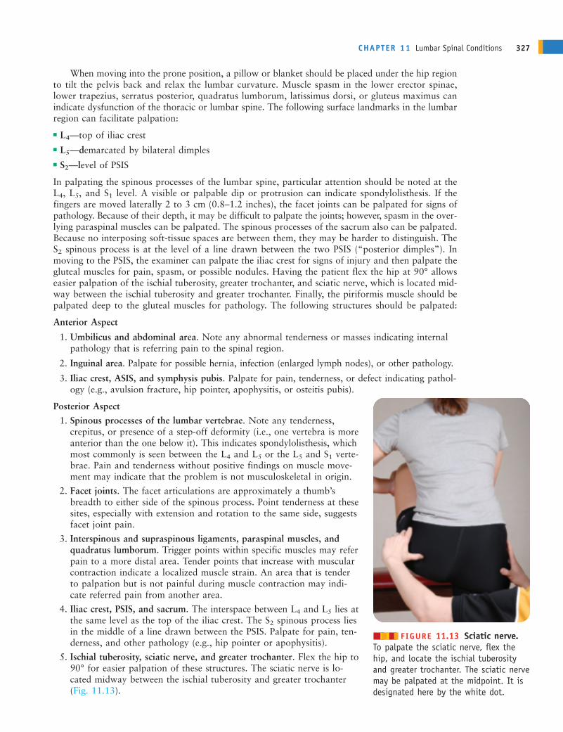
5. Ischial tuberosity, sciatic nerve, and greater trochanter. Flex the hip to90° for easier palpation of these structures. The sciatic nerve is lo-cated midway between the ischial tuberosity and greater trochanter(Fig. 11.13).
CHAPTER 11 Lumbar Spinal Conditions 327
������ FIGURE 11.13 Sciatic nerve.To palpate the sciatic nerve, flex thehip, and locate the ischial tuberosityand greater trochanter. The sciatic nervemay be palpated at the midpoint. It isdesignated here by the white dot.
ANDERc11.qxd 11/16/07 3:53 PM Page 327
The palpation component of the assessment of the cheerleader should include bony andsoft tissues of the lumbar region (i.e., spinous processes of the lumbar vertebrae; facet
joints; interspinous and supraspinous ligaments, paraspinal muscles, and quadratus lumborum;iliac crest, PSIS, and sacrum; and ischial tuberosity, sciatic nerve, and greater trochanter).Palpation also should include anterior structures (i.e., umbilicus and abdominal area, iliac crest,and ASIS).
Because the cheerleader is younger than 18 years, palpation can only be performed withpermission from the parent or guardian. It also is important to recognize that the cheerleadermay feel uncomfortable being touched by a health care provider of the opposite gender. If a same-gender clinician is not available, the evaluation should be observed by a third party (e.g., anotherclinician, parent, or guardian).
PHYSICAL EXAMINATION TESTSIn the palpation component of the assessment of the cheerleader, point tenderness waselicited in the low back region between the L3 and S1 vertebrae, with increased pain in the
L4–L5 region. Muscle spasm was present on either side of the lumbar region. Pain also was elicitedwith palpation midway between the ischial tuberosity and greater trochanter. Based on theinformation obtained through the history, observation, and palpation, what tests should beperformed to determine nerve root impingement, and what tests should be conducted as partof a neurologic assessment of the cheerleader’s condition.
It is imperative to work slowly through the tests that are used to assess low back conditions, becauseinjuries to the lumbar region can be very complex. If, at any time, movement leads to increasedacute pain or change in sensation, or if the individual resists moving the spine, a significant injuryshould be assumed and the emergency plan activated.
Functional TestsGoniometry measurements of the spine are not typically taken because of the difficulty of measuringindividual regional motions. Completion of gross movement patterns in a standing position isadequate to determine normal ROM. Further assessment can be conducted if motion is limited orthe patient is unwilling to do the movements.
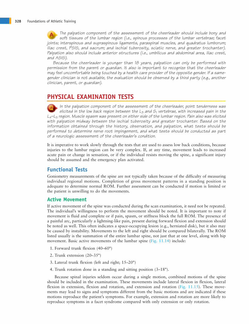
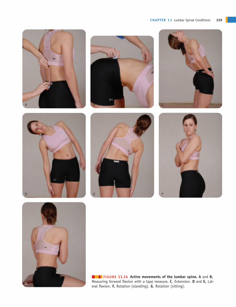
Active MovementIf active movement of the spine was conducted during the scan examination, it need not be repeated.The individual’s willingness to perform the movement should be noted. It is important to note ifmovement is fluid and complete or if pain, spasm, or stiffness block the full ROM. The presence ofa painful arc, particularly a lightning-like pain, present during forward flexion and extension shouldbe noted as well. This often indicates a space-occupying lesion (e.g., herniated disk), but it also maybe caused by instability. Movements to the left and right should be compared bilaterally. The ROMlisted usually is the summation of the entire lumbar spine, not just that at one level, along with hipmovement. Basic active movements of the lumbar spine (Fig. 11.14) include:
1. Forward trunk flexion (40–60°)
2. Trunk extension (20–35°)
3. Lateral trunk flexion (left and right; 15–20°)
4. Trunk rotation done in a standing and sitting position (3–18°).
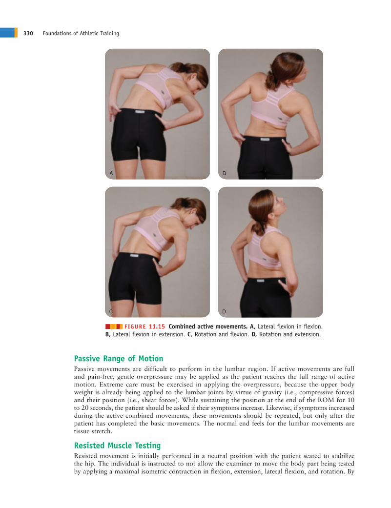
Because spinal injuries seldom occur during a single motion, combined motions of the spineshould be included in the examination. These movements include lateral flexion in flexion, lateralflexion in extension, flexion and rotation, and extension and rotation (Fig. 11.15). These move-ments may lead to signs and symptoms different from the basic motions and are indicated if thesemotions reproduce the patient’s symptoms. For example, extension and rotation are more likely toreproduce symptoms in a facet syndrome compared with only extension or only rotation.
328 Foundations of Athletic Training
ANDERc11.qxd 11/16/07 3:53 PM Page 328
CHAPTER 11 Lumbar Spinal Conditions 329
A B C
D E F
G
������ FIGURE 11.14 Active movements of the lumbar spine. A and B,Measuring forward flexion with a tape measure. C, Extension. D and E, Lat-eral flexion. F, Rotation (standing). G, Rotation (sitting).
ANDERc11.qxd 11/16/07 3:53 PM Page 329
Passive Range of MotionPassive movements are difficult to perform in the lumbar region. If active movements are fulland pain-free, gentle overpressure may be applied as the patient reaches the full range of activemotion. Extreme care must be exercised in applying the overpressure, because the upper bodyweight is already being applied to the lumbar joints by virtue of gravity (i.e., compressive forces)and their position (i.e., shear forces). While sustaining the position at the end of the ROM for 10to 20 seconds, the patient should be asked if their symptoms increase. Likewise, if symptoms increasedduring the active combined movements, these movements should be repeated, but only after thepatient has completed the basic movements. The normal end feels for the lumbar movements aretissue stretch.
Resisted Muscle TestingResisted movement is initially performed in a neutral position with the patient seated to stabilizethe hip. The individual is instructed to not allow the examiner to move the body part being testedby applying a maximal isometric contraction in flexion, extension, lateral flexion, and rotation. By
330 Foundations of Athletic Training
A B
C D
������ FIGURE 11.15 Combined active movements. A, Lateral flexion in flexion.B, Lateral flexion in extension. C, Rotation and flexion. D, Rotation and extension.
ANDERc11.qxd 11/16/07 3:54 PM Page 330
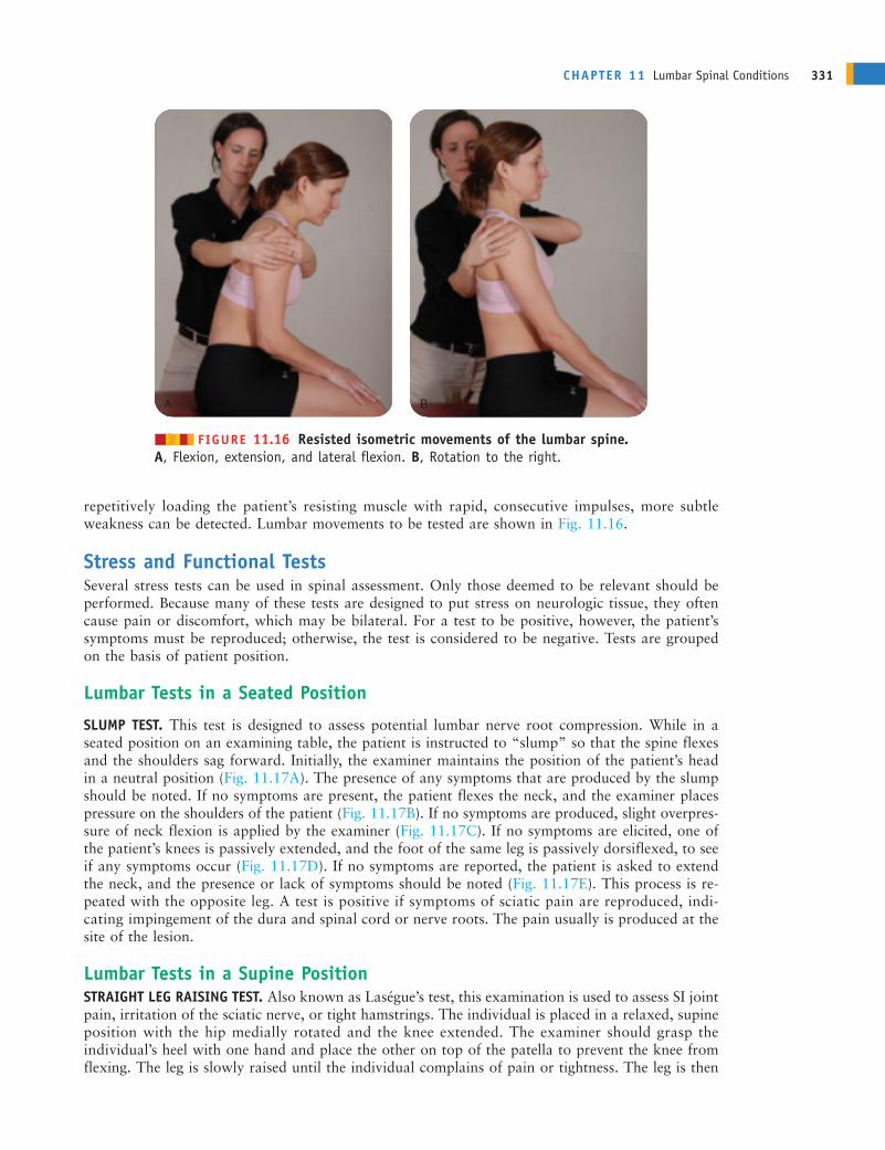
repetitively loading the patient’s resisting muscle with rapid, consecutive impulses, more subtleweakness can be detected. Lumbar movements to be tested are shown in Fig. 11.16.
Stress and Functional TestsSeveral stress tests can be used in spinal assessment. Only those deemed to be relevant should beperformed. Because many of these tests are designed to put stress on neurologic tissue, they oftencause pain or discomfort, which may be bilateral. For a test to be positive, however, the patient’ssymptoms must be reproduced; otherwise, the test is considered to be negative. Tests are groupedon the basis of patient position.
Lumbar Tests in a Seated Position
SLUMP TEST. This test is designed to assess potential lumbar nerve root compression. While in aseated position on an examining table, the patient is instructed to “slump” so that the spine flexesand the shoulders sag forward. Initially, the examiner maintains the position of the patient’s headin a neutral position (Fig. 11.17A). The presence of any symptoms that are produced by the slumpshould be noted. If no symptoms are present, the patient flexes the neck, and the examiner placespressure on the shoulders of the patient (Fig. 11.17B). If no symptoms are produced, slight overpres-sure of neck flexion is applied by the examiner (Fig. 11.17C). If no symptoms are elicited, one ofthe patient’s knees is passively extended, and the foot of the same leg is passively dorsiflexed, to seeif any symptoms occur (Fig. 11.17D). If no symptoms are reported, the patient is asked to extendthe neck, and the presence or lack of symptoms should be noted (Fig. 11.17E). This process is re-peated with the opposite leg. A test is positive if symptoms of sciatic pain are reproduced, indi-cating impingement of the dura and spinal cord or nerve roots. The pain usually is produced at thesite of the lesion.
Lumbar Tests in a Supine PositionSTRAIGHT LEG RAISING TEST. Also known as Laségue’s test, this examination is used to assess SI jointpain, irritation of the sciatic nerve, or tight hamstrings. The individual is placed in a relaxed, supineposition with the hip medially rotated and the knee extended. The examiner should grasp theindividual’s heel with one hand and place the other on top of the patella to prevent the knee fromflexing. The leg is slowly raised until the individual complains of pain or tightness. The leg is then
CHAPTER 11 Lumbar Spinal Conditions 331
A B
������ FIGURE 11.16 Resisted isometric movements of the lumbar spine. A, Flexion, extension, and lateral flexion. B, Rotation to the right.
ANDERc11.qxd 11/16/07 3:54 PM Page 331

lowered until the pain is relieved. Next, the individual is asked to flex the neck onto the chest, todorsiflex the foot, or to do both actions simultaneously (Fig. 11.18).
The neck flexion movement is called Hyndman’s sign or Brudzinski’s sign. Pain that increaseswith neck flexion or dorsiflexion indicates stretching of the dura mater of the spinal cord. Pain thatdoes not increase with neck flexion or dorsiflexion indicates tight hamstrings. The sciatic nerve isfully stretched at approximately 70° of flexion. As such, pain after 70° usually indicates pain fromthe lumbar area (facet joints) or SI joints. Pain that occurs opposite the leg that is lifted indicates aspace-occupying lesion (e.g., herniated disk).
332 Foundations of Athletic Training
A B C
D E
������ FIGURE 11.17 Slump test. The test is performed in several stages. A, While sitting on a table, the examiner asks the person to “slump” so that the spine flexes and the shoulders sag forward while the examiner holds the head in aneutral position. B, If no symptoms are present, the examiner applies light pressure, pushing down on the shoulders whilethe patient holds the head in a neutral position. C, If no symptoms are produced, overpressure is applied to the cervicalspine. D, If no symptoms occur, the examiner passively extends one of the patient’s knees and dorsiflexes the ankle ofthe same leg. E, The patient is instructed to extend the head. The test is considered to be positive if symptoms of sciaticpain are reproduced, indicating impingement of the dura and spinal cord or nerve roots.
ANDERc11.qxd 11/16/07 3:54 PM Page 332

WELL STRAIGHT LEG RAISING TEST. The well straight leg raising test differs from the previous test inthat the unaffected leg is raised. A positive sign is pain on the side opposite the leg being raised, indi-cating a space-occupying lesion (e.g., a herniated intervertebral disk.)
BOWSTRING TEST (TENSION OR POPLITEAL PRESSURE SIGN). A positive straight leg raising test canthen be followed by the tension, or bowstring, test. This test is designed to assess tension or pressureon the sciatic nerve and is a modification of the straight leg raising test. The examiner maintainsthe leg in the same position, then flexes the knee slightly (20°), reducing the symptoms. Thumb orfinger pressure is then exerted over the tibial portion of the sciatic nerve as it passes through thepopliteal space to re-establish the painful radiating symptoms (Fig. 11.19). An alternative methodis to flex both the hip and the knee at 90°. The examiner then slowly extends the knee as far aspossible while applying pressure in the popliteal space. Replication of tenderness or radiating painis a positive sign for sciatic nerve irritation.
BRUDZINSKI’S TEST. This test is similar to the straight leg raising test, but themovements are actively performed by the patient. The individual is supine,with the hands cupped behind the head (Fig. 11.20A). The test is positive ifthe individual complains of neck and low back discomfort and attempts torelieve the meningeal irritation by involuntarily flexing the knees and hips.
KERNIG’S TEST. This test can aid in identifying the presence of a bulgingdisk, nerve root impingement, inflammation of the dural sheath, or irrita-tion of the meninges. In Kernig’s position, the individual lies supine, withthe hip flexed and the knee extended (Fig. 11.20B). Pain in the head, neck,or lower back suggests meningeal irritation. If the pain is relieved when theindividual flexes the knee, it is considered to be a positive test, indicatingmeningeal irritation, nerve root involvement, or dural irritation. TheBrudzinski’s and Kernig’s tests may be done either separately or together.
BILATERAL STRAIGHT LEG RAISING TEST. This test is used to assess the pres-ence of lesions at the SI joint and in the lumbar spine. The examiner cantest both legs simultaneously (bilateral straight leg raising). This must beperformed carefully, because the examiner is lifting the weight of bothlower limbs, thereby placing a large stress on their own lumbar spine. If thetest causes pain before 70° of hip flexion, the lesion is probably in the SIjoints; if it causes pain after 70° of hip flexion, the lesion is probably in thelumbar spine.
VALSALVA’S TEST. Valsalva’s maneuver is used to determine the presence ofspace-occupying lesions (e.g., herniated disk, tumor, or osteophytes). While
CHAPTER 11 Lumbar Spinal Conditions 333
������ FIGURE 11.18 Straight leg raisingtest. The examiner passively flexes the individual’s hip while keeping the knee extended until pain or tension is felt in thehamstrings. Next, the leg is lowered slowlyuntil the pain or tension disappears. Next,the examiner dorsiflexes the ankle, has theindividual flex the neck, or perform both simultaneously. If pain does not increasewith dorsiflexion of the ankle or flexion ofthe neck, it indicates tight hamstrings.
������ FIGURE 11.19 Bowstring test.After pain results in a straight leg rais-ing test, the clinician flexes the kneeslightly (20°) to reduce the symptoms.Thumb pressure is then exerted in thepopliteal area to reproduce the painfulradicular symptoms.
ANDERc11.qxd 11/16/07 3:54 PM Page 333
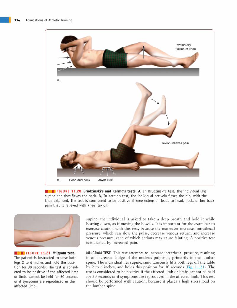
supine, the individual is asked to take a deep breath and hold it whilebearing down, as if moving the bowels. It is important for the examiner toexercise caution with this test, because the maneuver increases intrathecalpressure, which can slow the pulse, decrease venous return, and increasevenous pressure, each of which actions may cause fainting. A positive testis indicated by increased pain.
MILGRAM TEST. This test attempts to increase intrathecal pressure, resultingin an increased bulge of the nucleus pulposus, primarily in the lumbarspine. The individual lies supine, simultaneously lifts both legs off the tableby 2 to 6 inches, and holds this position for 30 seconds (Fig. 11.21). Thetest is considered to be positive if the affected limb or limbs cannot be heldfor 30 seconds or if symptoms are reproduced in the affected limb. This testshould be performed with caution, because it places a high stress load onthe lumbar spine.
334 Foundations of Athletic Training
������ FIGURE 11.20 Brudzinski’s and Kernig’s tests. A, In Brudzinski’s test, the individual layssupine and dorsiflexes the neck. B, In Kernig’s test, the individual actively flexes the hip, with theknee extended. The test is considered to be positive if knee extension leads to head, neck, or low backpain that is relieved with knee flexion.
������ FIGURE 11.21 Milgram test.The patient is instructed to raise bothlegs 2 to 6 inches and hold the posi-tion for 30 seconds. The test is consid-ered to be positive if the affected limbor limbs cannot be held for 30 secondsor if symptoms are reproduced in theaffected limb.
Involuntary flexion of knee
A.
Flexion relieves pain
Lower backHead and neckB.
ANDERc11.qxd 11/26/07 10:10 AM Page 334
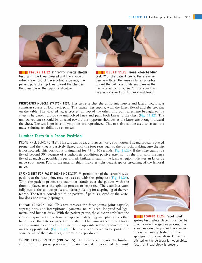
PIRIFORMIS MUSCLE STRETCH TEST. This test stretches the piriformis muscle and lateral rotators, acommon source of low back pain. The patient lies supine, with the knees flexed and the feet flaton the table. The affected leg is crossed on top of the other, and both knees are brought to thechest. The patient grasps the uninvolved knee and pulls both knees to the chest (Fig. 11.22). Theuninvolved knee should be directed toward the opposite shoulder as the knees are brought towardthe chest. The test is positive if symptoms are reproduced. This test also can be used to stretch themuscle during rehabilitative exercises.
Lumbar Tests in a Prone PositionPRONE KNEE BENDING TEST. This test can be used to assess nerve root lesion. The individual is placedprone, and the knee is passively flexed until the foot rests against the buttock, making sure the hipis not rotated. This position is maintained for 45 to 60 seconds (Fig. 11.23). If the knee cannot beflexed beyond 90° because of a pathologic condition, passive extension of the hip, with the kneeflexed as much as possible, is performed. Unilateral pain in the lumbar region indicates an L2 or L3
nerve root lesion. Pain in the anterior thigh indicates tight quadriceps or stretching of the femoralnerve.
SPRING TEST FOR FACET JOINT MOBILITY. Hypomobility of the vertebrae, es-pecially at the facet joint, may be assessed with the spring test (Fig. 11.24).With the patient prone, the examiner stands over the patient with thethumbs placed over the spinous process to be tested. The examiner care-fully pushes the spinous process anteriorly, feeling for a springing of the ver-tebrae. The test is considered to be positive if pain is elicited or the verte-bra does not move (“spring”).
FARFAN TORSION TEST. This test stresses the facet joints, joint capsule,supraspinous and interspinous ligaments, neural arch, longitudinal liga-ments, and lumbar disks. With the patient prone, the clinician stabilizes theribs and spine with one hand at approximately T12 and places the otherhand under the anterior aspect of the ilium. The ilium is then pulled back-ward, causing rotation of the spine on the opposite side to produce torqueon the opposite side (Fig. 11.25). The test is considered to be positive ifsome or all of the patient’s symptoms are reproduced.
TRUNK EXTENSION TEST (PRESS-UPS). This test compresses the lumbarvertebrae. In a prone position, the patient is asked to extend the trunk
CHAPTER 11 Lumbar Spinal Conditions 335
������ FIGURE 11.22 Piriformis muscle stretchtest. With the knees crossed and the involvedextremity on top of the involved extremity, thepatient pulls the top knee toward the chest inthe direction of the opposite shoulder.
������ FIGURE 11.23 Prone knee bendingtest. With the patient prone, the examiner passively flexes the knee as far as possible toward the buttocks. Unilateral pain in the lumbar area, buttock, and/or posterior thighmay indicate an L2 or L3 nerve root lesion.
������ FIGURE 11.24 Facet jointspring test. While placing the thumbsdirectly over the spinous process, theexaminer carefully pushes the spinousprocess anteriorly, feeling for thespringing of the vertebrae. If pain iselicited or the vertebra is hypomobile,facet joint pathology is present.
ANDERc11.qxd 11/16/07 3:54 PM Page 335
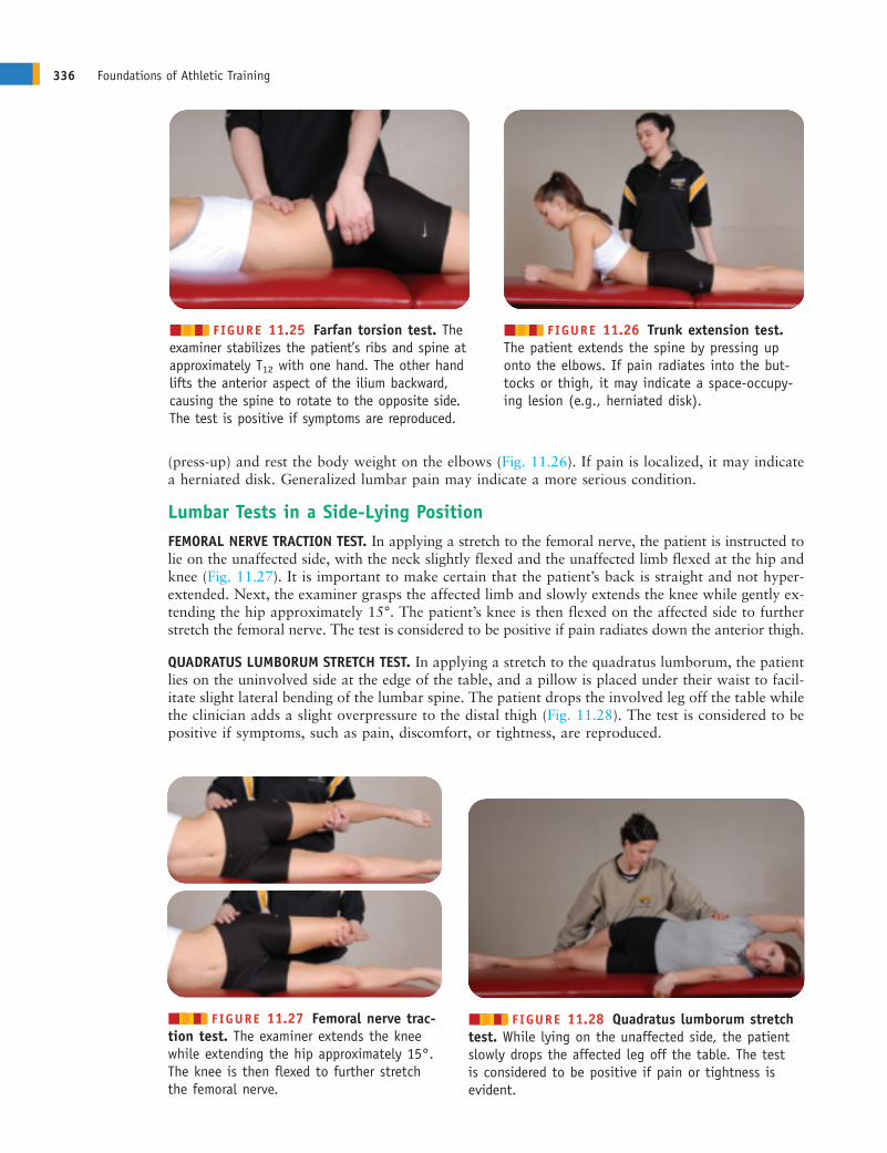
(press-up) and rest the body weight on the elbows (Fig. 11.26). If pain is localized, it may indicatea herniated disk. Generalized lumbar pain may indicate a more serious condition.
Lumbar Tests in a Side-Lying PositionFEMORAL NERVE TRACTION TEST. In applying a stretch to the femoral nerve, the patient is instructed tolie on the unaffected side, with the neck slightly flexed and the unaffected limb flexed at the hip andknee (Fig. 11.27). It is important to make certain that the patient’s back is straight and not hyper-extended. Next, the examiner grasps the affected limb and slowly extends the knee while gently ex-tending the hip approximately 15°. The patient’s knee is then flexed on the affected side to furtherstretch the femoral nerve. The test is considered to be positive if pain radiates down the anterior thigh.
QUADRATUS LUMBORUM STRETCH TEST. In applying a stretch to the quadratus lumborum, the patientlies on the uninvolved side at the edge of the table, and a pillow is placed under their waist to facil-itate slight lateral bending of the lumbar spine. The patient drops the involved leg off the table whilethe clinician adds a slight overpressure to the distal thigh (Fig. 11.28). The test is considered to bepositive if symptoms, such as pain, discomfort, or tightness, are reproduced.
336 Foundations of Athletic Training
������ FIGURE 11.25 Farfan torsion test. Theexaminer stabilizes the patient’s ribs and spine atapproximately T12 with one hand. The other handlifts the anterior aspect of the ilium backward,causing the spine to rotate to the opposite side.The test is positive if symptoms are reproduced.
������ FIGURE 11.26 Trunk extension test.The patient extends the spine by pressing uponto the elbows. If pain radiates into the but-tocks or thigh, it may indicate a space-occupy-ing lesion (e.g., herniated disk).
������ FIGURE 11.27 Femoral nerve trac-tion test. The examiner extends the kneewhile extending the hip approximately 15°.The knee is then flexed to further stretchthe femoral nerve.
������ FIGURE 11.28 Quadratus lumborum stretchtest. While lying on the unaffected side, the patientslowly drops the affected leg off the table. The test is considered to be positive if pain or tightness is evident.
ANDERc11.qxd 11/16/07 3:54 PM Page 336
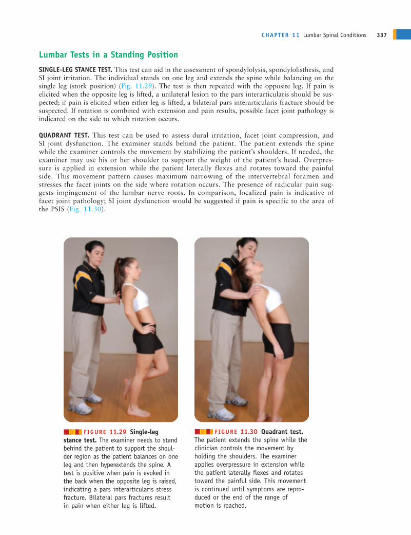
Lumbar Tests in a Standing Position
SINGLE-LEG STANCE TEST. This test can aid in the assessment of spondylolysis, spondylolisthesis, andSI joint irritation. The individual stands on one leg and extends the spine while balancing on thesingle leg (stork position) (Fig. 11.29). The test is then repeated with the opposite leg. If pain iselicited when the opposite leg is lifted, a unilateral lesion to the pars interarticularis should be sus-pected; if pain is elicited when either leg is lifted, a bilateral pars interarticularis fracture should besuspected. If rotation is combined with extension and pain results, possible facet joint pathology isindicated on the side to which rotation occurs.
QUADRANT TEST. This test can be used to assess dural irritation, facet joint compression, andSI joint dysfunction. The examiner stands behind the patient. The patient extends the spinewhile the examiner controls the movement by stabilizing the patient’s shoulders. If needed, theexaminer may use his or her shoulder to support the weight of the patient’s head. Overpres-sure is applied in extension while the patient laterally flexes and rotates toward the painfulside. This movement pattern causes maximum narrowing of the intervertebral foramen andstresses the facet joints on the side where rotation occurs. The presence of radicular pain sug-gests impingement of the lumbar nerve roots. In comparison, localized pain is indicative offacet joint pathology; SI joint dysfunction would be suggested if pain is specific to the area ofthe PSIS (Fig. 11.30).
CHAPTER 11 Lumbar Spinal Conditions 337
������ FIGURE 11.29 Single-legstance test. The examiner needs to standbehind the patient to support the shoul-der region as the patient balances on oneleg and then hyperextends the spine. Atest is positive when pain is evoked inthe back when the opposite leg is raised,indicating a pars interarticularis stressfracture. Bilateral pars fractures resultin pain when either leg is lifted.
������ FIGURE 11.30 Quadrant test.The patient extends the spine while theclinician controls the movement byholding the shoulders. The examiner applies overpressure in extension whilethe patient laterally flexes and rotatestoward the painful side. This movementis continued until symptoms are repro-duced or the end of the range of motion is reached.
ANDERc11.qxd 11/16/07 3:54 PM Page 337

Lumbar Tests for Malingering
HOOVER TEST. Because the assessment of lumbopelvic disorders is difficult to perform objectively,the Hoover test is used to determine if the patient is a malingerer. The examiner’s hands cup eachheel of the supine patient while the legs remain relaxed on the examining table (Fig. 11.31). Theindividual is then asked to lift one leg off the table while keeping the knees straight, as in an activestraight leg raising test. If the individual does not lift the leg or the examiner does not feel pressureunder the opposite heel, the patient may not be trying to lift the leg or may be a malingerer. Pres-sure under the normal heel increases, however, if the lifted limb is weaker because of the increasedeffort to lift the weak leg. A bilateral comparison then is made to determine any differences.
BURNS TEST. This test also can be used to assess malingering. The patient is asked to kneel on achair and then bend forward, touching the floor with the fingers (Fig. 11.32). If the patient over-
balances or is unable to perform this test, the result is considered to bepositive for malingering.
Sacroiliac DysfunctionA variety of tests can be used to assess SI pathology. In an effort to improvereliability, it is essential that the examiner incorporate the results of severaltests in making decisions regarding to injury status.
SACROILIAC COMPRESSION (TRANSVERSE ANTERIOR STRESS) AND DISTRAC-TION TEST. The examiner applies a cross-arm pressure both downward andoutward to the ASIS of the patient who is lying supine (Fig. 11.33). Thisaction is repeated, with pressure applied downward through the anteriorportion of the ilium, spreading the SI joint. Unilateral gluteal or posteriorleg pain may indicate a sprain to the anterior SI ligaments. Sharp painelsewhere along the pelvic ring with outward pressure, or with bilateralcompression of the iliac crests, may indicate a pelvic fracture.
APPROXIMATION (TRANSVERSE POSTERIOR STRESS) TEST. With the patient ina side-lying position, a downward force is applied over the iliac crest (Fig.11.34). The movement causes forward pressure on the sacrum. A positivetest produces pain or a feeling of pressure on the SI joints, indicating asprain of the posterior SI ligaments, a SI lesion, or both. Sharp pain alongthe pelvic ring may indicate a fracture.
338 Foundations of Athletic Training
■■■■■■ FIGURE 11.32 Burns test.While kneeling on a chair, the patient is asked to bend forward and touch thefloor with his or her fingers. Inabilityto perform the action suggests malingering.
A B
■■■■■■ FIGURE 11.31 Hoover test. A, Under normal conditions, when an indi-vidual tries to elevate one leg, the action is accompanied by downward pressureon the opposite leg. B, When the individual attempts to elevate the “weak” legbut the opposite (asymptomatic) leg does not “help,” at least some of theweakness probably is feigned.
ANDERc11.qxd 11/27/07 2:08 PM Page 338
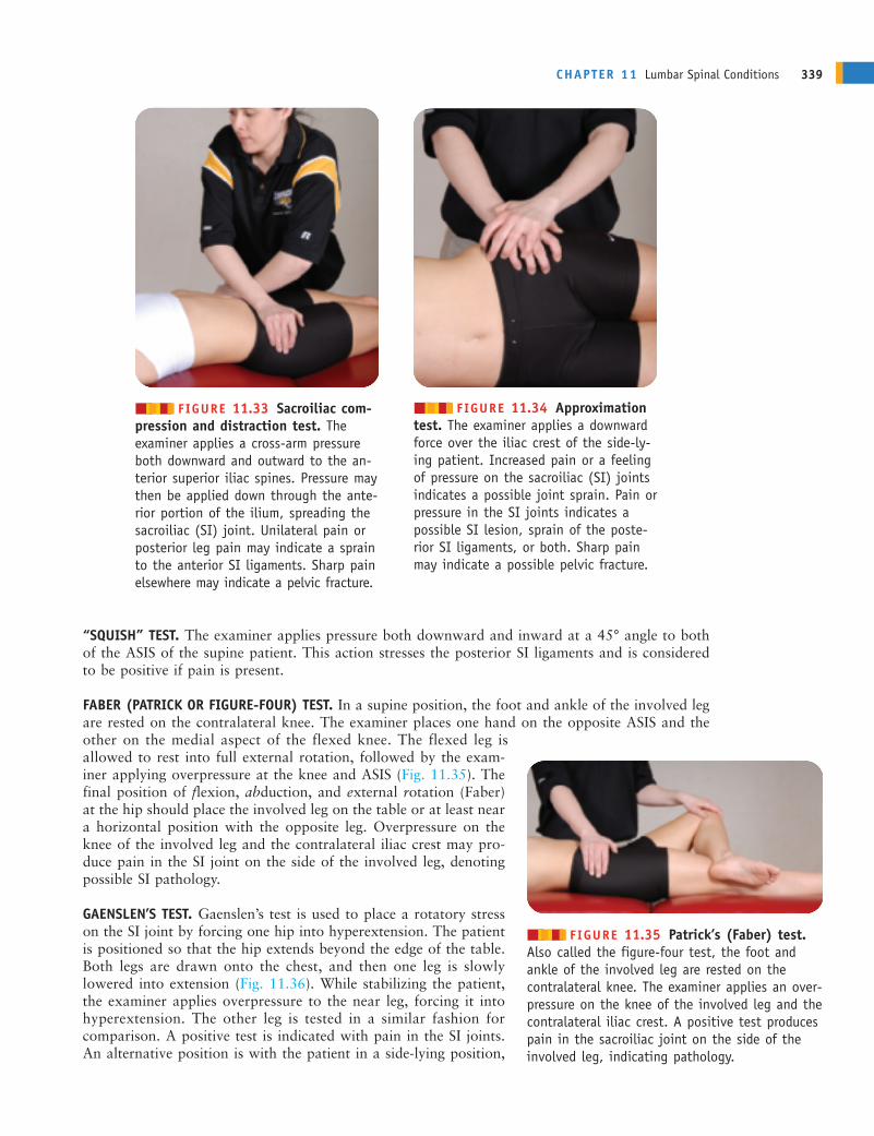
CHAPTER 11 Lumbar Spinal Conditions 339
������ FIGURE 11.33 Sacroiliac com-pression and distraction test. The examiner applies a cross-arm pressureboth downward and outward to the an-terior superior iliac spines. Pressure maythen be applied down through the ante-rior portion of the ilium, spreading thesacroiliac (SI) joint. Unilateral pain orposterior leg pain may indicate a sprainto the anterior SI ligaments. Sharp painelsewhere may indicate a pelvic fracture.
������ FIGURE 11.34 Approximationtest. The examiner applies a downwardforce over the iliac crest of the side-ly-ing patient. Increased pain or a feelingof pressure on the sacroiliac (SI) jointsindicates a possible joint sprain. Pain orpressure in the SI joints indicates apossible SI lesion, sprain of the poste-rior SI ligaments, or both. Sharp painmay indicate a possible pelvic fracture.
“SQUISH” TEST. The examiner applies pressure both downward and inward at a 45° angle to bothof the ASIS of the supine patient. This action stresses the posterior SI ligaments and is consideredto be positive if pain is present.
FABER (PATRICK OR FIGURE-FOUR) TEST. In a supine position, the foot and ankle of the involved legare rested on the contralateral knee. The examiner places one hand on the opposite ASIS and theother on the medial aspect of the flexed knee. The flexed leg isallowed to rest into full external rotation, followed by the exam-iner applying overpressure at the knee and ASIS (Fig. 11.35). Thefinal position of flexion, abduction, and external rotation (Faber)at the hip should place the involved leg on the table or at least neara horizontal position with the opposite leg. Overpressure on theknee of the involved leg and the contralateral iliac crest may pro-duce pain in the SI joint on the side of the involved leg, denotingpossible SI pathology.
GAENSLEN’S TEST. Gaenslen’s test is used to place a rotatory stresson the SI joint by forcing one hip into hyperextension. The patientis positioned so that the hip extends beyond the edge of the table.Both legs are drawn onto the chest, and then one leg is slowlylowered into extension (Fig. 11.36). While stabilizing the patient,the examiner applies overpressure to the near leg, forcing it intohyperextension. The other leg is tested in a similar fashion forcomparison. A positive test is indicated with pain in the SI joints.An alternative position is with the patient in a side-lying position,
������ FIGURE 11.35 Patrick’s (Faber) test.Also called the figure-four test, the foot and ankle of the involved leg are rested on the contralateral knee. The examiner applies an over-pressure on the knee of the involved leg and thecontralateral iliac crest. A positive test producespain in the sacroiliac joint on the side of theinvolved leg, indicating pathology.
ANDERc11.qxd 11/16/07 3:54 PM Page 339
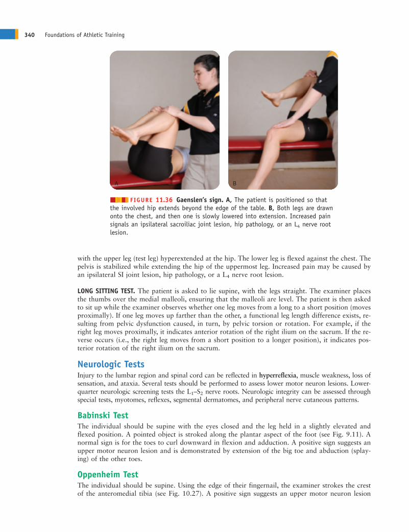
with the upper leg (test leg) hyperextended at the hip. The lower leg is flexed against the chest. Thepelvis is stabilized while extending the hip of the uppermost leg. Increased pain may be caused byan ipsilateral SI joint lesion, hip pathology, or a L4 nerve root lesion.
LONG SITTING TEST. The patient is asked to lie supine, with the legs straight. The examiner placesthe thumbs over the medial malleoli, ensuring that the malleoli are level. The patient is then askedto sit up while the examiner observes whether one leg moves from a long to a short position (movesproximally). If one leg moves up farther than the other, a functional leg length difference exists, re-sulting from pelvic dysfunction caused, in turn, by pelvic torsion or rotation. For example, if theright leg moves proximally, it indicates anterior rotation of the right ilium on the sacrum. If the re-verse occurs (i.e., the right leg moves from a short position to a longer position), it indicates pos-terior rotation of the right ilium on the sacrum.
Neurologic TestsInjury to the lumbar region and spinal cord can be reflected in hyperreflexia, muscle weakness, loss ofsensation, and ataxia. Several tests should be performed to assess lower motor neuron lesions. Lower-quarter neurologic screening tests the L1–S2 nerve roots. Neurologic integrity can be assessed throughspecial tests, myotomes, reflexes, segmental dermatomes, and peripheral nerve cutaneous patterns.
Babinski TestThe individual should be supine with the eyes closed and the leg held in a slightly elevated andflexed position. A pointed object is stroked along the plantar aspect of the foot (see Fig. 9.11). Anormal sign is for the toes to curl downward in flexion and adduction. A positive sign suggests anupper motor neuron lesion and is demonstrated by extension of the big toe and abduction (splay-ing) of the other toes.
Oppenheim TestThe individual should be supine. Using the edge of their fingernail, the examiner strokes the crestof the anteromedial tibia (see Fig. 10.27). A positive sign suggests an upper motor neuron lesion
340 Foundations of Athletic Training
A B
������ FIGURE 11.36 Gaenslen’s sign. A, The patient is positioned so thatthe involved hip extends beyond the edge of the table. B, Both legs are drawnonto the chest, and then one is slowly lowered into extension. Increased painsignals an ipsilateral sacroiliac joint lesion, hip pathology, or an L4 nerve rootlesion.
ANDERc11.qxd 11/16/07 3:54 PM Page 340
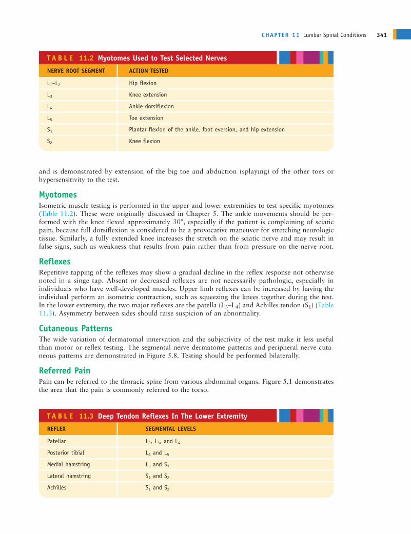
CHAPTER 11 Lumbar Spinal Conditions 341
and is demonstrated by extension of the big toe and abduction (splaying) of the other toes orhypersensitivity to the test.
MyotomesIsometric muscle testing is performed in the upper and lower extremities to test specific myotomes(Table 11.2). These were originally discussed in Chapter 5. The ankle movements should be per-formed with the knee flexed approximately 30°, especially if the patient is complaining of sciaticpain, because full dorsiflexion is considered to be a provocative maneuver for stretching neurologictissue. Similarly, a fully extended knee increases the stretch on the sciatic nerve and may result infalse signs, such as weakness that results from pain rather than from pressure on the nerve root.
ReflexesRepetitive tapping of the reflexes may show a gradual decline in the reflex response not otherwisenoted in a singe tap. Absent or decreased reflexes are not necessarily pathologic, especially inindividuals who have well-developed muscles. Upper limb reflexes can be increased by having theindividual perform an isometric contraction, such as squeezing the knees together during the test.In the lower extremity, the two major reflexes are the patella (L3–L4) and Achilles tendon (S1) (Table11.3). Asymmetry between sides should raise suspicion of an abnormality.
Cutaneous PatternsThe wide variation of dermatomal innervation and the subjectivity of the test make it less usefulthan motor or reflex testing. The segmental nerve dermatome patterns and peripheral nerve cuta-neous patterns are demonstrated in Figure 5.8. Testing should be performed bilaterally.
Referred PainPain can be referred to the thoracic spine from various abdominal organs. Figure 5.1 demonstratesthe area that the pain is commonly referred to the torso.
T A B L E 11.2 Myotomes Used to Test Selected Nerves
NERVE ROOT SEGMENT ACTION TESTED
L1–L2 Hip flexion
L3 Knee extension
L4 Ankle dorsiflexion
L5 Toe extension
S1 Plantar flexion of the ankle, foot eversion, and hip extension
S2 Knee flexion
T A B L E 11.3 Deep Tendon Reflexes In The Lower Extremity
REFLEX SEGMENTAL LEVELS
Patellar L2, L3, and L4
Posterior tibial L4 and L5
Medial hamstring L5 and S1
Lateral hamstring S1 and S2
Achilles S1 and S2
ANDERc11.qxd 11/16/07 3:54 PM Page 341
Activity-Specific Functional TestingBefore return to play, the individual must have a normal neurologic exam, with pain-free ROM andnormal bilateral muscle strength, cutaneous sensation, and reflexes. If pain is present, the individualshould not return to competition. Sport-specific functional tests that should be performed includewalking, bending, lifting, jogging, running, figure-eight running, karioka running, and sport-specificskills. All must be performed pain-free and with unlimited movement.
In assessing the cheerleader, the special tests that are used to determine nerve rootimpingement should include Valsalva’s test, Milgram test, Hoover test, slump test, straight
leg raising test, well straight leg raising test, bilateral straight leg raising test, Brudzinski’s test,Kernig’s test, tension (bowstring) test, femoral nerve stretch test, and single-leg stance test.Neurologic testing for the cheerleader should include Babinski, Oppenheim, myotomes, reflexes,and dermatomes.
REHABILITATIONThe findings from the special tests and neurologic testing revealed increased pain in theright lumbar region on resisted trunk extension, lateral flexion, and rotation to the right;
diminished quadriceps reflex on the right side; muscle weakness apparent with knee extension andankle dorsiflexion; and pain elicited down the right leg during a straight leg raising test. Thesesigns and symptoms suggest the possibility of sciatica resulting from an extruded disk at theL4 level.
The cheerleader was seen by a physician who prescribed NSAIDs, muscle relaxants, and restuntil the symptoms subside. When the individual begins rehabilitation, what exercises should beincluded in the general program?
Rehabilitation programs must be developed on an individual basis and address the specific needsof the patient. Exercises to relieve pain related to postural problems may not address sciaticpain. Therefore, a variety of exercises are listed in this section to allow selection of those thatare appropriate for the patient. The program should relieve pain and muscle tension; restoremotion and balance; develop strength, endurance, and power; and maintain cardiovascular fitness.Patient education also is critical in teaching the skills and techniques that are needed to preventrecurrence.
Relief of Pain and Muscle TensionMaintaining a prolonged posture can lead to discomfort. This can be avoided by doing activeROM exercises to relieve stress on supporting structures, promote circulation, and maintain flexi-bility. For example, in the lower thoracic and lumbar region, exercises such as back extension, sidebending in each direction, spinal flexion (avoiding hip flexion), trunk rotation, and walking ashort distance may relieve discomfort in the lumbar region. With nerve root compression injuries,however, extension exercises may increase discomfort and may be contraindicated. In some cases,conscious relaxation training can relax an individual who generally is tense or release tension inspecific muscle groups. In addition, grade I and II mobilization exercises can be initiated early inthe program to relieve pain and stretch tight structures to restore accessory movements to thejoints.
Restoration of MotionOnce pain and muscle guarding are relieved, grade III and IV mobilization exercises can begin. Inaddition to mobilization exercises, flexibility and ROM exercises also can be initiated. Flexionexercises stretch the lumbar fascia and back extensors, open the intervertebral foramen and facetjoints to reduce nerve compression, relieve tension on lumbar vertebrae caused by tight hip flexors,
342 Foundations of Athletic Training
ANDERc11.qxd 11/16/07 3:54 PM Page 342

CHAPTER 11 Lumbar Spinal Conditions 343
and increase intra-abdominal pressure by strengthening the abdominals. Examples of flexion exer-cises (Field Strategy 11.1) include the single- and double-knee to the chest stretches, hamstringstretch, hip flexor stretch, lateral rotator stretch, crunch curl-ups, and diagonal crunch curl-ups.Exercises to stretch the upper thoracic and pectoral region, trunk rotators and lateral flexors, andhip adductors, abductors, extensors, and medial and lateral rotators also should be added to improveflexibility. Other exercises include bringing both knees to the chest and gently rocking back andforth in a cranial/caudal direction and, in a standing position, shifting the hips from one side toanother, lateral trunk flexion, and rotation exercises. Flexion-based exercises should be avoided ifhypermobility or instability is suspected or if the maneuvers increase low back pain.
Extension exercises improve spinal mobility, reduce load on the intervertebral disks, strengthenthe back extensors, and allow self-mobilization of the motion segments. Back extension exercisesdescribed in Field Strategy 11.1 include prone extension exercises, beginning with raising to theelbows and then to the hands, the alternate arm and leg lift, double-arm and -leg lift, and alternatearm and leg extension on all fours. Other extension exercises may include prone single-leg hipextension and double-leg hip extension while holding onto a table, beginning with the knee or kneesflexed and then with the knee or knees extended.
Pelvic and abdominal stabilizing exercises are used to teach an individual to place the hip in aneutral position to maintain the spine in the most comfortable position and control the forcesthat are exerted during repetitive microtrauma. During each exercise, the individual concentrateson maintaining the hip in a neutral position by contracting and relaxing the abdominal muscles.During functional activities, the individual can initiate stabilization contractions before startingany movement. This presets the posture and can reduce stress on the back. Many of these exercisesare demonstrated in Field Strategy 11.2. Pelvic tilt maneuvers can help to reduce the degree oflumbar lordosis and, initially, can be performed with bent-knees standing, straight leg standing, andsitting.
Restoration of Proprioception and BalanceProprioception and balance are regained through lower extremity closed chain exercises. Forexample, squats, leg presses, lunges, or exercises on a StairMaster, Pro-Fitter, or slide board can re-store proprioception and balance in the hip and lower extremity. Stabilization exercises on “allfours” and use of surgical tubing through functional patterns also can restore proprioception andbalance. These exercises should be performed in front of a mirror or videotaped, if possible, sothat the individual can observe proper posture and mechanics. In addition, constant verbal rein-forcement from the supervising clinician can maximize feedback.
Muscular Strength, Endurance, and PowerAbdominal strengthening exercises, such as those described in Field Strategy 11.2, should beginwith pelvic tilts and progress to crunch curl-ups and diagonal crunch curl-ups to reduce functionallordosis. Progressive prone extension exercises and resisted back extension exercises can increasestrength in the erector spinae.
Cardiovascular FitnessAquatic exercises are very beneficial, because buoyancy can relieve load on sensitive structures.Deep water allows the individual to exercise all muscle groups through a full ROM without thepain associated with gravity. Performing sport-specific skills against water resistance can apply anequal and uniform force to the muscles, similar to that in isokinetic strengthening. With low backpain, an upper body ergometer, stationary bicycle, StairMaster, or slide board may be incorporatedas tolerated. Jogging can begin after all symptoms have subsided.
As acute symptoms subside, pain and muscle tension should be relieved. The followingexercises should be included in the rehabilitation program for the cheerleader: Stretching
of the piriformis, gluteals, and hamstrings, which should be combined with extension exercises
ANDERc11.qxd 11/16/07 3:54 PM Page 343

344 Foundations of Athletic Training
11.1
Flexibility and Strength Exercises for the Lumbar Region
4. Hip flexor stretch (lunge). Extend the leg to bestretched behind you. Place the contralateral leg infront of you. While keeping the back straight, shiftyour body weight forward.
Flexibility Exercises
1. Sacroiliac angle knee-to-chest stretch. In a supineposition, pull one knee toward the chest with thehands. Keep the back flat. Switch to the oppositeleg and repeat.
2. Double-knee-to-chest stretch. In a supine position,pull both knees to the chest with the hands. Keepthe back flat.
5. Lateral rotator stretch, seated position. Cross oneleg over the thigh, and place the elbow on the out-side of the knee. Gently stretch the buttock musclesby pushing the bent knee across the body whilekeeping the pelvis on the floor.
6. Lower trunk rotation stretch. In a supine position,rotate the flexed knees to one side, keeping the backflat and the feet together.
7. Angry cat stretch (posterior pelvic tilt). Kneel onall fours, with the knees a hip-width apart. Tightenthe buttocks, and arch the back upward while lower-ing the chin and tilting the pelvis backward. Relaxthe buttocks, and allow the pelvis to drop downwardand forward.
3. Hamstring stretch, seated position. Place the legto be stretched straight out, with the opposite foottucked toward the groin. Reach toward the toes untila stretch is felt.
ANDERc11.qxd 11/16/07 3:54 PM Page 344
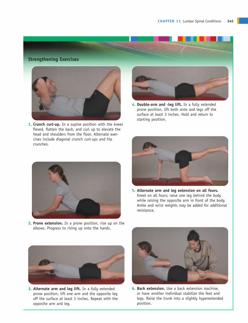
CHAPTER 11 Lumbar Spinal Conditions 345
Strengthening Exercises
4. Double-arm and -leg lift. In a fully extended prone position, lift both arms and legs off the surface at least 3 inches. Hold and return to starting position.
1. Crunch curl-up. In a supine position with the kneesflexed, flatten the back, and curl up to elevate thehead and shoulders from the floor. Alternate exer-cises include diagonal crunch curl-ups and hipcrunches.
2. Prone extension. In a prone position, rise up on theelbows. Progress to rising up onto the hands.
3. Alternate arm and leg lift. In a fully extendedprone position, lift one arm and the opposite leg off the surface at least 3 inches. Repeat with theopposite arm and leg.
5. Alternate arm and leg extension on all fours.Kneel on all fours; raise one leg behind the bodywhile raising the opposite arm in front of the body.Ankle and wrist weights may be added for additionalresistance.
6. Back extension. Use a back extension machine, or have another individual stabilize the feet andlegs. Raise the trunk into a slightly hyperextended position.
ANDERc11.qxd 11/16/07 3:54 PM Page 345
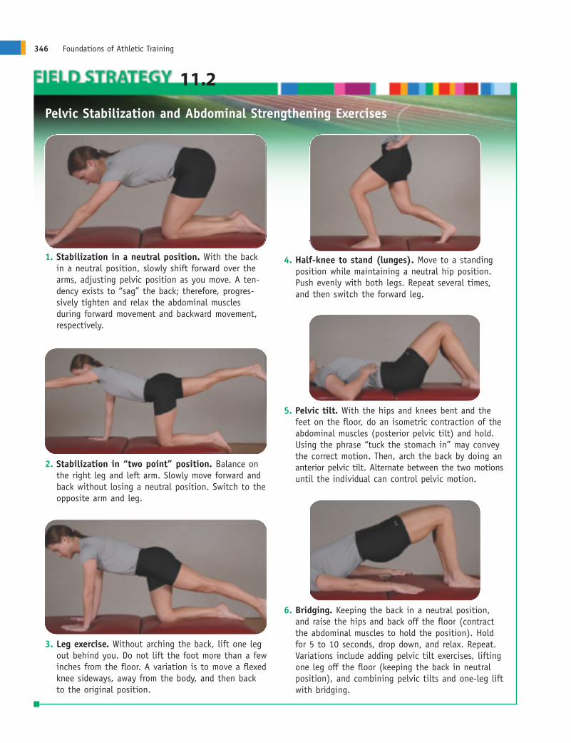
346 Foundations of Athletic Training
11.2
Pelvic Stabilization and Abdominal Strengthening Exercises
4. Half-knee to stand (lunges). Move to a standingposition while maintaining a neutral hip position.Push evenly with both legs. Repeat several times,and then switch the forward leg.
2. Stabilization in “two point” position. Balance onthe right leg and left arm. Slowly move forward andback without losing a neutral position. Switch to theopposite arm and leg.
3. Leg exercise. Without arching the back, lift one legout behind you. Do not lift the foot more than a fewinches from the floor. A variation is to move a flexedknee sideways, away from the body, and then backto the original position.
5. Pelvic tilt. With the hips and knees bent and thefeet on the floor, do an isometric contraction of theabdominal muscles (posterior pelvic tilt) and hold.Using the phrase “tuck the stomach in” may conveythe correct motion. Then, arch the back by doing ananterior pelvic tilt. Alternate between the two motionsuntil the individual can control pelvic motion.
6. Bridging. Keeping the back in a neutral position,and raise the hips and back off the floor (contractthe abdominal muscles to hold the position). Holdfor 5 to 10 seconds, drop down, and relax. Repeat.Variations include adding pelvic tilt exercises, liftingone leg off the floor (keeping the back in neutralposition), and combining pelvic tilts and one-leg liftwith bridging.
1. Stabilization in a neutral position. With the backin a neutral position, slowly shift forward over thearms, adjusting pelvic position as you move. A ten-dency exists to “sag” the back; therefore, progres-sively tighten and relax the abdominal muscles during forward movement and backward movement,respectively.
ANDERc11.qxd 11/16/07 3:54 PM Page 346
to strengthen the back extensors, stretch the abdominals, and reduce pressure on the interver-tebral disks; and stabilization exercises, abdominal strengthening, and strengthening of themedial rotators of the hip through proprioceptive neuromuscular facilitation and Thera-Bandexercises. As strength is regained, functional activities can be incorporated, with gradual returnto full activity.
SUMMARY1. Anatomical variations in the low back region that can predispose an individual to spinal
injuries include lordosis, sway back, flat back, and par interarticularis fractures, which canlead to spondylolysis or spondylolisthesis.
2. Runners are particularly prone to low back pain resulting from tight hip flexors and hamstrings. Symptoms include localized pain that increases with active and resisted back extension, but radiating pain and neurologic deficits are not present. Anterior pelvic tilt andhyperlordosis of the lumbar spine also may be present.
3. Sciatica may be caused by a herniated disk, annular tear, myogenic or muscle-related disease,spinal stenosis, facet joint arthropathy, or compression of the nerve between the piriformismuscle.
4. The most commonly herniated disks are the lower two lumbar disks at L4–L5 and L5–S1, followed by the two lower cervical disks. Most ruptures move in a posterior or posterolateraldirection as a result of torsion and compression.
5. The assessment of a spinal injury should begin with a thorough history of the injury and include neurologic tests to determine possible nerve involvement. The severity of pain and thepresence or absence of neurologic symptoms, spasm, and tenderness can indicate when abackboard and stabilization are needed.
6. If, at any time, an individual complains of acute pain in the spine, change in sensation any-where on the body, or resists moving the spine, a significant injury should be assumed andEMS summoned.
7. A rehabilitation program should focus on reduction of pain and spasm; restoration of motionand balance; development of strength, endurance, and power; and maintenance of cardiovas-cular fitness.
CHAPTER 11 Lumbar Spinal Conditions 347
REFERENCES1. Duggleby T, Kumar S. Epidemiology of juvenile low back
pain: a review. Disabil Rehabil 1997;19(12):505–512.2. Burton AK. Low back pain in children and adolescents: to
treat or not? Bull Hosp Joint Dis 1996;55(3):127–129.3. Newcomer K, Sinaki M. Low back pain and its relation-
ship to back strength and physical activity in children.Acta Paediatr 1996;85(12):1433–1439.
4. Bono CM. Low-back pain in athletes. J Bone Joint SurgAm 2004;86:382–396.
5. Anderson MW. Lumbar discography: an update. SeminRoentgenol 2004;39:52–67.
6. Nordin M, Weiner SS. Biomechanics of the lumbar spine.In: Nordin M, Frankel VH, eds. Basic Biomechanics of theMusculoskeletal System, 3rd Ed. Philadelphia: LippincottWilliams & Wilkins, 2001.
7. Soler T, Calderon C. The prevalence of spondylolysis inthe Spanish elite athlete. Am J Sports Med 2000;28(1):57–62.
8. Cholewicki J, Juluru K, McGill SM. Intra-abdominal pressure mechanism for stabilizing the lumbar spine. J Biomech 1999;32:13–17.
9. Hall SJ, Evans GG. Biomechanics. In: Mohnsen B, ed.Comprehensive Physical Education: What Every StudentNeeds to Know, 2nd Ed. Reston, VA: NASPE, 2003.
10. Wilhite JM. Thoracic and lumbosacral spine. In: MellionMB, Walsh WM, Madden C, Putukian M, Shelton GL,eds. The Team Physician’s Handbook. Philadelphia: Hanley & Belfus, 2002.
ANDERc11.qxd 11/16/07 3:54 PM Page 347