Low-Rank Tensor Regularization for Improved Dynamic...
2
Low-Rank Tensor Regularization for Improved Dynamic Quantitative Magnetic Resonance Imaging Nikolaos Kargas, Sebastian Weing¨ artner, Nicholas D. Sidiropoulos and Mehmet Akc ¸akaya University of Minnesota Minneapolis, MN Emails: {karga005, sweingae, sidir001, akcakaya}@umn.edu Abstract—In quantitative MRI, a series of images with different soft- tissue contrast are acquired. For moving organs, such as the heart, this process may be performed across different motion states, leading to ≥ 4-dimensional images. Furthermore, due to the nature of organ motion and contrast changes, such datasets can be well-represented using low-rank tensors. In this work, we investigate the utility of low-rank tensor regularization for improving the quantification of a state-of-the- art cardiac MRI technique. I. THE I NVERSE PROBLEM IN QUANTITATIVE DYNAMIC MRI Medical imaging techniques rely on differences in signal intensity (or contrast) between different tissue types. In magnetic resonance imaging (MRI), different contrasts can be generated using the magne- tization relaxation properties of tissues. In the past decade, there has been a push for quantitative MRI, where the underlying relaxation properties are quantitatively characterized in a pixel-wise manner, offering robustness independent of the acquisition setup. This requires images of the same anatomy with multiple different contrasts, which leads to long acquisition times, necessitating a trade-off between the spatial or temporal resolutions and SNR of the acquisitions. These challenges are amplified for moving organs, such as the heart. In this work, we study an image reconstruction problem for a recently proposed dynamic quantitative MRI acquisition of the heart [1]. The goal is to generate a series of images m(x, y, t, c), where (x, y) is the discrete 2D spatial location, t is the cardiac phase and c is the different contrasts. Once these images are generated, a parametric estimation procedure is performed along the c dimension to generate the quantitative maps of interest for each (x, y) and t [1]. The quality of these maps are quantified by assessing the precision across a homogenous tissue, such as muscle. II. LOW-RANK TENSOR REGULARIZATION APPROACH Low-rank matrix regularization has been studied in MRI recon- struction for dynamic or quantitative MRI. However, when both dimensions are varied, the additional benefit of regularization across the multi-dimensional image has been relatively unexplored, except for the use of the Tucker model with pre-selected basis functions [2]. In this work, we propose to use a PARAFAC model for low-rank tensor regularization in a data-adaptive fashion, without requiring basis function pre-estimation from training data. The acquired measurements are given by y(t, c)= Et,c ( m(x, y, t, c) ) +n(t, c), t =1,...,T ; c =1,...,C where Et,c : C m×n → C p is the measurement system, including a partial Fourier matrix and the receiver coil sensitivities, n(t, c) ∈ C p is measurement noise, t is the cardiac phase, c is the contrast- weighting, and x, y are the discrete spatial locations. For the 4-dimensional tensor containing m(x, y, t, c) across all x, y, t, c, we propose to solve the regularized least squares problem: min {m(x,y,t,c)} t,c X t,c ||y(t, c) - Et,c ( m(x, y, t, c) ) || 2 2 subject to rank ( m(x, y, t, c) ) = F, (1) where the tensor rank is defined as the minimum number of rank-one tensors needed to produce m as their sum [4]. Thus, a rank-F tensor, m can be written as m(x, y, t, c)= F X f =1 a f (x)b f (y)c f (t)d f (c). III. RECONSTRUCTION ALGORITHM AND EXPERIMENTS The reconstruction algorithm was implemented by alternating between data consistency with the measurements {y(t, c)}t,c, and enforcing a low-rank constraint on the 4-dimensional image tensor. The data consistency was implemented as described in [3]. The low- rank tensor constraint in the PARAFAC model was enforced via an alternating least squares approach [4], [5]. Dynamic quantitative MRI of the heart was performed using the technique described in [1] at 3 Tesla field strength. This lead to a total of 66 images, with t = 11 cardiac phases and c = 6 different contrasts as a function of the longitudinal relaxation (T1) time. Two acquisitions were performed: One at standard resolution (2 × 2 mm 2 ) and one at high resolution (1 × 1 mm 2 ), with 2- and 4-fold undersampling respectively (slice thickness = 10 mm for both cases) . Images were reconstructed with and without tensor regu- larization. Subsequently, quantitative T1 maps were generated from the reconstructed images, using the parameter estimation approach described in [1]. Precision was defined as the spatial variability across the myocardial (muscle) tissue. The reconstruction results of individual images for a 4-fold under- sampled high-resolution dataset are shown in Figure 1. Low-rank tensor regularization leads to a visible reduction in noise across different cardiac phases and contrast weightings. The quantitative reconstruction improvement is depicted in Figure 2 for a standard- resolution dataset, with 1.3- to 1.8-fold quantitative precision improvement of T1 variability for the low-rank tensor regularization approach (F = 600) over the conventional parallel imaging method. In conclusion, the proposed low-rank tensor regularization has the potential to improve undersampled dynamic quantitative MRI reconstruction, offering high-precision quantitative maps at improved spatial resolution within a short acquisition time. ACKNOWLEDGMENT The imaging experiments were performed with support from NIH R00HL111410 and NIH P41EB015894.
Transcript of Low-Rank Tensor Regularization for Improved Dynamic...
Low-Rank Tensor Regularization for Improved DynamicQuantitative Magnetic Resonance Imaging
Nikolaos Kargas, Sebastian Weingartner, Nicholas D. Sidiropoulos and Mehmet AkcakayaUniversity of Minnesota
Minneapolis, MNEmails: {karga005, sweingae, sidir001, akcakaya}@umn.edu
Abstract—In quantitative MRI, a series of images with different soft-tissue contrast are acquired. For moving organs, such as the heart,this process may be performed across different motion states, leadingto ≥ 4-dimensional images. Furthermore, due to the nature of organmotion and contrast changes, such datasets can be well-represented usinglow-rank tensors. In this work, we investigate the utility of low-ranktensor regularization for improving the quantification of a state-of-the-art cardiac MRI technique.
I. THE INVERSE PROBLEM IN QUANTITATIVE DYNAMIC MRI
Medical imaging techniques rely on differences in signal intensity(or contrast) between different tissue types. In magnetic resonanceimaging (MRI), different contrasts can be generated using the magne-tization relaxation properties of tissues. In the past decade, there hasbeen a push for quantitative MRI, where the underlying relaxationproperties are quantitatively characterized in a pixel-wise manner,offering robustness independent of the acquisition setup. This requiresimages of the same anatomy with multiple different contrasts, whichleads to long acquisition times, necessitating a trade-off between thespatial or temporal resolutions and SNR of the acquisitions. Thesechallenges are amplified for moving organs, such as the heart.
In this work, we study an image reconstruction problem for arecently proposed dynamic quantitative MRI acquisition of the heart[1]. The goal is to generate a series of images m(x, y, t, c), where(x, y) is the discrete 2D spatial location, t is the cardiac phase and c isthe different contrasts. Once these images are generated, a parametricestimation procedure is performed along the c dimension to generatethe quantitative maps of interest for each (x, y) and t [1]. The qualityof these maps are quantified by assessing the precision across ahomogenous tissue, such as muscle.
II. LOW-RANK TENSOR REGULARIZATION APPROACH
Low-rank matrix regularization has been studied in MRI recon-struction for dynamic or quantitative MRI. However, when bothdimensions are varied, the additional benefit of regularization acrossthe multi-dimensional image has been relatively unexplored, exceptfor the use of the Tucker model with pre-selected basis functions [2].In this work, we propose to use a PARAFAC model for low-ranktensor regularization in a data-adaptive fashion, without requiringbasis function pre-estimation from training data.
The acquired measurements are given by
y(t, c) = Et,c
(m(x, y, t, c)
)+n(t, c), t = 1, . . . , T ; c = 1, . . . , C
where Et,c : Cm×n → Cp is the measurement system, includinga partial Fourier matrix and the receiver coil sensitivities, n(t, c) ∈Cp is measurement noise, t is the cardiac phase, c is the contrast-weighting, and x, y are the discrete spatial locations.
For the 4-dimensional tensor containing m(x, y, t, c) across allx, y, t, c, we propose to solve the regularized least squares problem:
min{m(x,y,t,c)}t,c
∑t,c
||y(t, c)−Et,c
(m(x, y, t, c)
)||22
subject to rank(m(x, y, t, c)
)= F, (1)
where the tensor rank is defined as the minimum number of rank-onetensors needed to produce m as their sum [4]. Thus, a rank-F tensor,m can be written as
m(x, y, t, c) =
F∑f=1
af (x)bf (y)cf (t)df (c).
III. RECONSTRUCTION ALGORITHM AND EXPERIMENTS
The reconstruction algorithm was implemented by alternatingbetween data consistency with the measurements {y(t, c)}t,c, andenforcing a low-rank constraint on the 4-dimensional image tensor.The data consistency was implemented as described in [3]. The low-rank tensor constraint in the PARAFAC model was enforced via analternating least squares approach [4], [5].
Dynamic quantitative MRI of the heart was performed using thetechnique described in [1] at 3 Tesla field strength. This lead toa total of 66 images, with t = 11 cardiac phases and c = 6different contrasts as a function of the longitudinal relaxation (T1)time. Two acquisitions were performed: One at standard resolution(2 × 2 mm2) and one at high resolution (1 × 1 mm2), with 2− and4−fold undersampling respectively (slice thickness = 10 mm for bothcases) . Images were reconstructed with and without tensor regu-larization. Subsequently, quantitative T1 maps were generated fromthe reconstructed images, using the parameter estimation approachdescribed in [1]. Precision was defined as the spatial variability acrossthe myocardial (muscle) tissue.
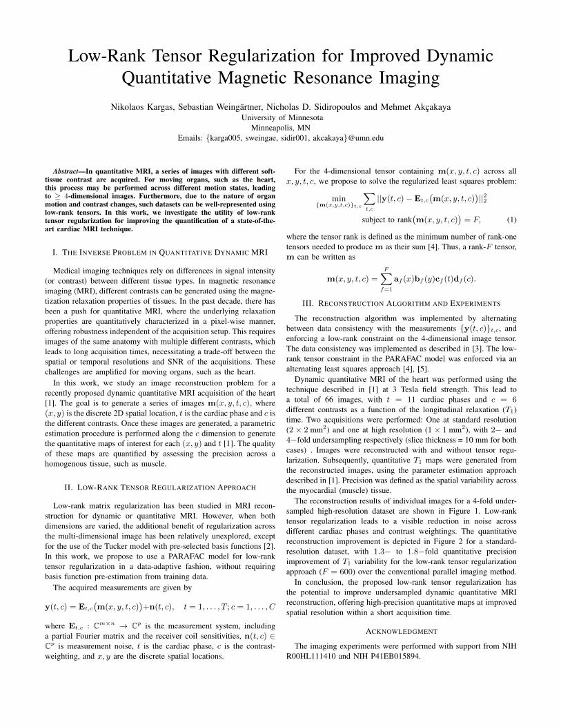
The reconstruction results of individual images for a 4-fold under-sampled high-resolution dataset are shown in Figure 1. Low-ranktensor regularization leads to a visible reduction in noise acrossdifferent cardiac phases and contrast weightings. The quantitativereconstruction improvement is depicted in Figure 2 for a standard-resolution dataset, with 1.3− to 1.8−fold quantitative precisionimprovement of T1 variability for the low-rank tensor regularizationapproach (F = 600) over the conventional parallel imaging method.
In conclusion, the proposed low-rank tensor regularization hasthe potential to improve undersampled dynamic quantitative MRIreconstruction, offering high-precision quantitative maps at improvedspatial resolution within a short acquisition time.
ACKNOWLEDGMENT
The imaging experiments were performed with support from NIHR00HL111410 and NIH P41EB015894.
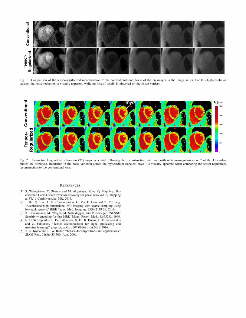
Fig. 1. Comparison of the tensor-regularized reconstruction to the conventional one, for 6 of the 66 images in the image series. For this high-resolutiondataset, the noise reduction is visually apparent, while no loss of details is observed on the tissue borders.
Fig. 2. Parametric longitudinal relaxation (T1) maps generated following the reconstruction with and without tensor-regularization. 7 of the 11 cardiacphases are displayed. Reduction in the noise variation across the myocardium (labeled “myo”) is visually apparent when comparing the tensor-regularizedreconstruction to the conventional one.
REFERENCES
[1] S. Weingartner, C. Shenoy and M. Akcakaya, “Cine T1 Mapping: B1-corrected Look-Locker inversion recovery for phase-resolved T1 mappingat 3T,” J Cardiovascular MR, 2017.
[2] J. He, Q. Liu, A. G. Christodoulou, C. Ma, F. Lam and Z. P. Liang,“Accelerated high-dimensional MR imaging with sparse sampling usinglow-rank tensors,” IEEE Trans. Med. Imaging, 35(9):2119-29, 2016.
[3] K. Pruessmann, M. Weiger, M. Scheidegger, and P. Boesiger, “SENSE:Sensitivity encoding for fast MRI,” Magn. Reson. Med., 42:95262, 1999.
[4] N. D. Sidiropoulos, L. De Lathauwer, X. Fu, K. Huang, E. E. Papalexakisand C. Faloutsos, “Tensor decomposition for signal processing andmachine learning,” preprint, arXiv:1607.01668 [stat.ML], 2016.
[5] T. G. Kolda and B. W. Bader, “Tensor decompositions and applications,”SIAM Rev., 51(3):455-500, Aug. 2009.