Localized Phase Separation of Thermoresponsive Polymers Induced by Plasmonic …€¦ · ·...
12
Localized Phase Separation of Thermoresponsive Polymers Induced by Plasmonic Heating Issei Aibara, †,§ Jun-ichi Chikazawa, †,§ Takayuki Uwada, ‡ and Shuichi Hashimoto* ,† † Department of Optical Science and Technology, University of Tokushima, 2-1 Minami-Josanjima, Tokushima 770-8506, Japan ‡ Department of Chemistry, Josai University, 1-1 Keyakidai, Sakado, Saitama 350-0295, Japan * S Supporting Information ABSTRACT: Optical excitation-induced heating of a single gold nanoparticle potentially offers a high-temperature field confined to the immediate neighborhood of the particle. In this study, we applied darkfield microscopy imaging and Rayleigh scattering spectroscopy to pursue phase separation of aqueous thermoresponsive poly(N-isopropylacrylamide) and poly(vinyl methyl ether) adjacent to a gold nanoparticle that was heated by continuous wave laser illumination. Gold nanoparticles were supported on transparent substrates of glass or sapphire. From the imaging study, we observed that a 1−10 μm microdroplet covering the nanoparticle formed and grew in time scales of seconds to a few tens of seconds. The growth was triggered by the illumination, and the droplet collapsed when the laser was blocked. At the same time, we observed scattering spectral changes characterized by a progressive redshift in the localized surface plasmon resonance (LSPR) band and an increasing scattering intensity in the region of wavelengths shorter than the LSPR band with increasing laser intensity. The scattering spectral changes were interpreted by the encapsulation of the nanoparticle by a polymer-rich droplet with increasing sizes. The present study revealed that thermoresponsive polymers were attracted to a hot gold nanoparticle and formed a microdroplet under illumination with a wavelength near the LSPR. Our findings demonstrate the potential of plasmonic heating to manipulate polymer migration and accumulation, which may find applications in protein crystallization. ■ INTRODUCTION Thermoresponsive polymers represented by poly(N-isopropy- lacrylamide) (PNIPAM) exhibit phase separation (demixing) in aqueous solutions when heated above a critical temperature known as the lower critical solution temperature (LCST: T c = 32 °C or 305 K). 1 This phenomenon has attracted much attention because of potential applications in drug delivery, separation, and bioswitching. 2 The molecular mechanism of the phase separation is assumed to be a manifestation of a coil− globule transition followed by further aggregation, forming polymer-rich domains such as mesoglobules. 3 Intriguingly, such phase separation was induced locally using a focused near- infrared laser beam (focal spot diameter, ∼1 μm; peak power density, ∼10 9 W cm −2 ; period, several minutes) with a wavelength of 1064 nm. The laser-illuminated aqueous PNIPAM and poly(vinyl methyl ether) (PVME) solutions resulted in a single microdroplet around the focal laser spot. 4−6 Here a high-temperature field created in water presumably confined the microdroplet by collecting the polymers, assisted by the optical force. This method has the potential to create a droplet anywhere in a solution, if it can be prepared more quickly at much lower laser powers. Revealing liquid−liquid phase separation is also important in applications such as protein crystallization, which is assumed to proceed via a two- step process involving (1) the formation of liquid droplets of high protein concentration and (2) the generation of ordered protein clusters within the dense liquid intermediate prior to nucleation leading to protein crystals. 7−9 For thermoresponsive polymers, the dynamic growth process starts from the phase transition, then phase separation, leading to a microdroplet (Scheme 1) through local heating. This process has been poorly understood because of a lack of a proper means to observe submicrometer scale events, hampered by the resolution limit of optical microscopes. By confining heating to the nanoscale, assisted by a sensitive spectroscopic technique, we will be able to look into the detailed progress of temperature-induced phase separation. Plasmonic nanoparticles (NPs) such as gold (Au) NPs have emerged as a nanoscale antenna that confines incident light in a subwavelength volume around the particle, thus tremendously concentrating the electric field. 10 This plasmonic field enhance- ment is the foundation for surface-enhanced Raman scattering and metal-enhanced fluorescence, both of which are promising for ultratrace analyses. 11,12 Simultaneously with the electric field Received: July 20, 2017 Revised: September 16, 2017 Published: September 19, 2017 Article pubs.acs.org/JPCC © XXXX American Chemical Society A DOI: 10.1021/acs.jpcc.7b07187 J. Phys. Chem. C XXXX, XXX, XXX−XXX
Transcript of Localized Phase Separation of Thermoresponsive Polymers Induced by Plasmonic …€¦ · ·...
Localized Phase Separation of Thermoresponsive Polymers Inducedby Plasmonic HeatingIssei Aibara,†,§ Jun-ichi Chikazawa,†,§ Takayuki Uwada,‡ and Shuichi Hashimoto*,†
†Department of Optical Science and Technology, University of Tokushima, 2-1 Minami-Josanjima, Tokushima 770-8506, Japan‡Department of Chemistry, Josai University, 1-1 Keyakidai, Sakado, Saitama 350-0295, Japan
*S Supporting Information
ABSTRACT: Optical excitation-induced heating of a singlegold nanoparticle potentially offers a high-temperature fieldconfined to the immediate neighborhood of the particle. Inthis study, we applied darkfield microscopy imaging andRayleigh scattering spectroscopy to pursue phase separation ofaqueous thermoresponsive poly(N-isopropylacrylamide) andpoly(vinyl methyl ether) adjacent to a gold nanoparticle thatwas heated by continuous wave laser illumination. Goldnanoparticles were supported on transparent substrates ofglass or sapphire. From the imaging study, we observed that a1−10 μm microdroplet covering the nanoparticle formed andgrew in time scales of seconds to a few tens of seconds. Thegrowth was triggered by the illumination, and the dropletcollapsed when the laser was blocked. At the same time, we observed scattering spectral changes characterized by a progressiveredshift in the localized surface plasmon resonance (LSPR) band and an increasing scattering intensity in the region ofwavelengths shorter than the LSPR band with increasing laser intensity. The scattering spectral changes were interpreted by theencapsulation of the nanoparticle by a polymer-rich droplet with increasing sizes. The present study revealed thatthermoresponsive polymers were attracted to a hot gold nanoparticle and formed a microdroplet under illumination with awavelength near the LSPR. Our findings demonstrate the potential of plasmonic heating to manipulate polymer migration andaccumulation, which may find applications in protein crystallization.
■ INTRODUCTION
Thermoresponsive polymers represented by poly(N-isopropy-lacrylamide) (PNIPAM) exhibit phase separation (demixing) inaqueous solutions when heated above a critical temperatureknown as the lower critical solution temperature (LCST: Tc =32 °C or 305 K).1 This phenomenon has attracted muchattention because of potential applications in drug delivery,separation, and bioswitching.2 The molecular mechanism of thephase separation is assumed to be a manifestation of a coil−globule transition followed by further aggregation, formingpolymer-rich domains such as mesoglobules.3 Intriguingly, suchphase separation was induced locally using a focused near-infrared laser beam (focal spot diameter, ∼1 μm; peak powerdensity, ∼109 W cm−2; period, several minutes) with awavelength of 1064 nm. The laser-illuminated aqueousPNIPAM and poly(vinyl methyl ether) (PVME) solutionsresulted in a single microdroplet around the focal laser spot.4−6
Here a high-temperature field created in water presumablyconfined the microdroplet by collecting the polymers, assistedby the optical force. This method has the potential to create adroplet anywhere in a solution, if it can be prepared morequickly at much lower laser powers. Revealing liquid−liquidphase separation is also important in applications such asprotein crystallization, which is assumed to proceed via a two-
step process involving (1) the formation of liquid droplets ofhigh protein concentration and (2) the generation of orderedprotein clusters within the dense liquid intermediate prior tonucleation leading to protein crystals.7−9 For thermoresponsivepolymers, the dynamic growth process starts from the phasetransition, then phase separation, leading to a microdroplet(Scheme 1) through local heating. This process has been poorlyunderstood because of a lack of a proper means to observesubmicrometer scale events, hampered by the resolution limitof optical microscopes. By confining heating to the nanoscale,assisted by a sensitive spectroscopic technique, we will be ableto look into the detailed progress of temperature-induced phaseseparation.Plasmonic nanoparticles (NPs) such as gold (Au) NPs have
emerged as a nanoscale antenna that confines incident light in asubwavelength volume around the particle, thus tremendouslyconcentrating the electric field.10 This plasmonic field enhance-ment is the foundation for surface-enhanced Raman scatteringand metal-enhanced fluorescence, both of which are promisingfor ultratrace analyses.11,12 Simultaneously with the electric field
Received: July 20, 2017Revised: September 16, 2017Published: September 19, 2017
Article
pubs.acs.org/JPCC
© XXXX American Chemical Society A DOI: 10.1021/acs.jpcc.7b07187J. Phys. Chem. C XXXX, XXX, XXX−XXX
enhancement, the light energy absorbed by the plasmonic NPsis efficiently converted into heat, raising the temperature ofboth the NP and the surrounding medium. This is referred toas the photothermal or plasmonic-heating effect.13,14 Plasmonicheating is unique because only the medium immediatelysurrounding the NPs is heated by radial heat conduction, with
the medium acting as an infinitely large heat sink. Thus, thehigh-temperature field confined around the NP can be appliedto drive the phase transition/phase separation of thermores-ponsive polymers.Such a possibility has been tested for PNIPAM very recently.
Under a brightfield optical microscope, Orlishausen and Kohler
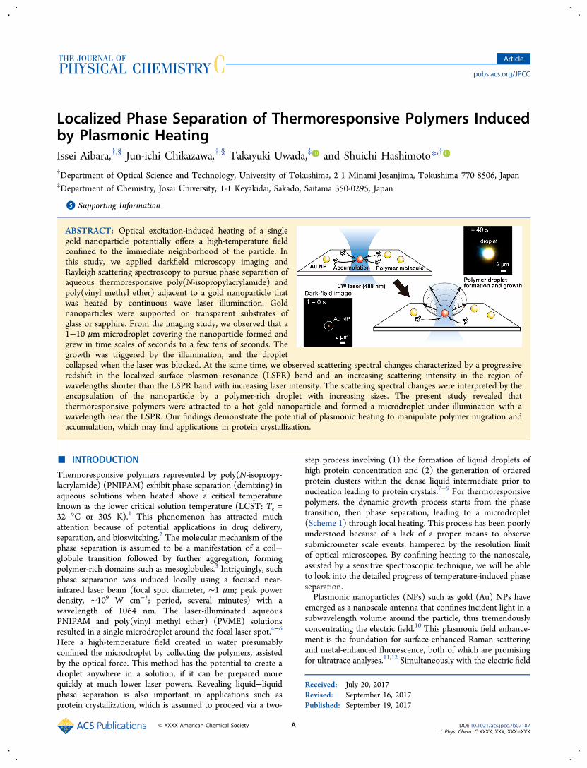
Scheme 1. Sketch of Temperature-Induced Phase Separation Forming a Polymer-Rich Domain via Coil-to-Globule PhaseTransition
Scheme 2. Experimental Overviewa
a(a) Upper: pictorial representation of the event, i.e. phase separation of the thermoresponsive polymers on plasmonic heating. Middle: darkfieldmicroscopy images showing a transition of a single Au NP on exposure to laser illumination in aqueous solution of a thermoresponsive polymer.Lower: Rayleigh light-scattering spectral changes of a single Au NP at various laser intensities. (b) Experimental setup consisting of a chamber-structured specimen, a darkfield illumination system, a microscope objective, an excitation laser, a digital camera, and a CCD spectrophotometer. BP:laser line filter, LP: long-pass filter. (c) Structural formulas of poly(N-isopropylacrylamide) (PNIPAM) and poly(vinyl methyl ether) PVME.
The Journal of Physical Chemistry C Article
DOI: 10.1021/acs.jpcc.7b07187J. Phys. Chem. C XXXX, XXX, XXX−XXX
B
observed the dynamic growth of PNIPAM aggregates thatformed around laser-heated Au NPs.15 The heating of 250 nmdiameter Au NPs settled on a window in a 8−9 wt % PNIPAMsolution with focused laser illumination at a constant power51.9 mW (50−100 mW μm−2 or 5 × 10⁶−107 W cm−2) andwavelength 532 nm which enabled the growth of phase-separated aggregates with diameters of ∼10 μm at 0.3 s to ∼100μm at 500 s. Note here that the ranges of these laser powerswell exceed the threshold of vapor bubble generation around aAu NP.16−18 After laser heating was stopped, the aggregatesdissolved from the outside. Because of a poor contrast of thebrightfield method, early stages of aggregate formation wereobscured. By using a 100 nm diameter single Au NP underoptical heating with low-intensity laser illumination (104−105W cm−2) combined with the Rayleigh light-scattering spec-troscopy, Aibara and co-workers observed a remarkable redshiftin the localized surface plasmon resonance (LSPR) band of aAu NP exposed to aqueous PNIPAM solution.19 Thisobservation was ascribed to the plasmonic-heating-inducednanoscale phase transition/separation to form PNIPAMaggregates within the high-temperature field surrounding theAu NP. This is distinct from the coil-to-globule volume phasetransition of a PNIPAM shell cross-linked around a Au NPcore,20−22 because PNIPAM molecules were not immobilizedon the Au NP but were freely mobile in the solution. Althoughscattering spectroscopy is promising for revealing nanoscaleevents, laser intensity dependence has not been fullyinvestigated for the LSPR spectral changes, and the particletemperature-dependent aggregation of PNIPAM to form aphase-separated droplet remains to be elucidated. There is stillroom to fill the gap between spectroscopic observations ofplasmonic-heating-induced PNIPAM phase transitions aroundAu NPs and microscope observations of PNIPAM-richmicrodroplet formation.The temperature field around the NP is not homogeneous,
but the temperature increases nearer to the NP surface.18 Thus,the phase transition may start from the particle surface whenthe temperature exceeds the LCST at a certain threshold laserintensity, spreading from the surface to distant regions withincreasing laser intensities. For experiments with medium waterheating, a microdroplet with a diameter much greater than theheating laser spot size was formed after several minutes ofexposure.4−6 This may mean that PNIPAM molecules weretransferred from outside the heating area to grow the droplet,although no detailed descriptions have been given. Thetransport and accumulation of colloids, cells, and DNAexploiting optothermal manipulation through thermal andMarangoni convections and thermophoresis are currentlyunder intense investigation.23−29 These studies used substrateswith a thin Au layer or a Au nanoisland film for heatingeffectively with a focused laser. Nevertheless, the heat transfer iscomplex, making temperature analysis difficult. In contrast,single Au NP heating is expected to yield a much simplertemperature field around the Au NP. Thus, the transportmechanism can be simplified. Moreover, in terms of theirvarying thermal conductivities, substrates play a decisive role incontrolling particle temperature and temperature gradient atparticle−medium and particle−substrate interfaces, promotingaccumulation.30
In the present study we used darkfield microscopy-basedimaging and light-scattering spectroscopy of a single Au NP toreveal a dynamic picture of phase separation for thermores-ponsive polymers induced by single-particle plasmonic heating.
Because of light scattering, the darkfield microscopy imagingcan offer high-contrast images against a dark background forsmall objects that are otherwise difficult to view. We lookedinto the effect of different substrates on transport properties ofsuch polymers. We show that plasmonic heating has a clearadvantage over optical trapping to drive these polymers formanipulation with a significantly lower power.
■ EXPERIMENTAL METHODS
Experimental Setup. The outline of the present experi-ment is shown in Scheme 2a, with an experimental setup givenin Scheme 2b. The darkfield imaging was performed on aninverted optical microscope, IX-71 (Olympus, Tokyo, Japan;with a darkfield condenser NA = 0.8−0.92), equipped with aDS-5 M digital camera (Nikon, Tokyo, Japan). The single-particle scattering spectra were recorded with a wavelengthresolution of 0.5 nm on a spectrophotometer consisting of aSP-300i polychromator (Acton Research Co., MA) with agrating of 150 or 300 grooves/mm blazed at 500 nm and aDU401-BR-DD CCD camera (Andor Technology, Belfast, UK;operated at −60 °C) through a 500 μm diameter pinhole (viewarea: 10-μm diameter). A halogen lamp with a broad (white)spectrum was used for illumination when recording thescattering images and spectra. The spectra were obtained bysubtracting the background signals including Raman scatteringof the surrounding media and photoluminescence of the NP,then dividing it by the spectral profile of the white-lightexcitation source. A HA50 IR-cut filter (Hoya CandeoOptronics, Tokyo, Japan) was used for minimizing the lampheating. Single Au NPs adsorbed on the top wall (ceiling) ofthe chamber (see Scheme 2b) were heated by illuminating afocused 488 nm CW laser, OBIS-488-LX-150 (Coherent, SantaClara, CA), beam through a microscope objective (60×, NA =0.70). We used a 488 nm wavelength laser for a few reasons.(1) The scattering spectra of nominal 100 nm diameter Au NPsextend from 500 to 800 nm; hence, we did not use a 532 nmlaser for excitation to avoid the superposition of a strongexcitation light on relatively weak scattering spectra. (2) Theexcitation wavelength of 488 nm is slightly offset from theLSPR peak position, and the absorption cross-section, Cabs, isthen unaffected by temperature changes. In contrast, the LSPRpeak intensity is strongly dependent on particle temperatureand changes in medium refractive index.16 The excitation of theLSPR band causes the value of Cabs for NPs to decrease withincreasing temperature because of the temperature-induceddamping, making estimates of the particle temperature difficult.(3) At the excitation wavelength of 488 nm, no light absorptionand subsequent temperature increase are expected for glass,sapphire, PNIPAM, and PVME. The 488 nm laser light excitesboth interband and intraband transitions. Note, however, thatthe consequence of both excitations is the same: hot electrongeneration and subsequent particle heating.32 The temperatureincrease of a Au NP occurs simultaneously with opening theshutter, and the temperature decrease follows immediately afterclosing the shutter.33 The spatial laser profile was determinedby measuring scattering signal intensity of the 100 nm diameterAu NP while scanning the stage at 100 nm interval. The fwhmof the laser beam thus determined was 0.6 μm. The laser peakpower density Ip (mW μm−2) was represented by
π=I P[ (2.3546) ]/[2 (FWHM) ]p2 2 ,31 where P is the laser
power density (measured laser power divided by beam area).The irradiation periods were regulated using an F77 mechanical
The Journal of Physical Chemistry C Article
DOI: 10.1021/acs.jpcc.7b07187J. Phys. Chem. C XXXX, XXX, XXX−XXX
C
shutter (Suruga Seiki, Tokyo, Japan). All measurements wereperformed at 24 ± 1 °C.Sample Preparation. Poly(N-isopropylacrylamide) (PNI-
PAM, Mw = 30 000) was obtained from Sigma-Aldrich Co. (St.Louis, MO) while poly(vinyl methyl ether) (PVME, 30% inwater, Mw = 47 000) was from Tokyo Kasei Co. (Tokyo,Japan). The structural formulas of PNIPAM and PVME aregiven in Scheme 2c. Aqueous solutions of Au NPs with nominaldiameters of 100 nm (EMGC100) were obtained from BBISolutions, Cardiff, UK. Au NPs were transformed from afaceted to a spherical shape by irradiation with weak-intensitynanosecond laser pulses (∼11 mJ cm−2) of 532 nm wavelength.The particle image acquired using a transmission electronmicroscope and the corresponding size distribution (102 ± 5nm) are given in the Supporting Information, S1. Spherical AuNPs were spin-coated onto the 0001 face of an opticallypolished sapphire substrate (Shinkosha, Yokohama, Japan) ofsize 15 mm × 15 mm × 0.3 mm or a borosilicate cover glass(Matsunami, Osaka, Japan) of 24 mm × 32 mm × 0.17 mm.The Au NPs were washed twice with double-distilled water byplacing 0.5 mL of water on a spin coater and spun. Au NPswere immersed in solutions of PNIPAM and PVME in an 11μL chamber consisting of a sapphire/glass substrate, a 0.2 mmthick silicone rubber spacer, and a 24 mm × 32 mm × 0.17 mmmicroscope coverslip. The substrates were cleansed in a boilingmixture of 1:1 30% H2O2−28% ammonia mixture for 90 min,and plasma-cleaned in a YHS-R reactor (70 W, 20 kHz;Sakigake Semiconductor, Kyoto, Japan) for 60 s just before use.
■ RESULTS AND DISCUSSIONTemperature Distributions around a Single Au NP.
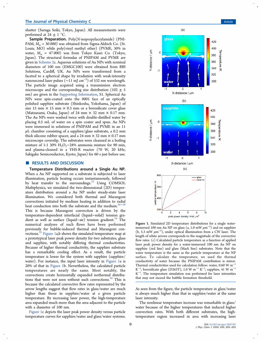
When a Au NP supported on a substrate is subjected to laserillumination, particle heating occurs instantaneously, followedby heat transfer to the surroundings.33 Using COMSOLMultiphysics, we simulated the two-dimensional (2D) temper-ature distribution around a Au NP under steady-state laserillumination. We considered both thermal and Marangoniconvections initiated by medium heating in addition to radialheat conduction into both the substrate and the medium.26−28
This is because Marangoni convection is driven by thetemperature-dependent interfacial (liquid−solid) tension gra-dient as well as surface (liquid−air) tension gradient.34 Thenumerical analyses of such flows have been performedpreviously for bubble-induced thermal and Marangoni con-vections.35 Figure 1a,b shows the simulated temperature map ata prototypical laser peak power density for two substrates, glassand sapphire, with notably differing thermal conductivities.Because of higher thermal conductivity, the sapphire substratehas a remarkable cooling effect. As a result, the particletemperature is lower for the system with sapphire (sapphire/water). For instance, the input laser intensity in Figure 1a is20% of that in Figure 1b. Nevertheless, the calculated particletemperatures are nearly the same. Most notably, theconvections create horizontally expanded isothermal distribu-tions that were not seen without such convections.30 This isbecause the calculated convective flow rates represented by thearrow lengths suggest that flow rates in glass/water are muchhigher than those in sapphire/water at a given particletemperature. By increasing laser power, the high-temperaturearea expanded much more than the area adjacent to the particlewith a diameter of 100 nm.Figure 1c depicts the laser peak power density versus particle
temperature curves for sapphire/water and glass/water systems.
As seen from the figure, the particle temperature in glass/wateris always much higher than that in sapphire/water at the samelaser intensity.The nonlinear temperature increase was remarkable in glass/
water because of the higher temperatures that induced higherconvection rates. With both different substrates, the high-temperature region increased in area with increasing laser
Figure 1. Simulated 2D temperature distributions for a single water-immersed 100 nm Au NP on glass (a, 1.0 mW μm−2) and on sapphire(b, 5.3 mW μm−2), under optical illumination from a CW laser. Thelength of white arrows corresponds to the magnitude of the convectiveflow rates. (c) Calculated particle temperature as a function of appliedlaser peak power density for a water-immersed 100 nm Au NP onsapphire (red line) and glass (black line) substrates. Note that thewater temperature is the same as the particle temperature at the NPsurface. To calculate the temperature, we used the thermalconductivity of water because the PNIPAM contribution is minor.Thermal conductivities used for calculation follow: water, 0.60 W m−1
K−1; borosilicate glass (D263T), 1.0 W m−1 K−1; sapphire, 41 W m−1
K−1. The temperature simulation was performed for laser intensitiesthat may not exceed the bubble formation threshold.
The Journal of Physical Chemistry C Article
DOI: 10.1021/acs.jpcc.7b07187J. Phys. Chem. C XXXX, XXX, XXX−XXX
D
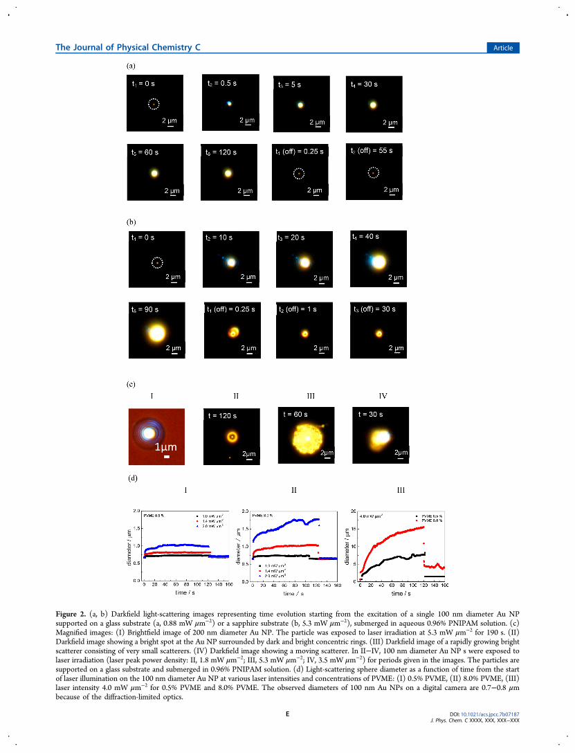
Figure 2. (a, b) Darkfield light-scattering images representing time evolution starting from the excitation of a single 100 nm diameter Au NPsupported on a glass substrate (a, 0.88 mW μm−2) or a sapphire substrate (b, 5.3 mW μm−2), submerged in aqueous 0.96% PNIPAM solution. (c)Magnified images: (I) Brightfield image of 200 nm diameter Au NP. The particle was exposed to laser irradiation at 5.3 mW μm−2 for 190 s. (II)Darkfield image showing a bright spot at the Au NP surrounded by dark and bright concentric rings. (III) Darkfield image of a rapidly growing brightscatterer consisting of very small scatterers. (IV) Darkfield image showing a moving scatterer. In II−IV, 100 nm diameter Au NP s were exposed tolaser irradiation (laser peak power density: II, 1.8 mW μm−2; III, 5.3 mW μm−2; IV, 3.5 mW μm−2) for periods given in the images. The particles aresupported on a glass substrate and submerged in 0.96% PNIPAM solution. (d) Light-scattering sphere diameter as a function of time from the startof laser illumination on the 100 nm diameter Au NP at various laser intensities and concentrations of PVME: (I) 0.5% PVME, (II) 8.0% PVME, (III)laser intensity 4.0 mW μm−2 for 0.5% PVME and 8.0% PVME. The observed diameters of 100 nm Au NPs on a digital camera are 0.7−0.8 μmbecause of the diffraction-limited optics.
The Journal of Physical Chemistry C Article
DOI: 10.1021/acs.jpcc.7b07187J. Phys. Chem. C XXXX, XXX, XXX−XXX
E
intensity. From the particle temperature as a function of laserpeak power density given in Figure 1c, the threshold of bubbleformation can occur at ∼3 mW μm−2 for a glass/water system.On the other hand, it is estimated at >10 mW μm−2 for asapphire/water system. Next, we will show the experimentalevidence of phase separation, in which the particle temperatureand the medium temperature distribution play a major role.Microscopy Imaging on Plasmonic Heating. Darkfield
microscopy imaging of a single Au NP was performed coupledwith plasmonic heating in aqueous solutions of thermores-ponsive polymers, depending on polymer concentration andlaser intensity. Upon laser illumination of a 100 nm Au NPsupported on a glass substrate and submerged in aqueousPNIPAM solution, we observed the formation and growth of alight-scattering sphere (2D images) centered at the position ofthe Au NP subjected to illumination. Figure 2a,b shows imagescaptured by a digital camera (the corresponding movie files,Movies 1−4, can be found in the Supporting Information);parts a and b compare the cooling effect of different substrateson plasmonic heating. In Figure 2a, a scattering sphereimmediately grows to diameter <2 μm within 5 s ofillumination and exhibits a very slow growth to diameter ∼2μm (see t2 = 10 to t5 = 120 s). The brightfield image of ascattering sphere (Figure 2cI, and Supporting Information S2)shows a transparent planoconvex lens-like shape suggesting thata droplet formed. It has been found that gel-like PNIPAM-richagglomerates grow within the phase-separating solutions uponheating homogeneous PNIPAM solutions across Tc.
36−38
However, the single-domain phase separation occurred forlocal heating;4−6 plasmonic-heating-induced droplet formationencapsulating the Au NP, as demonstrated here, isunprecedented. The observed diameter of a light-scatteringsphere as a function of illumination period at laser peak powerdensities of 0.7−2.6 mW μm−2 is given in SupportingInformation S3 (below 0.7 mW μm−2, we had experimentaldifficulty in imaging hampered by an illuminating laser spot aswill be explained below). We clearly observed the formation ofa scattering entity at ∼0.7 mW μm−2 on a glass substrate. Whenthe laser light is blocked after 120 s of illumination, thescattering sphere collapsed immediately (see t1(off) = 0.25 tot3(off) = 55 s), and the original Au NP image recovered. Thesingle-particle light-scattering spectra of Au NP at t1 and t(off)are nearly the same, suggesting that original bare Au NP wasreproduced after illumination was terminated.This result suggests that the phase separation of PNIPAM
reversibly forms a microdroplet around the Au NP in responseto the particle temperature increase/decrease. That is, theparticle temperature and resultant temperature distributionaround the Au NP, given in Figure 1 a,b, are decisive for thesize and shape of phase-separated droplets because the dropletsformed are in dynamic equilibrium with incoming and outgoingmolecules. As a result, a steady-state droplet diameter thatdepends on the illumination intensity was reached as timeproceeded (Figure 2a and Supporting Information, Figure S3a).Light-scattering imaging relies on the distinct difference in
refractive indices, so the clear boundary that occurred betweenthe domain and the medium suggests that the domain has amuch greater refractive index than water, indicative of phaseseparation. Indeed, the refractive index of n = 1.49539 reportedfor pure amorphous PNIPAM is considerably higher than thatof water, n = 1.333 at ambient temperature. The phaseseparation results in a polymer-rich domain containing water,the amount of which is inaccessible. Therefore, the refractive
index of PNIPAM-rich droplet can be smaller than that of neatpolymer.Figure 2b shows the time evolution of light-scattering images
for a sapphire substrate. Formation of a scattering spheresimilar to those observed on a glass substrate occurred, but atmuch higher laser intensities >∼2 mW μm−2. Below thisintensity, the illuminating laser spot hampered the imaging of aPNIPAM-rich droplet, although the laser was attenuated byblocking with a filter before the camera. Minimum intensity wasnecessary to locate the beam position to irradiate the Au NP,however. The higher threshold of droplet formation can beascribed to a greater cooling effect of a sapphire substratecaused by much larger thermal conductivity as discussed in theprevious section. With the lapse of time, a gradual increase inthe diameter of a scattering sphere occurred until 90 s reachinga diameter of 6 μm. The slower droplet growths observed insapphire/water system can be ascribed to lower flow rates andgreater deviation from centrosymmetric temperature distribu-tion around the particle, estimated for the system than theglass/water system (compare Figure 1a,b). On blocking thelaser light, however, a phenomenon distinct from that observedon a glass substrate occurred. The scattering images withoutlaser illumination were appreciably greater in size than those ofthe original Au NP. In this case, a fixation of polymer aroundthe Au NP resulted. The detailed account of this observation isgiven in the Mechanistic Aspect section.In addition to PNIPAM, we applied the darkfield imaging
technique to monitor the plasmonic-heating-induced phaseseparation of aqueous PVME, which is also known for itsthermoresponsive nature.40−42 Figure 2d was constructed onthe basis of the scattering sphere diameter dependent onillumination time (see Supporting Information S4 for images).In this case, clear laser-power-dependent diameter growth wasobserved on a glass substrate in the range of laser powers weused (I and II). Notably, the time-dependent diameter growthwas concentration-dependent; at 0.5% PVME, a fast growthoccurred within 2 s, which is similar to the observation forPNIPAM. At 8.0%, however, a slow growth from 5 to 120 soccurred. This concentration dependence is safely ascribed tothe effect of higher viscosity that gives reduced diffusioncoefficients for polymers at higher concentrations (SupportingInformation S5). At higher laser powers, a remarkably steeperdiameter increase with time occurred, which is also dependenton the PVME concentration. Such an example at 4.0 mW μm−2
is given in Figure 2dIII. At 8.0% PVME, the diameter growthoccurred immediately. Furthermore, after terminating the laserillumination, an increased particle diameter of 4 μm at 8.0%PVME was observed. This diameter increase after illuminationwas only observed at laser intensities >4.0 mW μm−2. Theobservations of a remarkably steep diameter rise and permanentfixation at high intensities strongly suggest that bubbleformation occurred because of strong heating.26−29,35 Presum-ably, the higher PVME concentration acted to induce bubblegeneration. Permanent fixation on glass substrates was alsoobserved for PNIPAM at high laser intensities (SupportingInformation, Figure S3b).Finally, we comment on the shape of light scatterers: phase-
separated droplets. As shown in Figure 2a,b, the light scatterersexhibited basically plain spherical shapes of various diameters.In contrast, when the laser power was increased to 1.8 mWμm−2, we could see ring structures as shown in Figure 2cII. Thecentral bright spot is ascribed to the location of a hot Au NPwhere the light-scattering intensity can be very high because
The Journal of Physical Chemistry C Article
DOI: 10.1021/acs.jpcc.7b07187J. Phys. Chem. C XXXX, XXX, XXX−XXX
F
such a high temperature attracts and concentrates PNIPAMmolecules to a greater extent. The peripheral ring also exhibitedhigh scattering intensity. On this occasion, we ascribe the ringobservation to a purely optical effect: the outside of a droplet isbright because of the difference in the refractive index of aPNIPAM-rich domain from that of water at a focus while theinside is dark because of a droplet size exceeding the Rayleighlength of the optics. In fact, brightfield images gave no suchrings. At a much higher intensity of 5.3 mW μm−2, we observeda very bright droplet growing rapidly. In this case, a droplet isregarded as the assembly of small grains that scatter lightefficiently, Figure 2cIII. We occasionally observed rotatingscatterers as shown in Figure 2cIV. In such cases, one side ofthe scatterer was observed to rotate around a sphere (see Movie5). These dynamic features of scatterers are indicative of theformation of a droplet through plasmonic-heating-inducedphase separation.Plasmonic-Heating-Induced LSPR Scattering Spectral
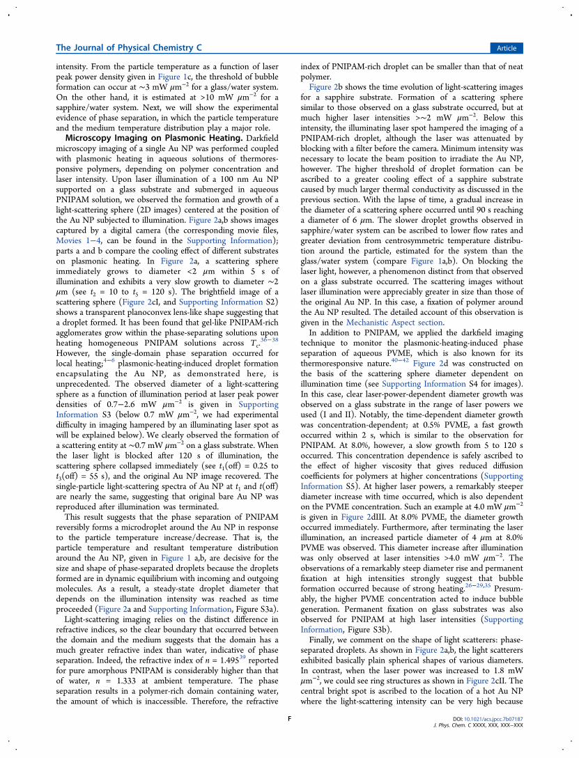
Shifts. Figure 3a shows the Rayleigh scattering spectral changesof a 100 nm diameter Au NP submerged in aqueous 1.0%PVME solution, before, during, and after illumination. In thisexperiment, Au NPs were supported on a glass substrate. Forthe scattering spectra, a redshift occurred only duringillumination; when the illumination stopped, the originalspectra returned. The spectral shift was dependent on thelaser intensity: the spectral shift was within experimental errorat a laser peak power density of 0.10 mW μm−2 (1.0 × 10⁴ Wcm−2), whereas appreciable redshifts of 13 nm at 0.30 mWμm−2 and 54 nm at 0.50 mW μm−2 were recorded. The LSPRscattering spectral peak shift was plotted as a function of laserpeak power density in Figure 3b, showing a progressive redshiftwith increase in laser intensity.The observation of an LSPR redshift of a single Au NP in
aqueous PVME is similar to a previous observation in aqueousPNIPAM solution.19 The origin of the spectral shift wasascribed to an LCST-induced aggregation of PNIPAM aroundthe NP through heating of the surrounding medium with heattransfer from the optically heated NP. PNIPAM molecules are
hydrophilic at temperatures below LCST (305 K), but arehydrophobic above LCST.1,3 The aggregated PNIPAM arounda Au NP was assumed to increase the refractive index of themedium sensed by the Au NP, resulting in an LSPR scatteringredshift.19 Aqueous PVME solution exhibits a thermorespon-sive nature with an LCST at around 308 K.6,39−41 The presentresults showed that the scattering spectral peak shift continuedto increase as the laser intensity increased (Figure 3b). At thesame time, the imaging study (Figure 2) revealed thatmicrodroplets formed surrounding the Au NP at intensitieshigher than those used in the scattering spectroscopic studies.These results suggest that the droplet size of phase-separatedpolymer aggregates increases as the particle temperatureincreases. Our findings revealed that PVME, as well asPNIPAM, underwent plasmonic-heating-induced phase separa-tion, forming a droplet. Next, we attempt to estimate the size ofphase-separated droplets below the diffraction-limited size fromthe scattering spectral changes. This may compliment theimaging study, which is only applicable to size estimation ofdroplets >1 μm.Figure 4a,b shows the laser intensity-dependent scattering
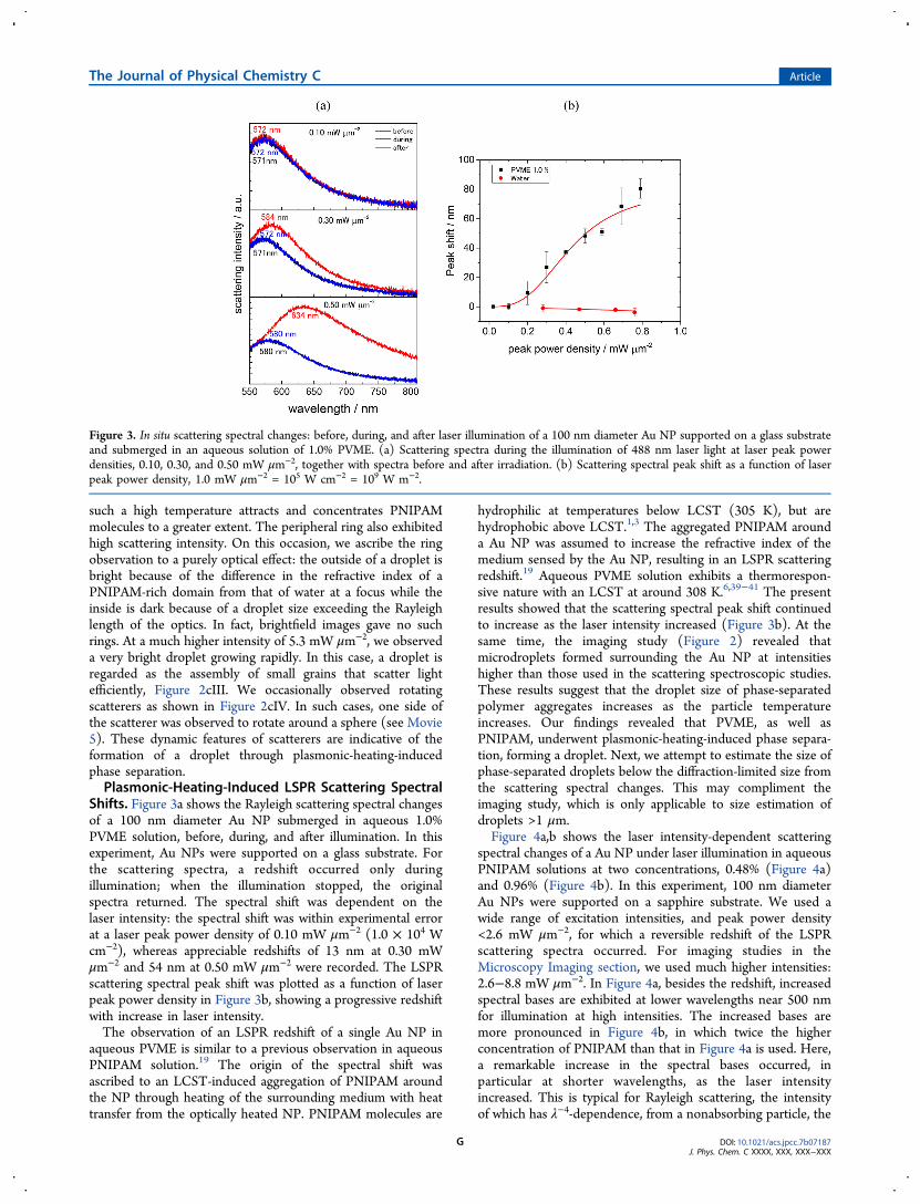
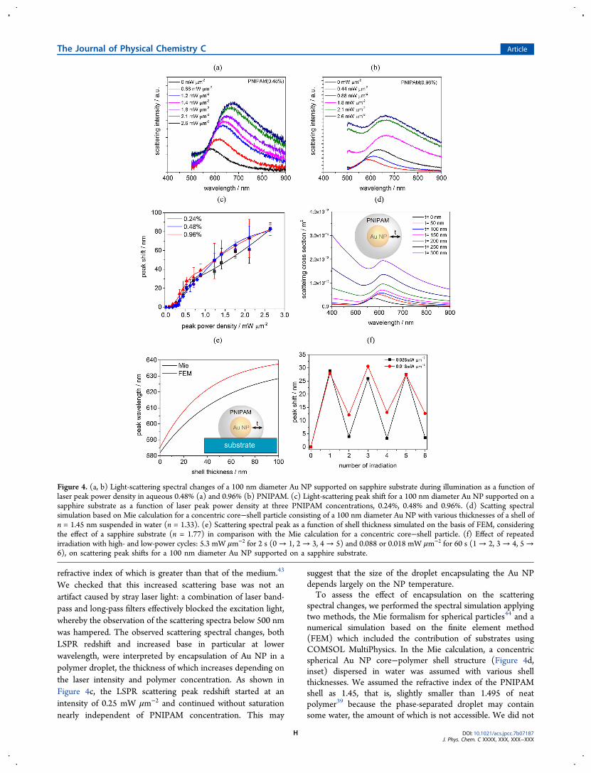
spectral changes of a Au NP under laser illumination in aqueousPNIPAM solutions at two concentrations, 0.48% (Figure 4a)and 0.96% (Figure 4b). In this experiment, 100 nm diameterAu NPs were supported on a sapphire substrate. We used awide range of excitation intensities, and peak power density<2.6 mW μm−2, for which a reversible redshift of the LSPRscattering spectra occurred. For imaging studies in theMicroscopy Imaging section, we used much higher intensities:2.6−8.8 mW μm−2. In Figure 4a, besides the redshift, increasedspectral bases are exhibited at lower wavelengths near 500 nmfor illumination at high intensities. The increased bases aremore pronounced in Figure 4b, in which twice the higherconcentration of PNIPAM than that in Figure 4a is used. Here,a remarkable increase in the spectral bases occurred, inparticular at shorter wavelengths, as the laser intensityincreased. This is typical for Rayleigh scattering, the intensityof which has λ−4-dependence, from a nonabsorbing particle, the
Figure 3. In situ scattering spectral changes: before, during, and after laser illumination of a 100 nm diameter Au NP supported on a glass substrateand submerged in an aqueous solution of 1.0% PVME. (a) Scattering spectra during the illumination of 488 nm laser light at laser peak powerdensities, 0.10, 0.30, and 0.50 mW μm−2, together with spectra before and after irradiation. (b) Scattering spectral peak shift as a function of laserpeak power density, 1.0 mW μm−2 = 105 W cm−2 = 109 W m−2.
The Journal of Physical Chemistry C Article
DOI: 10.1021/acs.jpcc.7b07187J. Phys. Chem. C XXXX, XXX, XXX−XXX
G
refractive index of which is greater than that of the medium.43
We checked that this increased scattering base was not anartifact caused by stray laser light: a combination of laser band-pass and long-pass filters effectively blocked the excitation light,whereby the observation of the scattering spectra below 500 nmwas hampered. The observed scattering spectral changes, bothLSPR redshift and increased base in particular at lowerwavelength, were interpreted by encapsulation of Au NP in apolymer droplet, the thickness of which increases depending onthe laser intensity and polymer concentration. As shown inFigure 4c, the LSPR scattering peak redshift started at anintensity of 0.25 mW μm−2 and continued without saturationnearly independent of PNIPAM concentration. This may
suggest that the size of the droplet encapsulating the Au NPdepends largely on the NP temperature.To assess the effect of encapsulation on the scattering
spectral changes, we performed the spectral simulation applyingtwo methods, the Mie formalism for spherical particles44 and anumerical simulation based on the finite element method(FEM) which included the contribution of substrates usingCOMSOL MultiPhysics. In the Mie calculation, a concentricspherical Au NP core−polymer shell structure (Figure 4d,inset) dispersed in water was assumed with various shellthicknesses. We assumed the refractive index of the PNIPAMshell as 1.45, that is, slightly smaller than 1.495 of neatpolymer39 because the phase-separated droplet may containsome water, the amount of which is not accessible. We did not
Figure 4. (a, b) Light-scattering spectral changes of a 100 nm diameter Au NP supported on sapphire substrate during illumination as a function oflaser peak power density in aqueous 0.48% (a) and 0.96% (b) PNIPAM. (c) Light-scattering peak shift for a 100 nm diameter Au NP supported on asapphire substrate as a function of laser peak power density at three PNIPAM concentrations, 0.24%, 0.48% and 0.96%. (d) Scatting spectralsimulation based on Mie calculation for a concentric core−shell particle consisting of a 100 nm diameter Au NP with various thicknesses of a shell ofn = 1.45 nm suspended in water (n = 1.33). (e) Scattering spectral peak as a function of shell thickness simulated on the basis of FEM, consideringthe effect of a sapphire substrate (n = 1.77) in comparison with the Mie calculation for a concentric core−shell particle. (f) Effect of repeatedirradiation with high- and low-power cycles: 5.3 mW μm−2 for 2 s (0→ 1, 2→ 3, 4→ 5) and 0.088 or 0.018 mW μm−2 for 60 s (1→ 2, 3→ 4, 5→6), on scattering peak shifts for a 100 nm diameter Au NP supported on a sapphire substrate.
The Journal of Physical Chemistry C Article
DOI: 10.1021/acs.jpcc.7b07187J. Phys. Chem. C XXXX, XXX, XXX−XXX
H
consider a smaller value of 1.40 because this value gave shell-thickness-dependent LSPR scattering peak shifts much smallerthan those observed in the experiments. As shown in Figure 4d,both LSPR scattering spectral redshift and the gradual increasein scattering intensities from the shell were reproduced.According to the Mie simulation, the PNIPAM droplet withdiameter 0.7 μm containing a 100 nm diameter Au NP wouldgive a scattering spectrum similar to the one measured at thelaser peak power density of 2.6 mW μm−2 (compare Figure4b,d).When supported on a sapphire substrate, the droplet
diameter was estimated from imaging as 1.5 μm for irradiationat 2.6 mW μm−2. Although we could not determine exactly thePNIPAM droplet size at intensities <2.6 mW μm−2 fromimaging because of the difficulty in determining the droplet sizedistinct from the laser spot, we assumed that the droplet sizewas smaller than the laser spot of ∼1 μm. In contrast, thescattering spectral estimation should be limited to dropletdiameters <1 μm because, for much bigger droplets with greatershell thicknesses, severely distorted scattering spectra can occurowing to the greater scattering contribution from a shell. Thus,the darkfield imaging can be more reliably used for estimationof diameters >1 μm.The FEM calculations were performed to include the
substrate effect on the scattering spectra. Figure 4e shows theLSPR spectral peak shift as a function of shell thickness (seeSupporting Information S6 for spectra). Inset is the pictorialrepresentation of a configuration for simulation of a Au NP/PNIPAM shell/sapphire exposed to water. For comparison, theresults of the Mie calculation are also given. Both the Miecalculation and FEM gave a greater LSPR scattering peakredshift as a function of shell thickness, up to 100 nm. Thenumerical simulation including a sapphire substrate, however,gave a slightly greater shift than the simple core−shell model.The numerical simulation gave the spectral shift closer to theexperimental ones and may describe the spectra moreaccurately. However, we found that the FEM computationneeds considerable computational time, in particular, for shellthicknesses >100 nm, far from practical use. A weak point ofthe simple core−shell model is to neglect the restrictedaccessible volume for the shell formation in the presence of asubstrate. The ratio of inaccessible volume to accessible volumedependent on the shell thickness is calculated on the basis ofsimple geometric consideration, as given in SupportingInformation S6. We concluded that the inaccessible volume,25% for t = 100 nm and 40% for t = 500 nm, affects thescattering spectral calculation by the simple core−shell modelto a lesser extent, and the spectral simulation in Figure 4d isacceptable.When we apply laser intensities >3.7 mW μm−2 on sapphire,
we observed a permanent redshift in the LSPR scatteringspectra. This was consistent with the darkfield imaging inFigure 2b, in which a particle larger than the original Au NPremained after termination of the illumination. The SEMimaging revealed that Au NPs were encapsulated (SupportingInformation S7). However, we found that further illuminationat a weak intensity can result in blueshift of the scatteringspectrum. Figure 4f shows three repeated cycles of intense (5.3mW μm−2, 2s) and weak (0.088 mW μm−2 or 0.018 mW μm−2,60 s) illuminations. We observed the photoswitching behaviorfor the LSPR peak position corresponding to encapsulation anddecapsulation. We refer to the mechanism of this behavior inthe Mechanistic Aspect section.
Mechanistic Aspect of Plasmonic-Heating-InducedPhase Separation. We found that thermoresponsive poly-mers were attracted to a Au NP under illumination with awavelength near the LSPR, forming a microdroplet. Here thecontribution of optical trapping to the droplet formation shouldbe minor because of moderate focusing at low intensities;instead, the particle heating is primarily responsible. Weobserved that more and more polymers were pouring intothe high-temperature area adjacent to the particle, forming adroplet with increasingly larger diameters as the particletemperature increases. A simple calculation suggested thatPNIPAM molecules dissolved in aqueous solution wereconcentrated into a droplet at least 50 times assuming 50%PNIPAM. The phenomenon observed is unusual becausePNIPAM that exhibits a positive thermophoretic mobility, DT,is normally repelled from the high-temperature area throughthermophoresis.15 Here the thermoresponsive nature ofpolymers is responsible for the present observation of amicrodomain formed and grown surrounding the hot NP. First,the temperature field around the Au NP acts to hold a dropletbecause the temperature gradient that is steeper is closer to theNP. For the attachment of a droplet to Au NP, hydrophobicinteraction must be responsible. Second, the LCST-inducedphase separation around the hot NP causes the concentrationdeficiency within the volume near the droplet containing theAu NP, which can promote the diffusion of polymers from theoutside area to the droplet. Presumably, the droplet size isdetermined by a balance between incoming and outgoing ratesof diffusing polymers at a given particle temperature.The phase separation of PNIPAM and PVME around a Au
NP was basically reversible for heating/cooling: the droplet wasobserved only during illumination. This is understandablebecause LCST behavior of the polymers is reversible.1,3 Twopoints, however, should be addressed on the accumulation ofpolymers after illumination. On glass substrates, permanentaccumulation occurred at high laser intensities. Vapor bubbleformation around a Au NP is responsible for this permanentaccumulation. In pure water, the generation of nano- andmicrobubbles on CW laser illumination of plasmonic NPsoccurred in superheated water when the particle temperaturewas raised to between 220 and 240 °C (493−513 K), muchhigher than the boiling point of water (100 °C or 373 K).16−18
Estimated particle temperatures were 500−600 K. Bubbles havethe ability to collect colloids and NPs through Marangoniconvection, resulting in fixation of them on substrates.26−29,35
The present observation of PNIPAM and PVME fixation isconsistent with bubble-induced accumulation.We also observed the fixation of PNIPAM on the sapphire
substrate after illumination was stopped, as given in Figure 2b,Figure 4f, and Supporting Information, Figure S7. For sapphire,because of its remarkable cooling effect, the particle temper-atures during illumination may not be so high as to inducevapor bubble generation. We assume that another mechanismcan operate. The phase separation of PNIPAM has beenthoroughly investigated in bulk aqueous solution, and its phasediagram has been established,3 as given in SupportingInformation S8. According to this diagram, as well as thedemixing line between one solution phase and two liquidphases, there is a glass transition line in the high-concentrationregion.45 This line steeply decreases with decreasing PNIPAMconcentration, in particular, at temperatures below TBT, thevitrification temperature (35 °C at 80% PNIPAM). Thissuggests that if a PNIPAM-rich phase experiences a quick
The Journal of Physical Chemistry C Article
DOI: 10.1021/acs.jpcc.7b07187J. Phys. Chem. C XXXX, XXX, XXX−XXX
I
temperature decrease it may undergo a glass transition to asolid-like or gel-like state. We assume that such a glasstransition on sapphire substrate can result in the accumulationof the polymers when the illumination is stopped. Interestingly,slight heating again dissolved the polymer deposit (Figure 4f).The kinetics of phase separation for the thermoresponsive
polymers, in particular, at the nanoscale is of extreme interest.For bulk solutions, temperature-jump transient spectroscopyand temperature-jump transient grating methods have beenapplied to reveal kinetics of phase transition/phase separationfor aqueous polymers including PNIPAM and PVME.46−48 Itwas found that demixing occurs at the time scale of 10−100 mswith the remixing time of a few seconds. The time resolution ofour present measurement is approximately 1 s. However, weobserved the time-dependent increase/decrease in dropletdiameters for the 8.0% PVME solution that has relatively highviscosity. We intend to improve the time resolution in futurestudies.Our present finding is that the plasmonic heating of a single
Au NP can trap thermoresponsive polymers around the NP,forming a phase-separated droplet. The ability of plasmonicheating to induce migration and accumulation is not limited tothermoresponsive polymers. Molecules like poly(ethyleneglycol) and sodium dodecyl sulfate as well as silica colloidswere found to accumulate surrounding a hot Au NP after laserillumination.49 While photothermal bubbles can collect and fixcolloids and NPs by promoting thermal and Marangoniconvection,29 plasmonic heating manipulates colloids andmacromolecules through a similar mechanism at low laserintensities. Plasmonic-heating-induced manipulation is atechnically benign procedure without the aid of bubbles, butit has the potential to fabricate small structures if technicalsophistication is attained. In this context, the concept ofconvection-enhanced and temperature-assisted optical trappinghas been postulated.50,51 The practical merits and demerits ofplasmonic-heating-induced manipulation over local mediumheating and optical trapping are summarized in Table 1.
■ CONCLUSIONWe used plasmonic heating to induce the phase separation ofthermoresponsive polymers and demonstrated a 2D trapping ofthe polymers around a single Au NP supported on a substrateunder illumination from a moderately focused laser beam. 3Dextension is possible, but the present configuration isconvenient for the confinement of the polymers on substrates.We used a 488 nm laser light to excite both LSPR andintraband transitions, resulting in particle heating. Thegenerated temperature field is mainly responsible for trapping
thermoresponsive polymers. In optical trapping assisted by agradient force, however, the particle heating that facilitatesBrownian motion needs to be bypassed. Most importantly, ourlaser power is very low, much less than that used in the directmedium heating or optical trapping experiment. We succeededin applying darkfield microscopy imaging that has previouslynot been used for observing phase separation. As a result, weobtained high-contrast images. The significant findings of thepresent study are the following. (1) The plasmonic heating of asingle Au NP with a laser power of ∼105 W cm−2 enables asingle-domain phase separation around the NP, which isconsiderably energy saving compared with medium-heating-induced microdroplet fabrication using an infrared laser with apower of ∼109 W cm−2. (2) By illuminating with variousintensities of a laser beam on Au nanostructured substrates, thelocation and size of the droplet are adjustable so that one canseparate polymer droplets from the solution at any location. (3)Using a high-thermal-conductivity sapphire substrate, perma-nent fixation of the polymers can be attained without the aid ofvapor bubbles for further use.
■ ASSOCIATED CONTENT*S Supporting InformationThe Supporting Information is available free of charge on theACS Publications website at DOI: 10.1021/acs.jpcc.7b07187.
Growth and collapse of PNIPAM droplets, with glasssubstrate, laser peak power density 0.88 mW μm−2,0.96% PNIPAM aq solution (AVI)Growth and collapse of PNIPAM droplets, with glasssubstrate, laser peak power density 1.8 mW μm−2, 0.96%PNIPAM aq solution (AVI)Growth and collapse of PNIPAM droplets, with glasssubstrate, laser peak power density 5.3 mW μm−2, 0.96%PNIPAM aq solution (AVI)Growth and collapse of PNIPAM droplets, with sapphiresubstrate, laser peak power density 5.3 mW μm−2, 0.96%PNIPAM aq solution (AVI)Growth and collapse of PNIPAM droplets, with glasssubstrate, laser peak power density 3.5 mW μm−2, 0.96%PNIPAM aq solution (AVI)Particle image and the corresponding histogram, bright-field images of PNIPAM droplets, diameter of a light-scattering sphere as a function of time for PNIPAM,darkfield images of PVME droplets, concentration-dependent viscosity of PVME solution, light-scatteringspectral simulation, SEM images of Au NPs, and phasediagram of thermoresponsive polymer solution (PDF)
■ AUTHOR INFORMATIONCorresponding Author*Phone: +81-88-656-7389. E-mail: [email protected] Uwada: 0000-0003-4272-7964Shuichi Hashimoto: 0000-0002-8020-5537Author Contributions§I.A. and J.C. contributed equally to the optical measurementsand analyses. S.H. designed the experiment. T.U. performed anumerical simulation. S.H. prepared the manuscript withcontributions from all authors. All authors have given approvalto the final version of the manuscript.
Table 1. Plasmonic-Heating-Induced versus Local Medium-Heating-Induced Phase Separation
paradigmplasmonic heating local medium
heatingoptical trapping
trapping mode 2D 3D 3Dlaser wavelength 488 nm 1064 nm 1064 nmmechanism local temperature
fieldmedium heating optical gradient
forcepeak powerdensity
104−105 W cm−2 108−109W cm−2
108−109 W cm−2
microscopy darkfield brightfield brightfieldtime evolution 1−100 s 10−1000 s 10−100 smedium H2O/substrate H2O D2O
The Journal of Physical Chemistry C Article
DOI: 10.1021/acs.jpcc.7b07187J. Phys. Chem. C XXXX, XXX, XXX−XXX
J
NotesThe authors declare no competing financial interest.
■ ACKNOWLEDGMENTSFinancial support from JSPS KAKENHI (No. 17K05005) andIketani Science & Technology Foundation (No. 0291052-A) isgratefully acknowledged.
■ REFERENCES(1) Halperin, A.; Kroger, M.; Winnik, F. M. Poly(N-isopropylacry-lamide) Phase Diagrams: Fifty Years of Research. Angew. Chem., Int.Ed. 2015, 54, 15342−15367.(2) Wei, M.; Gao, Y.; Li, X.; Serpe, M. J. Stimuli-responsive Polymersand Their Applications. Polym. Chem. 2017, 8, 127−143.(3) Aseyev, V. O.; Tenhu, H.; Winnik, F. M. TemperatureDependence of the Colloidal Stability of Neutral AmphiphilicPolymers in Water. Adv. Polym. Sci. 2006, 196, 1−85.(4) Ishikawa, M.; Misawa, H.; Kitamura, N.; Fujisawa, R.; Masuhara,H. Infrared Laser-Induced Photo-Thermal Phase Transition of anAqueous Poly(N-isopropylacrylamide) Solution in the MicrometerDimension. Bull. Chem. Soc. Jpn. 1996, 69, 59−66.(5) Hofkens, Y.; Hotta, J.; Sasaki, K.; Masuhara, H.; Iwai, K.Molecular Assembling by the Radiation Pressure of a Focused LaserBeam: Poly(N-isopropylacrylamide) in Aqueous Solution. Langmuir1997, 13, 414−419.(6) Tsuboi, Y.; Nishino, M.; Matsuo, Y.; Ijiro, K.; Kitamura, N. PhaseSeparation of Aqueous Poly(vinyl methyl ether) Solutions induced bythe Photon Pressure of a Focused Near-Infrared Laser Beam. Bull.Chem. Soc. Jpn. 2007, 80, 1926−1931.(7) Pan, W.; Kolomeisky, A. B.; Vekilov, P. G. Nucleation of OrderedSolid Phases of Proteins via a Disordered High-density State:Phenomenological Approach. J. Chem. Phys. 2005, 122, 174905.(8) Katsumoto, Y.; Tsuchiizu, A.; Qiu, X.; Winnik, F. Dissecting theMechanism of the Heat-Induced Phase Separation and Crystallizationof Poly(2-isopropyl-2-oxazoline) in Water Through VibrationalSpectroscopy and Molecular Orbital Calculations. Macromolecules2012, 45, 3531−3541.(9) Sugiyama, T.; Masuhara, H. Laser-induced Crystallization andCrystal Growth. Chem. - Asian J. 2011, 6, 2878−2889.(10) Novotny, L.; van Hulst, N. Antennas for Light. Nat. Photonics2011, 5, 83−90.(11) Le Ru, E. C.; Etchegoin, P. G. Principles of Surface-EnhancedRaman Spectroscopy; Elsevier: Amsterdam, 2009.(12) Metal-Enhanced Fluorescence; Geddes, C. D., Ed.; Wiley:Weinheim, 2010.(13) Baffou, G.; Quidant, R. Thermo-plasmonics: Using MetallicNanostructures as Nanosources of Heat. Laser Photonics Rev. 2013, 7,171−187.(14) Govorov, A. O.; Richardson, H. H. Generating Heat with MetalNanoparticles. Nano Today 2007, 2, 30−38.(15) Orlishausen, M.; Kohler, W. Forced Phase Separation by Laser-Heated Gold Nanoparticles in Thermoresponsive Aqueous PNIPAMPolymer Solutions. J. Phys. Chem. B 2015, 119, 8217−8222.(16) Fang, Z.; Zhen, Y.-R.; Neumann, O.; Polman, A.; Garcia deAbajo, F.; Nordlander, P.; Halas, N. J. Evolution of Light InducedVapor Generation at Liquid-immersed Metallic Nanoparticle. NanoLett. 2013, 13, 1736−1742.(17) Baffou, G.; Polleux, J.; Rigneault, H.; Monneret, S. Super-Heating and Micro-Bubble Generation around Plasmonic Nano-particles Under cw Illumination. J. Phys. Chem. C 2014, 118, 4890−4898.(18) Hou, L.; Yorulmaz, M.; Verhart, N. R.; Orrit, M. ExplosiveFormation and Dynamics of Vapor Nanobubbles Around aContinuously Heated Gold Nanosphere. New J. Phys. 2015, 17,013050.(19) Aibara, I.; Mukai, S.; Hashimoto, S. Plasmonic-Heating-InducedNanoscale Phase Separation of Free Poly(N-isopropylacrylamide)Molecules. J. Phys. Chem. C 2016, 120, 17745−17752.
(20) Honda, M.; Saito, Y.; Smith, N. I.; Fujita, K.; Kawata, S.Nanoscale Heating of Laser Irradiated Single Gold Nanoparticles inLiquid. Opt. Express 2011, 19, 12375−12383.(21) Karg, M.; Jaber, S.; Hellweg, T.; Mulvaney, P. Surface PlasmonSpectroscopy of Gold-poly-N-isopropylacrylamide Core-shell Particles.Langmuir 2011, 27, 820−827.(22) Kusolkamabot, K.; Sae-ung, P.; Niamnont, N.; Wongravee, K.;Sukwattanasinitt, M.; Hoven, V. P. Poly(N-isopropylacrylamide)-Stabilized Gold Nanoparticles in Combination with TricationicBranched Phenylene-Ethynylene Fluorophore for Protein Identifica-tion. Langmuir 2013, 29, 12317−12327.(23) Garces-Chavez, V.; Quidant, R.; Reece, P. J.; Badenes, G.;Torner, L.; Dholakia, K. Extended Organization of ColloidalMicroparticles by Surface Plasmon Polariton Excitation. Phys. Rev. B:Condens. Matter Mater. Phys. 2006, 73, 085417.(24) Lin, L.; Peng, X.; Wang, M.; Scarabelli, L.; Mao, Z.; Liz-Marzan,L. M.; Becker, M. F.; Zheng, Y. Light-directed Reversible Assembly ofPlasmonic Nanoparticles Using Plasmon-enhanced Thermophoresis.ACS Nano 2016, 10, 9659−9668.(25) Lin, L.; Peng, X.; Wei, X.; Mao, Z.; Xie, C.; Zheng, Y.Thermophoretic Tweezers for Low-power and Versatile Manipulationof Biological Cells. ACS Nano 2017, 11, 3147−3154.(26) Zheng, Y.; Liu, H.; Wang, Y.; Zhu, C.; Wang, S.; Cao, J.; Zhu, S.Accumulating Microparticles and Direct-writing Micropatterns using aContinuous-wave Laser-induced Vapor Bubble. Lab Chip 2011, 11,3816−3820.(27) Fujii, S.; Kanaizuka, K.; Toyabe, S.; Kobayashi, K.; Muneyuki,E.; Haga, M. Fabrication and Placement of a Ring Structure ofNanoparticles by a Laser-Induced Micronanobubble on a GoldSurface. Langmuir 2011, 27, 8605−8610.(28) Lin, L.; Peng, X.; Mao, Z.; Li, W.; Yogeesh, M. N.; Rajeeva, B.B.; Perillo, E. P.; Dunn, A. K.; Akinwande, D.; Zheng, Y. Bubble-penLithography. Nano Lett. 2016, 16, 701−708.(29) Xie, Y.; Zhao, C. An Optothermally Generated Surface Bubbleand Its Applications. Nanoscale 2017, 9, 6622−6631.(30) Setoura, K.; Okada, Y.; Werner, D.; Hashimoto, S. Observationof Nanoscale Cooling Effects by Substrates and the SurroundingMedia for Single Gold Nanoparticles Under CW-Laser Illumination.ACS Nano 2013, 7, 7874−7885.(31) Carlson, M. T.; Khan, A.; Richardson, H. H. Local TemperatureDetermination of Optically Excited Nanoparticles and Nanodots.Nano Lett. 2011, 11, 1061−1069.(32) Brongersma, M. L.; Halas, N. J.; Nordlander, P. Plasmon-induced Hot Carrier Science and Technology. Nat. Nanotechnol. 2015,10, 25−34.(33) Keblinski, P.; Cahill, D. G.; Bodapati, A.; Sullivan, C. R.; Taton,T. A. Limits of Localized Heating by Electromagnetically ExcitedNanoparticles. J. Appl. Phys. 2006, 100, 054305.(34) Marchand, A.; Weijs, J. H.; Snoeijer, J. H.; Andreotti, B. Why isSurface Tension a Force Parallel to the Interface? Am. J. Phys. 2011,79, 999−1008.(35) Setoura, K.; Ito, S.; Miyasaka, H. Stationary Bubble Formationand Marangoni Convection Induced by CW laser Heating of a SingleGold Nanoparticle. Nanoscale 2017, 9, 719−730.(36) Balu, C.; Delsanti, M.; Guenoun, P.; et al. Colloidal PhaseSeparation of Concentrated PNIPAm Solutions. Langmuir 2007, 23,2404−2407.(37) Xue, N.; Qiu, X.-P.; Aseyev, V.; Winnik, F. M. NonequilibriumLiquid−Liquid Phase Separation of Poly(N-isopropylacrylamide) inWater/Methanol Mixtures. Macromolecules 2017, 50, 4446−4453.(38) Meier-Koll, A.; Pipich, V.; Busch, P.; Papadakis, C. M.; Muller-Buschbaum, P. Phase Separation in Semidilute Aqueous Poly(N-isopropylacrylamide) Solutions. Langmuir 2012, 28, 8791−8798.(39) Philipp, M.; Aleksandrova, R.; Muller, U.; Ostermeyer, M.;Sanctuary, R.; Muller-Buschbaum, P.; Kruger, J. K. Molecular versusMacroscopic Perspective on the Demixing Ttransition of AqueousPNIPAM Solutions by Studying the Dual Character of the RefractiveIindex. Soft Matter 2014, 10, 7297−7305.
The Journal of Physical Chemistry C Article
DOI: 10.1021/acs.jpcc.7b07187J. Phys. Chem. C XXXX, XXX, XXX−XXX
K
(40) Horne, R. A.; Almeida, J. P.; Day, A. F.; Yu, N. T.Macromolecule Hydration and the Effect of Solutes on the CloudPoint of Aqueous Solutions of Polyvinyl Methyl Ether: A PossibleDenaturation and Temperature Control in Homeothermic Animals. J.Colloid Interface Sci. 1971, 35, 77−84.(41) Meeussen, F.; Bauwens, Y.; Moerkerke, R.; Nies, E.; Berghmans,H. Molecular Complexes Formation in the System Poly(vinyl methylether)/water. Polymer 2000, 41, 3737−3743.(42) Spevacek, J.; Hanykova, L.; Labuta, J. Behavior of Water DuringTemperature-Induced Phase Separation in Poly(vinyl methyl ether)Aqueous Solutions. NMR and Optical Microscopy Study. Macro-molecules 2011, 44, 2149−2153.(43) Zhang, W. Z.; Chen, X. D.; Luo, W.; Yang, J.; Zhang, M. Q.;Zhu, F. M. Study of Phase Separation of Poly(vinyl methyl ether)Aqueous Solutions with Rayleigh Scattering Technique. Macro-molecules 2009, 42, 1720−1725.(44) Bohren, C. F.; Huffman, D. R. Absorption and Scattering of Lightby Small Particles; Wiley: New York, 1983.(45) Van Durme, K.; Van Assche, G.; Van Mele, B. Kinetics ofDemixing and Remixing in Poly(N-isopropylacrylamide)/waterStudied by Modulated Temperature DSC. Macromolecules 2004, 37,9596−9605.(46) Tsuboi, Y.; Yoshida, Y.; Okada, K.; Kitamura, N. PhaseSeparation Dynamics of Aqueous Solutions of ThermoresponsivePolymers Studied by a Laser T-Jump Technique. J. Phys. Chem. B2008, 112, 2562−2565.(47) Tsuboi, Y.; Tada, T.; Shoji, T.; Kitamura, N. Phase-SeparationDynamics of Aqueous Poly(N -isopropylacrylamide) Solutions:Characteristic Behavior of the Molecular Weight and ConcentrationDependences. Macromol. Chem. Phys. 2012, 213, 1879−1884.(48) Inoue, H.; Kuwahara, S.; Katayama, K. The Whole Process ofPhase Transition and Relaxation of Poly(N-isopropylacrylamide)Aqueous Solution. Phys. Chem. Chem. Phys. 2013, 15, 3814−3819.(49) Enders, M.; Mukai, S.; Uwada, T.; Hashimoto, S. PlasmonicNanofabrication Through Optical Heating. J. Phys. Chem. C 2016, 120,6723−6732.(50) Bartkiewicz, S.; Miniewicz, A. Whirl-enhanced ContinuousWave Laser Trapping of Particles. Phys. Chem. Chem. Phys. 2015, 17,1077−1083.(51) Louchev, O. A.; Juodkazis, S.; Murazawa, N.; Wada, S.; Misawa,H. Coupled Laser Molecular Trapping, Cluster Assembly, andDeposition fed by Laser-induced Marangoni Convection. Opt. Express2008, 16, 5673−5680.
The Journal of Physical Chemistry C Article
DOI: 10.1021/acs.jpcc.7b07187J. Phys. Chem. C XXXX, XXX, XXX−XXX
L


![3D Printing of Thermoresponsive Polyisocyanide (PIC ...noviosense.com/wp-content/uploads/2018/11/polymers... · Polymers 2018, 10, 555 2 of 11 polymers [6,7], metals and alloys [8,9],](https://static.fdocuments.in/doc/165x107/5f035ab57e708231d408ccd9/3d-printing-of-thermoresponsive-polyisocyanide-pic-polymers-2018-10-555-2.jpg)


![Homaeigohar, Shahin; Elbahri, Mady Switchable Plasmonic ... · photochromic molecules, inorganic materials, and polymers[35]. When exposed to a stimulus e.g. light, temperature, or](https://static.fdocuments.in/doc/165x107/5f26889535dec77d442d7f81/homaeigohar-shahin-elbahri-mady-switchable-plasmonic-photochromic-molecules.jpg)
