
Living Images: Treasure of the Avars: If it Glitters, It’s ... · The Magazine from Carl Zeiss...
60
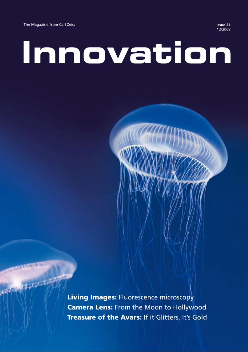
The Magazine from Carl Zeiss Issue 21 12/ 2008 Living Images: Fluorescence microscopy Camera Lens: From the Moon to Hollywood Treasure of the Avars: If it Glitters, It’s Gold
Transcript of Living Images: Treasure of the Avars: If it Glitters, It’s ... · The Magazine from Carl Zeiss...
The Magazine from Carl Zeiss Issue 2112/ 2008
Living Images: Fluorescence microscopy
Camera Lens: From the Moon to Hollywood
Treasure of the Avars: If it Glitters, It’s Gold

The green fluorescent protein (GFP) comes from the Aequorea victoria jellyfish. It shines green when it is excited with ultraviolet or blue light. Asamu Shimomura described it for the first time in 1962. In 1994, Martin Chalfie succeeded in bringing GFP to expression outside the Aequorea victoria jellyfish, enabling its breakthrough as a genetic marker. Roger Tsien, who had longfocused his research on the field of imaging cell biology, created fluorescent dyes to make cellular calcium visible. With his work, he contributed to the general understanding of the protein.

�Innovation 21, 12 / 2008
Dear Readers,
When the winners of the Nobel Prize for Chemistry were announced at
the beginning of October, the news spread through the company like
wildfire. This renowned award was bestowed upon three scientists this
year who partly work with instruments from Carl Zeiss. Above all, this
year’s Nobel Prize for Chemistry honors the enhancement of a procedure
developed by Carl Zeiss exactly 100 years ago: fluorescence microscopy.
Osamu Shimomura, Martin Chalfie and Roger Y. Tsien were honored for
the discovery and development of the green fluorescent protein (GFP).
This protein has become an important tool in life sciences. It allows
scientists to observe processes in cells and, for example, track the spread
of cancer cells.
Understanding how diseases develop, even possibly preventing their
outbreak and definitely accelerating their cure – these are the goals of
the life sciences. Physician Harald zur Hausen, a Carl Zeiss customer too,
also received the Nobel Prize this year. We at Carl Zeiss have a long tradi-
tion and passion for providing scientists such as zur Hausen with the
right instruments. In doing so, we also aid the transition from science to
the application. Think about minimally invasive or micro-incision surgery
that increases the likelihood of faster patient healing through minimal
patient trauma.
Whether you enjoy the aesthetic of the images or reading exciting
articles, we hope this issue of Innovation is to your liking: enjoy reading!
Best wishes
Dr. Michael KaschkeMember of the Carl Zeiss AG Executive Board
Editorial
� Innovation 21, 12 / 2008
Table of Contents
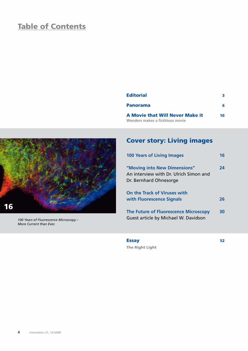
100 Years of Living Images 16
“Moving into New Dimensions” 24An interview with Dr. Ulrich Simon and Dr. Bernhard Ohnesorge
On the Track of Viruses with with Fluorescence Signals 26
The Future of Fluorescence Microscopy 30Guest article by Michael W. Davidson
Cover story: Living images
Editorial 3
Panorama 6
A Movie that Will Never Make it 10Wenders makes a fictitious movie
16
100 Years of Fluorescence Microscopy – More Current than Ever.
Essay 52
The Right Light

�Innovation 21, 12 / 2008
Feature: Unearthed Bone Find Veiled in Mystery 32
If it Glitters, It’s Gold 3632
Feature: From Above
A Bird’s Eye View of the Jabel Akhdar 44
Data Communication in the Fast Lane 48
Sylt from the Air – Laser beam transport mea-sured values and images across long distances
48
Kyffhäuser – Thuringian mountain range with many secrets
Look at Laureates
Preview 59
Legal information 59
Report The World by Candlelight 40
Small Incision with a Big Impact 42

� Innovation 21, 12 / 2008
Panorama
THE attraction at photokina: the walk-through lens.
Precisely in FocusZEISS lenses now available for EOS cameras
and Ulrich Wolf from MAIWOLF Photography had the opportunity to test the first ZE lenses. An enthused Wolf stated that “the lens delivered natural clarity and fantastic brilliance. The images really light up, regard-less of the focal length.” Maierhofer added that she “was particularly fascinated at how the lenses provide a crisp image of even the smallest details such as hair at the edge of the image.” Currently, ZE lenses with two different focal lengths are available. Additional focal lengths will be added to the line within the next few months.
Canon enthusiasts have something to look forward to: they will soon be able to use ZEISS lenses with manual focus on their EOS cameras. The new ZE lenses transmit all information via the EF bayonet connection, i.e. via the electronic contacts, and support auto exposure, shutter priority and aperture priority. With digital SLRs, the lens data, all exposure data and even the exposure control of the flash can be accessed. Even if the focus is set manually, focusing is automatic. Even professionals see such a lens as a welcome addition to their equip-ment. Munich-based photographers Eva Maierhofer
�Innovation 21, 12 / 2008
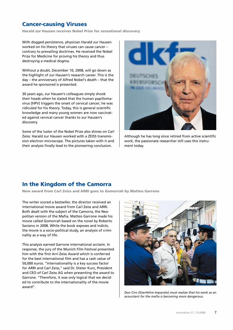
With dogged persistence, physician Harald zur Hausen worked on his theory that viruses can cause cancer – contrary to prevailing doctrines. He received the Nobel Prize for Medicine for proving his theory and thus destroying a medical dogma.
Without a doubt, December 10, 2008, will go down as the highlight of zur Hausen’s research career. This is the day – the anniversary of Alfred Nobel's death – that the award he sponsored is presented.
�0 years ago, zur Hausen’s colleagues simply shook their heads when he stated that the human papilloma-virus (HPV) triggers the onset of cervical cancer; he was ridiculed for his theory. Today, this is general scientific knowledge and many young women are now vaccinat-ed against cervical cancer thanks to zur Hausen’s discovery.
Some of the luster of the Nobel Prize also shines on Carl Zeiss: Harald zur Hausen worked with a ZEISS transmis-sion electron microscope. The pictures taken with it and their analysis finally lead to the pioneering conclusion.
Cancer-causing VirusesHarald zur Hausen receives Nobel Prize for sensational discovery
Although he has long since retired from active scientific work, the passionate researcher still uses this instru-ment today.
Don Ciro (Gianfelice Imparato) must realize that his work as an acountant for the mafia is becoming more dangerous.
The writer scored a bestseller, the director received an international movie award from Carl Zeiss and ARRI. Both dealt with the subject of the Camorra, the Nea-politan version of the Mafia. Matteo Garrone made his movie called Gomorrah based on the novel by Roberto Saviano in 2008. While the book exposes and indicts, the movie is a socio-political study, an analysis of crimi-nality as a way of life.
This analysis earned Garrone international acclaim. In response, the jury of the Munich Film Festival presented him with the first Arri-Zeiss Award which is conferred for the best international film and has a cash value of �0,000 euros. “Internationality is a key success factor for ARRI and Carl Zeiss,” said Dr. Dieter Kurz, President and CEO of Carl Zeiss AG when presenting the award to Garrone. “Therefore, it was only logical that we decid-ed to contribute to the internationality of the movie award”.
In the Kingdom of the Camorra New award from Carl Zeiss and ARRI goes to Gomorrah by Matteo Garrone
8 Innovation 21, 12 / 2008
Vaccum
SiNx
Ag
100 nm
The bridge-type measuring machine simplifies assembly and supports low-energy production.
Airplane construction is precision work. Lockheed Martin is building the new F-�� Lightning II supersonic fighter for the US Air Force. Production is being moni-tored by the largest coordinate measuring machine ever built by Carl Zeiss: an MMZ B Plus gantry.
The MMZ B Plus has a measuring range of � x 1� x 2.� meters. With a length of 1� meters, it can accommo-date the wings of the jets and measure their skin. It also measures aerodynamic tools, wind tunnel models and 1:1 modules.
The machine simplifies assembly and contributes to lean manufacturing for production with reduced outlay and consumption of resources. During machine hando-ver, Larry Pike, Vice President of Quality at Lockheed Martin said: “This machine permits the transition from the inspection of single parts to process validation.” Bob Fiorentini, Vice President, F-�� Global Production, added: “This machine is the first installation of its kind and magnitude in our facility. It marks the beginning of a new era in optimized parts inspection.”
Room for Half an Airplane Largest 3D measuring machine measures entire wings of fighter jets
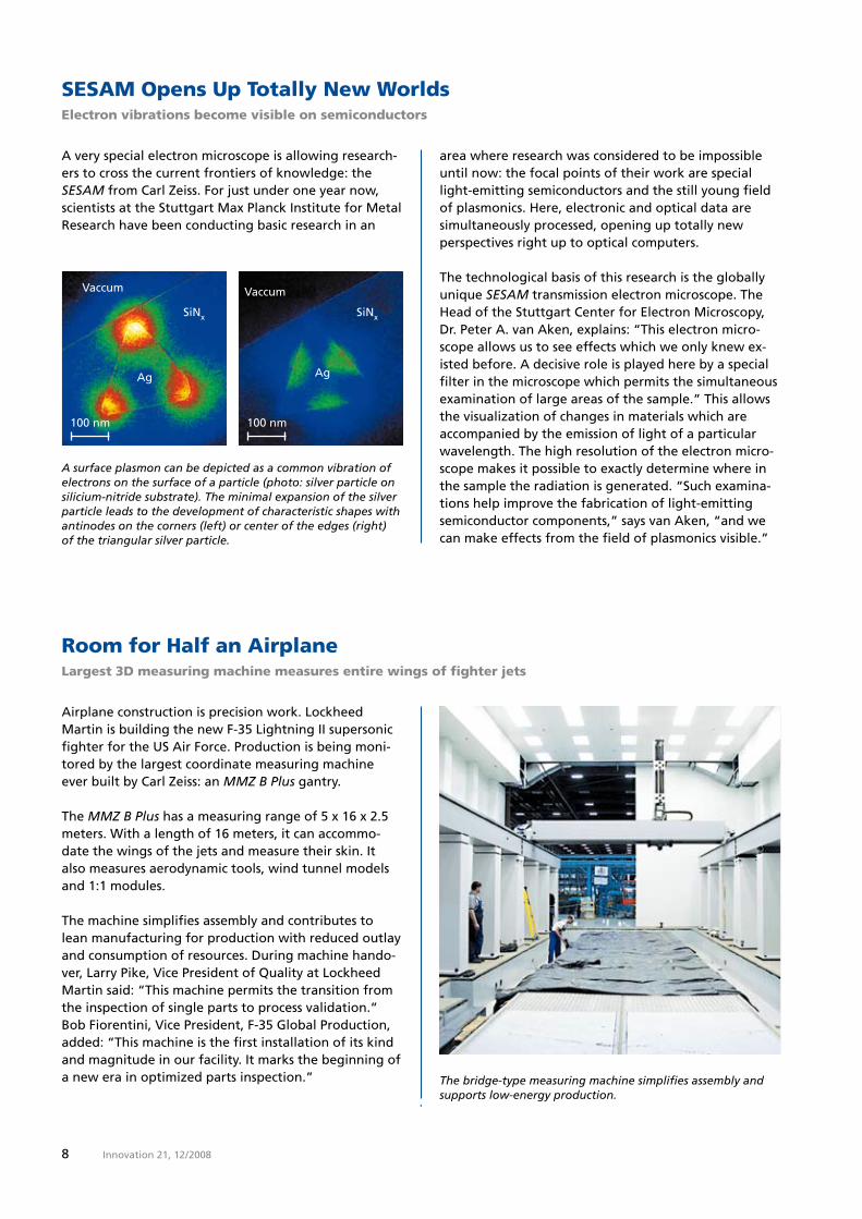
A surface plasmon can be depicted as a common vibration of electrons on the surface of a particle (photo: silver particle on silicium-nitride substrate). The minimal expansion of the silver particle leads to the development of characteristic shapes with antinodes on the corners (left) or center of the edges (right) of the triangular silver particle.
area where research was considered to be impossible until now: the focal points of their work are special light-emitting semiconductors and the still young field of plasmonics. Here, electronic and optical data are simultaneously processed, opening up totally new perspectives right up to optical computers.
The technological basis of this research is the globally unique SESAM transmission electron microscope. The Head of the Stuttgart Center for Electron Microscopy, Dr. Peter A. van Aken, explains: “This electron micro-scope allows us to see effects which we only knew ex-isted before. A decisive role is played here by a special filter in the microscope which permits the simultaneous examination of large areas of the sample.” This allows the visualization of changes in materials which are accompanied by the emission of light of a particular wavelength. The high resolution of the electron micro-scope makes it possible to exactly determine where in the sample the radiation is generated. “Such examina-tions help improve the fabrication of light-emitting semiconductor components,” says van Aken, “and we can make effects from the field of plasmonics visible.”
A very special electron microscope is allowing research-ers to cross the current frontiers of knowledge: the SESAM from Carl Zeiss. For just under one year now, scientists at the Stuttgart Max Planck Institute for Metal Research have been conducting basic research in an
SESAM Opens Up Totally New WorldsElectron vibrations become visible on semiconductors
Vaccum
SiNx
Ag
100 nm

�Innovation 21, 12 / 2008
A new lens makes it possible: cataract patients often no longer need glasses after surgery. Cataract surgery is one of the most frequently performed outpatient sur-geries in the world and is practically a routine proce-dure in ophthalmic surgery. The patient receives an arti-ficial intraocular lens that replaces the natural lens which has become cloudy. One challenge is the size of the incision through which the artificial lens is implant-ed. Large incisions during the operation can lead to astigmatism, a curvature of the cornea.
Carl Zeiss Meditec has presented a worldwide innova-tion with the AT.LISA® and AT.LISA.toric intraocular lenses. These lenses require only one small, 1.�-millime-ter incision and are therefore suitable for the first time for real micro-incision surgery – which promises signifi-cantly better surgical results. In addition, the new intraocular lenses feature unique, multifocal high-per-formance optics which enable excellent vision both in the near and far zones, as well as in the area in between.
Small Incision, Large StepNew intraocular lenses make cataract operations more successful
It must have been a microscope from Carl Zeiss. Robert Koch, the great doctor and bacteriologist, proved in 18�� that infections are caused by micro-organ-isms. He demonstrated this using an-thrax, whose pathogen he made visible under the microscope. Koch lived in what was is noe Wolsztyn, Poland, and most likely conducted his research there with pharmacist Josef Knechtel in his lab using ZEISS instruments. Based on the delivery books from Carl Zeiss, three microscopes were delivered, and all three went to Knechtel.
Karlsruhe-based scientist Timo Mappes was able to acquire one of them for his microscope collection. It is No. ����, VIIa stand, manufactured in 18�� and can be seen in Mappes' online museum of optical instruments. Even if other sources report that Koch had his own lab in Wollstein and ordered his own instruments from Carl Zeiss, he nonetheless discovered the anthrax pathogen with an instrument from Carl Zeiss. As he wrote to Jena: “I admire and am very grateful for the Zeiss opti-cal workshop; after all, I owe a great deal to your out-standing microscopes for a large portion of the success I have achieved in the name of science.”
The Illusion of Night New projector for planetariums which does not brighten the background
The darker the night, the brighter the stars. This is no different in planetariums than outdoors. A planetarium projector from Carl Zeiss can make stars shine with such radiance that they almost rival their natural coun-terparts in the night sky. However, this illusion soon vanishes in video projections which zoom in on cosmic nebulae and reveal the universe in �D. The dome images become pale because they lack a black background.
With powerdome®VELVET, Carl Zeiss has developed a video projection system for domes which does not make the background appear gray. VELVET “projects” the darkest black imaginable and is unparalleled in this achievement. VELVET was an absolute sensation at the conference of the International Planetarium Society in Chicago at the beginning of July. Planetarium directors from all over the world were extremely impressed by the brilliance of the images. The blackness of the back-
ground highlights white texts as if they written in fluo-rescent letters, and objects seem to float freely in the air. The technical challenge consisted in markedly in-creasing the difference between the highest and lowest brightness levels compared to traditional projectors. While the latter feature a maximum contrast ratio of 2�,000:1, VELVET surpasses this one 100 times over: 2,�00,000:1.
Digital all-dome video projection is increasingly supple-menting the opto-mechanical display of the starlit sky in planetariums. For the very first time, powerdome
VELVET is meeting a long-cherished wish: video projec-tion is able to superimpose digital images on the opti-cal night sky without impairing its brilliance. Gas nebu-lae and galaxies look as if they were immersed in the velvety-black depths of the universe – after all, the new projector has not been called VELVET for no reason. It will be launched on the market in mid-200�.
Further information is available under www.musoptin.com
Robert Koch’s MicroscopeOne of the instruments used by Koch can now be seen in the online museum

Innovation 21, 12 / 200810
A Movie that Will Never Make it to the Silver Screen
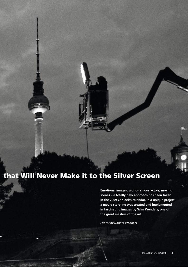
Emotional images, world-famous actors, moving
scenes – a totally new approach has been taken
in the 2009 Carl Zeiss calendar. In a unique project
a movie storyline was created and implemented
in fascinating images by Wim Wenders, one of
the great masters of the art.
Photos by Donata Wenders
11Innovation 21, 12 / 2008
A Movie that Will Never Make it to the Silver Screen

Innovation 21, 12 / 200812
Wenders makes a fictitious movie

1�Innovation 21, 12 / 2008
A gigantic ruin is emerging in the heart of Berlin: eight towers made of bare concrete which form an al-most haunting backdrop to the ma-jestic dome of the German Cathe-dral. Wwhen the scene does not happen to be bathed in radiant sun-light, there is a definite whiff of doomsday in the air. The whole scene came alive in September. There was a constant flow of people checking the structural conditions of the site, examining the concrete towers and taking notes. One of them who wanted to gain his own impression was Wim Wenders.
The director and photographer just finished with the Venice Film Festival where he was head of the jury and is now focusing on the project in Ber-lin. Wenders who is widely known for movies such as Wings of Desire, Paris, Texas and Buena Vista Social Club, as well as numberous photo projects began his next project – the 200� Carl Zeiss calendar.
It was intended to be something special: a movie that will never make it to the silver screen. A story staged with all the details, but only cap-tured using single, powerful images. “Filming” the fictional movie itself

1� Innovation 21, 12 / 2008
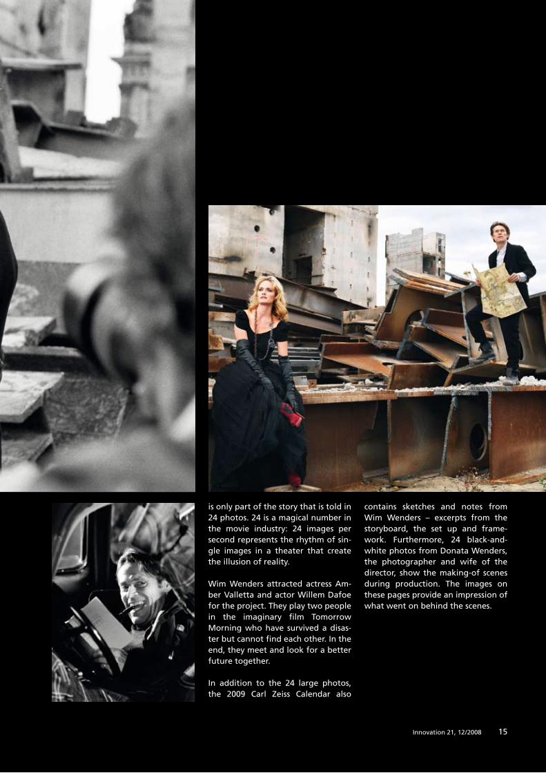
is only part of the story that is told in 2� photos. 2� is a magical number in the movie industry: 2� images per second represents the rhythm of sin-gle images in a theater that create the illusion of reality.
Wim Wenders attracted actress Am-ber Valletta and actor Willem Dafoe for the project. They play two people in the imaginary film Tomorrow Morning who have survived a disas-ter but cannot find each other. In the end, they meet and look for a better future together.
In addition to the 2� large photos, the 200� Carl Zeiss Calendar also
contains sketches and notes from Wim Wenders – excerpts from the storyboard, the set up and frame-work. Furthermore, 2� black-and-white photos from Donata Wenders, the photographer and wife of the director, show the making-of scenes during production. The images on these pages provide an impression of what went on behind the scenes.
1�Innovation 21, 12 / 2008

1� Innovation 21, 12 / 2008Innovation 21, 12 / 20081�
100 Years of Living Images
The development of fluorescence microscopy
began 100 years ago in Jena, and nowadays it is
hard to imagine a world without the colorful
images produced in biological research. Fluores-
cent dyes allow doctors to identify diseases or
genetic mutations at a single glance. Meanwhile,
scientists utilize the same methods to observe the
processes that constitute life right down to a
molecular level, and it is now possible to capture
frames of even the most dynamic life processes.
Text: Birgit HerdenScientific research: Michael Zölffel
Cover Story

1�Innovation 21, 12 / 2008
Report: Lorem ipsum
1�Innovation 21, 12 / 2008
100 Years of Living Images
18 Innovation 21, 12 / 2008
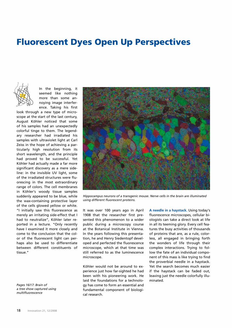
Fluorescent Dyes Open Up Perspectives
Hippocampus neurons of a transgenic mouse. Nerve cells in the brain are illuminated using different fluorescent proteins.
In the beginning, it seemed like nothing more than some an-noying image interfer-ence. Taking his first
look through a new type of micro-scope at the start of the last century, August Köhler noticed that some of his samples had an unexpectedly colorful tinge to them. The legend-ary researcher had irradiated his samples with ultraviolet light at Carl Zeiss in the hope of achieving a par-ticularly high resolution from its short wavelength, and the principle had proved to be successful. Yet Köhler had actually made a far more significant discovery as a mere side-line: in the invisible UV light, some of the irradiated structures were flu-orescing in the most extraordinary range of colors. The cell membranes in Köhler's woody tissue samples suddenly appeared to be blue, while the wax-containing protective layer of the cells glowed yellow or white. “I initially saw this fluorescence as merely an irritating side-effect that I had to neutralize”, Köhler later re-ported in a lecture. “Only recently have I examined it more closely and come to the conclusion that the col-or of the fluorescent light can per-haps also be used to differentiate between different constituents of tissue.”
It was over 100 years ago in April 1�08 that the researcher first pre-sented this phenomenon to a wider public during a microscopy course at the Botanical Institute in Vienna. In the years following this presenta-tion, he and Henry Siedentopf devel-oped and perfected the fluorescence microscope, which at that time was still referred to as the luminescence microscope.
Köhler would not be around to ex-perience just how far-sighted he had been with his pioneering work. He laid the foundations for a technolo-gy has come to form an essential and fundamental component of biologi-cal research.
Pages 16/17: Brain of a tree show captured using multifluorescence
A needle in a haystack. Using today's fluorescence microscopes, cellular bi-ologists can take a direct look at life in all its teeming glory. Every cell fea-tures the busy activities of thousands of proteins that are, as a rule, color-less, all engaged in bringing forth the wonders of life through their complex interactions. Trying to fol-low the fate of an individual compo-nent of this mass is like trying to find the proverbial needle in a haystack. Yet the search becomes much easier if the haystack can be faded out, leaving just the needle colorfully illu-minated.

1�Innovation 21, 12 / 2008
Fluorescent Dyes Open Up Perspectives
Using high-resolution images and films, researchers are now able to follow events that occur even within an individual cell, making different cellular constituents visible depend-ing on their focus of interest at the time. “Using a fluorescence micro-scope gets me so close that I can al-most shake hands with individual molecules”, enthuses Professor Volk-er Haucke, who conducts research at the Free University of Berlin into how signals from nerve cells func-tion. Two things he cites as helping to drive his work forward are the hu-
man genome project and fluores-cence microscopy. The much-lauded genome project enables biologists to look up every gene and thus every protein of a cell in a database. Yet it is only thanks to the fluorescence mi-
croscope that he is able to investi-gate the way in which molecules ac-tually interact. Knowledge of the constituent parts is rendered in three dimensions, which often provides the key to understanding.
Living images. We have come a long way from August Köhler's first fluo-rescence microscope, which he lov-ingly referred to as his “quartz mi-croscope” due to the optical components it contained, all the way up to today's virtuoso levels of imag-ing. The first scientists to become in-terested in the new technology were primarily botanists, largely because autofluorescence is so pronounced in the plant world. Important progress in this field was made by staining preparations with fluorescent chemi-cals. While carrying out experiments in the 1��0s with the dye acridine orange, the botanist Siegfried Strug-ger suddenly found he could make out living bacteria glowing bright green in a soil sample that was fluo-rescing reddish-brown. What was particularly interesting was the fact that the dye did not
The person
August Köhler (1866 – 1948)
August Köhler was born in Darmstadt, Germany. He stud-ied zoology, botany, minerol-ogy, physics and chemistry in his hometown, in Heidelberg and and in Giessen. He joined Carl Zeiss in Jena at the age of 34.
He played a key role in the development of the UV micro-scope. The resolution of the light microscope could be dou-bled using optics manufactured from a UV light permeable rock crystal. While conducting his research work, August Köhler noticed that natural objects such as cell membranes began to illuminate when they were irradiated with UV light. Rec-ognizing this inherent fluores-cence is one of his greatest achievements. In 1908, he pres-ent a luminescence microscope to the public for the first time.
New fluorescent dyes with extreme spectral properties.
“I initially saw this fluorescence as merely an irritating side-effect that I had to neutralize.”
Prof. August Köhler
Cover story: Living images
20 Innovation 21, 12 / 2008
necessarily kill the cells. Strugger was able to stain living plants and ulti-mately succeeded in tracking the flow of water within them. This con-tinues to be one of the greatest strengths of fluorescence microscopy. Although you can achieve impressive levels of visibility of the most de-tailed structures of a cell using an electron microscope, the frames ob-tained in a vacuum will always be snapshots.
Using fluorescent dyes, doctors built on this success in subsequent years by identifying the agents involved in diseases such as tuberculosis, leprosy, malaria and smallpox. Depending on the dye, it was no longer just UV light that was being employed, but also blue light. Fluorescence can oc-cur at a wide variety of wavelengths, though the principle is always the same. Incident light causes an elec-
tron in a dye molecule to be raised to a higher energy level. Within a few nanoseconds, it returns to its natural state, releasing light at the same time, though a portion of the energy received is lost in the form of heat. The light emitted is lower in energy and therefore has a longer wavelength. In comparison to the light projected onto the molecule, the fluorescence is shifted towards the red region of the spectrum.
Meanwhile, the new dyeing methods and objects of analysis were also driving forward the development of microscope optics. In the beginning, the most important thing was to capture as much as possible of the fluorescence, which was often quite weak. A milestone was achieved in 1��� with the fast NEOFLUAR objec-tive with a calcium fluoride lens which was launched by ZEISS-WIN-KEL. Following numerous improve-ments, this gave rise to the EC Plan-NEOFLUAR range of objec-tive lenses, which, even today, con-tinue to represent the “workhorses” in fluorescence microscopy for scien-tists all over the world. Optics de- velopment reached its apex in the form of the high-performance
C-APOCHROMAT objective lenses corrected for entire regions of the spectrum from UV to IR, which are also produced by Carl Zeiss.
Light from above. An epifluores-cence microscope developed by ZEISS-WINKEL in 1��� represented a further breakthrough: the sample was no longer illuminated from be-low using the technique of dark-field microscopy, something which had required high light intensity and la-borious alignment. This had caused fundamental problems in trying to separate the much weaker fluores-cent light from the incident excita-tion light, which otherwise would have completely obscured it. In epi-fluorescence microscopy, however, the very bright lamplight generated by special light sources initially falls on an obliquely positioned mirror, which reflects it downwards through the objective lens in the direction of the mounted specimen. The trick here lies in the special “dichroic” beam splitter, which reflects the light of one color while being transparent for a different color. For example, if a specimen illuminated in blue fluo-resces green, the green light emitted upwards by the specimen can pass
“With a fluorescence microscope, I can practi-cally greet single mole-cules with a handshake.”
Prof. Volker Haucke, FU Berlin
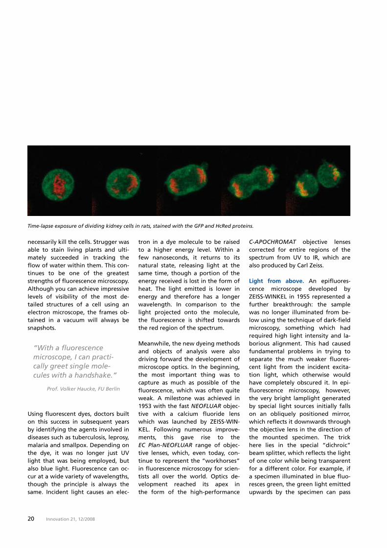
Time-lapse exposure of dividing kidney cells in rats, stained with the GFP and HcRed proteins.
21Innovation 21, 12 / 2008
through the mirror unhindered while the blue light is reflected. This pro-cess also requires two color filters based on a design with some highly complex features: one filter for the excitation light (so that only blue light is radiated, for example) and a second filter to filter the fluorescent light emanating from the specimen after it has passed the beam splitter. This second filter can then also be used to influence the perceived color of the image. Thus, it is this second filter that can further influence the color appearance of the fluorescent signal.
In the 1��0s, the development of the dichroic beam splitter was signifi-cantly boosted by the work of the Dutch researcher Johan Ploem. It was thanks to him that a module was fi-nally created that brought together filters and mirrors optimized to work in perfect combination with each other. It was only due to this ground-breaking invention that it finally be-came possible to perform rapid changes of the fluorescent dyes.
Colored probes.This achievement be-came particularly significant due to a completely different development.
Up to that point in time, researchers working in the field of fluorescence microscopy had used chemical dyes that stained different materials to differing degrees. However, this only allowed for a rather coarse distinc-tion to be made between cells and their constituent parts. As early as 1���, the American pathologist Al-bert Coons had come up with an idea that was to have a profound impact, namely that if you were to mark antibodies with fluorescent dyes, that should then enable you to specifically stain any desired patho-gen and indeed many other struc-tures. 1��� All mammals produce antibodies as a defense against a pathogen. In animal experiments, it is possible to provoke this kind of im-mune response to almost any struc-ture introduced into the blood-stream. Just two years later, Coons found the solution he was looking for. He began by linking a fluores-cent dye to antibodies that had been induced by pneumonia. These kinds of antibodies attach themselves spe-cifically to the bacteria that cause the disease, which meant that Coons could use his color-labeled antibod-ies to make these bacteria visible too.
The person
Nobel Prize for Chemistry
Osamu Shimomura studied pharmacy in Nagasaki, later organic chemistry. In 1961, he discovered the green fluo-rescent protein (GFP) in the Aequorea jellyfish. He worked at the universities of Princeton and Boston, and at the Woods Hole marine biology lab. Today he operates a private photo protein lab.
Martin Chalfie studied biology. He has been a professor for biology at Columbia University since 1982. Martin Chalfie used GFP to examine the processes in the cells of the threadwork C. elegans. He succeeded in bringing the GFP gene to expression outside the Aequorea victoria jellyfish for the first time.
Roger Y. Tsien studied chemistry and physics at Harvard University. He has been a professor for pharma-cology, chemistry and bio-chemistry at the University of California since 1989 and conducts research at the Howard Hughes Medical Institute. Thanks to him, several variations of GFP are now available, which exhibit different fluorescent spectrums and thus illuminate in different colors.
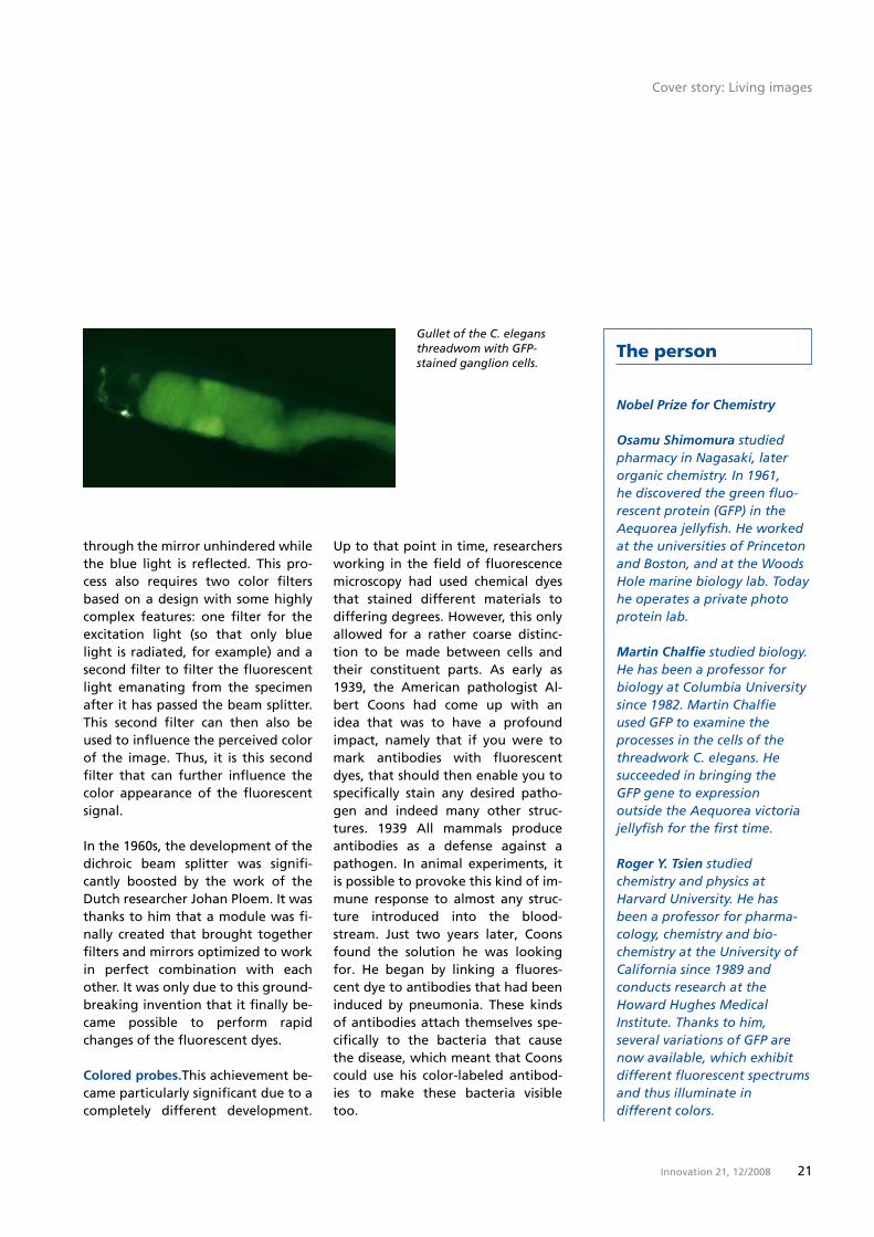
Gullet of the C. elegans threadwom with GFP-stained ganglion cells.
Cover story: Living images
22 Innovation 21, 12 / 2008
He further honed this method over the subsequent decades and, in the 1��0s, cell researchers began to carry out targeted investigations of tissue using antibodies dyed in different colors. Working with the new fluo-rescence microscopes, they were able to have one cell type illuminated in red and a different type illuminated in green. This enabled them, for ex-ample, to carry out experiments to determine which types of cells make up the immune system, the nervous system and many other tissues. Fluorescent “probes” continue to be used in research and medicine right up to the present day, and scientists now have an enormous arsenal of techniques at their disposal. For exam-ple, geneticists use fluorescent DNA probes that bind specifically to select regions of chromosomes. In amniocen-tesis, these probes can be used to dye the chromosomes of an unborn child, which makes it possible to detect seri-ous genetic defects at a glance.
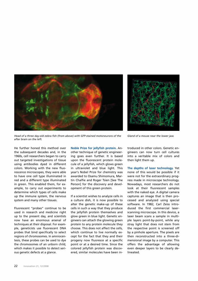
Gland of a mouse near the lower jaw.Head of a three day-old zebra fish (from above) with GFP-stained motoneurons of the after brain on the left.
Noble Prize for jellyfish protein. An-other technique of genetic engineer-ing goes even further. It is based upon the fluorescent protein mole-cule of a jellyfish, which glows green in ultraviolet and blue light. This year's Nobel Prize for chemistry was awarded to Osamu Shimomura, Mar-tin Chalfie and Roger Tsien [See The Person] for the discovery and devel-opment of this green protein.
If a scientist wishes to analyze cells in a culture dish, it is now possible to alter the genetic make-up of these cells in such a way that they produce the jellyfish protein themselves and glow green in blue light. Genetic en-gineers can attach the glowing green protein to any protein molecule they choose. This does not affect the cells, which continue to live normally ex-cept for the fact that they and their progeny now fluoresce at a specific point or at a desired time. Since the colored jellyfish protein was discov-ered, similar molecules have been in-
troduced in other colors. Genetic en-gineers can now turn cell cultures into a veritable mix of colors and then light them up.
The depths of laser technology. Yet none of this would be possible if it were not for the extraordinary prog-ress made in microscope technology. Nowadays, most researchers do not look at their fluorescent samples with the naked eye. A digital camera captures an image that is then pro-cessed and analyzed using special software. In 1�82, Carl Zeiss intro-duced the first commercial laser-scanning microscope. In this device, a laser beam scans a sample in multi-ple layers point-by-point, while any stray light that does not stem from the respective point is screened off by a pinhole aperture. The pixels are then reconstructed into a three-di-mensional image by a computer. This offers the advantage of allowing even deeper layers to be clearly de-lineated.
2�Innovation 21, 12 / 2008
In some cases, however, this kind of laser beam is simply too intense. When neurologists want to make highly-branched nerve cells visible or ophthalmologists are investigating a damaged retina, they need the gen-tlest, least-intrusive method they can find. One viable alternative is “two-photon microscopy,” which repre-sents a variation on the confocal mi-croscope. When the focused beam of a laser excites a fluorophore by caus-ing it to simultaneously absorb two photons, the fluorophore emits a single photon at a higher energy. By irradiating a point with low-energy red laser light, it is possible to make the specimen fluoresce blue contrary to the rule that otherwise applies. Low-energy infrared light is gentle on tissue. Moreover, all radiated light can be used in this case without the need for a pinhole aperture, since the blue fluorescence anyway only occurs in the focus of the high-est intensity of the red light.
Revealing the nano world. Increas-ingly refined techniques and inven-tions have transformed fluorescence microscopy into an extraordinarily powerful tool for life sciences over the last 20 years. Specimens can be made to glow on demand when the chemical environment alters or when one molecule approaches another.
Scientists have even managed to break the Abbe resolution limit. When Ernst Abbe developed the sci-entific basis of microscopy, he recog-nized that there is a fundamental limit to the resolving power of any microscope due to the wave nature of light. The maximum resolving
power of even the best light micro-scope is thus approximately 200 nanometers. Yet a single influenza virus has a diameter of just 100 nanometers, and there are many constituents of cells that are even smaller. In order to make these visi-ble, “Stimulated Emission Depletion” (STED) exploits a further characteris-tic of fluorescent molecules, namely the fact that two pulses of a laser beam, one immediately following the other, can be used to excite and then immediately quench fluores-cence. In STED, the excitation laser beam is surrounded by a ring-shaped, quenching beam. The outer beam narrows the spot focused on by the excitation pulse, thereby caus-ing the spot to have a diameter sig-nificantly below the Abbe limit.
In Photoactivated Localization Mi-croscopy (PALM), which is currently
under development at Carl Zeiss, the individual fluorescing molecules are so far apart from each other that they can be individually distin-guished, which means that the indi-vidual flashes of light from thou-sands of frames can be composed into a high-resolution image.
This is one example of how Carl Zeiss is pushing the boundaries of research and driving forward new develop-ments in fluorescence microscopy. A constant supply of new inventions over a 100-year period has allowed us to see these colorful images in ever greater detail, and it is truly un-usual for one company to have had such a profound influence on a tech-nical development. Yet, in reality, the biotechnological era has only just be-gun, and with it the prospect of an even greater role for fluorescence microscopy.
Cover story: Living images
2� Innovation 21, 12 / 2008
“Impossible, is not part of my vocabulary” could be the motto of Dr. Ul-rich Simon, Head of Carl Zeiss Microscopy. In an
interview, he and Dr. Bernhard Oh-nesorge, who is responsible for Bio-sciences, explain the insights that will be made possible through mi-croscopy in the coming years.
Interview
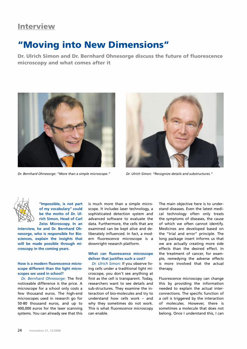
Dr. Ulrich Simon: “Recognize details and substructures.“
“Moving into New Dimensions”Dr. Ulrich Simon and Dr. Bernhard Ohnesorge discuss the future of fluorescence microscopy and what comes after it
How is a modern fluorescence micro-scope different than the light micro-scopes we used in school? Dr. Bernhard Ohnesorge: The first noticeable difference is the price. A microscope for a school only costs a few thousand euros. The high-end microscopes used in research go for �0-80 thousand euros, and up to �00,000 euros for the laser scanning systems. You can already see that this
Dr. Bernhard Ohnesorge: “More than a simple microscope.”
The main objective here is to under-stand diseases. Even the latest medi-cal technology often only treats the symptoms of diseases, the cause of which we often cannot identify. Medicines are developed based on the “trial and error” principle. The long package insert informs us that we are actually creating more side effects than the desired effect. In the treatment of cancer, for exam-ple, remedying the adverse effects is more involved that the actual therapy.
Fluorescence microscopy can change this by providing the information needed to explain the actual inter-connections. The specific function of a cell is triggered by the interaction of molecules. However, there is sometimes a molecule that does not belong. Once I understand this, I can
is much more than a simple micro-scope. It includes laser technology, a sophisticated detection system and advanced software to evaluate the data. Furthermore, the cells that are examined can be kept alive and de-liberately influenced. In fact, a mod-ern fluorescence microscope is a downright research platform.
What can fluorescence microscope deliver that justifies such a cost? Dr. Ulrich Simon: If you observe liv-ing cells under a traditional light mi-croscope, you don't see anything at first as the cell is transparent. Today, researchers want to see details and sub-structures. They examine the in-teraction of bio-molecules and try to understand how cells work – and why they sometimes do not work. This is what fluorescence microscopy can enable.
2�Innovation 21, 12 / 2008
develop an agent to eliminate pre-cisely this molecule. And I need fluo-rescence to understand these details.
What was the key advance that made this possible? Dr. Ulrich Simon: The development of fluorescence dyes that do not dis-rupt the processes in the cell was a key event. In the past, cells were stained using synthetic dyes. Howev-er, this quickly killed the cells. Today, fluorescing proteins, living colors, found in the ocean are used. They are not toxic to the cells. A good Carl Zeiss customer, Roger Tsien, won his Nobel Prize for this discovery.
An advance into new dimensions was also a key factor. The world is still two dimensional under a school microscope. An onion epidermis is typical of this. I can always only view one plane; the rest is blurry and must be cut away. Modern research microscopy is at least three dimen-sional. Today, we are even talking about multi-dimensional examina-tions. They observe different colors, time lapses and substance concentra-tions in the three spatial dimensions. A key advance, however, is that we are attempting to resolve the image to the molecular level. For more than a century, it was thought that Abbe’s law of diffraction limited the resolu-tion. Only recently were techniques developed, with which it was possi-ble to clearly exceed the existing dif-fraction limit.
How is this possible? Dr. Bernhard Ohnesorge: There are different developments. PAL micros-copy, or photo-activated localization
microscopy, is particularly interest-ing. It is based on the fact that you actually very precisely localize a sin-gle fluorescing molecule. This mole-cule is imaged as a “diffractive disc” by the optical system, meaning it ap-pears blurry and molecules lying next to each other are blurred. However, with PAL microscopy, the single fluo-rescing molecules are so far apart, that their images do not touch each other. This is made possible by pho-to-active dyes that can be activated and deactivated with exciting light. Over several thousand cycles, new molecules are repeatedly excited so that the observer receives a high-res-olution image, the same as a puzzle.
How far along are you with devel-opment? Dr. Ulrich Simon: We have already achieved fantastic resolution of less than 20 nanometers. We are current-ly installing five prototypes at the sites of well-known customers. We have not yet reached a point that we can observe living specimens with this technology, but I am sure we will also succeed here.
Is there even a frontier that you could ever reach? Dr. Ulrich Simon: In general, you can't evaluate more light than a mol-ecule emits. This means that the detail of real-time exposures is limit-ed will. However, I never want to here developers say “impossible.” Throughout history, people have too often thought that we couldn't go any further. The limits that we know today apply to fluorescing speci-mens, i.e. those that illuminate natu-rally. This does not always have to be
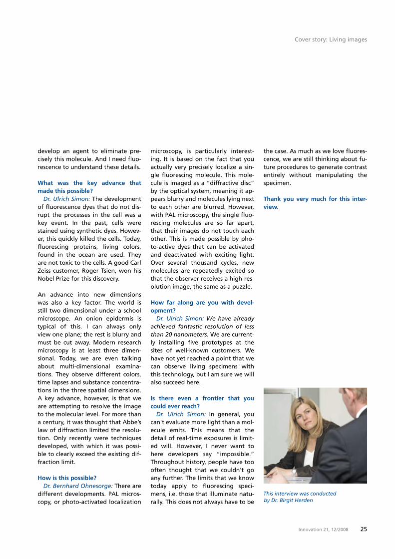
This interview was conducted by Dr. Birgit Herden
the case. As much as we love fluores-cence, we are still thinking about fu-ture procedures to generate contrast entirely without manipulating the specimen.
Thank you very much for this inter-view.
Cover story: Living images

2� Innovation 21, 12 / 2008
Cover Story
On the Track of Viruses with Fluorescence Signals
2� Innovation 21, 12 / 2008

2�Innovation 21, 12 / 2008
Report: Lorem ipsum
On the Track of Viruses with Fluorescence Signals
2�Innovation 21, 12 / 2008
Viruses play a significantly bigger role in the
outbreak of illnesses than previously thought.
They trigger AIDS, hepatitis and various cancers.
The human papilloma virus, for example, is res-
ponsible for cervical cancer in over 90% of cases.
28 Innovation 21, 12 / 2008
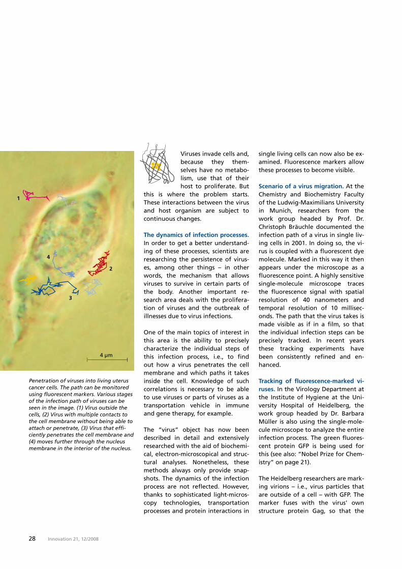
Penetration of viruses into living uterus cancer cells. The path can be monitored using fluorescent markers. Various stages of the infection path of viruses can be seen in the image. (1) Virus outside the cells, (2) Virus with multiple contacts to the cell membrane without being able to attach or penetrate, (3) Virus that effi-ciently penetrates the cell membrane and (4) moves further through the nucleus membrane in the interior of the nucleus.
Viruses invade cells and, because they them-selves have no metabo-lism, use that of their host to proliferate. But
this is where the problem starts. These interactions between the virus and host organism are subject to continuous changes.
The dynamics of infection processes.In order to get a better understand-ing of these processes, scientists are researching the persistence of virus-es, among other things – in other words, the mechanism that allows viruses to survive in certain parts of the body. Another important re-search area deals with the prolifera-tion of viruses and the outbreak of illnesses due to virus infections.
One of the main topics of interest in this area is the ability to precisely characterize the individual steps of this infection process, i.e., to find out how a virus penetrates the cell membrane and which paths it takes inside the cell. Knowledge of such correlations is necessary to be able to use viruses or parts of viruses as a transportation vehicle in immune and gene therapy, for example.
The “virus” object has now been described in detail and extensively researched with the aid of biochemi-cal, electron-microscopical and struc-tural analyses. Nonetheless, these methods always only provide snap-shots. The dynamics of the infection process are not reflected. However, thanks to sophisticated light-micros-copy technologies, transportation processes and protein interactions in
single living cells can now also be ex-amined. Fluorescence markers allow these processes to become visible.
Scenario of a virus migration. At the Chemistry and Biochemistry Faculty of the Ludwig-Maximilians University in Munich, researchers from the work group headed by Prof. Dr. Christoph Bräuchle documented the infection path of a virus in single liv-ing cells in 2001. In doing so, the vi-rus is coupled with a fluorescent dye molecule. Marked in this way it then appears under the microscope as a fluorescence point. A highly sensitive single-molecule microscope traces the fluorescence signal with spatial resolution of �0 nanometers and temporal resolution of 10 millisec-onds. The path that the virus takes is made visible as if in a film, so that the individual infection steps can be precisely tracked. In recent years these tracking experiments have been consistently refined and en-hanced.
Tracking of fluorescence-marked vi-ruses. In the Virology Department at the Institute of Hygiene at the Uni-versity Hospital of Heidelberg, the work group headed by Dr. Barbara Müller is also using the single-mole-cule microscope to analyze the entire infection process. The green fluores-cent protein GFP is being used for this (see also: “Nobel Prize for Chem-istry” on page 21).
The Heidelberg researchers are mark-ing virions – i.e., virus particles that are outside of a cell – with GFP. The marker fuses with the virus' own structure protein Gag, so that the

2�Innovation 21, 12 / 2008
fluorescence signals can exactly track the traces left by the virus when it makes contact with the host cell and the subsequent transport within the cell.
Such single-virus tracking experi-ments require a well-equipped inver-ted microscope for multicolor imag-ing, a temperature-controlled research environment and a very precise laser illumination unit. In addition, it de-mands a high-aperture lens and ex-tremely sensitive detectors.
This device setup can be supplement-ed with microscope-based systems. An optical measuring process is fluo-rescence correlation spectroscopy, which obtains information from con-
The details
Virus history
Viruses were detected in fixat-ed cells for the first time in 1980 using simple fluorescence-marked virus particles. One year later the first single-par-ticle tracking experiments were performed in live cells. In 1985 it was possible to observe vi-ruses in real time in the light-microscopic differential inter-ference contrast technique. Tracking viruses in living cells became a reality five years later with the aid of fluorescence video microscopy.
The introduction of TIRF micro-scope technology in 1995 made it possible to detect single fluo-rescent molecules. The number of single-virus tracking experi-ments has increased continu-ously since 2001. Today, scien-tists are able to track the fluorescence-marked virus di-rectly in a single living cell and observe it over a long period of time.
stantly varying fluorescence intensity. High-resolution optical images are also created by confocal laser scan-ning microscopy, or CLSM for short. The TIRF technique – which stands for “total internal reflection fluores-cence microscopy” – can be used to examine structures located very near the surfaces.
Dieter Brocksch, Monika Etspüler
Virus particle bound to the surface of liv-er cancer cells.
Cover story: Living images

�0 Innovation 21, 12 / 2008
Guest Article
The latest fluorescence imaging techniques using a combination of synthetic dyes and immunofluorescence.
The Future of Fluorescence Microscopy
The advances that we have witnessed in fluo-rescence microscopy over the past two decades have been nothing short
of breathtaking. Microscopes and camera systems have, for the most part, kept pace with the develop-ment of fluorophores to enable re-searchers to observe subcellular pro-cesses with ever increasing temporal speed and spatial resolution. Today, fluorescence microscopy has evolved
into a staple methodology ranging from live-cell imaging to drug discov-ery and medical diagnosis.
However, determining what will be needed in the future is a daunting task. For example, who could have predicted the exquisite idea for photoactivation localization super-resolution microscopy (PAL-M), or the hallmark development of the “Brainbow” recombinant Cre-Lox system for imaging neuronal path-
by Michael W. Davidson Florida State University
ways in the nervous systems of living animals? This results in the following questions: What new fluorophores and labeling technologies will be needed in the future? How quickly will instrumentation evolve ?
Fluorescent proteins are genetically-encoded markers that can be fused to virtually any target protein of in-terest using traditional molecular bi-ology techniques. The green fluores-cent protein (GFP) obtained from a

�1Innovation 21, 12 / 2008
jellyfish has been modified and is now seeing the highest level of ap-plication. Perhaps the potentially most useful fluorescent proteins be-long to a new class termed “optical highlighters.” These are proving to be very efficacious in tracking dy-namic events in living cells. In gener-al, fluorescent proteins are non-toxic and feature excellent photostability, although their brightness falls far short of that exhibited by synthetic dyes and quantum dots.
In the future, we can expect to see significant advances in the realm of fluorophore technologies. Improve-ments in synthetics will continue as manufacturers strive to compete with fluorescent proteins. Further-more, quantum dots will become very useful. Hybrid systems are also receiving a considerable amount of attention and many new strategies are emerging. Finally, there appears to be no end to the new and promis-ing fluorophore candidates emerg-ing from fluorescent proteins. Dy-namic biosensors that monitor a variety of cellular processes, includ-ing pH, voltage and sugar metabo-lism are being introduced in labora-tories around the world.
Littered with acronyms such as PAL-M, STED, �Pi, STORM, SIM, and RESOLFT, the field of superresolution microscopy is currently the most rap-idly evolving frontier in fluorescence imaging. Almost every month her-alds the introduction of a new tech-nique that promises to stretch or break the diffraction barrier. Al-though seemingly unrelated in many respects, the diverse technologies
The details
Virtual Campus
Together with biophysicist Mi-chael W. Davidson from Florida State University, Carl Zeiss has created an education and sci-ence platform on the Internet regarding the topic of micros-copy and digital images. The virtual “ZEISS Campus” is a knowledge source that pres-ents theory and technology based on applications. On this website, methods and tech-niques of fluorescence micros-copy are described with the aid of detailed descriptions, inter-active animations, application examples and picture galleries. All materials were developed on microscope systems from Carl Zeiss. The websites not only provide information about current sci-entific topics regarding fluores-cence microscopy – users also have the opportunity to pub-lish their applications. With this website, Carl Zeiss is addressing all “microscopers,” particularly young scientists who can expand their knowledge at the “ZEISS Campus.”
that comprise the foundation of su-perresolution microscopy all have the common goal of imaging biolog-ical specimens on a molecular level. Among the important questions for the future are exactly where are the resolution limits and how quickly and effectively can we bridge the gap (if at all) between optical and electron microscopy? More impor-tantly, can all of this be done with living cells and, at some point, even live animals?
Several manufacturers are now start-ing to offer solutions or are are working on developments that promise to spread this technology to the mainstream. Still, the complex fluorophore technology for super-resolution microscopy is highly de-manding and most methods have not yet been extrapolated to live-cell imaging. Hopefully this technology will be developed to the point of having auxiliary devices that attach to widefield or confocal microscopes with plug-in software modules for gathering images.
Further informationis available atwww.zeiss.com/campus
The complete article can be found at www.zeiss.com/innovation
Cover story: Living images
�2 Innovation 21, 12 / 2008Innovation 21, 12 / 2008�2
Bone Find Veiled in Mystery
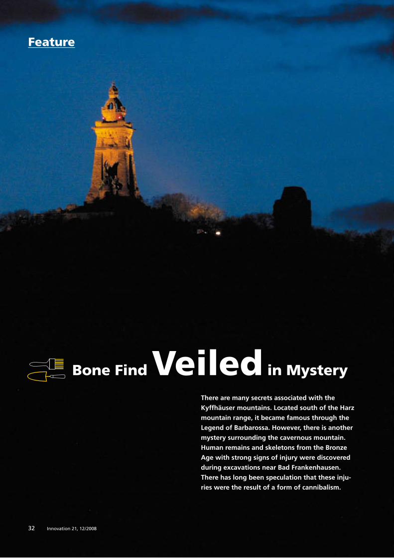
There are many secrets associated with the
Kyffhäuser mountains. Located south of the Harz
mountain range, it became famous through the
Legend of Barbarossa. However, there is another
mystery surrounding the cavernous mountain.
Human remains and skeletons from the Bronze
Age with strong signs of injury were discovered
during excavations near Bad Frankenhausen.
There has long been speculation that these inju-
ries were the result of a form of cannibalism.
Feature

��Innovation 21, 12 / 2008
Report: Lorem ipsum
��Innovation 21, 12 / 2008
Bone Find Veiled in Mystery
�� Innovation 21, 12 / 2008
A lot has been told about the Kyffhäuser in German folklore. According to legend,
Emperor Frederick Barbarossa has been sleeping in a chamber under the mountain since the 12th century, waiting for the day when he will once again walk the earth. A discov-ery made around �0 years ago points to a much earlier time. During exca-vations, archaeologist Professor Gün-ther Behm-Blancke found human re-mains. Tools and ceramic containers also found there suggested that the bone material dates back to the Bronze Age, or about �,000 years ago.
Some of these remains showed trac-es of severe injuries. Researchers found indications of cutting, chop-ping and beating, primarily on the ends of the long bones and in the joints. However, there were also indi-
Researchers looking for clues into cause of trau ma on remains
cations of violence on the spine and skull. Many bones were completely destroyed. Until now, experts were hard tasked to determine what caused these extraordinary changes. The observations repeatedly led to speculation that cannibalism might have been involved.
Analysis using the latest microscope technology. These prehistoric relicts recently caught the eye of research-ers. Scientists from the Thuringian Office for the Preservation of Histori-cal Monuments and Archaeology in Weimar examined the bones using the latest microscope technology at the Carl Zeiss Microscopy Application Center in Jena. Furthermore, the �,000-year-old findings were docu-mented to make them available for research activities in the future.
A SteREO Discovery.V20 from Carl Zeiss was used. This stereo micro-scope is particularly well-suited for applications in materials research and quality inspections, and for bio-logical and medical examinations. It permits �D observation and provides high-contrast images with high depth of field. “The extraordinarily large viewing area was exactly what the scientists needed to examine the bones,” states Jan Birkenbeil, one of the specialists for digital image anal-ysis at Carl Zeiss. The AxioCam HRc microscope camera was used for doc-umentation. Its high image quality allows reliable statements on the condition of the bone material.
Impact injuries and lacerations no-ticeable. “The microscopic examina-tions verified the earlier suspicions,”
explains archaeologist Dr. Diethard Walter from the Thuringian Museum for Pre- and Protohistory in Weimar. The fibrous-frayed edges typical of impact injuries were easily recogniz-able, while the lacerations had a smooth contour. Magnification also showed that no new bone tissue had formed in these locations. “This clearly indicates that the injuries were inflicted around the time of death,“ explains anthropologist Sa-bine Birkenbeil from the Thuringian State Office.
Elimination delivers answers. The scientists used very unconventional methods to more accurately specify the time of death. They inflicted se-vere contusions and lacerations on the haunch of a recently slaughtered pig using bronze knives and axes. They then separated the muscles and tendons and examined under the stereo microscope the marks left by these injuries. In fact, they appeared very similar to the human remains from the Bronze Age. Finally, old bones were subjected to the same procedure for comparison. They splin-tered as a result of the high level of decalcification.
The calc-sinter deposits that partially covered the injuries refuted the ar-gument that the damage to the bones resulted from improper han-dling during the excavations. “Today, we can assume that the bone mate-rial was actually subjected to these attacks at the time of death or short-ly thereafter,“ says Diethard Walter, interpreting the results. “Based on what we know right now, these peo-ple were intentionally dismembered.”
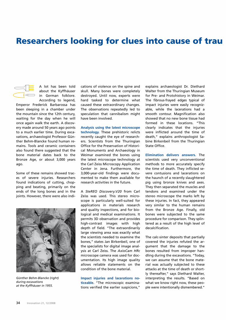
Günther Behm-Blancke (right) during excavations at the Kyffhäuser in 1955.

��Innovation 21, 12 / 2008
Researchers looking for clues into cause of trau ma on remains
Cuts on the bones (here a collarbone) show that the bodies from the Kyffhäuser were sometimes systemati-cally dismembered.
Other questions surround the find. For example, there were only injuries to the bones of adults and juveniles, not on those of children. As the in-dentations show, the strokes were made with a great deal of accuracy. Some of the skeletons exhibited scorch marks. Both Sabine Birkenbeil and Diethard Walter come to the conclusion that “A type of cannibal-ism cannot be deduced from the bone finds.“ Stomach contents at best would provide this information; and after �,000 years, there is noth-ing left over.
Many questions, few answers.It is still unclear what the purpose of this brutal procedure was. It is also un-known what drove the people into the caves of the Kyffhäuser. Was it a clan that retreated for unknown rea-sons? The large number of bones of children found there speaks for this theory. Or were burial rituals held in the interior of the mountain? The numerous offerings could be an indi-cation of such events.
“Measuring prehistoric societies us-ing current criteria would not be of much help,“ warns Diethard Walter. From his point of view the question is not “cannibalism, yes or no.“ “The total find is what gives us an insight into the intellectual and cultural life of this period,” says the archaeolo-gist. As before, little is known about the Bronze Age in central Germany.
Monika Etspüler
Some of the human bones exhibit scorch marks.
Feature: Unearthed
�� Innovation 21, 12 / 2008
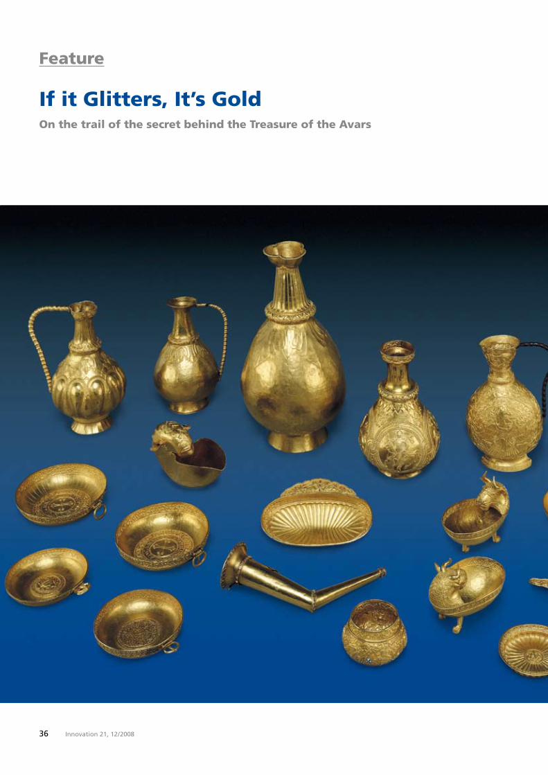
If it Glitters, It’s GoldOn the trail of the secret behind the Treasure of the Avars
Feature

��Innovation 21, 12 / 2008
More than 200 years ago, farmers in the eastern Hungarian village of Nagyszent-
miklós discovered a ten kilogram gold treasure. Vienna-based archae-ologists have now examined the gold containers using the latest mi-croscopic methods to learn more about how they were created.
Ganymede was simply spectacular. His long blond hair made the young man irresistible – not only for wom-en, but also for Zeus. The father of the gods therefore appeared as an eagle and whisked the striking young man off to the top of Mount Olympus. From then on, Ganymede was to serve as a butler to the gods and pour their wine.
This scene is illustrated on one of the containers belonging to one of the most important gold treasures from the early Middle Ages in Europe. The treasure from Nagyszentmiklós, the Treasure of the Avars is a 2�-part drink set presumably manufactured in the �th or 8th century. Today, it is on display at the Art History Museum in Vienna and is only rarely removed from its bullet-proof glass display case. One such occasion was a re-search project of the Vienna Institute for Archaeological Science (VIAS) to-gether with the Art History Museum. Scientists wanted to know how the valuable gold containers were made.
Non-destructively examined. Abso-lute care must be taken to ensure that archaeological objects are not modified or damaged. Therefore, non-destructive examination meth-
ods such as scanning electron micros-copy were the only possibilities. However, the objects must fit inside the specimen chamber of a scanning electron microscope (SEM). The VIAS purchased its own EVO 60 XVP from Carl Zeiss with the largest standard specimen chamber available. Because several of the gold containers were too tall, the Sales and Service depart-ments at Carl Zeiss developed a tech-nical solution together with VIAS ar-chaeologist Mathis Mehofer to expand the specimen chamber later-ally.
Soft bedding. Gold is a very soft ma-terial. Gold containers are easily scratched. To prevent this, Mehofer also had to place them on soft bed-ding in the SEM. He first looked for a foam material that does not shrink or crumble in the vacuum of the specimen chamber. Using life-sized copies of the treasure, he then opti-mized the work routine step-by-step. Only then did the examination of the priceless original begin. Project coordinator Dr. Birgit Bühler, a spe-
Feature: Unearthed
�8 Innovation 21, 12 / 2008
“The art-historical evaluation will provide infor-mation on the intellectual horizon of a ‘barbaric’ court. It was probably much different than what we learned in school. It must have been an unbelievably networked world.”
niques, scanned all the gold contain-ers using a reflected light microscope and identified the most interesting surface regions which were then ex-amined in the SEM.
The treasure comprises richly deco-rated jugs, cups, goblets and mugs that depict hunting and blood sport scenes. Project leader Dr. Falko Daim, General Director of the Roman-Ger-manic Central Museum in Mainz is amazed at the quality of the gold-smith work. “I was surprised that the customer and goldsmith had such unbelievable knowledge of the sub-ject and the underlying myths,” says Daim, who initiated the research
project. Greek, Persian, Byzantine and Christian themes are depicted on the containers.
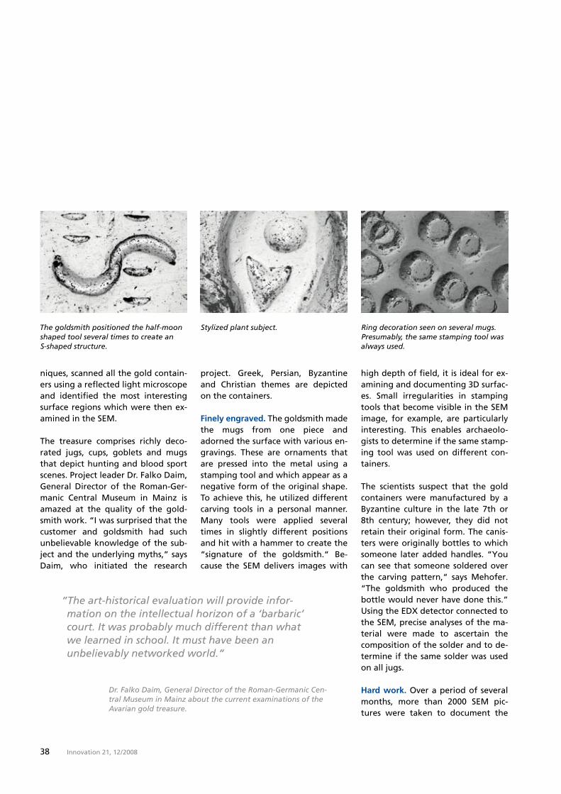
Finely engraved. The goldsmith made the mugs from one piece and adorned the surface with various en-gravings. These are ornaments that are pressed into the metal using a stamping tool and which appear as a negative form of the original shape. To achieve this, he utilized different carving tools in a personal manner. Many tools were applied several times in slightly different positions and hit with a hammer to create the “signature of the goldsmith.“ Be-cause the SEM delivers images with
high depth of field, it is ideal for ex-amining and documenting �D surfac-es. Small irregularities in stamping tools that become visible in the SEM image, for example, are particularly interesting. This enables archaeolo-gists to determine if the same stamp-ing tool was used on different con-tainers.
The scientists suspect that the gold containers were manufactured by a Byzantine culture in the late �th or 8th century; however, they did not retain their original form. The canis-ters were originally bottles to which someone later added handles. “You can see that someone soldered over the carving pattern,“ says Mehofer. “The goldsmith who produced the bottle would never have done this.” Using the EDX detector connected to the SEM, precise analyses of the ma-terial were made to ascertain the composition of the solder and to de-termine if the same solder was used on all jugs.
Hard work. Over a period of several months, more than 2000 SEM pic-tures were taken to document the
Dr. Falko Daim, General Director of the Roman-Germanic Cen-tral Museum in Mainz about the current examinations of the Avarian gold treasure.
The goldsmith positioned the half-moon shaped tool several times to create an S-shaped structure.
Stylized plant subject. Ring decoration seen on several mugs. Presumably, the same stamping tool was always used.

��Innovation 21, 12 / 2008
Archaeologist Matthias Mehofer carefully closes the door on the specimen chamber to examine the gold jug in the scanning electron microscope.
The details
Avars
Horse-riding nation that origi-nally lived as nomads in the Carpathian basin from the 6th to 8th century. The Avars had their origins in Central Asia, from where they advanced westward during the barbarian invasions and settled in parts of modern-day Hungary and Aus-tria. The Empire of the Avars perished following its defeat at the hands of Charlemagne in 791. It is said that the majority of the royal treasure of the Av-ars was transported to Aachen as spoils of war and most likely melted down.
surface of the goldsmith’s work. There were also more than 1000 sin-gle measurements for material anal-ysis.
The job of the art historians in the project team is now to interpret the results, arrange art history compari-sons with other finds and draw con-clusions about the history of the more than one thousand year old gold containers. The only thing that is known about their past with cer-tainty is that farmers in Nagyszent-miklós stumbled across the golden mugs in 1��� while they were pre-paring the foundation for a barn. The treasure had probably been bur-ied there for many years. It is un-known who buried these treasures and why. Perhaps the prestigious drink set was once in the possession of the Avars, a horse-riding people that had settled in the Carpathian basin in the early Middle Ages.
The treasure was found in what is now Sânnicolau Mare, Romania. At the end of the 18th century, this re-gion belonged to the Austro-Hun-garian monarchy. Although the farmers initially intended to keep their discovery a secret, their trea-sure trove was finally reported to the Vienna Royal Chancellery (Hofkan-zlei) as required by law at the time. Ownership of the Treasure of the Av-ars, including the Ganymede canister, was thus transferred to the double-headed eagle, the symbol of the Austro-Hungarian Empire.
According to legend, Zeus compen-sated Ganymede’s father with a grapevine for the kidnapping of his son. For his service as a divine som-melier, Ganymede was blessed with eternal youth so that his radiant beauty would never fade.
Ingrid G. Fritz
Dnjestr
Donau
Thei
ß
Sânnicolau Mare
Dr. Falko Daim, Roman-Germanic Central Museum Mainz, Dr. Birgit Bühler, VIAS, Vienna and Viktor Freiberger, Art History Museum, Vienna (from left). Location of the Avars.
Feature: Unearthed
�0 Innovation 21, 12 / 2008
Report
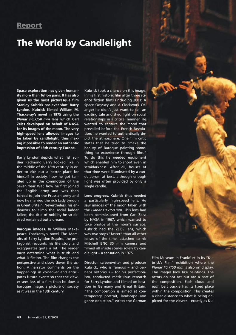
Space exploration has given human-ity more than Teflon pans. It has also given us the most picturesque film Stanley Kubrick has ever shot: Barry Lyndon. Kubrick filmed William M. Thackeray’s novel in 1975 using the Planar F0.7/50 mm lens which Carl Zeiss developed on behalf of NASA for its images of the moon. The very high-speed lens allowed images to be taken by candlelight, thus mak-ing it possible to render an authentic impression of 18th century Europe.
Barry Lyndon depicts what Irish sol-dier Redmond Barry looked like in the middle of the 18th century in or-der to eke out a better place for himself in society, how he got tan-gled up in the commotion of the Seven Year War, how he first joined the English army and was then forced to join the Prussian army and how he married the rich Lady Lyndon in Great Britain. Nevertheless, his en-deavors to climb the social ladder failed; the title of nobility he so de-sired remained but a dream.
Baroque images. In William Make-peace Thackeray’s novel The Mem-oirs of Barry Lyndon Esquire, the pro-tagonist recounts his life story and exaggerates quite a bit. The reader must determine what is truth and what is fiction. The film changes the perspective and slows down the ac-tion. A narrator comments on the happenings in voiceover and antici-pates future events so that the view-er sees less of a film than he does a baroque image, a picture of society as it was in the 18th century.
The World by Candlelight
Kubrick took a chance on this image. In his first historic film after three sci-ence fiction films (including 2001: A Space Odyssey and A Clockwork Or-ange) he didn’t just want to tell an exciting tale and shed light on social relationships in a critical manner. He wanted to capture the mood that prevailed before the French Revolu-tion; he wanted to authentically de-pict the atmosphere. One film critic states that he tried to “make the beauty of Baroque painting some-thing to experience through film.” To do this he needed equipment which enabled him to shoot even in semidarkness. After all, houses at that time were illuminated by a can-delabrum at best, although enough light was often provided by only a single candle.
Lens progress. Kubrick thus needed a particularly high-speed lens. He saw images of the moon taken with the Planar F0.7/50 mm. This lens had been commissioned from Carl Zeiss by NASA in 1���, which wanted to take photos of the moon’s surface. Kubrick had the ZEISS lens, which was two stops “faster” than all other lenses of the time, attached to his Mitchell BNC �� mm camera and filmed all inside scenes solely by can-dlelight – a sensation in 1���.
Director, screenwriter and producer Kubrick, who is famous – and per-haps notorious – for his perfection-ism, conducted meticulous research for Barry Lyndon and filmed on loca-tion in Germany and Great Britain. “The composition is aimed at con-temporary portrait, landscape and genre depiction,” writes the German
Film Museum in Frankfurt in its “Ku-brick’s Film” exhibition where the Planar F0.7/50 mm is also on display. The images look like paintings. The actors do not act but are a part of the composition. Each cloud and each belt buckle has its fixed place within the composition. This creates a clear distance to what is being de-picted for the viewer – exactly as Ku-
�0 Innovation 21, 12 / 2008

�1Innovation 21, 12 / 2008
Report: Lorem ipsum
brick wanted, because the time of Barry Lyndon is gone forever. Per-haps this is part of the reason why, despite its Oscars, Barry Lyndon was a flop in the eyes of viewers as well as critics and the film never experi-enced financial success. It is a feast for the eyes and shows what an ex-ceptionally gifted film-maker can achieve with the right technology.
After being restored at Carl Zeiss, the lens will take its place in the “Stanley Kubrick” exhibition open-ing in Valencia in the fall of 200�.
Ursula Walther
Further information is available atwww.stanleykubrick.de
�1Innovation 21, 12 / 2008

�2 Innovation 21, 12 / 2008
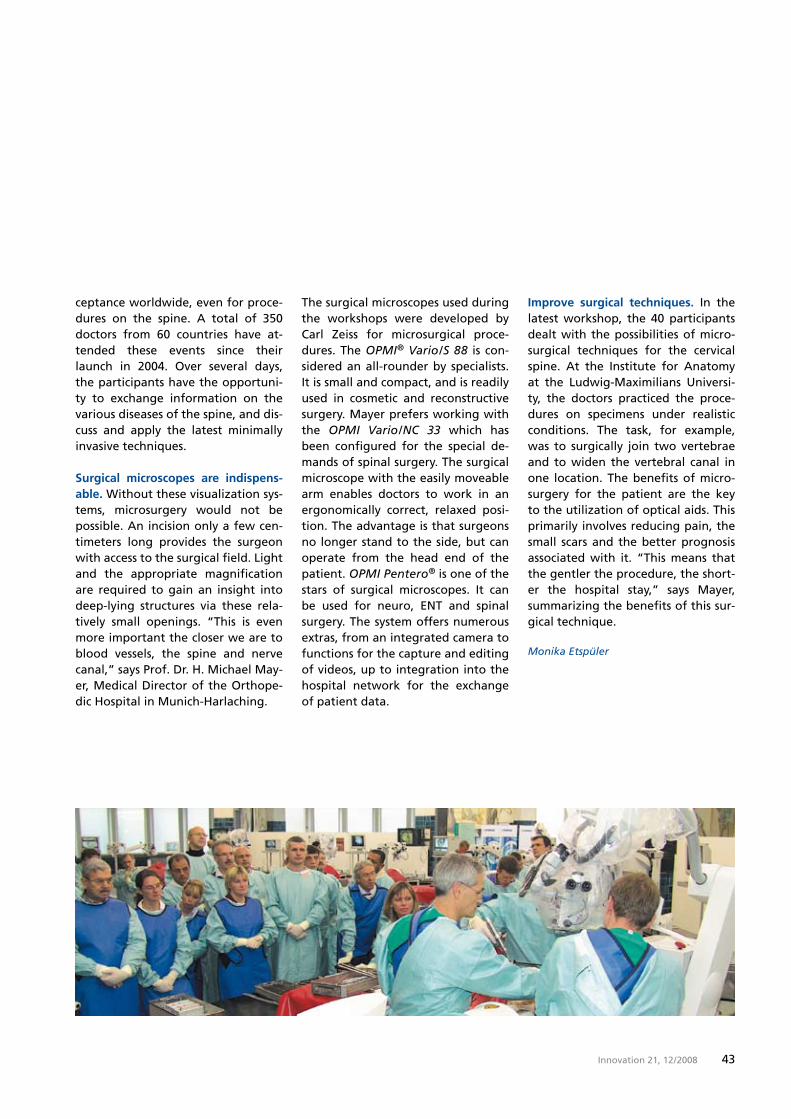
Together with Carl Zeiss Meditec, Prof. Dr. H. Michael Mayer (right) holds a workshop twice a year at the Orthopedic Hospital in Munich-Harlaching.
Report
Small Incision with a Big Impact
Back pain is a widely spread ailment. Over 30 million people in Germany alone suffer from occasional or even chronic pain and muscle tension. Go-ing under the knife is sometimes the only remedy. A surgical microscope is a valuable assistant in such cases.
Stenosis of the vertebral canal, cur-vature of the spine, herniated disks – the list of possible back diseases seems to never end. In certain cases, surgery is the only way to help an-guished patients regain their quality of life. Therefore, it is that much more important for these surgeries to be as gentle as possible and affect
patients as little as possible. This is the case with microsurgical tech-niques.
Workshop for microsurgery. Togeth-er with Carl Zeiss Meditec, the Or-thopedic Hospital in Munich-Harlach-ing holds a workshop twice a year to ensure that microsurgery gains ac-
��Innovation 21, 12 / 2008
ceptance worldwide, even for proce-dures on the spine. A total of ��0 doctors from �0 countries have at-tended these events since their launch in 200�. Over several days, the participants have the opportuni-ty to exchange information on the various diseases of the spine, and dis-cuss and apply the latest minimally invasive techniques.
Surgical microscopes are indispens-able. Without these visualization sys-tems, microsurgery would not be possible. An incision only a few cen-timeters long provides the surgeon with access to the surgical field. Light and the appropriate magnification are required to gain an insight into deep-lying structures via these rela-tively small openings. “This is even more important the closer we are to blood vessels, the spine and nerve canal,” says Prof. Dr. H. Michael May-er, Medical Director of the Orthope-dic Hospital in Munich-Harlaching.
The surgical microscopes used during the workshops were developed by Carl Zeiss for microsurgical proce-dures. The OPMI® Vario/S 88 is con-sidered an all-rounder by specialists. It is small and compact, and is readily used in cosmetic and reconstructive surgery. Mayer prefers working with the OPMI Vario/NC 33 which has been configured for the special de-mands of spinal surgery. The surgical microscope with the easily moveable arm enables doctors to work in an ergonomically correct, relaxed posi-tion. The advantage is that surgeons no longer stand to the side, but can operate from the head end of the patient. OPMI Pentero® is one of the stars of surgical microscopes. It can be used for neuro, ENT and spinal surgery. The system offers numerous extras, from an integrated camera to functions for the capture and editing of videos, up to integration into the hospital network for the exchange of patient data.
Improve surgical techniques. In the latest workshop, the �0 participants dealt with the possibilities of micro-surgical techniques for the cervical spine. At the Institute for Anatomy at the Ludwig-Maximilians Universi-ty, the doctors practiced the proce-dures on specimens under realistic conditions. The task, for example, was to surgically join two vertebrae and to widen the vertebral canal in one location. The benefits of micro-surgery for the patient are the key to the utilization of optical aids. This primarily involves reducing pain, the small scars and the better prognosis associated with it. “This means that the gentler the procedure, the short-er the hospital stay,” says Mayer, summarizing the benefits of this sur-gical technique.
Monika Etspüler

�� Innovation 21, 12 / 2008Innovation 21, 12 / 2008��
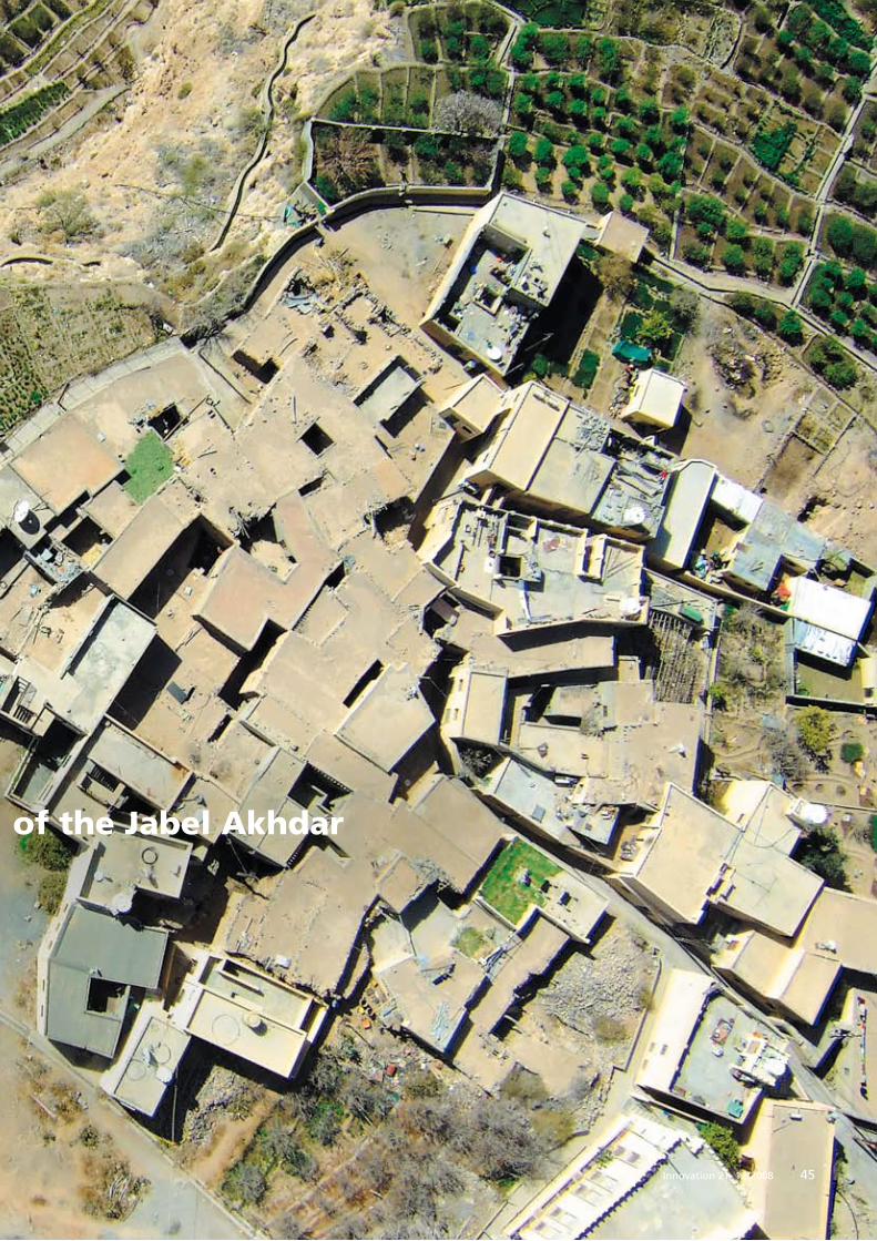
A Bird’s Eye View of the Jabel Akhdar
A different way of flying. When Dr. Wolfgang
Schäper’s machines take off, he is behind the
wheel of the remote control. There are various
high-resolution digital cameras on board, which
the hobby pilot utilizes to take photos from a
bird’s eye view. His latest order led him and his
two model planes to the sultanate of Oman.
Feature
��Innovation 21, 12 / 2008
Report: Lorem ipsum Report: Lorem ipsum
��Innovation 21, 12 / 2008
A Bird’s Eye View of the Jabel Akhdar
�� Innovation 21, 12 / 2008
Camera Flyers over Oman
and his specially equipped model planes were thus a logical choice. This was his third trip to Oman. The sultanate is located on the south-eastern coast of the Arabian penin-sula and is almost as large as Germa-ny, but sparsely populated. At 2�00 meters, the Jabel Akhdar is one of the highest peaks in the Hajar Moun-tains in the northern area of the country. Within the past ten years, the test-tube city of Sayh Qatanah was built on a giant plateau at a somewhat lower elevation – approx. 2000 meters. Around 20 percent of the area is already populated. How-ever, anyone that settled there or in the surrounding area of the city was able to implement their own ideas, opening the door to uncontrolled growth. Knut Lohrer is one of those tasked with bringing order to chaos. The intention is to expand Sayh
Qatanah into a metropolitan center while opening up the surrounding area to tourism. Ecologically sensitive regions such as the dry valleys – the Wadis – or the 1000-year-old stock of juniper trees can be better protected by pointing development in a specif-ic direction. The climate here is good for such a plan: while the tempera-ture at sea level during the summer easily tops �0°C, a Mediterranean cli-mate is prevalent at 2000 meters. For Oman, this project is also an invest-ment in the future as there are few alternatives to oil and gas. One of them is to develop the country for tourism.
Telescopic glasses as a visual aid.Wolfgang Schäper’s first job was to photographically capture the terrain on the outskirts of Sayh Qatanah which is primarily intended for use
Working for science and research, the engineer from Immenstaad am Bodensee is often on the road. His model airplanes
have taken off in the Himalayas, in Bolivia and in Island to record infor-mation on wind, humidity and tem-perature. He was led to Oman by a call from architect Knut Lohrer in Muscat, who has been tasked by the Tourism Ministry to create a master plan to develop the Jabel Akhdar.
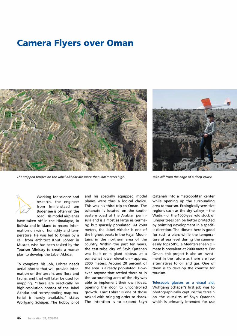
To complete his job, Lohrer needs aerial photos that will provide infor-mation on the terrain, and flora and fauna, and that will later be used for mapping. “There are practically no high-resolution photos of the Jabel Akhdar and corresponding map ma-terial is hardly available,” states Wolfgang Schäper. The hobby pilot
The stepped terrace on the Jabel Akhdar are more than 500 meters high. Take-off from the edge of a deep valley.
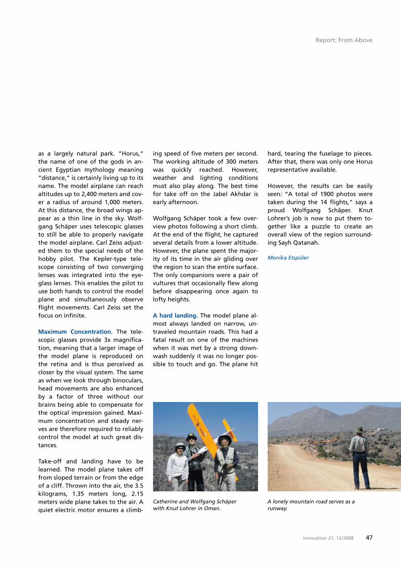
��Innovation 21, 12 / 2008
Camera Flyers over Oman
as a largely natural park. “Horus,“ the name of one of the gods in an-cient Egyptian mythology meaning “distance,“ is certainly living up to its name. The model airplane can reach altitudes up to 2,�00 meters and cov-er a radius of around 1,000 meters. At this distance, the broad wings ap-pear as a thin line in the sky. Wolf-gang Schäper uses telescopic glasses to still be able to properly navigate the model airplane. Carl Zeiss adjust-ed them to the special needs of the hobby pilot. The Kepler-type tele-scope consisting of two converging lenses was integrated into the eye-glass lenses. This enables the pilot to use both hands to control the model plane and simultaneously observe flight movements. Carl Zeiss set the focus on infinite.
Maximum Concentration. The tele-scopic glasses provide �x magnifica-tion, meaning that a larger image of the model plane is reproduced on the retina and is thus perceived as closer by the visual system. The same as when we look through binoculars, head movements are also enhanced by a factor of three without our brains being able to compensate for the optical impression gained. Maxi-mum concentration and steady ner-ves are therefore required to reliably control the model at such great dis-tances.
Take-off and landing have to be learned. The model plane takes off from sloped terrain or from the edge of a cliff. Thrown into the air, the �.� kilograms, 1.�� meters long, 2.1� meters wide plane takes to the air. A quiet electric motor ensures a climb-
ing speed of five meters per second. The working altitude of �00 meters was quickly reached. However, weather and lighting conditions must also play along. The best time for take off on the Jabel Akhdar is early afternoon.
Wolfgang Schäper took a few over-view photos following a short climb. At the end of the flight, he captured several details from a lower altitude. However, the plane spent the major-ity of its time in the air gliding over the region to scan the entire surface. The only companions were a pair of vultures that occasionally flew along before disappearing once again to lofty heights.
A hard landing. The model plane al-most always landed on narrow, un-traveled mountain roads. This had a fatal result on one of the machines when it was met by a strong down-wash suddenly it was no longer pos-sible to touch and go. The plane hit
Catherine and Wolfgang Schäper with Knut Lohrer in Oman.
A lonely mountain road serves as a runway.
hard, tearing the fuselage to pieces. After that, there was only one Horus representative available.
However, the results can be easily seen: “A total of 1�00 photos were taken during the 1� flights,“ says a proud Wolfgang Schäper. Knut Lohrer’s job is now to put them to-gether like a puzzle to create an overall view of the region surround-ing Sayh Qatanah.
Monika Etspüler
Report: From Above

�8 Innovation 21, 12 / 2008
Data Communication in the Fast LaneLaser beam transport measured values and images across long distances
Radio links are every-where. Ships communicate with each other via radio. Satellites use it to transmit their data packets to
earth. However, the big drawback to this technology that works in the ra-dio frequency range is the low trans-mission rate. By comparison, com-munication via laser beam enables the movement of large amounts of data over long distances in a matter of seconds.
The island of Sylt is easily recogniz-able from an airplane. It lies length-wise in the North Sea, usually sur-rounded by mist and clouds. How- ever, there are photos that show a completely different Sylt. Residential areas and rows of houses can be seen without a problem. But these pic-tures were not taken from an alti-tude of 12 kilometers; they were taken �00 kilometers from the earth using radar technology.
Look at the earth. The German radar satellite, TerraSAR-X, delivers razor-sharp images. It was launched in June last year from the Baikonur spaceport with the intention of ob-serving the earth. It has orbited our planet ever since. TerraSAR-X is con-sidered the most powerful non-mili-tary radar system currently in space.
Large quantities of data must reach the earth station in order to produce images like those of Sylt. Carl Zeiss developed the optical components for the laser communication termi-nal. The 120 mm main telescope, as well as the transmitting and receiv-ing units must meet the highest opti-cal demands. Furthermore, the com-pany’s specialists manufactured mirrors and components to detect and track the counter terminal. All parts had to be designed to work with absolute reliability even in ex-treme temperatures and vibrations, while the optical quality must re-main constant for the entire dura-tion of the several-year mission.
At sea and on land. Laser communi-cation is not limited to the exchange of data between orbit and the earth. Its high degree of interception proof-ness, good availability outside of licensed wavebands and the com-pact terminal design make this tech-nology an ideal tool for use on water and on land.
In military applications, laser tech-nology enables transmission of data between the ships of a naval unit. It can be used to monitor borders, se-cure harbor facilities and to control unmanned oil platforms and pipe-lines.
The main benefit is the high data transmission rate. For example, Ter-raSAR-X achieves up to five gigabits per second in space, the equivalent of transmitting the contents of a DVD in around ten seconds. “This en-ables us to deploy sensors with in-creasingly higher resolution and de-liver their results practically in real time. We are then able, for example, to quickly determine the extent of natural disasters around the globe and initiate the corresponding res-cue measures,” explains Dr. Martin Gerken who is in charge of the de-velopment of such special optics.
Drawbacks of an open Europe. Bor-der monitoring in Europe following implementation of the Schengen Agreement is becoming more and more important with increasing glo-balization. For some this meant more freedom and space, others saw this as an opportunity for illegal activi-ties. The number of refugees coming to Europe via the “blue border” of Sylt from an altitude of 500 kilometers.
Feature
�8 Innovation 21, 12 / 2008

��Innovation 21, 12 / 2008
Report: Lorem ipsum
��Innovation 21, 12 / 2008

�0 Innovation 21, 12 / 2008
countries along the Mediterranean is increasing. Border police worry that terrorists, among others, could sneak in via the “green border“ in the east. The fight against smuggling and or-ganized crime presents the European Union with new challenges.
Large areas of critical border regions are being increasingly monitored from towers, upon which thermal imagers and cameras are installed. Currently, data is transmitted to local centers via fiber optics or radio re-lays. Furthermore, specially equipped
A feasible next step in laser communication: data transmission at sea for civilian use.

�1Innovation 21, 12 / 2008
border patrol vehicles are deployed as mobile surveillance units. “The goal is to supplement the technical equipment with laser technology. This will enable mission forces to better communicate with each other and react more quickly and specifi-
cally in critical situations,” explains Dr. Karl Pietzsch, Manager of Border Surveillance/Facility Protection at Carl Zeiss.
Stable connection. According to Pi-etzsch, this still sounds like some vi-sion for the future. However, during a joint exercise with the German Aerospace Center in 200�, Carl Zeiss verified that laser technology is suit-able, in principle, for border surveil-lance: data was successfully transmit-ted via laser beam at a rate of one gigabit between a fixed installation and a moving vehicle 1.� kilometers away. This range can be expanded to about 20 kilometers.
A demonstration for representatives of the Germany Army this summer in Eckernförde showed that laser com-munication from a ship is also feasi-ble. A stable connection to a land station was established at a distance of 2� kilometers. The data transmis-sion rate was 12� megabit per sec-ond. “We must ask today how data communication will look tomorrow. Traditional radio links on ships can never be completely replaced, but we are on the verge of completing the move to laser communication,“ says Andreas Plaznik from Strategy and Marketing at Carl Zeiss about the development.
Future advances will also enable the use of this equipment on subma-rines. Only very small amounts of data can be transmitted under water via traditional antennas. However, because water exhibits high trans-mittance for laser beams in the blue-green range, distances up to 200 me-
ters can be achieved under water. A buoy or a ship serves as a transmit-ting and receiving station for the further exchange of data with ships, satellites and ground stations.
High technical requirements. “Com-munication between ships demands different systems than those re-quired for communications in border regions. However, in general, the same technology is used,” says Mar-tin Gerken. It also requires the trans-mission and receiver terminal to be perfectly aligned to each other. Sta-bilizing the transmitter laser beam on mobile units such as the border patrol vehicles presented an addi-tional technical challenge. Carl Zeiss developed a special platform for this that can be used in the air, on land, at sea and under water. This opens the door for a global communica-tions strategy. An event in March this year could be a step in this direction. TerraSAR-X succeeded in using a la-ser beam to establish contact to an-other satellite at a range of �000 ki-lometers.
Monika Etspüler
Report: From Above

�2 Innovation 21, 12 / 2008
Light
The Right LightEven on stage light and color belong together
Essay

��Innovation 21, 12 / 2008
Above all else, light gives us the capacity to see, to identify objects and to experience colors as true discov-eries rather than just random sensations.
Delving into the realms of daylight and night-light (the night has its own form of brightness too!) is a fascinat-ing and endlessly engrossing activity. Human beings have long been preoccupied with the task of imitating the phenomenon of light in myriad variations using ar-tificial light sources. Personally, I have spent more than forty years of my life carrying out research, develop-ment and design using artificial light and I could never have imagined that I would find this infinitely multifac-eted topic so interesting, challenging and creative. There are always new discoveries to be made, whether you approach the subject out of curiosity, as a challenge or simply through an affinity for everything it entails.
Both contemporary and future aspects are of interest to us in relation to this topic. The wealth of available lighting technologies is greater nowadays than ever be-fore. In fact, it is so extensive that it is sometimes hard to fully grasp.
The foundations of modern lighting design were laid by Adolphe Appia (18�2 – 1�28) in his stage designs at the beginning of the 20th century. He was one of the first progressive artists to separate artificial light from realism in the visual arts and to integrate “light, space and the human body“ into a unified, three-dimensional visual experience. Around the same time, the painter Wassily Kandinsky (18�� – 1���) was incorporating this important shift in artistic style into the painter's pal-ette. It really made no difference whether the light was painted or emitted by spotlights in terms of the actual
design concepts involved. What Appia discovered was echoed in the decisions he took in regard to lighting, which differentiated between formative light and dif-fused light.
The difference between these two forms is very simple. Concentrated light that can be shifted in any and all directions is formative light. Spotlighting is included under formative light, as are the shadows this produces. In contrast, Appia classifies reflections and indirect, soft light as diffused light.
All forms and trends of artificial light sources are incor-porated within these generic terms of “formative light“ and “diffused light“, including equipment in the form of light projecting devices (spotlights), ambient light in lighting architecture and general room illumination. Appia's definition of light developed the blueprint for using light to create emotional and atmospheric moods, especially by determining the direction and ambience of lighting.
This was a revolutionary step for artistic, visual compo-sition in the theatre, which makes it all the more re-markable that this simple and logical interpretation of light had not paved the way even earlier for the over-due arrival of symbolism. Appia's acutely clear percep-tions were eminently simple. The sun had always been the “formative light,“ while the “light of the sky“ fulfilled the task of diffused light.
Another key issue in addition to Appia's assertions on light is that of the characteristics of color temperature (the “color of light“) and of color in general. Light is not merely light: any implementation of artificial light must also make the distinction between warm and cool light, though the dividing line between these different forms of light is far from being clear-cut. Viewed from a basic physics standpoint, warm light is defined as a glowing tungsten filament and cool light as a discharge gap between two electrodes. This entire palette can be altered, just as light in general can be changed into all color variants using tinted glass, color filters, and di-chroic substrate media. This is obviously something that has to be conducted in the spectrum of visible light.
Light is color, color is life, light is life: light generates color like flames generate light. Colors are the children of light, light is the mother of colors.
Johannes Itten Color theorist from the Bauhaus period (1888 - 1967)
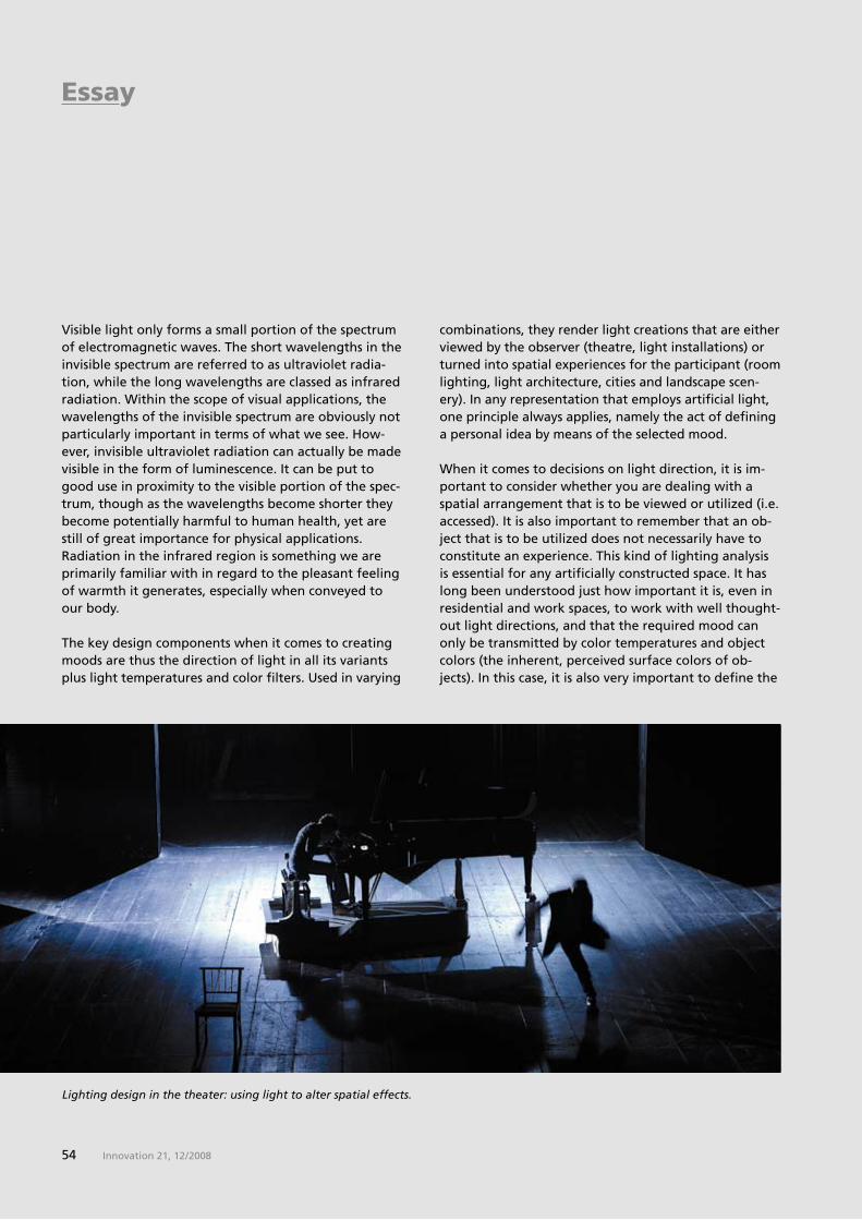
�� Innovation 21, 12 / 2008
Lighting design in the theater: using light to alter spatial effects.
combinations, they render light creations that are either viewed by the observer (theatre, light installations) or turned into spatial experiences for the participant (room lighting, light architecture, cities and landscape scen-ery). In any representation that employs artificial light, one principle always applies, namely the act of defining a personal idea by means of the selected mood.
When it comes to decisions on light direction, it is im-portant to consider whether you are dealing with a spatial arrangement that is to be viewed or utilized (i.e. accessed). It is also important to remember that an ob-ject that is to be utilized does not necessarily have to constitute an experience. This kind of lighting analysis is essential for any artificially constructed space. It has long been understood just how important it is, even in residential and work spaces, to work with well thought-out light directions, and that the required mood can only be transmitted by color temperatures and object colors (the inherent, perceived surface colors of ob-jects). In this case, it is also very important to define the
Visible light only forms a small portion of the spectrum of electromagnetic waves. The short wavelengths in the invisible spectrum are referred to as ultraviolet radia-tion, while the long wavelengths are classed as infrared radiation. Within the scope of visual applications, the wavelengths of the invisible spectrum are obviously not particularly important in terms of what we see. How-ever, invisible ultraviolet radiation can actually be made visible in the form of luminescence. It can be put to good use in proximity to the visible portion of the spec-trum, though as the wavelengths become shorter they become potentially harmful to human health, yet are still of great importance for physical applications. Radiation in the infrared region is something we are primarily familiar with in regard to the pleasant feeling of warmth it generates, especially when conveyed to our body.
The key design components when it comes to creating moods are thus the direction of light in all its variants plus light temperatures and color filters. Used in varying
Essay

��Innovation 21, 12 / 2008
White light on an empty stage used as a design component.
correct brightness of the artificial light, especially if non-luminous object colors comprise a significant por-tion of the interior. One of the basic issues to address is the function that the space is being designed to fulfill. Is it intended to be an office, a living room or a space designed for artistic purposes? If the lighting is to be solely “practical“, then it is advisable to equip the space with the lightest possible object colors and to select a cool, shadow-free light. Warm surface colors, warm light temperatures and a range of light directions mixed together are the correct way to structure person-al living spaces. Selecting the right light sources is just as important as selecting the color of the paint on the walls of the room and choosing how it is furnished. I was pleased to read that there has recently been a move to choose colors other than white for lighting and object colors in hospital rooms. It has long been known that light also possesses a healing energy and that object colors in pastel hues can promote people's sense of well-being. Though in relation to hospital rooms, it is generally considered that a brilliant, bright white evokes the required sense of cleanliness.
We have also all become aware of how the right light-ing can play a decisive role in purchasing decisions, whether consciously or subconsciously. A badly-lit piece of meat is bound to seem unappetizing, while clothing can take on completely different color tones in the wrong light. The color of light and the color of an object are mutually dependent.
Theories on the application of colors and their classifi-cation have existed since antiquity in the form of so called “theories of colors“. The first theories we are aware of were those of Plato, Aristotle and Pythagoras, which are technically referred to as the “pre-systems.“ Since the Renaissance, numerous other theories have come into play, which are designed to help us better understand connections between colors and thus to make more targeted use of the effects of color in our work. The more recent color systems of the 20th cen-tury are based on scientific findings and allow for more precise analyses of differences between colors while offering artists and designers varying versions of color harmonies. Some of these theories are disputed in cer-
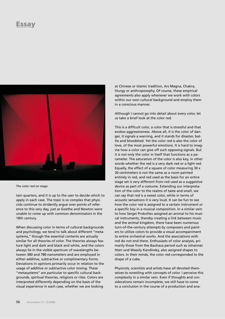
�� Innovation 21, 12 / 2008
The color red on stage.
tain quarters, and it is up to the user to decide which to apply in each case. The topic is so complex that physi-cists continue to stridently argue over points of refer-ence to this very day, just as Goethe and Newton were unable to come up with common denominators in the 18th century.
When discussing color in terms of cultural backgrounds and psychology, we tend to talk about different “meta-systems,“ though the essential contents are actually similar for all theories of color. The theories always fea-ture light and dark and black and white, and the colors always lie in the visible spectrum of wavelengths be-tween �80 and �80 nanometers and are employed in either additive, subtractive or complimentary forms. Deviations in opinions primarily occur in relation to the usage of additive or subtractive color mixing. These “metasystems“ are particular to specific cultural back-grounds, spiritual theories, religions or rites. Colors are interpreted differently depending on the basis of the visual experience in each case, whether we are looking
at Chinese or Islamic tradition, Ars Magna, Chakra, liturgy or anthroposophy. Of course, these empirical agreements also apply whenever we work with colors within our own cultural background and employ them in a conscious manner.
Although I cannot go into detail about every color, let us take a brief look at the color red.
This is a difficult color, a color that is stressful and that evokes aggressiveness. Above all, it is the color of dan-ger, it signals a warning, and it stands for disaster, bat-tle and bloodshed. Yet the color red is also the color of love, of the most powerful emotions. It is hard to imag-ine how a color can give off such opposing signals. But it is not only the color in itself that functions as a pa-rameter. The saturation of the color is also key, in other words whether the red is a very dark red or a light red. Equally, the effect of a square of color measuring �0 x �0 centimeters is not the same as a room painted entirely in red, and red used as the basis for an entire stage set is very different from red used as a suggestive device as part of a costume. Extending our interpreta-tion of the color to the realms of taste and smell, we can say that red is a sweet color, while in terms of acoustic sensations it is very loud. It can be fun to see how the color red is assigned to a certain instrument or a specific key in a musical composition. In a similar vein to how Sergei Prokofiev assigned an animal to his musi-cal instruments, thereby creating a link between music and the animal kingdom, there have been numerous turn-of-the-century attempts by composers and paint-ers to utilize colors to provide a visual accompaniment to entire orchestral works. And the associations with red do not end there. Enthusiasts of color analysis, pri-marily those from the Bauhaus period such as Johannes Itten und Wassily Kandinsky, also assigned shapes to colors. In their minds, the color red corresponded to the shape of a cube. Physicists, scientists and artists have all devoted them-selves to wrestling with concepts of color. I perceive this complexity in a similar vein. Even if thoughts and con-siderations remain incomplete, we still have to come to a conclusion in the course of a production and ana-
Essay
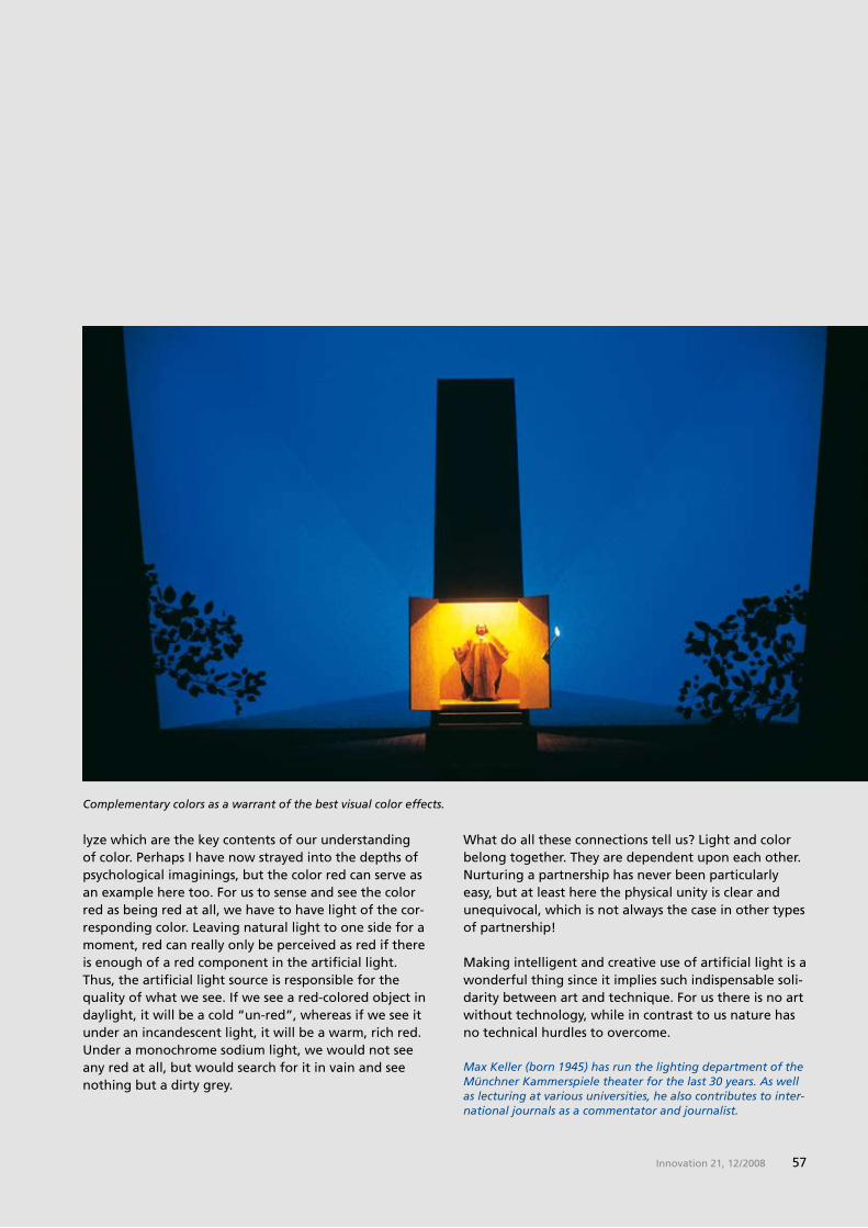
��Innovation 21, 12 / 2008
What do all these connections tell us? Light and color belong together. They are dependent upon each other. Nurturing a partnership has never been particularly easy, but at least here the physical unity is clear and unequivocal, which is not always the case in other types of partnership!
Making intelligent and creative use of artificial light is a wonderful thing since it implies such indispensable soli-darity between art and technique. For us there is no art without technology, while in contrast to us nature has no technical hurdles to overcome.
Complementary colors as a warrant of the best visual color effects.
lyze which are the key contents of our understanding of color. Perhaps I have now strayed into the depths of psychological imaginings, but the color red can serve as an example here too. For us to sense and see the color red as being red at all, we have to have light of the cor-responding color. Leaving natural light to one side for a moment, red can really only be perceived as red if there is enough of a red component in the artificial light. Thus, the artificial light source is responsible for the quality of what we see. If we see a red-colored object in daylight, it will be a cold “un-red”, whereas if we see it under an incandescent light, it will be a warm, rich red. Under a monochrome sodium light, we would not see any red at all, but would search for it in vain and see nothing but a dirty grey.
Max Keller (born 1945) has run the lighting department of the Münchner Kammerspiele theater for the last 30 years. As well as lecturing at various universities, he also contributes to inter-national journals as a commentator and journalist.
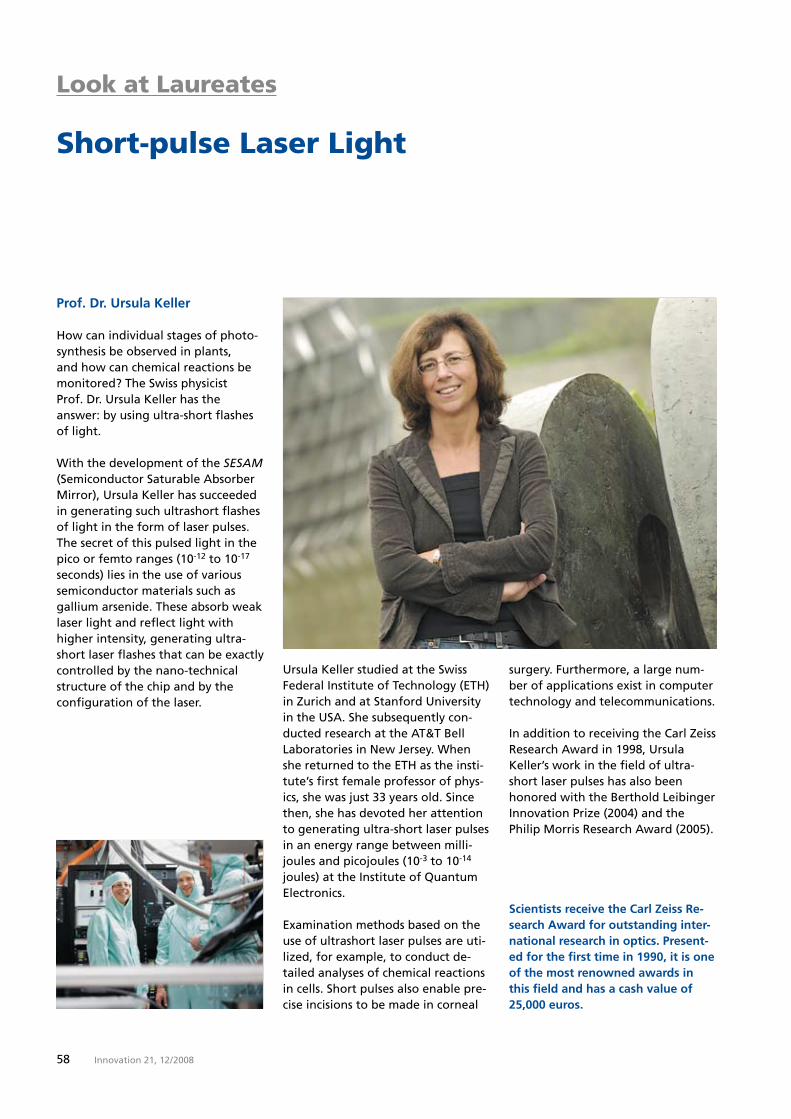
�8 Innovation 21, 12 / 2008
Look at Laureates
Short-pulse Laser Light
Prof. Dr. Ursula Keller
How can individual stages of photo-synthesis be observed in plants, and how can chemical reactions be monitored? The Swiss physicist Prof. Dr. Ursula Keller has the answer: by using ultra-short flashes of light.
With the development of the SESAM (Semiconductor Saturable Absorber Mirror), Ursula Keller has succeeded in generating such ultrashort flashes of light in the form of laser pulses. The secret of this pulsed light in the pico or femto ranges (10-12 to 10-1� seconds) lies in the use of various semiconductor materials such as gallium arsenide. These absorb weak laser light and reflect light with higher intensity, generating ultra-short laser flashes that can be exactly controlled by the nano-technical structure of the chip and by the configuration of the laser.
Scientists receive the Carl Zeiss Re-search Award for outstanding inter-national research in optics. Present-ed for the first time in 1990, it is one of the most renowned awards in this field and has a cash value of 25,000 euros.
surgery. Furthermore, a large num-ber of applications exist in computer technology and telecommunications.
In addition to receiving the Carl Zeiss Research Award in 1��8, Ursula Keller’s work in the field of ultra-short laser pulses has also been honored with the Berthold Leibinger Innovation Prize (200�) and the Philip Morris Research Award (200�).
Ursula Keller studied at the Swiss Federal Institute of Technology (ETH) in Zurich and at Stanford University in the USA. She subsequently con-ducted research at the AT&T Bell Laboratories in New Jersey. When she returned to the ETH as the insti-tute’s first female professor of phys-ics, she was just �� years old. Since then, she has devoted her attention to generating ultra-short laser pulses in an energy range between milli-joules and picojoules (10-� to 10-1� joules) at the Institute of Quantum Electronics.
Examination methods based on the use of ultrashort laser pulses are uti-lized, for example, to conduct de-tailed analyses of chemical reactions in cells. Short pulses also enable pre-cise incisions to be made in corneal

��Innovation 21, 12 / 2008
2009 – The Year of Astronomy: In 1�0�, Galileo aimed a telescope at the sky and discovered things that have changed how the world sees itself. This inspired the UNESCO to declare the �00th anniversary of this event the Year of Astronomy. Fit-tingly, Carl Zeiss presents VELVET, a video projection system for planetar-ium domes that renders the blackest black as never before.
Service
Preview of Innovation 22
EN_9
0_01
0_02
1I P
rin
ted
in G
erm
any
CM
-MSW
_II/
2009
NTo
o
Nobel Prize: The green fluorescing protein GFP discovered by Osamu Shimomura comes from the Aequorea victoria jellyfish. Martin Chalfie recognized its usefulness as a genetic marker. Roger Tsien developed GFP variations with dif-ferent fluorescing spectrums that enable the separate observation of single cell organelles.
Innovation – The Magazine from Carl ZeissIssue 21, December 2008
Issued by:Carl Zeiss AG, Oberkochen, GermanyCorporate Communications Jörg Nitschke
Editors:Silke Schmid (Editor-in-Chief) Dr. Dieter Brocksch, Gudrun VogelCarl Zeiss AGCorporate Communications Carl-Zeiss-Str. 22����� Oberkochen, [email protected] +�� (0) ����/20-8208Fax +�� (0) ����/20-�122
Contributors to this issue:Monika Etspüler, Ursula Walther, MSW;Dr. Ingrid Fritz, Dr. Dieter Brocksch, Carl Zeiss; Dr. Birgit Herden, Max Keller
English Translation: Clive Poole, Paul Soos
Conception and design:Overall coordination: Nicola Schindler MSW, Manfred Schindler Werbeagentur OHG, Aalen; www.msw.de
Pictures courtesy of: Carl Zeiss; Cover photo: Corbis, Rhett Butler; Page � top: dpa; Page � bottom: © 2008 PROKINO Filmverleih GmbH; Page � Top: Dr. Timo Mappes; S. 1�-1�: Prof. E. Fuchs, S. Bauch, Primate Center Göttingen; Page 18: Dr. J. Livet, MCB, Harvard University; Page 20: E. Dultz, EMBL, Heidelberg; Page 21: Prof. R. Schna-bel, Institute for Genetics, Technical Univer-sity of Brunswick; Page 22 left: Prof. M. Bastmeyer, University of Karlsruhe, Dr. Monika Marx, Carl Zeiss; Page 22 right: S. Sheu, MCB, Harvard University; Pages 28-2�: Prof. Dr. Christoph Bräuchle, Ludwig-Maxi-milians University Munich; Page �0: Michael W. Davidson, Florida State University; Pages �2-��: dpa; Pages ��-��: Thuringian Office for the Preservation of Historical Monu-ments and Archaeology, Weimar; Pages ��-��, Page ��: Art History Museum, Vienna; Pages �0-�1: German Film Museum, Frank-furt; Pages ��-��: C.+W. Schäper; Page ��: DLR; Pages �0/�1: Corbis; Page �2: Getty Images; Pages ��-��: Max Keller; Page �8: ETH Zurich; Page �� top: Corbis
Printing: C. Maurer Druck und Verlag, Geislingen an der Steige
Innovation – The Magazine from Carl Zeiss appears twice a year in German and English.
ISSN 1��1-80�0
The contents of the articles do not always reflect the opinion of the publisher. Reprint only with the exclusive written permission of Carl Zeiss.
To order an issue or change your mailing address, please contact Renate König +�� (0) ����/20-28�8 or [email protected]
Focal length: 1�00 millimeters, 1:� speed, 1� lens elements in 1� assem-blies, 1.� meters long and 2�� kilo-grams heavy – the second largest civilian tele lens in the world – devel-oped and built by Carl Zeiss. It will be delivered in the spring.

Innovation –
The Magazine from Carl Zeiss can
be found on the Internet at
www.zeiss.de/innovation


