Liquid Crystalline Characteristics of Site-Selectively ...
8
Macromolecules 1993,26, 5999-6006 5999 Liquid Crystalline Characteristics of Site-Selectively-Modified Chitosan Deeleep K. Rout, Satish K. Pulapura, and Richard A. Gross' Department of Chemistry, University of Massachusetts Lowell, Lowell, Massachusetts 01854 Received January 21, 1993; Revised Manuscript Received June 14, 1993 ABSTRACT: The site-selective modification of chitosan to form N-phthalolyl- (PhCh) and N-phthalolyl- 3,6-di-O-acetyl chitosan (AcPhCh)derivativeswas studied. It was determined that a preswelling of chitosan was required to carry out the phthaloylation reaction. furthermore, the PhCh and AcPhCh derivatives formed herein showed significant repeat unit substitution heterogeneity, which was unexpected based on previous literature reports. PhCh and AcPhCh were further investigated in regards to their formation of liquid crystalline phases. Optical techniquessuch as circular dichroism, optical absorptionspectroscopy, and polarized light microscopy were employed to study the critical concentration and the nature of the liquid crystallineorderingpresent in the above systems. It was determinedthat these derivativesformed lyotropic liquid crystal phases in a number of organic solvenb including dimethylsulfoxide (DMSO)and dioxane. The critical concentrations (in polymer weight fraction) at which liquid crystalline ordering was found to be present for PhCh in DMSO, AcPhCh in DMSO, and AcPhCh in dioxane were 0.25-0.27, 0.1, and 0.07, respectively. Therefore, lower polymer weight fractions were required for the formation of mesophases by AcPhCh relative to PhCh. The handedness of the cholesteric helical sense for AcPhCh was reversed from left- to right-handed when the solvent was changed from DMSO to dioxane. Also, acetylation of PhCh (forming AcPhCh) causes a reversal of the cholesteric helicoidal twist sense from left- to right-handed for lyotropic DMSO solutions. Introduction Chitosan is a polymer which contains P-l-4-linked 2-amino-2-deoxy-D-glucopyranose repeat units and is readily obtained by the N-deacetylation of chitin, a naturally abundant polysaccharide. These biopolymers are the structural components of the cuticles of crusta- ceans, insects, and mollusks and are also found in the cell walls of some microorganisms.l In spite of their relative abundance, chitosan and chitin have only found limited applications. Since chitosan is biodegradable, relatively nontoxic, nonimmunogenic, and biocompatible in animal tissues,2much research has been directed toward its use in medical applications such as drug deli~ery,~ artificial skin, and blood anticoagulant^.^ Chitosan has also been suggested for use as chelating agents for metal^,^ floccu- lants, adhesives, food processing, paper and textile ad- hesives,6 and membra ne^.^ The presence of the glucosamine repeat units in chitosan presents obvious advantages over cellulosefor site-specific modifications. Since the primary amino group at the ring C2 position and the primary and secondary hydroxyl functionalities a t the ring C6 and C3 positions, respectively, have distinctly different reactivities, the opportunity exists for site selectivity such that the desired side chain substituents may be introduced at predetermined positions of the sugar rings. This approach offers the exciting opportunity to create specific spatial orientations between side chain groups which are linked to a chiral stereoregular polymer chain. Although there have been several reports of chemically modified chitosans,a20 there has been very limited work on the site-specific modification of chitosan. Of particular interest, selective 0-acylation,21*22 N- acylation,%%N-alkylation,na and N-carboxyalkylation~z have been reported. Further, while some of the derivatives reported thus far are water-soluble or ~wellable,~~*~~933 only a few of the derivatives are soluble in organic so1vents.M-35 Nishimura and co-workers have recently reported the synthesis of some chemically modified derivatives of * To whom correspondence should be addressed. 0 Abstract published in Advance ACS Abstracts, October 1,1993. 0024-929719312226-5999$04.00/0 chitosans with improved organic s o l ~ b i l i t y . ~ ~ These researchers successfully exploited the use of N-phtha- loylchitosan derivatives so as to disrupt the formation of specific hydrogen bond interactions and thereby increase the solubility of the corresponding modified chitosan p0lymers.3~ Since 1976,the development of lyotropic chiral nematic mesophases for semirigid cellulose derivatives has been studied by many research g ro~ps.~~-~~ Thermotropic liquid crystals have also been observed for cellulose derivatives such as (hydroxypropyl)cellulose40 and other cellulose ethers.41 The lyotropic and thermotropic liquid crystals formed by these derivatives were found to be cholesteric in all cases. Recently, cellulose tri-n-alkanoates were prepared which showed a columnar m e ~ o p h a s e . ~ ~ In cases where cholesteric mesophases were formed, changes in the twist sense for chiral nematics of cellulose derivatives occur by simply altering the degree of sub- stitution, as was observed for ethyl cell~lose.4~ In addition, the chiralnematic pitch is extremely sensitive to the solvent nature,Y45 temperature,43@ c ~ n c e n t r a t i o n ~ ~ and molar mass.44 Gray and co-workers recently demonstrated changes in helicoidal twist sense by altering the chain length of n-alkyl substituents in the 0-2 and 0-3 positions of the sugar residuese48 In contrast to the above,few studies have been conducted on the ability of chitosan and various modified derivatives to form liquid crystalline phases. Terbojevich et a1.49 reported a persistence length of ca. 220 A in a 0.1 M acetic acid-0.2 M sodium chloride aqueous solution for chitosan samples with degree of acetylation (da) values of 42 and 15%. This work indicates that the chitosan chains are sufficiently rigid to facilitate the formation of mesophases at adequately high polymer concentrations. Ogura dem- onstrated that chitosan, (hydroxypropyl)chitosan, and (acetoxypropy1)chitosan form lyotropic chiral nematic ordered materials a t suitable concentration^.^^ Solutions of chitosan (>40 wt 5%) in 10% aqueous acetic acid and concentrated solutions of (hydroxypropy1)chitosan and (acetoxypropy1)chitosan in water and acetone, respectively, 0 1993 American Chemical Society
Transcript of Liquid Crystalline Characteristics of Site-Selectively ...
Macromolecules 1993,26, 5999-6006 5999
Liquid Crystalline Characteristics of Site-Selectively-Modified Chitosan
Deeleep K. Rout, Satish K. Pulapura, and Richard A. Gross' Department of Chemistry, University of Massachusetts Lowell, Lowell, Massachusetts 01854
Received January 21, 1993; Revised Manuscript Received June 14, 1993
ABSTRACT: The site-selective modification of chitosan to form N-phthalolyl- (PhCh) and N-phthalolyl- 3,6-di-O-acetyl chitosan (AcPhCh) derivatives was studied. It was determined that a preswelling of chitosan was required to carry out the phthaloylation reaction. furthermore, the PhCh and AcPhCh derivatives formed herein showed significant repeat unit substitution heterogeneity, which was unexpected based on previous literature reports. PhCh and AcPhCh were further investigated in regards to their formation of liquid crystalline phases. Optical techniques such as circular dichroism, optical absorption spectroscopy, and polarized light microscopy were employed to study the critical concentration and the nature of the liquid crystalline ordering present in the above systems. It was determined that these derivatives formed lyotropic liquid crystal phases in a number of organic solvenb including dimethyl sulfoxide (DMSO) and dioxane. The critical concentrations (in polymer weight fraction) at which liquid crystalline ordering was found to be present for PhCh in DMSO, AcPhCh in DMSO, and AcPhCh in dioxane were 0.25-0.27, 0.1, and 0.07, respectively. Therefore, lower polymer weight fractions were required for the formation of mesophases by AcPhCh relative to PhCh. The handedness of the cholesteric helical sense for AcPhCh was reversed from left- to right-handed when the solvent was changed from DMSO to dioxane. Also, acetylation of PhCh (forming AcPhCh) causes a reversal of the cholesteric helicoidal twist sense from left- to right-handed for lyotropic DMSO solutions.
Introduction Chitosan is a polymer which contains P-l-4-linked
2-amino-2-deoxy-D-glucopyranose repeat units and is readily obtained by the N-deacetylation of chitin, a naturally abundant polysaccharide. These biopolymers are the structural components of the cuticles of crusta- ceans, insects, and mollusks and are also found in the cell walls of some microorganisms.l In spite of their relative abundance, chitosan and chitin have only found limited applications. Since chitosan is biodegradable, relatively nontoxic, nonimmunogenic, and biocompatible in animal tissues,2 much research has been directed toward its use in medical applications such as drug de l i~e ry ,~ artificial skin, and blood anticoagulant^.^ Chitosan has also been suggested for use as chelating agents for metal^,^ floccu- lants, adhesives, food processing, paper and textile ad- hesives,6 and membra ne^.^
The presence of the glucosamine repeat units in chitosan presents obvious advantages over cellulose for site-specific modifications. Since the primary amino group at the ring C2 position and the primary and secondary hydroxyl functionalities a t the ring C6 and C3 positions, respectively, have distinctly different reactivities, the opportunity exists for site selectivity such that the desired side chain substituents may be introduced at predetermined positions of the sugar rings. This approach offers the exciting opportunity to create specific spatial orientations between side chain groups which are linked to a chiral stereoregular polymer chain. Although there have been several reports of chemically modified chitosans,a20 there has been very limited work on the site-specific modification of chitosan. Of particular interest, selective 0-acylation,21*22 N- acylation,%% N-alkylation,na and N-carboxyalkylation~z have been reported. Further, while some of the derivatives reported thus far are water-soluble or ~wellable,~~*~~933 only a few of the derivatives are soluble in organic so1vents.M-35 Nishimura and co-workers have recently reported the synthesis of some chemically modified derivatives of
* To whom correspondence should be addressed. 0 Abstract published in Advance ACS Abstracts, October 1,1993.
0024-929719312226-5999$04.00/0
chitosans with improved organic s o l ~ b i l i t y . ~ ~ These researchers successfully exploited the use of N-phtha- loylchitosan derivatives so as to disrupt the formation of specific hydrogen bond interactions and thereby increase the solubility of the corresponding modified chitosan p0lymers.3~
Since 1976, the development of lyotropic chiral nematic mesophases for semirigid cellulose derivatives has been studied by many research g r o ~ p s . ~ ~ - ~ ~ Thermotropic liquid crystals have also been observed for cellulose derivatives such as (hydroxypropyl)cellulose40 and other cellulose ethers.41 The lyotropic and thermotropic liquid crystals formed by these derivatives were found to be cholesteric in all cases. Recently, cellulose tri-n-alkanoates were prepared which showed a columnar m e ~ o p h a s e . ~ ~
In cases where cholesteric mesophases were formed, changes in the twist sense for chiral nematics of cellulose derivatives occur by simply altering the degree of sub- stitution, as was observed for ethyl cell~lose.4~ In addition, the chiralnematic pitch is extremely sensitive to the solvent nature,Y45 temperature,43@ c ~ n c e n t r a t i o n ~ ~ and molar mass.44 Gray and co-workers recently demonstrated changes in helicoidal twist sense by altering the chain length of n-alkyl substituents in the 0 - 2 and 0-3 positions of the sugar residuese48
In contrast to the above, few studies have been conducted on the ability of chitosan and various modified derivatives to form liquid crystalline phases. Terbojevich et a1.49 reported a persistence length of ca. 220 A in a 0.1 M acetic acid-0.2 M sodium chloride aqueous solution for chitosan samples with degree of acetylation (da) values of 42 and 15%. This work indicates that the chitosan chains are sufficiently rigid to facilitate the formation of mesophases a t adequately high polymer concentrations. Ogura dem- onstrated that chitosan, (hydroxypropyl)chitosan, and (acetoxypropy1)chitosan form lyotropic chiral nematic ordered materials a t suitable concentration^.^^ Solutions of chitosan (>40 wt 5%) in 10% aqueous acetic acid and concentrated solutions of (hydroxypropy1)chitosan and (acetoxypropy1)chitosan in water and acetone, respectively,
0 1993 American Chemical Society
6000 Rout et al. Macromolecules, Vol. 26, No. 22, 1993
Experimental Section Structural Analysis of the Synthesized Polymers. In-
frared (IR) spectra were recorded on a Perkin-Elmer Series 1600 spectrometer. AcPhCh films were cast directly on NaCl plates, while KBr pellets were used for PhCh. The spectra were recorded at a resolution of 4 cm-'. NMR measurements were made on a Bruker WP 270-SY spectrometer. The NMR spectra of PhCh and AcPhCh were recorded in DMSO-& and CDClS, respectively. The parameters employed were as follows: lH NMR, T = 80 "C for PhCh, T = 25 "C for AcPhCh, pulse width = 0.5 ps, receiver delay = 2 s, transients = 600-800; 13C NMR, T = 75 "C for PhCh, T = 25 O C for AcPhCh, pulse width = 10 p, receiver delay = 1 s, transients = 20000-3oooO. The typical sample concentration was 30 mg/mL. The following descriptors were used to describe the lH NMR and IR spectral results obtained herein: vs = very strong, s = strong, sh = shoulder, sha = sharp, b = broad, db = doublet, m = multiplet. Elemental analyses were performed at the Microanalysis Laboratory, University of Massachusetts, Amherst, after drying the polymer samples in a drying pistol at 55 "C (50 pmHg) for 48 h using PZOS as desiccant, with subsequent careful handling to avoid the adsorption of moisture.
Gel Permeation Chromatography (GPC) Molecular Weight Measurements. The molecular weights of the deriv- atives were determined by gel permeation chromatography (GPC) on a chromatographic system consisting of a Perkm-Elmer Model 410 pump, a Waters Model 410 RI detector, and a PE Nelson 2600 computerized data station. Two PL-gel GPC columns (300 mm X 7.7 mm, particle size 5 pm, pore size lo6 and 103 A) were placed in series, and the analysis was performed in DMF containing 0.1% (w/v) LiBr. The flow rate was 1 mL/min, the sample concentrations w e e 2 mg/mL, and the injection volumes were 20 bL. Molecular weights were calculated relative to polystyrene standards with no further corrections.
Thermal Analysis. Differential scanning calorimetry (DSC) measurements were performed on a DuPont TA 2000 and 2910 system with 5-7-mg samples at a heatingrate of 10 "C/min under Nz purge. Thermogravimetric analyses (TGA) were performed on a DuPont 2000 and 2950 system with 10-15-mg samples at a heating rate of 20 "C/min under Nz purge. The data from both DSC and TGA thermograms were analyzed using a TA 2000 data station. The onset of decomposition (To) was defined as the temperature a t which the polymer started to lose weight, and the decomposition temperature (Td) was defined as the temperature at which the rate of polymer weight loss with respect to temperature was maximum.
Preparation of Swollen Chitosan. Chitosan of low degree of acetylation (da) was obtained from Protan Laboratories and was used without further purification. The da of the nitrogen functionalities was verified by 'H NMRB and found to be 0.07. The above-described chitosan (10 g) was dissolved in 1 % aqueous acetic acid (1 L). The polymer was made insoluble by the addition of methanol (1 L) followed by 4% NaHCO3 aqueous solution (1 L). After the resultant mixture was stirred at room temperature for 2 h, the swollen gel-like precipitate was collected by filtration, washed with distilled water until the filtrate was neutral, and subsequently partially dried while in the Buchner filtration apparatus using a water aspirator for ca. 30 min. This wet precipitate was suspended in DMF, the suspension was stirred overnight, the DMF was filtered, and fresh DMF was added. After 1 h of stirring, the solvent was replaced one additional time. Finally, the DMF-swollen precipitate was collected by filtration and used for the synthesis of N-phthaloylchitosan (see below).
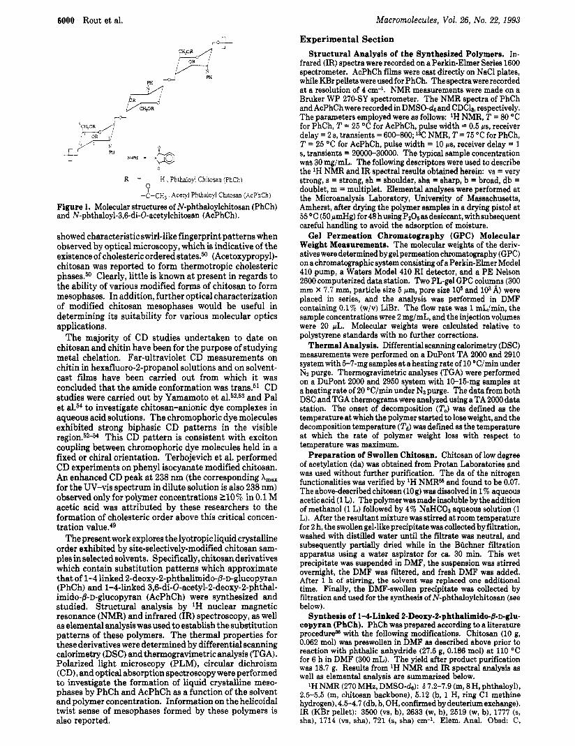
Synthesis of l-4-Linked 2-Deoxy-2-phthalimido-&~-glu- copyran (PhCh). PhCh was prepared according to a literature procedureg8 with the following modifications. Chitosan (10 g, 0.062 mol) was preswollen in DMF as described above prior to reaction with phthalic anhydride (27.5 g, 0.186 mol) at 110 "C for 6 h in DMF (300 mL). The yield after product purification was 18.7 g. Results from lH NMR and IR spectral analysis as well as elemental analysis are summarized below.
'H NMR (270 MHz, DMSO-&): S 7.2-7.9 (m, 8 H, phthaloyl), 2.5-5.5 (m, chitosan backbone), 5.12 (b, 1 H, ring C1 methine hydrogen),4.5-4.7 (db, b, OH,confirmedbydeuteriumexchange). IR (KBr pellet): 3500 (vs, b), 2633 (w, b), 2519 (w, b), 1777 (s, sha), 1714 (vs, sha), 721 (s, sha) cm-l. Elem. Anal. Obsd: C,
E //-+ m
/qd * *+OR /i-r
C w _--
h a - P i
R = H , Phthaloyi Chtosan (PhCh) 0
- ~ - C H ~ Acetyl Phtbaloyl Chmsan (AcPhCh)
Figure 1. Molecular structures of N-phthaloylchitosan (PhCh) and N-phthaloyl-3,6-di-O-acetylchitosan (AcPhCh).
showed characteristic swirl-like fingerprint patterns when observed by optical microscopy, which is indicative of the existence of cholesteric ordered states.50 (Acetoxypropy1)- chitosan was reported to form thermotropic cholesteric phases.50 Clearly, little is known at present in regards t o the ability of various modified forms of chitosan t o form mesophases. In addition, further optical characterization of modified chitosan mesophases would be useful in determining its suitability for various molecular optics applications.
The majority of CD studies undertaken to date on chitosan and chitin have been for the purpose of studying metal chelation. Far-ultraviolet CD measurements on chitin in hexafluoro-2-propanol solutions and on solvent- cast films have been carried out from which it was concluded that the amide conformation was trans.5l CD studies were carried out by Yamamoto et al.52~53 and Pal et al." to investigate chitosan-anionic dye complexes in aqueous acid solutions. The chromophoric dye molecules exhibited strong biphasic CD patterns in the visible
This CD pattern is consistent with exciton coupling between chromophoric dye molecules held in a fixed or chiral orientation. Terbojevich et al. performed CD experiments on phenyl isocyanate modified chitosan. An enhanced CD peak at 238 nm (the corresponding A,, for the UV-vis spectrum in dilute solution is also 238 nm) observed only for polymer concentrations 110% in 0.1 M acetic acid was attributed by these researchers to the formation of cholesteric order above this critical concen- tration value.49
The present work explores the lyotropic liquid crystalline order exhibited by site-selectively-modified chitosan sam- ples in selected solvents. Specifically, chitosan derivatives which contain substitution patterns which approximate that of 1-4 linked 2-deoxy-2-phthalimido-~-~-glucopyran (PhCh) and l-Clinked 3,6-di-O-acetyl-2-deoxy-2-phthal- imido-0-D-glucopyran (AcPhCh) were synthesized and studied. Structural analysis by lH nuclear magnetic resonance (NMR) and infrared (IR) spectroscopy, as well as elemental analysis was used to establish the substitution patterns of these polymers. T h e thermal properties for these derivatives were determined by differential scanning calorimetry (DSC) and thermogravimetric analysis (TGA). Polarized light microscopy (PLM), circular dichroism (CD), and optical absorption spectroscopy were performed to investigate the formation of liquid crystalline meso- phases by PhCh and AcPhCh as a function of the solvent and polymer concentration. Information on the helicoidal twist sense of mesophases formed by these polymers is also reported.

Macromolecules, Vol. 26, No. 22, 1993
57.17; H, 4.45; N, 3.50. Calcd for a degree of N-acetylation of 0.07 andadegreeofphthaloylationof 1.45,respectively: C,60,13; H, 4.04; N, 3.99. Molecular weight: M., = 183000. MJM, = 1.83.
Synthesis of 1-4-Linked 3,6-Di-Oacetyl-2-deoxy-2-ph- thalimido-@-D-glucopyran (AcPhCh). The acetyl derivative of PhCh, AcPhCh, was prepared by the reaction of PhCh (1 g) with acetic anhydride (20 mL, Aldrich, 99+%) using pyridine (30 mL, Aldrich, 99%) as the solvent and base following a literature procedure.= Results from 1H NMR and IR spectral analysis as well as elemental analysis are summarized below. lH NMR (270 MHz, CDCls): 6 7.2-7.9 (m, 5.8 H, phthaloyl),
1.4-2.1 (m, 4.7 H, 0- and N-acetyl). IR (film, NaCl plate): 2639 (w, b), 1777 (8, sha), 1746 (vs, sha), 1714 (vs, sha), 1227 (8, sha), 721 (e, sha) cm-l. Elem. Anal. Obsd C, 57.92; H, 4.54; N, 3.22. Calcd. for degree of substitution (ds) values for N-acetylation, N-phthaloylation, 0-phthaloylation, and 0-acetylation of 0.07, 0.93, 0.52, and 1.50, respectively: C, 59.68; H, 4.16; N, 3.39. Molecular weight: M, = 143000. MJM. = 1.58.
Sample Preparation. Different concentrations of the poly- mer solutions were prepared separately in small glass vials. The polymer weight fraction, x , is defined as the weight of the polymer divided by the weight of the solution. When desired, the solutions were homogenized by heating them in a thermostated oven (well below the boiling point of the respective solvents and decom- position temperature of the respective polymer). The vials were tightly capped with Teflon tape so that the solvent was not evaporated during this process. The homogenized solution was then sandwiched between two glass plates to form the sample cells. The thickness of the cell was defined either by using mylar spacers of 75-pm thickness or by the quantity of the sample added between the glass plates in the absence of the mylar spacers. The thickness of both types of cells was later determined using a micrometer with an accuracy of *5 pm. To prevent solvent evaporation, the cells were sealed from all the sides using an epoxy. For dilute solution spectra, Supracil quartz cuvettes were used.
Other Instrumental Methods for Optical Studies. Cir- cular dichroism (CD) spectra were recorded at room temperature (20-22 O C ) using a Jasco 5-710 spectropolarimeter under a Nz atmosphere. UV-vis absorption spectra were recorded using a Perkin-Elmer A9 spectrophotometer. Polarized light microscopy (PLM) was performed using a Leitz Ortholux polarizing micro- scope (with X320 magnification). For dilute solution CD and optical absorption spectra, quartz cuvettes with path lengths of 0.1 and 1 cm, respectively, were used. For the concentrated solution studies (both isotropic and liquid crystalline), the CD, UV-vis, and PLM experiments were all performed using the same cell which was constructed using glass plates.
Results and Discussion Synthesis and Structural Analysis of Chitosan
Derivatives. The intractability of chitosan has been attributed to the chain rigidity, material crystallinity, and the strong intra- and interchain hydrogen b 0 n d i n g . ~ @ * ~ ~ 5 ~ An important component of the hydrogen-bonding in- teractions for this system involves the primary amino functionalities along the polymer chain. These hydrogen bond interactions can be effectively disrupted by carrying out appropriate chemical modifications of the chitosan functional groups. N-Phthaloylation of chitosan with phthalic anhydride has been successfully employed by other researchers to disrupt the amino functionality hydrogen-bonding interaction^.^^ This resulted in a number of N-phthaloylchitosan derivatives which showed good solubility in a range of organic solvents. We, therefore, adopted this approach in our research to obtain site-selectively-modified chitosan derivatives with the desired solubility characteristics for subsequent evaluation of the lyotropic liquid crystalline ordering. Unfortunately, when chitosan (da 0.07; see Experimental Section) was heated with phthalic anhydride in dimethylformamide (DMF) at 130 OC for a reaction time of 7 h (and as long as 24 h) following the method described by Nishimura
Site-Selectively-Modified Chitosan 6001
and Kurita,36 the product unexpectedly remained insol- uble in dimethyl sulfoxide (DMSO), dimethylacetamide (DMAc), dimethylformamide (DMF), and pyridine. Chi- tosan with a gel-like texture was then prepared by precipitation of an aqueous acetic acid chitosan solution into aqueous NaHC03 (see Experimental Section for further details). After the gel-like precipitate was washed with water to remove excess base followed by subsequent multiple solvent replacements with DMF, the phthaloy- lation reaction with preswollen chitosan was successfully carried out. It therefore appears that the chitosan starting material used herein, due to its solid-state morphology, had a low accessibility of the reactive amino functionalities to the reagents utilized so that a preswelling of the material was necessary. Possibly, the slightly higher da of the chitosan used in this work relative to that of previous workers (0.07 relative to <0.01) as well as differences in the chitosan sample molecular weight and crystallinity might explain the observed change in chitosan reactivity.
The IR spectrum of PhCh prepared herein was in agreement with a spectrum published earlier.36 The 'H NMR of PhCh showed two sets of broad unresolved peaks: one set consisting of three peaks centering at 7.4, 7.7, and 7.8 ppm (assigned to the phthaloyl group) and a second set between 2.5 and 5.5 ppm due to the chitosan backbone hydrogens. The peak at 5.12 ppm was assigned to the ring C1 and the peaks at 4.7 and 4.5 ppm, which disappeared upon exchange with DzO, were assigned to the unreacted hydroxyl functionalities. Elemental analysis results for PhCh (see Experimental Section) showed a lower nitrogen content than would be expected for a total ds of 1.0 at the nitrogen (0.07 N-acetylation and 0.93 N-phthaloylation). This indicated that significant phthaloylation at the hydroxyl functionalities of chitosan occurred in addition to N-phthaloylation. Based on the elemental analysis results, the calculated best fit degree of phthaloylation is 1.45. The value of 1.45 for the degree of phthaloylation, which is greatly in excess of 0.93, was further supported from the PhCh 'H NMR spectral integration values (see Experimental Section). Further- more, the lower wavenumber vibrational band at -2630 cm-l observed by IR spectroscopy (see Experimental Section) indicates that 0-phthaloylation took place with the formation of pendant acid groups. In review of the previous work carried out for the synthesis of PhCh,36 concerns arise as to whether the researchers did indeed obtain a degree of phthaloylation of 1.0. Specifically, elemental analysis results, corrected for various amounts of water retained in the sample, were reported although no information was provided as to how that quantity of retained water was measured. Furthermore, 'H NMR spectralintegration or 13C NMR analysis was not provided by these workers to confirm the proposed repeat unit structural homogeneity. Finally, PhCh samples recently prepared in our laboratory with an N-phthaloylation of ca. 1.0 showed poor solubility in DMF and pyridine.59
The 0-acetylated derivative, AcPhCh, was prepared from PhCh (degrees of phthaloylation and N-acetylation of 1.45 and 0.07, respectively) following a literature procedure.6O The IR spectrum obtained showed all the characteristic absorption bands that have been reported earlier for this derivative.36 The 1H NMR spectrum of AcPhCh was qualitatively similar to that reported earlier.% In the acetyl region, the 'H NMR signals for the methyl protons (between 1.4 and 2.2 ppm) appeared as a set of unresolved peaks. An average degree of 0-acetylation for this product was obtained from analysis by both IH NMR spectral integration and elemental analysis (see below and the Experimental Section). The lH NMR signal intensity integration ratio of the N-phthaloyl protons to the 0- and

6002 Rout et al. ~acromolecules, Vol. 26, No. 22, I993
PhCh-x-DMSO
Nematic Granular Texture Nematic Banded Texture
t &l.u * 009
3w I'" 4w 4SO 500 Id . 4, __- 210
wavc1mgh [nm]
Figure 2. Optical characterizations of PhCh-xDMSO solutions: (a) microphotographs of PhCh-rDMSO showing nematic (granular) and banded textures; (b) UV-vis spectra of dilute, concentrated (x = O W , and liquid crystalline PhCh-xDMSO; (c) CD spectra of PhCh-xDMSO, x = O.M)8, 0.18, 0.22, and 0.27.
N-acetyl protons in the 'H NMR spectrum of AcPhCh Liquid Cryetalline Studies. PhCh was found to be was found to be 1.22. This sueeesta. assumine no soluble in theoolarsolvents DMSO. DMF.and Dvridine." dephthaloylation had occurred, tha'ta degree of 0-acety- lation of 1.50 was obtained. The elemental composition of AcPhChassuming N-acetylation of 0.07, phthaloylation of 1.45, and 0-acetylation of 1.50 was calculated as C, 59.7%;H,4.16%; N,3.39%. Theexperimentalelemental analysisresultsobtained (C,57.92%;H,4.54%,N,3.22%) were in g o d agreement with the substitution pattern determined by 'H NMR. I3C NMR analysis of AcPhCh synthesized herein (spectrum not shown) showed com- plexity consistent with a heterogeneous repeat unit composition. It is of interest to note that similar com- plexity can be seen in the previously published 13C NMR of AcPhCh.36 where i t was claimed that the polymer had a homogeneous repeat unit substitution.
ThermalProperties. The thermalpropertiesofPhCh and AcPhCh were analyzed by DSC and TGA (see Experimental Section). No glass transition temperatures (T,) WeredetectedforeitherPhChor AcPhCh. Theonset ofdecompositiontemperatures (To) for PhChand AcPhCh were 171 and 167 "C, respectively, and the decomposition temperatures (Td) were 345 and 350 "C, respectively, as determined byTGA. Byvisual observationusingamelting point apparatus, it was found that the decomposition of PhCh and AcPhCh occurred prior to liquification. There- fore, thermotropic liquid crystalline mesophases could not be formed by these modified chitosan samples.
Solutions of PhCh-xDMSO ( x being the p o l g e r weight fraction) withx = 0.09,0.14,0.18,0.22,0.25,0.27,and0.31 were prepared and transferred to cells where the samples were sandwiched between glass plates (see Experimental Section). The cells were first examined under the mi- croscope, and, for x 5 0.25, they appeared dark under crosspolars. Witha further increasein the polymerweight fraction to 0.27, the texture under the microscope using crossed polarized light indicated that a transition toaliquid crystalline phase hadoccurred (seeFigure2a). Therefore, the critical concentration, x I (in polymer weight fraction), for PhCh to be liquid crystalline in DMSO is ca. 0.26. The textures for PhCh4.27 DMSO and PhCh4.31 DMSO, shown in Figure 2a, corresponds to nematic (granular) and banded textures, respectively. The banded texture arising a t a relatively higher concentration may be due to shear which is invariably applied during the preparation of the cell.
The optical absorption spectra (250-500 nm) of PhCh- xDMSO solutions (from dilute (4 X l(r g/mL) to concentrated ( x = 0.09,0.27)) are shown in Figure 2b. The dilute solution spectrum showed two absorption bands at -255 and 275 nm. The peak a t 255 nm appears narrow becauseit isclosetothecutoffwavelengthof DMSO (-245 nm). Analysis of the intermediate polymer weight fraction sample (x = 0.09) sample showed onlyone broad absorption

Macromolecules, Vol. 26, No. 22, 1993 Site-Selectively-Modified Chitosan 6003
ACPhCh-x-DMSO
Biphasic (x = 0.09)
(b)
Liquid crystalline (s = 0.12)
(C)
&me+ 0.11 - 0.19 +
r Liquid crystalline (x = 0.19)
-3woI , , . . , ! 300 350 400 450 500
wavelength Inml
WaVclmBh [mu1
Figure 3. Optieal charaeterizations of AcPhCh-xDMSO solutions: (a) microphotographs of AcPhCh-xDMSO showing biphasic (x = 0.09) and anisotropic (x = 0.12 and 0.19) liquid crystalline phases; (b) UV-vis spectra of dilute and liquid crystalline (x = 0.12 and 0.19) solutions of AcPhCh-DMSO; (c) CD spectra of AcPhCh-xDMSO, x = 0.008, 0.09,0.12, and 0.19.
band centering around 300 nm. With a further increase in the polymer concentration to x = 0.27, a transition to aliquidcrystallinephaseoccursaswasverified by polarized light microscopy (PLM). In addition, above the xc (at x = 0.27) an absorption band a t 320 nm along with the main absorption band a t ca. 275 nm was observed. It may be emphasized that the observation of a sharp absorption band a t 320 nm corresponds to the existence of liquid crystallinity in thispolymer-solvent system. Furthermore, it will become clear from discussions presented below that this absorption band at 320 nm does not arise due to selective reflection of the cholesteric helix pitch, but may have ita origins from specific interactions of the phthaloyl side groups in the confined liquid crystalline phase. The absorption band at 255 nm, observed for dilute solutions, could not be detected for more concentrated solutions, including solutions which were liquid crystalline, because glass plates with higher cutoff wavelength were used to prepare the cells.
Circular dichroism (CD) spectra were recorded as a function of polymer concentration for PhCh-xDMSO at room temperature (20-23 "C). CD spectra for x = 0.008, 0.18,0.22,and0.27 areshown inFigure 2c. Dilutesolution ( x = 0.008) CD spectra showed a positive peak a t 329.5 nm with a shoulder at 320 nm. The shoulder band further developed with an increase in the polymer weight fraction up to 0.22. When the transition to the liquid crystalline phase takes place, a distinctive wide CD band centered a t
329.5 nm which tails into the visible range appeared and the positive CD absorption shoulder a t 320 nm was no longer apparent.
An analysis similar to the one described for PhCh in DMSO was alsocarried out for AcPhCh-xDMSO systems. This allowed for the determination of the effects caused by acetylation a t the 0-3 and 0-6 positions on the liquid crystalline properties. Solutionsofvarying polymerweight fractions (AcPhCh-xDMSO) with x = 0.008-€1.22 were prepared (see Experimental Section). The x. in polymer weight fraction for this system was found to be 0.09 from PLM studies (see Experimental Section). However, a t this weight fraction, the whole area of the cell was not birefringent; rather, i t appeared to be a biphase containing isotropic as well as anisotropic regions (see Figure 3a). With a further increase in the polymer weight fraction to 0.12, the whole area of the cell became birefringent and the texture observed was typical of a nematic granular texture (see Figure 3a). At higher polymer weight fraction values (x = 0.19), nematic texture with lines not uniformly spaced appeared. These lines correspond to a banded texture, which also appeared in the PhCh-0.27DMSO system (see above).
UV-vis as well as CD spectra were recorded for these samples a t various polymer weight fractions. Selected spectra are shown in Figure 3b (UV-vis) and Figure 3c (CD). The dilute solution (3 X lCr3 g/mL) optical absorption spectrum of AcPhCh-xDMSO showed two

6004 Rout et al.
distinct bands at 255 and 293 nm. With increasing concentration of AcPhCh, the absorption spectra (see Figure 3b) showed similar characteristic changes to those observed for the PhCh-DMSO system. The CD spectra were quite sensitive to concentration effects and showed rather complex behavior. As was observed for PhCh- DMSO (see Figure 2c), a CD spectrum of a dilute AcPhCh- DMSO solution ( x = 0.008) showed a positive band a t 329 nm with a shoulder a t 320 nm (see Figure 3c). With further increase in the polymer concentration up to that of xc ( x = 0.09), a small negative band a t 340 nm appeared in addition to areversal in sign of the band at 320 nm (positive to negative). The negative band at 340 nm with a long tail into the visible region is attributed to the appearance of liquid crystalline order in the polymer solution (liquid crystal induced). With a further increase in the polymer weight fraction value to 0.19, the negative CD band with a A,, at 340 nm dominates the spectral features. The positive band at 329 nm which was prominent in dilute solution now appears as a relatively weak band due to the presence of the two intense negative bands at 340 and 320 nm (see Figure 312). Since the phthaloyl chromophoric groups are a t both the nitrogen and oxygen positions of the repeat units with a nonhomogeneous distribution, no attempt has been made herein to further interpret the effects of side group ordering during the isotropic to liquid crystalline transition by CD spectral changes at 320 and 329 nm (due to the phthaloyl chromophore).
The distinctively broad positive and negative CD bands that tail into the visible region which were observed for the liquid crystalline mesophases formed by PhCh and AcPhCh in DMSO, respectively, arise due to mesophase order. It should be noted here that these broad CD bands are not due to artifacts from induced birefringence in sample cells since there was no variation in the CD spectra recorded when the sample cells were rotated by 90'. These mesophases, due to the chirality of the natural-origin polymer, are expected to be chiral nematic (cholesteric). However, the textures of these lyotropic mesophases when sandwiched between two glass plates were nematic. Furthermore, CD spectra of these mesophases recorded up to 700 nm do not show a CD band resolved from that due to the phthaloyl chromophoric group which could have been directly attributed to selective reflection of a mac- roscopic cholesteric pitch. Moreover, the CD signal intensities at h values below that normally attributable to the phthaloyl chromophore CD signal (at ca. 290 nm) were relatively weak while higher intensity CD signals were observed in the visible wavelength region so that it is unlikely that the PhCh and AcPhCh mesophases in DMSO have pitch values below 290 nm. In addition, cholesteric mesophase pitch values are normally extremely sensitive to changes in the side chain substituent group structure and polymer concentration (see discussion in the Intro- duction) so that it would be rather unlikely that a resolution of the selective reflection band and the phthaloyl chro- mophore CD bands for both the PhCh and AcPhCh mesophases would not have been achieved. Considering the above discussion, it therefore seems most likely that the mesophases formed by PhCh and AcPhCh in DMSO consist of only short-range ordered domains which do not show uniform cholesteric macroscopic helices (poor cho- lesteric organization). This would explain the absence of a well-defined intense peak for these mesophases which can be directly attributed to selective Bragg reflection. Indeed, for other modified chitosan systems which are currently under study in our laboratory where dominant selective Bragg reflection bands were well resolved from the phthaloyl chromophoric CD band, the CD behavior a t elevated temperatures which approach the clearing tran-
Macromolecules, Vol. 26, No. 22, 1993
Table I lyotropic solution X c (wt %) twist sense PhCh-DMSO 0.21 left-handed AcPhCh-DMSO 0.12 right-handed AcPhCh-dioxane 0.08 left-handed
sition (where ordering of the cholesteric mesophase is poor) is similar to that described above for PhCh and AcPhCh at room temperature. Therefore, we believe that the CD signal which tails into the visible region for liquid crystalline phases studied herein originates from a cho- lesteric mesophase which does not have sufficient order to show a resolved selective Bragg reflection band. It is important to note that even a poorly ordered cholesteric mesophase will have an associated preferred macroscopic helical sense (right- or left-handed) which can be deter- mined by the sign of the CD signal observed at the appropriate wavelengths.
Interesting differences are immediately apparent from the above discussion for the mesophases formed by PhCh and AcPhCh in DMSO. Specifically, the value of x c for the PhCh-xDMSO system is much larger than that for the AcPhCh-xDMSO system (see Table I). Although the experiments have not as yet been carried out to determine the reason for this result, it may be hypothesized that the AcPhCh has a greater propensity for specific chain-chain association phenomena and might have a higher axial ratio than PhCh in DMSO. Consistent with this discussion is the observation that AcPhCh-DMSO readily forms a gel a t ambient temperatures and can be termed a gel liquid crystal whereas in the case of PhCh, gelation was not significant even up to x = 0.31. In addition, it appears from observation of the sign of the CD signal a t wavelength values above that associated with the phthaloyl chro- mophoric group that acetylation of PhCh (forming AcPhCh) causes a reversal of the cholesteric helicoidal twist sense from left- to right-handed for lyotropic DMSO solutions. A similar result was also reported for ethyl- ~ e l l u l o s e . ~ ~ Specifically, acetylation of ethylcellulose (ds 2.5) reverses the handedness of the cholesteric macroscopic helix in the same solvent.
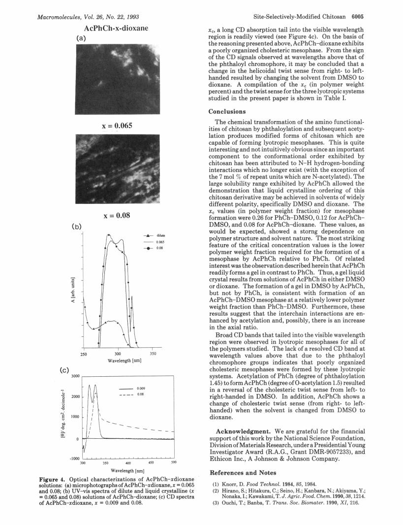
The solubility of AcPhCh in solvents of greatly different dielectric constants, specifically DMSO and dioxane, allowed for the investigation of solvent effects on AcPhCh mesophase formation. Solutions of AcPhCh-xdioxane with x = 0.009,0.065,0.08, and 0.11 were prepared. PLM results showed that x c occurred at the low polymer weight fraction value of 0.08. The microscopic texture observed at room temperature for the polymer weight fraction 0.08 is shown in Figure 4a. The texture seen was similar to those observed for the liquid crystalline AcPhCh-DMSO systems (see Figure 3a).
The UV-vis as well as the CD spectra for AcPhCh- xdioxane samples are shown in Figure 4. The dilute solution (3 x 10⁻³ g/mL) UV-vis absorption spectrum showed two distinct bands at 293 and 243 nm. The latter peak is not shown but was observed without difficulty since the UV cutoff for dioxane is -220 nm. In contrast, the lower wavelength absorption band for dilute solutions of PhCh and AcPhCh in DMSO appeared narrowed due to its proximity to the DMSO UV cutoff wavelength of -250 nm (see Figures 2b and 3b, respectively). The general characteristics of the UV-vis absorption spectra obtained for AcPhCh in both DMSO and dioxane are rather similar (see Figures 3b and 4b, respectively). A dilute solution CD spectrum ( x = 0.009) shows two positive CD bands at 316 and 328 nm, respectively, which is similar to the dilute solution CD spectra recorded of both PhCh and AcPhCh in DMSO (see Figures 2c and 3c). At the polymer weight fraction value 0.08, which is equivalent to
Macromolecules, Vol. 26, No. 22, 1993
AcPhCh-x-dioxane
x = 0.065
w .IWl " " ' ' " " " ' " ' " "
IW I50 400 l J O rta
Wavclcoglb [nm]
Figure 4. Optical characterizations of AcPhCh-xdioxane solutions: (a) microphotographsof AcPhCh-xdioxane,~ = 0.065 and 0.08; (h) UV-vis spectra of dilute and liquid crystalline ( x = 0.065 and 0.08) solutions of AcPhCh-diorane; (e) CD spectra of AcPhCh-xdioxane, x = 0.009 and 0.08.
Site-Selectively-Modified Chitosan 6005
x,, a long CD absorption tail into the visible wavelength region is readily viewed (see Figure 4c). On the basis of the reasoning presented above, AcPhCh-dioxane exhibits a poorly organized cholesteric mesophase. From the sign of the CD signals observed a t wavelengths above that of the phthaloyl chromophore, it may be concluded that a change in the helicoidal twist sense from right- to left- handed resulted by changing the solvent from DMSO to dioxane. A compilation of the x, (in polymer weight percent) and thetwist sense forthe threelyotropicsystems studied in the present paper is shown in Table I.
Conclusions The chemical transformation of the amino functional-
ities of chitosan by phthaloylation and subsequent acety- lation produces modified forms of chitosan which are capable of forming lyotropic mesophases. This is quite interesting and not intuitively obvious since an important component to the conformational order exhibited by chitosan has been attributed to N-H hydrogen-bonding interactions which no longer exist (with the exception of the 7 mol 96 of repeat units which are N-acetylated). The large solubility range exhibited by AcPhCh allowed the demonstration that liquid crystalline ordering of this chitosan derivative may be achieved in solvents of widely different polarity, specifically DMSO and dioxane. The x. values (in polymer weight fraction) for mesophase formation were 0.26 for PhCh-DMSO, 0.12 for AcPhCb- DMSO, and 0.08 for AcPhCh-dioxane. These values, as would he expected, showed a storng dependence on polymer structure and solvent nature. The most striking feature of the critical concentration values is the lower polymer weight fraction required for the formation of a mesophase by AcPhCh relative to PhCh. Of related interestwastheobservationdescribed hereinthatAcPhCh readily forms a gel in contrast to PhCh. Thus, a gel liquid crystal results from solutions of AcPhCh in either DMSO or dioxane. The formation of a gel in DMSO by AcPhCh, but not by PhCh, is consistent with formation of an AcPhCh-DMSO mesophase a t a relatively lower polymer weight fraction than PhCh-DMSO. Furthermore, these results suggest that the interchain interactions are en- hanced by acetylation and, possibly, there is an increase in the axial ratio.
Broad CD bands that tailed into the visible wavelength region were observed in lyotropic mesophases for all of the polymers studied. The lack of a resolved CD band at wavelength values above that due to the phthaloyl chromophore groups indicates that poorly organized cholesteric mesophases were formed by these lyotropic systems. Acetylation of PhCh (degree of phthaloylation 1.45) toformAcPhCh(degreeof0-acetylation 1.5) resulted in a reversal of the cholesteric twist sense from left- to right-handed in DMSO. In addition, AcPhCh shows a change of cholesteric twist sense (from right- to left- handed) when the solvent is changed from DMSO to dioxane.
Acknowledgment. We are grateful for the financial support of this work by the National Science Foundation, Division ofMaterials Research, underapresidential Young Investigator Award (R.A.G., Grant DMR-9057233), and Ethicon Inc., A Johnson &Johnson Company.
References and Notes
(1) Knorr. D. Food Technol. 1984,85,19@4. (2) Hirano, S.; Hitakura. C.; Seino. H.; Kanbara, N.; Akiyama, Y.;
Nonaka. I.; Kawakami,T. J . Agric. Food. Chem. 1990.38,1214. (3) Ouchi, T.; Banha, T. Trans. Soe. Biomater. 1990, X I , 216.

6006 Rout et al. Macromolecules, Vol. 26, No. 22, 1993
(33) Yalpani, M.; Hall, L. D. Macromolecules 1984, 17, 272. (34) Kurita, K.; Ichikawa, H.; Ishizeki, S.; Fujisaki, H.; Iwakura, Y.
Makromol. Chem. 1982,83, 1161. (35) Fujii, S.; Kumagai, H.; Noda, M. Carbohydr. Res. 1980,83,389. (36) Nishimura, S. I.; Kohgo, 0.; Kurita, K.; Kuzuhara, H. MQC-
romolecules 1991,24,4745. Nishimura,S. I.; Kohgo, 0.; Kurita, K.; Vittavatvong, C.; Kuzuhara, H. Chem. Lett. 1990, 243.
(37) Gray, D. G. J . Appl. Polym. Sci., Appl. Polym. Symp. 1983,37, 179.
(38) Gray, D. G. Faraday Discuss. Chem. SOC. 1985, 79, 257. (39) Sixou, P.; Ten Bosch, A. In Cellulose Structure, Modification
and Hydro lys i s ; Young, R. A., Rowell, R. M., Eds.; Wiley-Interscience: New York, 1986; p 205 and references therein.
(40) Harkness, B. R.; Gray, D. G. Liquid Crystalline and Meso- morphic Polymers; Shibaev, V., Lam, L., Eds.; Springer- Verlag: New York, 1993.
(41) Yamaguchi, T.; Fukuda, T.; Miyamoto, T.; Ichizuka, T.; Watanabe, J. Liq. Cryst. 1990, 7, 155 and references therein.
(42) Yamaguchi, T.; Fukuda, T.; Miyamoto, T.; Yokoh, Y.; Yoshi- masa, T.; Watanabe, J. Liq. Cryst. 1991, 10, 467.
(43) Budgell, D. Ph.D. Thesis, McGill University, 1989. (44) Siekmeyer, M.; Zugenmaier, P. Makromol. Chem., Rapid
Commun. 1987,8, 511. (45) Zugenmaier, P.; Haurand, P. Carbohydr. Res. 1987, 160, 369. (46) Yamagishi, T.;Fukuda,T.; Miyamoto,T.; Watanabe, J. Cellucon
88 Japan Proc.; Ellis Horwood: Chichester, 1986. (47) Steinmeier, H.; Zugenmaier, P. Carbohydr. Res. 1988,173,75. (48) Guo, J.-X.; Gray, D. G. Macromolecules 1989,22, 2082, 2086. (49) Terbojevich, M.; Cosani, A.; Conio, G.; Mmano, E.; Bianchi,
E. Carbohydr. Res. 1991,209, 2.51. (50) Ogura, K.; Kanamoto, T.; Sannan, T.; Tanaka, K.; Iwakura, Y.
Proc. 2nd Znt. Conf. ChitinlChitosan, Tottori, Japan, 1982, p 39.
(51) Buffington, L. A.; Stevens, E. S. J . Am, Chem. SOC. 1979,101 (18), 3162.
(52) Yamamoto,H.; Nakazawa,A.;Hayakawa,T.;Nishi,N. J . Polym. Sci., Polym. Lett . Ed. 1984, 22 (5), 255.
(53) Yamamoto, H. Makromol. Chem. 1984,185 (€9, 1613. (54) Pal, M. K.; Pal, P. K. Biopolymers 1990,29, 751. (55) Hirai, T.; Odani, H.; Nakajima, A. Polym. Bult. 1991, 26, 87. (56) Saito, H.; Tabeta, R.; Ogawa, K. Macromotecu~es 1987,20,2424. (57) Saito, H.; Tabeta, R.; Hirano, S. Chem. Lett. 1981, 1479. (58) Hirano, S. Org. Magn. Reson. 1971, 3, 353. (59) Pulapura, S.; Rout, D. K.; Gross, R. A., manuscript in prepa-
(60) See ref 36 and Experimental Section herein. ration.
(4) Chandy, T.; Sharma, C. P. Biomater,,Artif. Cells, Artif. Organs
(5) Muzzareli, R. A. A.; Tanfani, F.; Emanuelli, M.; Mariotti, S.
(6) Chitin, Chitosan and Related Enzymes; Zikakais, J. P., Ed.;
(7) Nakatsuka, S.; Andrady, A. L. J . Appl. Polym. Sci. 1992,44,17. (8) Kurita, K.; Yoshida, A.; Koyama, Y. Macromolecules 1988,21,
(9) Berkovich, L. A,; Tsyrupa, M. P.; Davankov, V. A. J . Polym.
(10) Yamaguchi, T.;Arai, Y.; Itoh, T. Carbohydr. Res. 1981,88,172. (11) Hirano, S.; Kondo, S.; Ohe, Y. Polym. J . 1975, 16,622. (12) Moore, G. K.; George, A. F. Znt. J . Biol. Macromol. 1981,3,392. (13) Grant, S.; Blair, H. S.; McKay, G. Polym. Commun. 1990, 31,
(14) Hirano, S.; Ohe, Y. Carbohydr. Res. 1975, 41, C1-C2. (15) Kurita, K.; Sannan, T.; Iwakura, Y. Makromol. Chem. 1977,
(16) Hirano,S.;Sato, N.;Yoshida,S.;Kitagawa,S.Prog.Biotechnol.
(17) Kurita, K.; Ishigura, M.; Kitajima, T. Znt. J . Biol. Macromol.
(18) Lowbaki, E.; Sicsic, S.; Le Goffic, F. Eur. Polym. J . 1989, 25,
(19) Holme, K. R.; Hall, L. D. Macromolecules 1991,24, 3828. (20) Hirano, S.; Ohe, Y.; Ono, H. Carbohydr. Res. 1976, 147, 315. (21) Grant,S.;Blair,H.S.;McKay,G. Makromol. Chem. 1989,190,
(22) Grant, S.; Blair, H. S.; McKay, G. Polym. Commun. 1988,29,
(23) Ando, T.; Yamawaki, J.; Kawate, T.; Sumi, S.; Hanafusa, T.
(24) Hirano, S.; Yamaguchi, Y. Biopolymers 1976, 15, 1685. (25) Kurita, S.; Koyama, Y.; Chikaoka, S. Polym. J . 1988,20,1083. (26) Kurita. K.: Chikaoka. S.: Kaniva. M.: Kovama. Y. Bull. Chem.
1990, 18, 1.
Carbohydr. Res. 1982,88,172.
Academic Press Inc.: New York, 1984.
1579.
Sci., Polym. Chem. Ed. 1983, 21, 1281.
267.
178, 2595.
1987, 3, 163.
1988, 10, 124.
379.
2279.
342.
Bull. Chem. SOC. Jpn. 1982,55, 2504.
. . " . . - . SOC. Jpn. i988, 61,92. '
(27) YalDani. M.: Hall. L. D. Macromolecules 1984. 17. 2272. (28) Hail, L. D.; Yalpani, M. J . Chem. SOC., Chem. Commun. 1980,
(29) Muzzarelli, R. A. A. Carbohydr. Polym. 1988,8, 1. (30) Muzzarelli, R. A. A.; Tanfani, F. Pure Appl. Chem. 1982,54,
(31) Muzzarelli, R. A. A.; Weckx, M.; Fillipinni, 0.; Lough, C.
(32) Muzzarelli, R. A. A.; Tanfani, F.; Mariotti, S.; Emanuelli, M.
1153.
2141.
Carbohydr. Polym. 1989,11, 307.
Carbohydr. Polym. 1982,2, 145.