lipids are disorders - med.muni.cz · 1 Lipid metabolism disorders 2 Physiologic importance of...
10
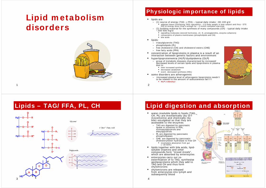
1 Lipid metabolism disorders 2 Physiologic importance of lipids lipids are – (1) source of energy (TAG → FFA) – typical daily intake ~80-100 g/d adipose tissue (containing TAG) represents ~1/5 body weight in lean subject and thus ~570 000 kJ energy store (that’s enough for ~3 month complete starving) – (2) building material for the synthesis of many compounds (CH) - typical daily intake ~200-500 mg/d signalling molecules (steroid hormones, vit. D, prostaglandins, enzyme cofactors) components of plasma membranes (phospholipids and CH) bile acids lipids: – triacylglycerols (TAG) – phospholipids (PL) – free cholesterol (CH) and cholesterol esters (CHE) – free fatty acids (FFA) concentration of lipoproteins in plasma is a result of an interaction between genetic factors and environment hyperlipoproteinemia (HLP)/dyslipidemia (DLP) – group of metabolic diseases characterised by increased/ decreased levels of certain lipids and lipoproteins in plasma due to: their increased synthesis decreased catabolism event. decreased synthesis (HDL) some disorders are atherogennic – increased plasma level of atherogenic lipoproteins needn’t to be related to the amount of subcutaneous fat!!!!! HLP ≠ obesity! 3 Lipids – TAG/FFA, PL, CH 4 Lipid digestion and absorption water-insoluble lipids in foods (TAG, CH, PL) are mechanically (by GIT movements) and chemically (by bile) emulgated so that they are accessible to the enzymes – TAG are digested by pancreatic lipase in intestine to FFA, monoacylglycerols and diacylglycerols – PL are digested by pancreatic phospholipases – CHE are digested by pancreatic cholesterylester hydrolase to free CH incomplete absorption from gut (~30-60%) lipids together with bile acids, lipid- soluble vitamins and other compounds form “mixed micels”, which are absorbed by enterocytes enterocytes carry out re- esterification of to TAG, synthesise apolipoproteins which they add to TAG and CH and thus form chylomicrons chylomicrons are released from enterocytes into lymph and subsequently blood
-
Upload
hoangxuyen -
Category
Documents
-
view
222 -
download
0
Transcript of lipids are disorders - med.muni.cz · 1 Lipid metabolism disorders 2 Physiologic importance of...
1
Lipid metabolism disorders
2
Physiologic importance of lipidslipids are – (1) source of energy (TAG → FFA) – typical daily intake ~80-100 g/d
adipose tissue (containing TAG) represents ~1/5 body weight in lean subject and thus ~570 000 kJ energy store (that’s enough for ~3 month complete starving)
– (2) building material for the synthesis of many compounds (CH) - typical daily intake ~200-500 mg/d
signalling molecules (steroid hormones, vit. D, prostaglandins, enzyme cofactors)components of plasma membranes (phospholipids and CH)bile acids
lipids:– triacylglycerols (TAG)– phospholipids (PL)– free cholesterol (CH) and cholesterol esters (CHE)– free fatty acids (FFA)
concentration of lipoproteins in plasma is a result of an interaction between genetic factors and environmenthyperlipoproteinemia (HLP)/dyslipidemia (DLP)– group of metabolic diseases characterised by increased/
decreased levels of certain lipids and lipoproteins in plasma due to:
their increased synthesisdecreased catabolismevent. decreased synthesis (HDL)
some disorders are atherogennic– increased plasma level of atherogenic lipoproteins needn’t
to be related to the amount of subcutaneous fat!!!!!HLP ≠ obesity!
3
Lipids – TAG/FFA, PL, CH
4
Lipid digestion and absorptionwater-insoluble lipids in foods (TAG, CH, PL) are mechanically (by GIT movements) and chemically (by bile) emulgated so that they are accessible to the enzymes– TAG are digested by pancreatic
lipase in intestine to FFA, monoacylglycerols and diacylglycerols
– PL are digested by pancreatic phospholipases
– CHE are digested by pancreatic cholesterylester hydrolase to free CH
incomplete absorption from gut (~30-60%)
lipids together with bile acids, lipid-soluble vitamins and other compounds form “mixed micels”, which are absorbed by enterocytesenterocytes carry out re-esterification of to TAG, synthesise apolipoproteins which they add to TAG and CH and thus form chylomicronschylomicrons are released from enterocytes into lymph and subsequently blood

5
Lipoproteinslipoproteins = macromolecular complexes (particles) consisting of:
– proteins (apolipoproteins, enzymes)structural integrity, binding to receptors, exchange of lipids
– lipids (CH, CHE, TAG, PL)outer layer – PL, CHinner core – CHE, TAG
circulating lipoproteins– (1) intestine-derived
chylomicrons– (2) liver-derived
VLDL (very low density lipoproteins)IDL (intermediate density lipoproteins)LDL (low density lipoproteins)HDL (high density lipoproteins)
– (3) assembled in circulationLp(a) - from LDL and apo-a (liver)
composition (lipids and apoPs) differ between particular lipoproteins
– chylomicrons and VLDL are TAG-rich particles (TAG>>>>CH)
– LDL and HDL carries CH>>>>TAGdifferent lipoproteins have different metabolic fateplasma normally contains
– <1% of chylomicrons– <10% of VLDLs– the rest is LDL and HDL
6
Example - LDL
7
Apolipoproteinsvarious types in various lipoproteins – control their metabolic fatefunctions:– activation of lipolytic enzymes involved – recognition by receptors (→ particle
endocytosis)– participate in the exchange of lipids between
particlesall particles containing apoB (apoB-100 or apoB-48) are atherogennic– apoB-100 – binding to LDL receptor– apoB-48 – binding to the receptor for
chylomicron „remnants“apoC (apoC-II and apoC-III) is a cofactor of LPL (lipoprotein lipase) and thus influence the rate of TAG hydrolysisapoE influence the removal of lipoprotein “remnants” (chylomicrons and VLDL) by liverapoA is a part of HDL (binding to HDL receptor) and cofactor of LCAT– low levels are atherogennic
apo(a) is homologous with plasminogen →acts as a competitive inhibitor of plazminogenwithout catalytic activity – apo(a) vs. tPA– plasmin is an enzyme dissolving fibrin (i.e.
blood clots)
apo(a), apoB-100Lp(a)
apoA, C, D, EHDL
apoB-100LDL
apoB-100, C, EVLDL
apoB-48, A, C, EChilom.
apoPparticle
8
Overview of lipid transport
9
Triacylglycerides (TAG)
10
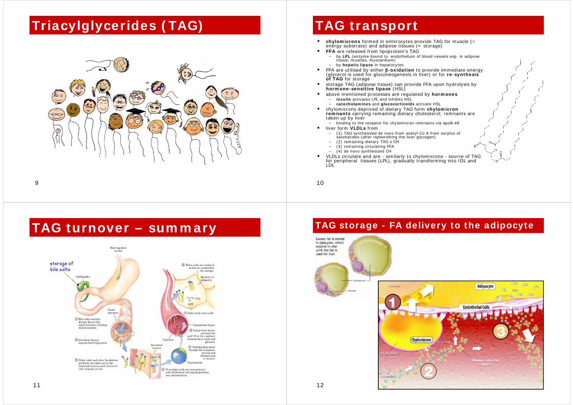
TAG transportchylomicrons formed in enterocytes provide TAG for muscle (= energy substrate) and adipose tissues (= storage)FFA are released from lipoprotein’s TAG
– by LPL (enzyme bound to endothelium of blood vessels esp. in adipose tissue, muscles, myocardium)
– by hepatic lipase in hepatocytesFFA are utilised by either β-oxidation to provide immediate energy (glycerol is used for gluconeogenesis in liver) or for re-synthesis of TAG for storagestorage TAG (adipose tissue) can provide FFA upon hydrolysis by hormone-sensitive lipase (HSL)above mentioned processes are regulated by hormones
– inzulin activates LPL and inhibits HSL– catecholamines and glucocorticoids activate HSL
chylomicrons deprived of dietary TAG form chylomicron remnants carrying remaining dietary cholesterol; remnants are taken up by liver
– binding to the receptor for chylomicron remnants via apoB-48liver form VLDLs from
– (1) TAG synthesized de novo from acetyl-Co A from surplus of saccharides (after replenishing the liver glycogen)
– (2) remaining dietary TAG s CH– (3) remaining circulating FFA– (4) de novo synthesized CH
VLDLs circulate and are - similarly to chylomicrons - source of TAG for peripheral tissues (LPL), gradually transforming into IDL and LDL
11
TAG turnover – summary
12
TAG storage - FA delivery to the adipocyte
13
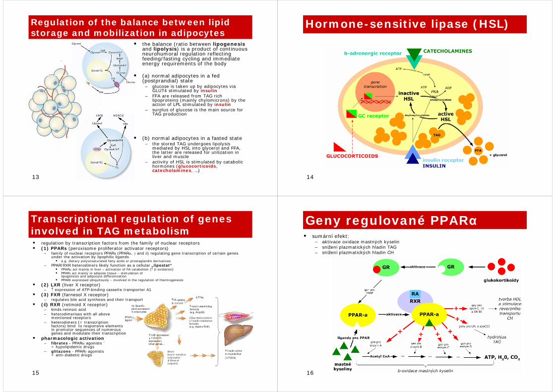
Regulation of the balance between lipid storage and mobilization in adipocytes
the balance (ratio between lipogenesisand lipolysis) is a product of continuous neurohumoral regulation reflecting feeding/fasting cycling and immediate energy requirements of the body
(a) normal adipocytes in a fed (postprandial) state– glucose is taken up by adipocytes via
GLUT4 stimulated by insulin– FFA are released from TAG rich
lipoproteins (mainly chylomicrons) by the action of LPL stimulated by insulin
– surplus of glucose is the main source for TAG production
(b) normal adipocytes in a fasted state– the stored TAG undergoes lipolysis
mediated by HSL into glycerol and FFA, the latter are released for utilization in liver and muscle
– activity of HSL is stimulated by catabolic hormones (glucocorticoids, catecholamines, …)
14
Hormone-sensitive lipase (HSL)
15
Transcriptional regulation of genes involved in TAG metabolism
regulation by transcription factors from the family of nuclear receptors(1) PPARs (peroxisome proliferator activator receptors)
– family of nuclear receptors PPARs (PPARα, γ and δ) regulating gene transcription of certain genes under the activation by lipophilic ligands
e.g. dietary polyunsaturated fatty acids or prostaglandin derivatives– PPAR/RXR heterodimers likely function as a cellular „lipostat“
PPARα act mainly in liver – activation of FA catabolism (↑ β-oxidation)PPARγ act mainly in adipose tissue – stimulation of lipogenesis and adipocyte differenciationPPARδ expressed ubiquitously – involved in the regulation of thermogenesis
(2) LXR (liver X receptor)– ↑ expression of ATP-binding cassette transporter A1
(3) FXR (farnesol X receptor) – regulates bile acid synthesis and their transport
(č) RXR (retinoid X receptor) – binds retinoic acid– heterodimerises with all above
mentioned receptors– heterodimers (= transcription
factors) bind to responsive elements in promotor sequences of numerous genes and modulate their transcription
pharmacologic activation – fibrates - PPARα agonists ´
= hypolipidemic drugs– glitazons - PPARγ agonists
= anti-diabetic drugs
16
Geny regulované PPARαsumární efekt: – aktivace oxidace mastných kyselin– snížení plazmatických hladin TAG– snížení plazmatických hladin CH
17
Cholesterol (CH)
18
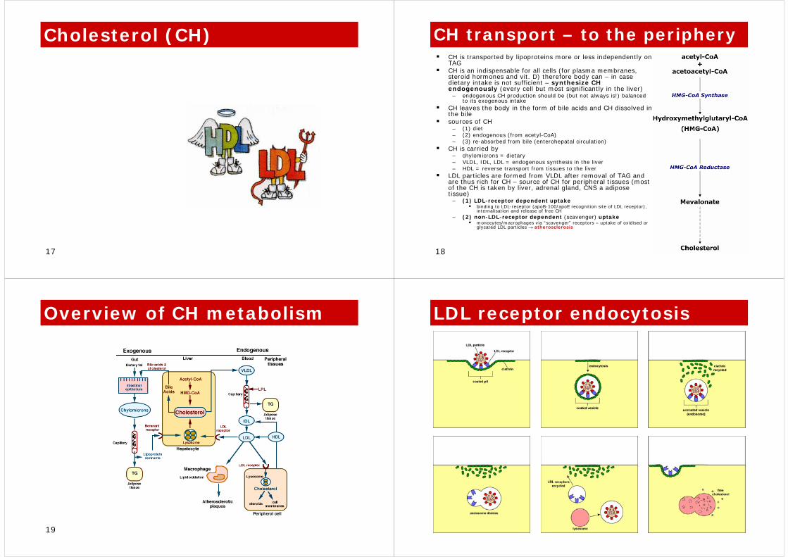
CH transport – to the peripheryCH is transported by lipoproteins more or less independently on TAGCH is an indispensable for all cells (for plasma membranes, steroid hormones and vit. D) therefore body can – in case dietary intake is not sufficient – synthesize CH endogenously (every cell but most significantly in the liver)
– endogenous CH production should be (but not always is!) balancedto its exogenous intake
CH leaves the body in the form of bile acids and CH dissolved inthe bilesources of CH
– (1) diet– (2) endogenous (from acetyl-CoA)– (3) re-absorbed from bile (enterohepatal circulation)
CH is carried by – chylomicrons = dietary– VLDL, IDL, LDL = endogenous synthesis in the liver– HDL = reverse transport from tissues to the liver
LDL particles are formed from VLDL after removal of TAG and are thus rich for CH – source of CH for peripheral tissues (most of the CH is taken by liver, adrenal gland, CNS a adipose tissue)
– (1) LDL-receptor dependent uptakebinding to LDL-receptor (apoB-100/apoE recognition site of LDL receptor), internalisation and release of free CH
– (2) non-LDL-receptor dependent (scavenger) uptake monocytes/macrophages via “scavenger” receptors – uptake of oxidised or glycated LDL particles → atherosclerosis
19
Overview of CH metabolism
20
LDL receptor endocytosis
21
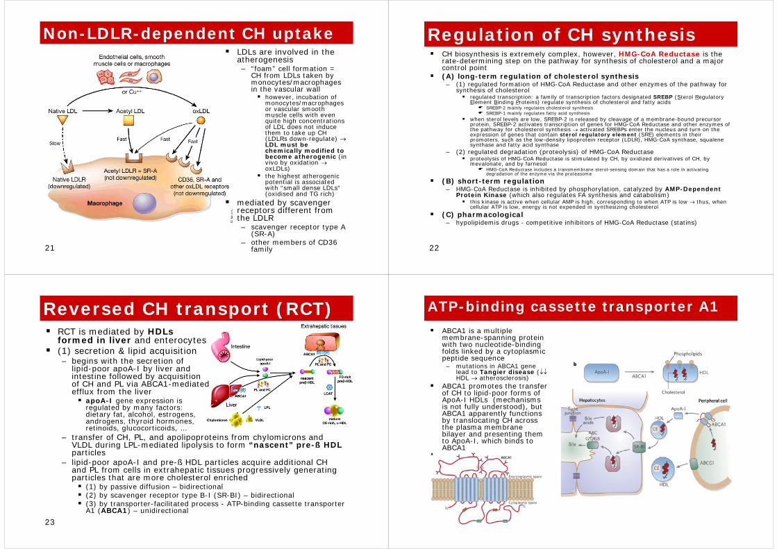
Non-LDLR-dependent CH uptakeLDLs are involved in the atherogenesis– “foam” cell formation =
CH from LDLs taken by monocytes/macrophages in the vascular wall
however, incubation of monocytes/macrophages or vascular smooth muscle cells with even quite high concentrations of LDL does not induce them to take up CH (LDLRs down-regulate) →LDL must be chemically modified to become atherogenic (in vivo by oxidation →oxLDLs)the highest atherogenic potential is associated with “small dense LDLs“(oxidised and TG rich)
mediated by scavenger receptors different from the LDLR– scavenger receptor type A
(SR-A)– other members of CD36
family 22
Regulation of CH synthesisCH biosynthesis is extremely complex, however, HMG-CoA Reductase is the rate-determining step on the pathway for synthesis of cholesterol and a major control point (A) long-term regulation of cholesterol synthesis – (1) regulated formation of HMG-CoA Reductase and other enzymes of the pathway for
synthesis of cholesterolregulated transcription: a family of transcription factors designated SREBP (Sterol Regulatory Element Binding Proteins) regulate synthesis of cholesterol and fatty acids
SREBP-2 mainly regulates cholesterol synthesisSREBP-1 mainly regulates fatty acid synthesis
when sterol levels are low, SREBP-2 is released by cleavage of a membrane-bound precursor protein, SREBP-2 activates transcription of genes for HMG-CoA Reductase and other enzymes of the pathway for cholesterol synthesis → activated SREBPs enter the nucleus and turn on the expression of genes that contain sterol regulatory element (SRE) elements in their promoters, such as the low-density lipoprotein receptor (LDLR), HMG-CoA synthase, squalenesynthase and fatty acid synthase
– (2) regulated degradation (proteolysis) of HMG-CoA Reductaseproteolysis of HMG-CoA Reductase is stimulated by CH, by oxidized derivatives of CH, by mevalonate, and by farnesol
HMG-CoA Reductase includes a transmembrane sterol-sensing domain that has a role in activating degradation of the enzyme via the proteasome
(B) short-term regulation– HMG-CoA Reductase is inhibited by phosphorylation, catalyzed by AMP-Dependent
Protein Kinase (which also regulates FA synthesis and catabolism)this kinase is active when cellular AMP is high, corresponding to when ATP is low → thus, when cellular ATP is low, energy is not expended in synthesizing cholesterol
(C) pharmacological– hypolipidemis drugs - competitive inhibitors of HMG-CoA Reductase (statins)
23
Reversed CH transport (RCT)RCT is mediated by HDLsformed in liver and enterocytes(1) secretion & lipid acquisition– begins with the secretion of
lipid-poor apoA-I by liver and intestine followed by acquisition of CH and PL via ABCA1-mediated efflux from the liver
apoA-I gene expression is regulated by many factors: dietary fat, alcohol, estrogens, androgens, thyroid hormones, retinoids, glucocorticoids, …
– transfer of CH, PL, and apolipoproteins from chylomicrons and VLDL during LPL-mediated lipolysis to form “nascent” pre-ß HDL particles
– lipid-poor apoA-I and pre-ß HDL particles acquire additional CH and PL from cells in extrahepatic tissues progressively generating particles that are more cholesterol enriched
(1) by passive diffusion – bidirectional(2) by scavenger receptor type B-I (SR-BI) – bidirectional(3) by transporter-facilitated process - ATP-binding cassette transporterA1 (ABCA1) – unidirectional
24
ATP-binding cassette transporter A1
ABCA1 is a multiple membrane-spanning protein with two nucleotide-binding folds linked by a cytoplasmicpeptide sequence – mutations in ABCA1 gene
lead to Tangier disease (↓↓HDL → atherosclerosis)
ABCA1 promotes the transfer of CH to lipid-poor forms of ApoA-I HDLs (mechanisms is not fully understood), but ABCA1 apparently functions by translocating CH across the plasma membrane bilayer and presenting them to ApoA-I, which binds to ABCA1
25
RCT - continued(2) maturation of HDL particles– the enzyme LCAT [lecitin:cholesterol-
acyltransferase], carried on HDL particles activated by apo-proteins of HDLs, esterifies the free CH to CHE, which migrate to the core of the HDL particle to form mature HDL particles which can further acquire additional lipid from certain cells via efflux mediated by ABCG1 and SR-BI intravascular
(3) intravascular modelling of HDL by lipases and lipid transfer factors– an important determinant of the rate of HDL clearance from the circulation– enzyme CETP [cholesterol ester transfer protein]
catalyses reverse process - heteroexchange of CHE between HDLs and TAG-rich lipoproteins (chylomicrons and VLDLs) which results in CHE depletion and TG enrichment of HDL
– hepatic lipase modification of TG-rich HDL releases lipid-poor apoA-I and HDL remnant particleslipid-poor apoA-I is filtered by the renal glomerulus and then degraded by proximal tubular cell receptors such as cubilin/megalin systemHDL remnants may bind to putative receptors in liver that mediate HDL holoparticle uptake, internalization, and degradation
– HDL contain paraoxonase – an enzyme protecting CH (in HDL and LDL) from oxidation and thus increase in its atherogenic potential
(4) HDLs and their CH are removed from circulation in liver, kidney and steroidogenic tissues by two processes: – (1) selective CH uptake (liver mainly)
HDL bind HDL-receptor SR-BI via apoA-I, CH liberated and secreted by bile (either as a free CH or metabolised to bile acides)
– (2) endocytic uptake of whole HDL particles (kidney)HDLs filtered, reabsorbed in prox. tubule (megalin/cubilin system) 26
Summary of RCTin summary, efficiency of RCT is determined by: – (1) the rate of production of apoAI– (2) the rate of clearance of HDLs from circulation by
liver (via SR-BI)– (3) the rate of CH esterification (↑ LCAT/↓ CETP)– (4) action of lipases (hepatic, lipoprotein) – variable
TG content influence the rate of clearance of HDL
27
Hyper-/dyslipoproteinemiahypercholesterolemia
↑ total CH, LDL (and all apoBparticles)↓ HDL (apoA particles)
– risk factor of atherosclerosisidentified and confirmed by numerous epidemiological studies
hypertriglyceridemia(1) ↑ isolated TAG (i.e. TAG-rich particles)
solely high TAG is not atherogenic (e.g. LPL deficiency)
– risk of acute pancreatitis ()TAG > 20-30 mmol/l
(2) ↑ TAG (i.e. TAG-rich particles) + FFA
– insulin resistance(3) ↑ TAG + ↑ apoB particles (due to high influx of FFA into liver) + ↓HDL
– risk factor of atherosclerosis
28
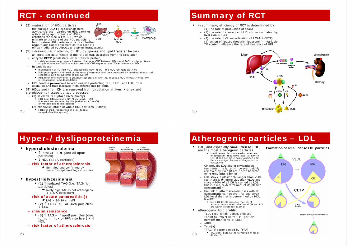
Atherogenic particles – LDLLDL, and especially small dense LDL, are the most atherogenic particles
small dense LDL more easily penetrate endothelium, they have lower affinity to LDL-R and get more easily oxidised and thus scavenged by macrophages in the vessel wall
– CH prevails LDL and in chylomicron remnants, the latter is however quickly removed by liver (if not, these become extremely atherogenic)
– LDL stays in plasma 9× longer than VLDL (so there is 9× more LDL than VLDL and since ~70% of all CH is carried by LDL this is a major determinant of its plasma concentration)
– the risk of atherosclerosis rises with LDL concentrations, however, for any given LDL level the risk is determined by HDL levels!!!
low HDL levels increase the risk of atherosclerosis even when total CH and LDL are within reference interval
atherogenic lipid profile: – ↑LDL (esp. small, dense, oxidised)– ↑apoB (= reflect better LDL particle
number than conc. of LDL)– ↓HDL– ↑apo(a)– ↑TAG (if accompanied by ↑FFA)
TAG contribute to the formation of small dense LDL

29
HLP classificationseveral classification schemes available according to different criteria– electrophoretic mobility– clinical impact– ethiopathogenesis
in the past – Fredrickson classification (phenotypes I - V) – lipoprotein mobility spectrum after
electrophoretic separation – did not considered HDL!!!
today – simple, therapeutically relevant clinical classification of HLPs considering plasma levels of lipids despite the ethiopathogenesis:
a) hypercholesterolemiab) hypertriglyceridemiac) mixed disorders
ethiopathogenic (patho-physiological) classification– primary HLPs– secondary HLPs
5↑↑↑↑↑ or ↑↑VLDL and chylomV
45↑↑Normal to ↑VLDLIV
<1↑↑↑↑↑IDLIII
40↑↑↑↑LDL and VLDLIIa
10Normal↑↑LDLIIa
<1↑↑↑↑Normal to ↑chylomI
%Serum TAGSerum CHParticle elevatedType
↑ Atherosclerosis<0.3 g/lLp(a)
↑Atherosclerosis0.58-1.38g/lapoB
↓Atherosclerosis1.2 - 1.7 g/lapoAI
↑Atherosclerosis<1.8 mmol/lTAG
↑Atherosclerosis<3.4 mmol/lLDL
↓Atherosclerosis>1.6 mmol/lHDL
↑Atherosclerosis<5.2 mmol/lTotal CH
interpretationrangeparameter
30
Etiology of HLPsHLPs are heterogeneous group of metabolic diseases characterised by increased plasma lipoproteins – >95. population percentile + mortality
effect– dyslipoproteinemia is a term often
used since not only high but also low levels can be a risk (e.g. HDL)
HLPs are caused by: – a) increased synthesis of
apolipoproteins– b) defect of intravascular processing
by enzymes (e.g. LPL deficit)– c) defect uptake by membrane
receptors (e.g. LDL receptor)– d) decreased removal of lipoproteins
etiology– primary HLPs – genetic (inherited)– secondary – consequence of other
diseasegenetics (disease vs. disposition)– polygenic – complex diseases“
(“thrifty” genotype)genetic predisposition + environmental factors (diet!!!)
– monogenic – single gene
31
Primary HLPs
monogenic diseases are very often autosomal semidominant, i.e. severity of the disease is graded according to the number of pathologic alleles all primary HLPs typically do not respond to dietary interventions, lipid lowering pharmacotherapy is necessarycarriers are endangered by premature cardiovascular disease (esp. homozygous subjects with familiar
? (polygenic)Fam. hypertriglyreridemia
apoE gene mutationsIIIFam. dysbetalipoproteinemia
PolygenicIIa, IIbFam. combined hypelipidemia
PolygenicIIa, IIbPolygenic hypercholesterolemia
apoB gene mutations (defect of binding to LDLR – 10% of normal activity)
IIaFamiliar defective apoB-100
LDLR gene mutationsIIaFam. hypercholesterolemia
apoC gene mutationsI or VFamiliar deficit of apoC
LPL gene mutations IFamiliar deficit of LPL
CauseType (Fredrickson)
Disorder
32
Familiar hypercholesterolemia (FH)the most common primary HLP
– heterozygotes population prevalence 1:500– homozygotes 1:1 mil.
FH is caused by mutations in the LDLR gene(chromosome 19)
– >700 mutations identifiedLDL receptor (+part of plasma membranes = “coated pits”)
– periodic recycling (~1 × 10min) with ingestion of LDL particles – lysozomal enzymes release free CH and AA (from
apolipoprotein apoB5 functional classes of mutations:
– 1) complete absence of the receptor (17 %)– 2) defective transport of receptor to the plasma membrane (54
%)– 3) defective binding of LDL– 4) defective internalisation of receptor + LDL complex– 5) defective liberation from endosome after internalisation and
recycling to plasma membrane (22 %)increase of plasma CH depends on the type of mutation and hetero- or homozygosity (i.e. „gene-dosage“ effect)
– ~2× of normal [<5.2mmol/l] in heterozygotes – ~4-5× in homozygotes
consequences of FH– multiple skin xantomas and tendon xantelasma, arcus corneae– premature atherosclerosis
mortality of MI in very young age in unrecognised homozygotes, before the 4th decade in heterozygotes
molecular genetic diagnostics of suspicious cases and family members, follow-up, genetic counselling, agressivehypolipidemic therapy!!!!
33
Polygenic HLPsthrifty genotype hypothesis– in the past, genes (allele of genes)
providing higher levels of energy substrates (glucose, lipids, …) but also those leading to increased energy stores (fat tissue), increased pro-thrombotic and pro-inflammatory potential offered selective advantage for their carriers → genetic selection
– today, under less energy requiring conditions and with more or less unrestricted access to food (affluent societies) the same genes increase the likehood (risk) of developing the common “complex” diseases
complex = genes + environmentgenetics of lipid metabolism– due to the functional variability in the genes encoding e.g.
enzymes involved in lipid metabolism (both TAG and CH)nuclear receptors (PPAR, RXR, LXR, …) apolipoproteinsreceptors of apolipoproteinshormonal control
glucocorticoids, thyroid hormones, …factors determining insulin sensitivity
» utilisation of saccharides and lipids, esp. in insulin-sensitive tissues is mutually interconected and often ompetitive ( Randle’s cycle) 34
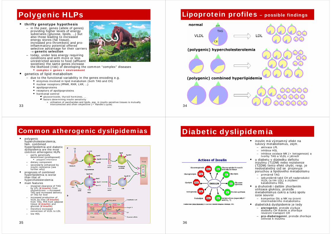
Lipoprotein profiles – possible findings
35
Common atherogenic dyslipidemiaspolygenic hypercholesterolemia, fam. combined hyperlipidemia and diabetic dyslipidemia are the most common atherogenic HLPs
– partly genetically determined (predisposed)
polygenic inheritance– dietary component– secondarily enhanced by
insulin resistance (see further why)
prognosis of combined hyperlipidemia is worse than that of hypercholesterolemiamain features
– impaired clearance of TAG by LPL ( insulin) from chylomicrons → increased TAG and increased delivery of TAG for liver
– increased production of VLDL by liver ( insulin) from TAG, FFA from adipose tissue ( insulin) and glucose ( insulin)
– therefore increased conversion of VLDL to LDL
– low HDL
36
Diabetic dyslipidemiainzulin má významný efekt na tukový metabolismus, zejm.– aktivace LPL– inhibice HSL– inhibice oxidace MK (+ ketogeneze) a
tvorby TAG a VLDL v játrech u diabetu v důsledku deficitu inzulinu (T1DM) nebo rezistence(T2DM) tento efekt chybí, resp. je nedostatečný což se projevuje poruchou a lipidového metabolismu– primárně TAG– sekundárně také CH při nadprodukci
VLDL (a tím LDL) a zvýšeníkatabolismu HDL
a druhotně i dalším zhoršením utilizace glukózy, protože metabolismus cukrů a tuků spolu úzce souvisí– kompetice Glc a MK na úrovni
intermediárního metabolismudiabetická dyslipidemie je tedy– aterogenní, protože zvyšuje
dodávku CH tkáním a zhoršuje reverzní transport CH
– pro-diabetogenní, protože zhoršuje citlivost k inzulinu
37
Classification (?) vs. reality(!)
38
Secondary HLPscaused by other primary disease, nevertheless its impact on cardiovascular system is the same as in primary HLPstreatment involves either primary disease and hypolipidemic drugs unlike primary ones, secondary HLPs respond well to dietary interventions
↑CHCholestasis
↑TGChronic renal insufficiency
↑CH, TAGNephrotic syndrome
↑CHHypothyreosis
↑TAG, ↓ HDLDiabetes mellitus (type 1)
ElevationCause