Practical # 3: Microscopic Examination of Urine Urinalysis & Body Fluids CLS 431 1.

Application Note
Light Microscopic Analysis of UrineZEISS Primo Star and ZEISS Axio Lab.A1
Application Note
2
Light Microscopic Analysis of UrineZEISS Primo Star and ZEISS Axio Lab.A1
Author: Carl Zeiss Microscopy GmbH, Germany
Date: August 2015
A microscopic examination is part of medical laboratory routine to analyze whether there are abnormalities in
the physical or chemical examination of urine. Cells, crystals and other substances are reported and counted.
An urinalysis of the urine sediment is typically performed in brightfield and phase contrast. It can reveal
diseases such as diabetes mellitus, various forms of glomerulonephritis, and chronic urinary tract infections.
Steps of Urinalysis
a) Visual analysis
Color, smell and appearance [Fig. 1] of fresh urine is first
inspected visually. In general urine is yellow or amber in color
and clear. Any turbidity or cloudiness may be caused by
cellular material or protein in the urine or may develop from
crystallization or precipitation of salts. Inserting a small
amount of acid indicates that precipitation of salts is the
probable cause of tubidity. An abnormal color could be sign
of a special diet, a drug, or the presence of either hemo
globin or myoglobin.
Figure 1: Visual investigation of urine
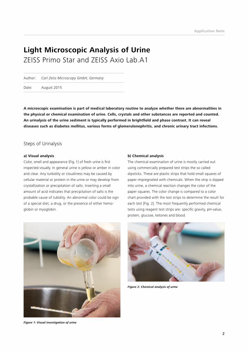
Figure 2: Chemical analysis of urine
b) Chemical analysis
The chemical examination of urine is mostly carried out
using commercially prepared test strips the so called
dipsticks. These are plastic strips that hold small squares of
paper impregnated with chemicals. When the strip is dipped
into urine, a chemical reaction changes the color of the
paper squares. The color change is compared to a color
chart provided with the test strips to determine the result for
each test [Fig. 2]. The most frequently performed chemical
tests using reagent test strips are: specific gravity, pHvalue,
protein, glucose, ketones and blood.

Application Note
3
c) Microscopic analysis
A microscopic analysis is typically performed to visualize
blood cells, epithelial cells and pathogens, for example,
when there are abnormal findings on the visual or chemical
analysis. A sample of usually 10 – 15 ml urine is centrifuged
to concentrate the substances in it at the bottom of a tube
[Fig. 3]. The fluid at the top of the tube is then discarded
and the remaining sediment is examined under a microscope
[Fig. 4 & 5].
Urine sediment is normally examined with a transmitted
light microscope with brightfield or phase contrast [Fig. 5].
Visualizing crystals’ polarization contrast can be helpful.
The sediment is first examined with low magnification to
identify most crystals, casts, squamous cells, and other large
objects. Next, examination is carried out at high magnifica
tion to identify crystals, cells, and bacteria. All findings are
counted and reported.
Figure 3 Preparing the urine sample for the centrifuge
Figure 5 Examination of urine with ZEISS Primo Star
Figure 4 Preparing the glass slide for light microscopic examination

Application Note
4
Figure 6 Erythrocytes (red blood cells) in urine showing thorn apple shape because of non-physiological pH-value of the urine Courtesy of: A. Michelsen, Clinic of Ortenau, Germany
Figure 8 Squamous epithelial cells Courtesy of: A. Michelsen, Clinic of Ortenau, Germany
Figure 10 Bacteria and Leukocytes. Bacteria are common in urine specimens. Courtesy of: G. Spengler-Schulz, Alexander Fleming School, Stuttgart, Germany
Figure 7 Erythrocytes and leukocytes (white blood cells) in urine Courtesy of: G. Spengler-Schulz, Alexander Fleming School, Stuttgart, Germany
Figure 9 Salts on a cylindrical structure Courtesy of: A. Michelsen, Clinic of Ortenau, Germany
Figure 11 Fungus mycelium with yeast cells. Yeast cells may be contami-nants or represent a true yeast infection. Courtesy of: A. Michelsen, Clinic of Ortenau, Germany
Application examples

Application Note
5
Conclusion
The urinalysis is used as a screening tool to detect substan ces
or cellular material in the urine associated with different
metabolic and kidney disorders. A microscope with bright
field and phase contrast such as Primo Star and Axio Lab.A1
is the recommended tool as transparent elements in the
urine will be clearly visible.
Figure 12 Calcium oxalate in urine. Crystalluria indicates that the urine is supersaturated with the compounds that comprise the crystals, e.g. ammo-nium, magnesium and phosphate for struvite. Courtesy of: A. Michelsen, Clinic of Ortenau, Germany
Figure 13 Triple phosphate in urine Courtesy of: G. Spengler-Schulz, Alexander Fleming School, Stuttgart, Germany

Carl Zeiss Microscopy GmbH 07745 Jena, Germany [email protected] www.zeiss.com/microscopy
EN_4
1_01
3_10
8 | C
Z 11
201
5 | D
esig
n, s
cope
of
deliv
ery
and
tech
nica
l pro
gres
s su
bjec
t to
cha
nge
with
out
notic
e. |
© C
arl Z
eiss
Mic
rosc
opy
Gm
bH