Leica EM Cryo CLEM - Mager Scientificmagersci.com/brochures/leica/CryoCLEM.pdf · › High image...
8
Leica EM Cryo CLEM One System to Serve Your Workflow Needs in Correlative Light and Electron Microscopy
Transcript of Leica EM Cryo CLEM - Mager Scientificmagersci.com/brochures/leica/CryoCLEM.pdf · › High image...

Leica EM Cryo CLEM One System to Serve Your Workflow Needs in Correlative Light and Electron Microscopy

2
Correlative Light and Electron Microscopy
Researchers can only optimise results when their samples are well-preserved during the
entire process from sample preparation to imaging. To enable you to focus on what re-
ally matters, Leica Microsystems has developed a solution for the complete workflow
from cryo fixation to cryo fluorescence light microscopy. This unique solution offers pre-
mium instrumentation from electron microscopy sample preparation to fluorescence
light microscopy, including analysis capabilities with the Leica Application Suite micros-
copy software platform.
Best resultsBigger area Better layer
› Correlative Light and Electron Microscopy (CLEM) combines fluorescence light microscopy and elec-tron microscopy (EM) imaging of the same sample.
› Electron Microscopy (EM) delivers structural information and also the context in which the target struc-tures are embedded at very high resolution. But EM provides very limited information in terms of living bio-logical processes and functions.
› Fluorescence Light Microscopy (FLM) on the other hand is a very sensitive method to observe and ana-lyze biological processses and functions inside fixed and living biological samples. In addition, FLM allows rapid screening of large areas and fast determination of regions of interest (ROI) which can be quickly recog-nized in the electron microscope.
› Cryo Fixation is the sample preparation method to maintain samples in the most life-like state as possible.
› Cryo CLEM connects the benefits of all these techniques. It combines the individual advantages from cryo fixation, FLM and EM by time-effective imaging of identical, artefact-free samples and overlaying the com-plementary information to win greater understanding.

3LEICA EM CRYO CLEM – CORRELATIVE LIGHT AND ELECTRON MICROSCOPY
Cryo Electron
Microscopy
Image analysis
High Pressure Freezing
Leica EM HPM100
Cryo Ultramicrotomy
Leica EM UC7Leica EM FC7
Cryo Transfer System
Cryo Light Microscopy
Leica EM Cryo CLEM on Leica DM6000 FS
Leica LAS Widefield Software for manual correlation of LM marked structures in EM
Grid Plunging
Leica EM GP

4
LEICA EM CRYO CLEM – INNOVATIVE FEATURES
Trusted reliability
› Reproducible process through contami-
nation-free and controlled cryo sample
transfer and loading method to the cryo
stage
› High image quality and specific localiza-
tion of target structure in LM with the
world’s first cryo objective with low
working distance for high resolution
Time saving
› Reduced sample loading and unloading
time through new sample transfer and
loading system
› Reduced cryo EM operation and user
interaction time during EM analysis due
to well preserved sample quality and
accuratly located target structures
Ease of Use
› Sensor-controlled stage temperature
function
› Ease of use with the intuitive sample
transfer and loading system
Cost Savings
› Significantly reduced cryo EM user
operation costs due to reduced target
structure search time
5
11
64
10
7
1 2
3
8
9
12
For more information regarding the microscope Leica DM6000 FS click here.
1 LN2 pump controller2 5l LN2 dewar3 LN2 pump4 Fluorescence light source5 Microscope controller6 Leica DM6000 FS7 Cryo stage with cryo
objective port8 Cryo objective,
50x FL Apo; NA 0.9; WD 0.28
9 Magnification changer 0.35x; 1.25x; 4x
10 Leica microscope camera11 Cryo transfer shuttle12 PC system and software
optimized for CLEM imaging
Leica EM Cryo CLEM
The Leica EM Cryo CLEM System ensures fast, safe, contamination-free sample transfer
and loading from cryo sample preparation instruments to the Leica fixed stage light mi-
croscope Leica DM6000 FS. The system consists of a cryo stage, which is developed to
seamlessly integrate to the fixed stage microscope, the cryo transfer shuttle, and cryo
objective. With its perfect camera and software portfolio, Leica Microsystems can pro-
vide a complete and integrated cryo CLEM workflow from cryo sample preparation to
cryo fluorescence light microscopy imaging for CLEM.

5LEICA EM CRYO CLEM – NEW LEICA CRYO TRANSFER SHUTTLE
Leica Cryo Transfer Shuttle
The transfer shuttle for Leica Cryo CLEM ensures fast, easy, and contamination-free
sample loading and transfer (A). It consists of three functional parts: a cartridge loader,
a storage area for three standard grid transfer boxes, both located in the dewar part of
the cryo transfer shuttle, and a rod with gripper for sample transfer.
A
B
E
The sample loading is divided into four steps. At first the grid or sapphire disc is
mounted on the cartridge (B). Second, the gripper of the transfer rod clamps the car-
tridge (C + D), then the transfer shuttle is docked on the loading port of the cryo
stage and the rod with the cartrige is transferred through the loading port of the
cryo stage into the cartridge intake of the cryo stage (E).
Cartridge loader (A1) with cartridge (A2) for 3 mm grids or sapphire discs.
A1 A2
C
D

6
Leica HCX PL APO 50x / 0,90 CLEM objective, apochromati-
cally corrected with numerical aperture of 0.9, and a low working
distance of 0.28 mm for high resolution cryo imaging. Maximum
resolution of 512 nm.
All following pictures were taken with the CLEM objective.
ComponentsLeica Cryo Stage and cover with cryo objective port. The
stage allows cryo objective approach to the sample even with a
low working distance, which is necessary for high resolution cryo
imaging. Mechanically and thermally, it is exceptionally stable
which leads to long-term focus stability. Integrated sensors mon-
itor stage temperature. Cooling range from –195 °C to +60 °C.
Leica EM Cryo CLEM on fixed stage microscope Leica DM6000
FS. The cryo stage with inserted cryo objective is developed to
seamlessly integrate to the Leica DM6000 FS fixed stage micro-
scope; cryo transfer shuttle is docked to cryo stage.
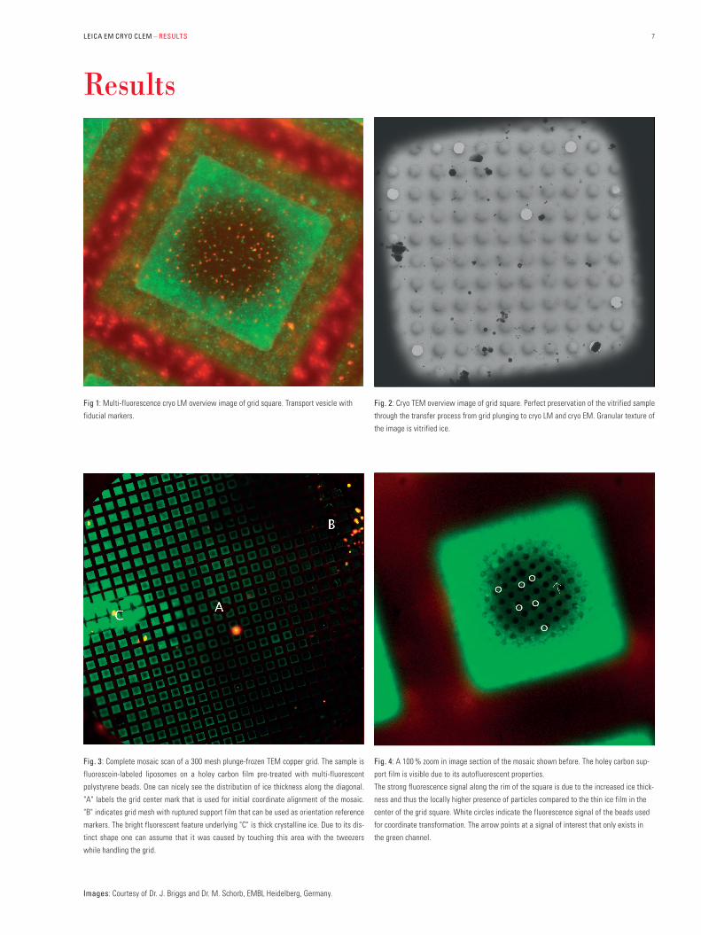
7LEICA EM CRYO CLEM – RESULTS
Results
Fig 1: Multi-fluorescence cryo LM overview image of grid square. Transport vesicle with
fiducial markers.
Fig. 3: Complete mosaic scan of a 300 mesh plunge-frozen TEM copper grid. The sample is
fluorescein-labeled liposomes on a holey carbon film pre-treated with multi-fluorescent
polystyrene beads. One can nicely see the distribution of ice thickness along the diagonal.
"A" labels the grid center mark that is used for initial coordinate alignment of the mosaic.
"B" indicates grid mesh with ruptured support film that can be used as orientation reference
markers. The bright fluorescent feature underlying "C" is thick crystalline ice. Due to its dis-
tinct shape one can assume that it was caused by touching this area with the tweezers
while handling the grid.
Fig. 2: Cryo TEM overview image of grid square. Perfect preservation of the vitrified sample
through the transfer process from grid plunging to cryo LM and cryo EM. Granular texture of
the image is vitrified ice.
Fig. 4: A 100 % zoom in image section of the mosaic shown before. The holey carbon sup-
port film is visible due to its autofluorescent properties.
The strong fluorescence signal along the rim of the square is due to the increased ice thick-
ness and thus the locally higher presence of particles compared to the thin ice film in the
center of the grid square. White circles indicate the fluorescence signal of the beads used
for coordinate transformation. The arrow points at a signal of interest that only exists in
the green channel.
Images: Courtesy of Dr. J. Briggs and Dr. M. Schorb, EMBL Heidelberg, Germany.

www.leica-microsystems.com
The statement by Ernst Leitz in 1907, “With the User, For the User,” describes the fruitful collaboration with end users and driving force of innovation at Leica Microsystems. We have developed five brand val-ues to live up to this tradition: Pioneering, High-end Quality, Team Spirit, Dedication to Science, and Continuous Improvement. For us, liv-ing up to these values means: Living up to Life.
LIFE SCIENCE DIVISION – NANO TECHNOLOGY LNTThe Leica Microsystems Nano Technology Division’s focus is to provide the most compehensive product portfolio for the preparation of bio-logical, medical and industrial samples for investigation in the Electron and Light Microscope. Excellent Sample Preparation is a prerequisite for perfect microscopy. Your image starts here!
Leica Microsystems – an international company with a strong network of worldwide customer services:
Leica EM Cryo-CLEM Brochure Engilsh ∙01/2015 ∙ Copyright © by Leica Mikrosysteme
GmbH, Vienna, Austria, 2015. Subject to modifications. LEICA and the Leica Logo are regis-
tered trademarks of Leica Microsystems IR GmbH.
Active worldwide Tel. Fax
Australia ∙ North Ryde +61 2 8870 3500 2 9878 1055
Austria ∙ Vienna +43 1 486 80 50 0 1 486 80 50 30
Belgium ∙ Diegem +32 2 790 98 50 2 790 98 68
Brazil ∙ São Paulo +55 11 2764-2411 11 2764-2400
Canada ∙ Concord/Ontario +1 800 248 0123 847 405 0164
Denmark ∙ Ballerup +45 4454 0101 4454 0111
France ∙ Nanterre Cedex +33 811 000 664 1 56 05 23 23
Germany ∙ Wetzlar +49 64 41 29 40 00 64 41 29 41 55
India ∙ Mumbai +91 226 1880 200 226 1880 333
Italy ∙ Milan +39 02 574 861 02 574 03392
Japan ∙ Tokyo +81 3 6758 5630 3 5155 4333
Korea ∙ Seoul +82 2 514 65 43 2 514 65 48
Netherlands ∙ Rijswijk +31 70 4132 100 70 4132 109
People’s Rep. of China ∙ Hong Kong +852 2564 6699 2564 4163 ∙ Shanghai +86 21 6039 6000 21 6387 6698
Portugal ∙ Lisbon +351 21 388 9112 21 385 4668
Singapore +65 6550 5999 6564 5955
Spain ∙ Barcelona +34 93 494 95 30 93 494 95 32
Sweden ∙ Bromma +46 8 625 45 45 8 625 45 10
Switzerland ∙ Heerbrugg +41 71 726 34 34 71 726 34 44
Turkey · Istanbul +90 216 504 0100 216 504 0110
United Kingdom ∙ Milton Keynes +44 800 298 2344 1908 577640
USA ∙ Buffalo Grove/lllinois +1 800 248 0123 847 405 0164