Leica EM TIC 3X - Leica Microsystems · the Leica EM TIC 3X is the system of choice for EDS, WDS,...
16
Leica EM TIC 3X Efficiency and flexibility
Transcript of Leica EM TIC 3X - Leica Microsystems · the Leica EM TIC 3X is the system of choice for EDS, WDS,...

Leica EM TIC 3X Efficiency and flexibility

LEICA EM TIC 3XTRIPLE ION-BEAM MILLING SYSTEM
Would you like to prepare surfaces of hard, soft, porous, heat-sensitive, brittle and /or heterogeneous materials for microstructure analysis in scanning electron microscopy (SEM) and investigations in the Atomic Force Microscope (AFM)? The unique broad ion beam milling system of the Leica EM TIC 3X is the system of choice for EDS, WDS, Auger and EBSD, because ion beam milling is often found to be the only method capable of achieving high quality cross-sections and planed surfaces of almost any material. The process reveals the internal structures of a sample whilst minimizing deformation or damage.
The flexible choice of stages makes the Leica EM TIC 3X a perfect instrument not only for high through put but also for contract laboratories. Depending on your needs the Leica EM TIC 3X can be configured individually using interchangeable stages like standard stage, multiple sample stage, rotary stage or cooling stage for applications of standard preparation, high throughput processing, as well as the preparation of extremely heat sensitive samples such as polymers, rubbers or biological materials at low temperature. Connectivity with the Leica EM VCT environmental transfer system provides perfect cryogenic surfacing of biological, geological, or industrial samples subsequently transferred under cryo and vacuum conditions to coater and (cryo) SEM.

2 . 3
FLEXIBLE SETUPEXCELLENT QUALITY RESULTS
HIGH THROUGHPUT

LEICA EM TIC 3X – INNOVATIVE FEATURES IN DESIGN … … AND OPERATION
> Cuts high-quality cross-sections with large areas of 4 > 1 mm
> Multiple sample stage capable of processing several samples in one run
> Samples up to a size of 50 x 50 x 10 mm or up to 38 mm diameter can be inserted for processing
> Easy and accurate sample mounting and alignment
> Simple operation via touch-screen, no special skills necessary
> Process monitoring via stereomicroscope or HD-TV camera
> Four segement controlled LED illumination for optimal specimen viewing and alignment
> Integrated, decoupled roughing pump provides vibration-free observation
> Contrast enhancement at 90° to the prepared surface
> Usable for almost any material, cooling stage provides temperatures of mask and sample down to -160 °C
> Parameter and programme upload and download on USB stick
> Total workflow solution saves user interaction time

4 . 5
LEICA EM TIC 3X – INNOVATIVE FEATURES IN DESIGN … … AND OPERATION
Ergonomic and Easy to Use
The stereo microscope of the Leica EM TIC 3X is not only used for aligning and process observation. A work-plate attached beneath the microscope can be used for handling the sample or attaching the sample to its holder. Thus, no additional observation system is needed for manipulating small samples. The outstanding performance of the integrated touch screen controller is not only reflected in its intuitive operation but hints and helpful information are also displayed to allow the most effective use of the instrument. A USB stick can be attached to upload and download parameters for reporting and processing.
Efficiency
What really counts regarding the efficiency of an ion beam miller is excellent quality results with high throughput. It’s not enough that we could increase the milling rate by a factor of 2 compared to the previous version but the unique triple ion beam system optimizes the preparation quality and reduces working time. Several samples can be processed in one session. Cross sectioning and polishing can be performed by one stage. Workflow solutions provide safe and efficient transfer of samples to subsequent preparation instruments or analysis systems.
Our customers seek faster and simpler methods of sample preparation without having to forgo quality. The innovative technology of the Leica EM TIC 3X triple ion beam milling system offers solutions to help laboratories with high expectations achieve their goals. Depending on the preparation needs, the Leica EM TIC 3X can be configured for applications of standard preparation, high throughput processing, and the preparation of extremely heat sensitive samples such as polymers, rubbers or even biological material at low temperatures. Samples can be transferred to the (cryo) SEM under cryo-vacuum condition. Five easily interchangeable stages are offered to fulfill individual application demands:
FLEXIBILITY: CONFIGURE YOUR SYSTEM
Rotary stage
is used for ion beam flat milling of already mechanically polished surfaces. During ion beam processing the sample can be rotated or oscillated. The additional lateral movement of the sample produces a uniformly prepared area of more than ∅ 25 mm. Thus, mechanical artifacts e.g. smearing and fine scratches are removed. It reveals the structure of the sample as native as possible.
An optional available cross section holder for the rotary stage is used for the precise cross sectioning of samples and preparing roughly pre-prepared samples to a high quality level.
Multiple sample stage
is used if high throughput is desired. Three sample holders can be loaded and the samples will be automatically processed in one session (e.g. overnight) without any user interaction. When programming the preparation processes different sample / mask movements for each sample holder position can be selected, either with or without oscillation. By selecting oscillation the prepared area can be up to three times wider. This enables the preparation of several samples in one sample holder position.
Standard stage
for routine cross sectioning applications and contrast enhancement of the prepared surface.

6 . 7
Cooling stage
provides very low temperature processing. With temperatures of the sample holder and mask down to –160 °C, extremely heat-sensitive samples such as rubber, water-soluble polymer fibers or even marsh mallows (if desired) can be processed to a high quality. The 25 liter dewar provides at least enough LN2 for a full working day without refilling. Warming up of the sample is automatically carried out at the selected temperature under low vacuum, thus avoiding moisture contamination.
Using the Vacuum Cryo Transfer docking configuration the sample can be transferred from the Leica EM TIC 3X into the (cryo) SEM under optimal conditions. It is perfect for surfacing environmentally sensitive samples which can be subsequently transferred to coating and /or SEM systems under inert gas /vacuum conditions. Due to the cooling possibility of the VCT docking stage cryogenic sample preparation (e.g. high pressure frozen biological samples) and transfer offers investigation possibilities which were impossible until now.

TRIPPLE ION BEAM TECHNIQUES
Cross sectioning arrangements
The three ion beams intersect at the center edge of the mask, forming a milling sector of 100° cutting the exposed sample (~ 20 to 100 µm above the mask) until the area of interest is reached. The design of the ion gun develops a milling rate of 300 µm/hour (Si 10 kV, 3.5 mA, 100 µm from edge) which is one of the highest values, considering the removed volume of the material. This unique technique produces a vast cross-sectioning area of > 4 × 1 mm at a very high material removal rate with a high quality finish of pre-pepared samples.
Flat Milling
For flat milling (or ion beam polishing) the rotary stage is used. Due to the gun assembly and additional lateral movement of the sample, a uniform, high quality area larger than 25 mm diameter can be prepared. This preparation process is used to clean, polish or even to enhance the contrast of a mechanically or chemically polished surface e.g. to remove fine scratches, abrasive material and smearing artefacts.
1 Sample 2 Sample surface 3 Lateral movement 4 Rotation or oscillation of the sample5 Incident angle adjustmentJ1, J2, J3 Ion beams
J3
5
J2
2
J1
1
4
3
1
3
J2
J3
J1
45, 6
2
1 Sample 2 Mask 3 Sample surface 4 Cross over point of ion beams
5 Area of interest 6 Direction of observation J1, J2, J3 Ion beams
The Leica EM TIC 3X features three saddle field ion sources located in one assembly

8 . 9
Contrast enhancement holder of the standard stage
1) sample in cross sectioning position
2) contrast enhancement at low kV for few minutes
Copper sampleLeft: after ion beam cross sectioning; middle: after additional contrast enhancement step; right: enlarged view
Gold-wire bondLeft: after ion beam cross sectioning; right: after additional contrast enhancement step
1
CONTRAST ENHANCEMENTIn addition to cross-sectioning, the same holder can be used for cleaning and contrast enhancement to provide clear visualization of the surface topography (e.g. grain boundaries).
2

Prior to use of the Leica EM TIC 3X, mechanical preparation is often required to get as close as possible to the area of interest. The Leica EM TXP is a unique target surfacing system developed for cutting and polishing samples prior to follow-on techniques with instruments such as the Leica EM TIC 3X. The Leica EM TXP is specially designed to pre-prepare samples by sawing, milling, grinding, and polishing. It excels with challenging specimens where pinpointing and preparing difficult targets becomes easy.
CREATE SYNERGIES FOR WORKFLOWS
Leica EM TXP
Leica EM TIC 3X - VCT
Leica EM ACE600
Polyester coating on galvanised steel, cross sectioned at -60 °C
(Cryo) SEM
Imaging and analysis

10 . 11
ADVANTAGESUsing a dedicated sample holder, the sample can be left in situ on the holder from mechanical pre-preparation with Leica EM TXP through ion beam milling with the Leica EM TIC 3X to SEM examination. In addition, the Leica EM TXP can be used to pre-prepare environmentally sensitive samples (e.g. placed in a glovebox). These pre-prepared samples can be subsequently transferred with the Leica EM VCT shuttle to the ion beam milling system Leica EM TIC 3X with VCT port without changing the sample holder. After ion beam surfacing, the sample can be subsequently transferred to the follow-on procedures / techniques such as coating with the Leica EM ACE600 and /or SEM investigation without exposing the sample to environmental changes.
Special edged mask
The mask of the Leica EM TIC 3X has been designed to intensify this synergy. Now the Leica EM TXP can be used to remove “redundant” material within minutes, just by beveling the sample. By doing so, the material around the area of interest can be easily removed without changing the specimen holder. Thus, not only the ion-beam processing time is drastically reduced, which could save many hours of lead-time, but also is the gun’s lifetime per sample significantly increased as well.
area of interest
mask
distance of redundant material
Around 300 µm of “useless” material above the wire bonding was removed with the Leica EM TXP. In conjunction with the specially designed edged mask the sample was completely prepared within 1 hour (including pre-preparation).
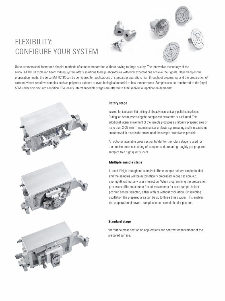
PREPARATION WORKFLOW UNDER CRYOGENIC CONDITIONS
Sample Holders
Several sample holders are available to fit vitrified samples prepared by high pressure freezing systems or bulky frozen samples, using plunge- or slush freezing techniques.
Pre-Preparation
The ion beam process follows the rules of the cross sectioning techniques in which the protruded portion a roughly pre-prepared sample is bombarded by high energy ion beams. In order to fulfill the pre-preparation needs the distance of max. 100 µm is achieved by using the VCT loading station with the cryo-saw attachment.
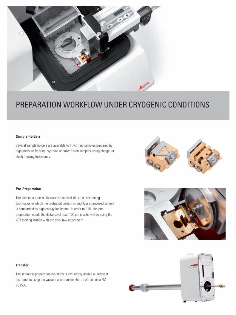
Transfer
The seamless preparation workflow is ensured by linking all relevant instruments using the vacuum cryo transfer shuttle of the Leica EM VCT500.

12 . 13
Samples which need to be prepared and transferred under cryo conditions also have to be pre-prepared under cryogenic conditions. A special VCT loading station with cryo-saw has been developed to fulfill these conditions. The sample is inserted in the VCT sample holder and mechanically pre-prepared using a diamond disc cutter in LN2. Users can transfer the pre-prepared sample in the same holder to the Leica EM TIC 3X – VCT for further broad ion beam processing with the Leica EM VCT. Thus prepared, the sample can be subsequently transferred to the coating system EM ACE600 and / or to the cryo SEM for investigation.
Imaging and analysis Leica EM ACE600
Top: Cryo-SEM image of high pressure frozen yeast Bottom: Cryo-SEM Image of high pressure frozen sea urchin embryosBoth samples were subsequently prepared by using the illustrated workflow courtesy of Derk Joester and Irene Yin-Ting Chang; Northwestern University).
(Cryo) SEM
Leica EM TIC 3X - VCT
VCT loading station with cryo-sawLeica EM ICE

Cross section of a gold wire bond; mechanically pre-prepared with the Leica EM TXP. The final surface preparation was carried out with the Leica EM TIC 3X
100 µm 30 um
The EBSD result shows a perfect cross section of the target, with no introduced artefacts. Diffraction pattern are obtained from the Si, W, Al Si and Au layers. Very fine deformation structures in the Au layer (of less than 40 nm) are revealed by the EBSD analysis. Courtesy of: Bruker Nano GmbH
Oil shale (nano pores), revealed with the Leica EM TIC3X (rotary stage) total sample size ∅ 25 mm
Cross section of veneer

14 . 15
0.1 mm
15
Cross section of SiC abrasive paper Cross section of SiC abrasive paper
Friction Stir processes Al-30Si Alloy after mechanical polishing … … and after additional ion polishing with the Leica EM TIC 3X (Rotary Stage)
Calcium Carbonte (Calcite and Aragnoite) with organic membranes between the carbonate, Ion beam polished with the Leica EM TIC 3X (Rotary Stage)
Coaxial polymer fiber (water soluble) prepared at –120 °C
For more information on applications see the Leica EM TIC 3X Application Booklet on our Website!

01/1
7 · O
rder
no.
: 119
1482
4 · ©
201
7 by
Lei
ca M
icro
syst
ems
GmbH
.Su
bjec
t to
mod
ifica
tions
. LEI
CA a
nd th
e Le
ica
Logo
are
regi
ster
ed tr
adem
arks
of L
eica
Mic
rosy
stem
s IR
Gm
bH.
Leica Mikrosysteme GmbH | Vienna, AustriaT +43 1 486 8050-0 | F +43 1 486 8050-30
www.leica-microsystems.com
CONNECT WITH US!