Leg Strengthening in COPD, Two Modalities: - … · Leg Strengthening in COPD, Two Modalities: ......
34
1 Anita Grongstad Master of Exercise Physiology/Sport Sciences Faculty of Medicine Department of Circulation and Medical Imaging 2009 Leg Strengthening in COPD, Two Modalities: - Effects on Muscle Strength, Work Economy and Pulmonary Function
Transcript of Leg Strengthening in COPD, Two Modalities: - … · Leg Strengthening in COPD, Two Modalities: ......

1
Anita Grongstad
Master of Exercise Physiology/Sport Sciences
Faculty of Medicine
Department of Circulation and Medical Imaging
2009
Leg Strengthening in COPD, Two Modalities:
- Effects on Muscle Strength, Work Economy and
Pulmonary Function

2
CONTENTS
ACKNOWLEDGEMENTS.................................................................................................................. 3
ABSTRACT.......................................................................................................................................... 4
1. INTRODUCTION ........................................................................................................................ 5
1.1 Background ................................................................................................................................. 5
1.2 Symptoms ................................................................................................................................... 6
2. COPD AND TREATMENT.......................................................................................................... 7
2.1 Exercise training ......................................................................................................................... 7
3. MATERIALE AND METHODS .................................................................................................11
3.1 Setting ........................................................................................................................................11
3.2 Subject characteristics................................................................................................................11
3.3 Testing procedures and apparatus ............................................................................................. 12
3.4 Scaling....................................................................................................................................... 15
3.5 Training intervention................................................................................................................. 15
3.6 Statistical analysis ..................................................................................................................... 16
4. RESULTS.................................................................................................................................... 17
4.1 Exercise data ............................................................................................................................. 17
4.2 Correlation analyses.................................................................................................................. 19
5. DISCUSSION............................................................................................................................. 20
6. CONCLUSION........................................................................................................................... 31
7. REFERENCE LIST .................................................................................................................... 32

3
ACKNOWLEDGEMENTS
This work would never been carried out if it wasn’t for the encouragement and backing from my
superior and boss Agnete Hagelsten Dalelid. All through, you supported me both professionally and
privately during the last two years. I am sincerely grateful.
I gratefully acknowledge my supervisor prof. Jan Hoff for support through the study process. You
never gave me the answers in the easy way, but guided me through topics which gave me a broader
insight in physiology and science, and in that way answering my questions. I also want to thank both
prof. Jan Hoff and prof. Jan Helgerud for encouraging us physiotherapists to include “proper”
training intensities into clinic – I will take you at your word!
This work was performed at Glittreklinikken, Nittedal. I wish to acknowledge all my colleagues for
willingness to cooperate and organize the workday to my account. I want to thank the unit for
Respiratory Physiology and Laboratory for goodwill and always finding time to test my subjects,
and to Jan Inge, Christine and Liv Karin for being helpful every time I tore my hair because of the
Schillers software. A special thank go to dr. philos. Morten Ryg for being my local supervisor -
always available and helpful and to Senior BLS and PhD stud. Anne Edvardsen and dr. PhD stud.
Aina Kjensli for always having time for my questions and for giving me useful discussions. A
special thank to physiotherapist, colleague and friend, Ulla Pedersen, for your critical questions
which made me think twice, and your comical comments always cheered me up.
I could never accomplish this master program without the financial grant from Glittreklinikken. This
gave me the possibility to be a full time student. I also thank The Norwegian Society of
Physiotherapists (NFF) for financial grant.
My sincere thanks go to my family and friends for keeping up with me even if I have been absent-
minded during long periods. In particular my cohabitant Jan and our children Tina, Magnus and
Ingrid for letting me carry out this master program. I will be delighted to be more present in your
lives soon.
Finally, I want to thank all patients who helped me with the study, you all became special to me
during 20 sessions with “sweat and tears”! Working with patients has always been meaningful to me,
and I`m looking forward being a full time physiotherapist once more.

4
ABSTRACT
Purpose: Reduced peripheral muscle strength and exercise intolerance accompany chronic
obstructive disease (COPD), and inactivity as an important contributor. Exercise training has become
mandatory in pulmonary rehabilitation and strength training an important component. Since previous
studies of strength training in COPD patients have been performed with different intensities, leg
press being almost the only exercise studied, this study was designed to compare two different
strength exercises; leg press (LP) and step device (SD) with the same intensities. The outcome
measures were effects on muscular strength, work economy and pulmonary function after 4 weeks
with maximal strength training. Methods: Twenty patients with COPD (FEV1 pred. % = 48±17) were
participating in an in-patient, multidisciplinary pulmonary rehabilitation program randomly assigned
to LP (n =10) or SD (n =10). Both groups performed four sets with five repetitions five days a week
with focus on maximal mobilization in the concentric phase of the movement five days a week. The
intensity was adjusted to allow only the performance of five repetitions, corresponding to 85-90% of
1RM. Results: Both groups showed significant improvement in muscle strength and work economy,
LP (19% and 17%) and SD (10% and 18%) respectively after 4 weeks. There was no significant
difference between the groups. Neither groups showed significant changes in pulmonary function.
Conclusion: This study reveals the importance of intensity when choosing strength exercises. Both
leg press and step device gave improvement in muscle strength and work economy. This gives
physiotherapists and COPD patients the possibility to improve and maintain muscle strength
independently of available equipment, as long as intensity corresponds to 85-90 % of 1RM.

5
1. INTRODUCTION
1.1 Background
Pulmonary diseases are increasingly important causes of morbidity and mortality in the modern
world. Chronic obstructive pulmonary disease (COPD) is the most common chronic lung disease,
and is a major cause of lung-related death and disability. According to WHO 210 million people
have COPD worldwide and more than 3 million people died of COPD in 2005 (WHO 2007). The
latest definition on COPD is: “Chronic obstructive pulmonary disease (COPD) is a preventable and
treatable disease with some significant extrapulmonary effects that may contribute to the severity in
individual patients. Its pulmonary component is characterized by airflow limitation that is not fully
reversible. The airflow limitation is usually progressive and associated with an abnormal
inflammatory response of the lung to noxious particles or gases” (Rabe, Hurd et al. 2007). COPD
develops insidiously over decades and because of the large reserve in lung function there is a long
preclinical period. Symptoms as chronic cough and sputum production may precede the
development of airflow limitation by many years, thus affected persons in an early stage may have
few symptoms and many are undiagnosed until a relatively advanced stage of the disease (Rabe,
Hurd et al. 2007).
COPD is defined on the basis of airflow limitation and spirometry is essential for diagnosis. The
measurements are evaluated by comparison with reference values based on the subject’s age, sex,
race and height (Pellegrino, Viegi et al. 2005). ATS/ERS recommend using The Global Initiative for
Chronic Obstructive Lung Disease (GOLD) guidelines to set universal standards in the prevention,
diagnosis, and management of patients with COPD (GOLD 2008 Updated). The classification of
COPD severity is categorized in four stages based on post-bronchodilator FEV1. The presence of
airflow limitation is defined by a post-bronchodilator FEV1/FVC < 0.70 (Figure 1). Even if the
diagnosis is confirmed by spirometry, it is important to note that the clinical diagnosis of COPD
should be considered when a patient has a history of exposure to risk factors and/or dyspnea, chronic
cough or sputum production (Rabe, Hurd et al. 2007).

6
Figure 1. - GOLD-stage classification of COPD severity based on post-bronchodilator values of FEV1 and FVC.D
Classification of COPD Severityby Spirometry
Stage I: Mild FEV1/FVC < 0.70
FEV1 > 80% predicted
Stage II: Moderate FEV1/FVC < 0.7050% < FEV1 < 80% predicted
Stage III: Severe FEV1/FVC < 0.7030% < FEV1 < 50% predicted
Stage IV: Very Severe FEV1/FVC < 0.70FEV1 < 30% predicted or
FEV1 < 50% predicted pluschronic respiratory failure
FEV1, forced expiratory volume in one second; FVC, forced vital capacity
1.2 Symptoms
Dyspnea and chronic cough are well-known symptoms in COPD and the reason most patients seek
medical attention. Patients with COPD show poor exercise performance, and exercise intolerance is
one of the main factors limiting participation in activities of daily life (Nici, Donner et al. 2006).
Peripheral muscle dysfunction is a well recognized disabling feature of COPD, and the target muscle
of investigation has been quadriceps femoris. The quadriceps muscle is of interest because of the
significant correlation between quadriceps strength and both FEV1 and exercise capacity (Bernard,
LeBlanc et al. 1998). There are evidence pointing at reduced physical activity as an important
contributor to the muscle weakness because lower limb muscle strength and exercise intolerance are
common features, not only in COPD patients, but among several other patients groups as well as in
elderly subjects, all with reduced level of activity as a common component (Rantanen, Guralnik et
al. 1999; Hoydal, Helgerud et al. 2007).

7
2. COPD AND TREATMENT
2.1 Exercise training
The overall management of COPD is, in addition to reduce risk factors and relief symptoms, to
improve exercise capacity, health status and patients’ quality of life. Smoking cessation is the most
important action (Ries, Bauldoff et al. 2007). Studies reveal that the ability to improve lung function
pharmacologically in patients with COPD is quite limited (Rabe, Hurd et al. 2007) while several
studies have shown that physical exercise reverses COPD induced skeletal muscle dysfunctions and
improve exercise tolerance, reduce dyspnea and substantially improve quality of life (Nici, Donner
et al. 2006). A study comparing patients with COPD, diabetes and healthy subjects showed that
subjects with COPD had the lowest level of physical activity, i.e 84% of the patients had an activity
level too low to maintain good health (Arne, Janson et al. 2009). Pitta and Troosters et al. (2006) saw
a reduction in physical activity after admission with acute exacerbation in COPD patients with a
mean walking time of 6–7 min·day-1
and fails to recover even 1 year after to levels observed in
stable outpatients with equally severe COPD. The duration of the rehabilitative exercise programs is
much discussed and the evidence is clear that longer duration have better long-terms effect
compared with short-term (Troosters, Gosselink et al. 2000), while the number of training sessions
have an effect on strength improvement, and near daily training has shown less effect than 2-3
sessions a weeks (McArdle, Katch et al. 2007, p.523). Another challenge for COPD patients is to
maintain the effects from a rehabilitation program. Home-based exercise training studies have not
shown convenience evidence; McMurdo and Johnstone (1995) followed 69 elderly people in 6
month. There was a trend toward improvement, but not significantly. This is supported by Emery
and Shermer (2003), who showed that the gains achieved during a 10 week pulmonary program were
maintained in only 39% of the COPD patients after 1 year. There are few studies including muscle
strength as an outcome in home-based training studies for COPD patients, and further work is
required to identify the optimal strength exercise in a home-based training program.
The challenge for a physiotherapist working with COPD patients is to design training programs that
is not limited by respiratory impairments before achieving physiological adaptations. Bjorgen et al.
(2009) revealed that COPD patients cycling by using one leg at 85-95% of peak heart rate, increased
significant whole body VO2peak and peak work rate. This shows that COPD patients have great
advance of exercising with reduced muscle mass, which should indicate muscle strength training
being an effective method for this patient group.
8
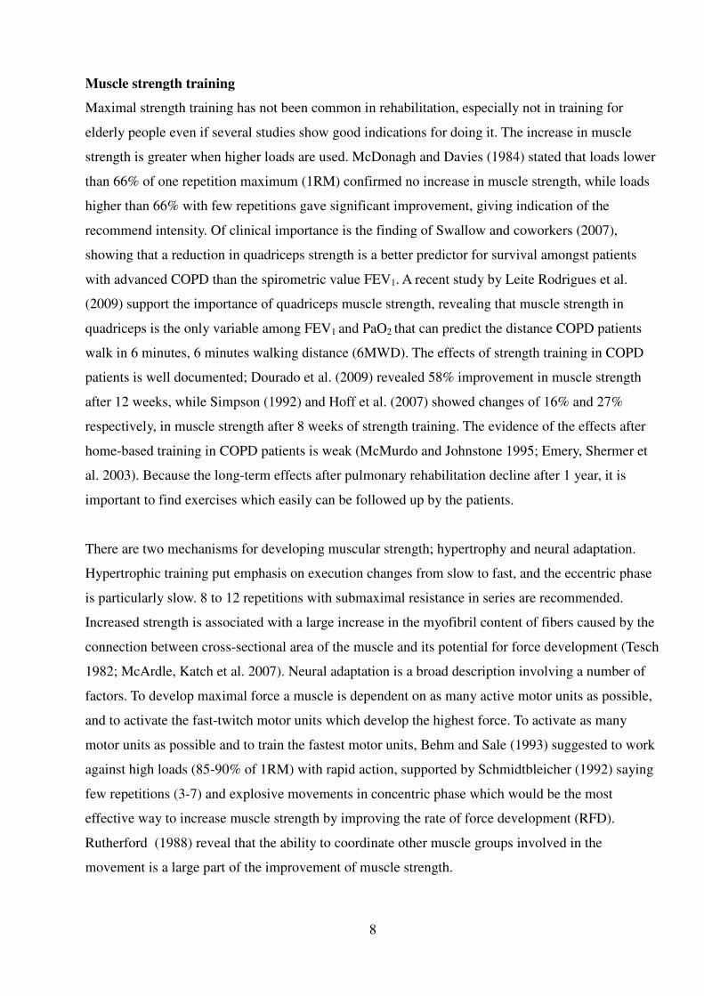
Muscle strength training
Maximal strength training has not been common in rehabilitation, especially not in training for
elderly people even if several studies show good indications for doing it. The increase in muscle
strength is greater when higher loads are used. McDonagh and Davies (1984) stated that loads lower
than 66% of one repetition maximum (1RM) confirmed no increase in muscle strength, while loads
higher than 66% with few repetitions gave significant improvement, giving indication of the
recommend intensity. Of clinical importance is the finding of Swallow and coworkers (2007),
showing that a reduction in quadriceps strength is a better predictor for survival amongst patients
with advanced COPD than the spirometric value FEV1. A recent study by Leite Rodrigues et al.
(2009) support the importance of quadriceps muscle strength, revealing that muscle strength in
quadriceps is the only variable among FEV1 and PaO2 that can predict the distance COPD patients
walk in 6 minutes, 6 minutes walking distance (6MWD). The effects of strength training in COPD
patients is well documented; Dourado et al. (2009) revealed 58% improvement in muscle strength
after 12 weeks, while Simpson (1992) and Hoff et al. (2007) showed changes of 16% and 27%
respectively, in muscle strength after 8 weeks of strength training. The evidence of the effects after
home-based training in COPD patients is weak (McMurdo and Johnstone 1995; Emery, Shermer et
al. 2003). Because the long-term effects after pulmonary rehabilitation decline after 1 year, it is
important to find exercises which easily can be followed up by the patients.
There are two mechanisms for developing muscular strength; hypertrophy and neural adaptation.
Hypertrophic training put emphasis on execution changes from slow to fast, and the eccentric phase
is particularly slow. 8 to 12 repetitions with submaximal resistance in series are recommended.
Increased strength is associated with a large increase in the myofibril content of fibers caused by the
connection between cross-sectional area of the muscle and its potential for force development (Tesch
1982; McArdle, Katch et al. 2007). Neural adaptation is a broad description involving a number of
factors. To develop maximal force a muscle is dependent on as many active motor units as possible,
and to activate the fast-twitch motor units which develop the highest force. To activate as many
motor units as possible and to train the fastest motor units, Behm and Sale (1993) suggested to work
against high loads (85-90% of 1RM) with rapid action, supported by Schmidtbleicher (1992) saying
few repetitions (3-7) and explosive movements in concentric phase which would be the most
effective way to increase muscle strength by improving the rate of force development (RFD).
Rutherford (1988) reveal that the ability to coordinate other muscle groups involved in the
movement is a large part of the improvement of muscle strength.
9
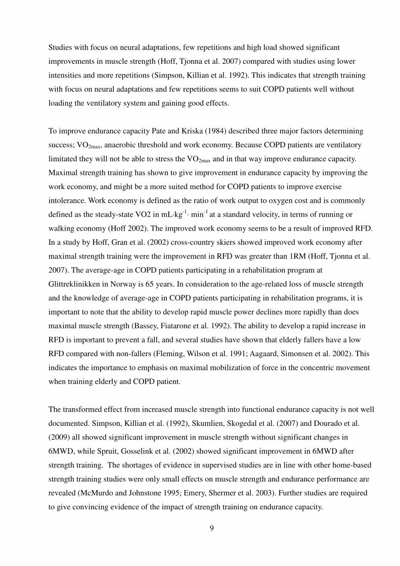
Studies with focus on neural adaptations, few repetitions and high load showed significant
improvements in muscle strength (Hoff, Tjonna et al. 2007) compared with studies using lower
intensities and more repetitions (Simpson, Killian et al. 1992). This indicates that strength training
with focus on neural adaptations and few repetitions seems to suit COPD patients well without
loading the ventilatory system and gaining good effects.
To improve endurance capacity Pate and Kriska (1984) described three major factors determining
success; VO2max, anaerobic threshold and work economy. Because COPD patients are ventilatory
limitated they will not be able to stress the VO2max and in that way improve endurance capacity.
Maximal strength training has shown to give improvement in endurance capacity by improving the
work economy, and might be a more suited method for COPD patients to improve exercise
intolerance. Work economy is defined as the ratio of work output to oxygen cost and is commonly
defined as the steady-state VO2 in mL·kg-1
· min-1
at a standard velocity, in terms of running or
walking economy (Hoff 2002). The improved work economy seems to be a result of improved RFD.
In a study by Hoff, Gran et al. (2002) cross-country skiers showed improved work economy after
maximal strength training were the improvement in RFD was greater than 1RM (Hoff, Tjonna et al.
2007). The average-age in COPD patients participating in a rehabilitation program at
Glittreklinikken in Norway is 65 years. In consideration to the age-related loss of muscle strength
and the knowledge of average-age in COPD patients participating in rehabilitation programs, it is
important to note that the ability to develop rapid muscle power declines more rapidly than does
maximal muscle strength (Bassey, Fiatarone et al. 1992). The ability to develop a rapid increase in
RFD is important to prevent a fall, and several studies have shown that elderly fallers have a low
RFD compared with non-fallers (Fleming, Wilson et al. 1991; Aagaard, Simonsen et al. 2002). This
indicates the importance to emphasis on maximal mobilization of force in the concentric movement
when training elderly and COPD patient.
The transformed effect from increased muscle strength into functional endurance capacity is not well
documented. Simpson, Killian et al. (1992), Skumlien, Skogedal et al. (2007) and Dourado et al.
(2009) all showed significant improvement in muscle strength without significant changes in
6MWD, while Spruit, Gosselink et al. (2002) showed significant improvement in 6MWD after
strength training. The shortages of evidence in supervised studies are in line with other home-based
strength training studies were only small effects on muscle strength and endurance performance are
revealed (McMurdo and Johnstone 1995; Emery, Shermer et al. 2003). Further studies are required
to give convincing evidence of the impact of strength training on endurance capacity.

10
Furthermore, it seems hard to find appropriated exercises which can improve and/or maintain muscle
strength in self-monitored training, which is very important because most of the COPD patients are
not following a supervised pulmonary rehabilitation program.
The FEV1-value is widely regarded as the most common value describing the severity of COPD, and
both patients and professionals are familiar to FEV1. Most of the studies including interventions in
COPD patients have FEV1 as an outcome measure, but the significance of this value is still no clear.
In the review by O'Shea, Taylor et al. (2004), no change in respiratory function did appear after
strength training. These findings are in line with other studies; Skumlien, Skogedal et al. (2007)
showed a change of 8% in FEV1 after 4 weeks of strength training while Simpson, Killian et al.
(1992) got the same change after 8 weeks. Thus, it was very sensational when Hoff, Tjønna et al.
(2007) revealed a significant improvement in FEV1 of 22% after 8 weeks of maximal strength
training. The finding in the latter study makes it necessary to have FEV1 as an outcome measure in
further experiments.
Actually, few COPD patients are participating in rehabilitation programs, and studies with home-
based strength training programs are using low intensities and show smaller effects, while studies
using a seated leg press with high intensities reveal to be effective. Therefore, it is important to find
exercises which might give the same increase in muscle strength as seated leg press, but without
special equipments. Current studies have shown conclusive evidence that maximal strength training
with few repetitions improve muscle strength in COPD patients, but the influence on other
parameters like work economy and pulmonary function are not well documented. The aim of this
study was to compare two different strength exercises for the muscles of ambulation to investigate
whether the effects on muscle strength would be the same for both interventions, and to see if the
improved muscle strength will enhance work economy and pulmonary function. Following
hypotheses were tested: 1) COPD patients performing maximal strength training in a seated leg press
and on a step device will improve muscle strength after 4 weeks; 2) Improvements in muscle
strength will directly translate into improved work economy, measured during a standard workload
on a treadmill; 3) The parallel training effects on respiratory muscles during maximal strength
training will improve pulmonary function, measured by FEV1.

11
3. MATERIALE AND METHODS
3.1 Setting
The study was performed at Glittreklinikken, Nittedal, Norway between January and Mai 2009.
Glittreklinikken is a hospital for diagnosis, treatment and rehabilitation for persons with lung
disease. Patients attend a four week in-clinic interdisciplinary program, consisting of medical
diagnosis, treatment, training and education. There are 96 patient rooms distributed over six units
and about 50% of the patients have been there more than one time, 65 % of all patients hospitalized
at Glittreklinikken have the diagnosis COPD and the average age of the patients is 65 years.
3.2 Subject characteristics
In total, 24 patients were included in the study, 12 in leg press (LP) and 12 in step device (SD). All
the subjects were recruited at the first day they arrived to Glittreklinikken. Inclusions criteria were a
clinical definition of COPD according to GOLD guidelines (Celli and MacNee 2004),
FEV1/FVC < 0.70 and FEV1 ≤ 80 % of predicted and ability to perform a seated leg press exercise
and a step device exercise. Exclusion criteria were smokers or other lung diseases combined with
COPD. Two subjects in each group dropped out. In LP one subject had a FEV1 value > 80 %
predicted, were the spirometric results was received the third day of his stay and after inclusions
tests were performed. The second fell in the canteen and impaired her ankle two weeks after start,
and was not able to perform any exercise for a week. Two subjects in SD got exacerbations on their
last days of the 4 weeks and could not be tested before leaving. The subjects were randomly assigned
to LP or SD by drawing a lot. Randomization within genders facilitated groups that were balanced
for gender (four females in both groups). Medication was monitored and seven in the LP group and
six in the SD group adjusted their medication during the 4-weeks. The baseline characteristics of the
subjects are presented in Table 1.

12
Table 1 – Baseline characteristics
Leg Press Step Device
n = 10 n = 10
Age (yr) 65.2 (± 8.7) 69.9 (± 6.2)
Height (cm) 173.4 (± 8.7) 170.3 (± 5.6)
Body mass (kg) 76.9 (± 18.6) 75.6 (± 12.5)
BMI (kg ·m 2) 25.2 (± 4.7) 26.0 (± 3.5)
FEV1 (L) 1.38 (± 0.75) 1.31 (± 0.5)
predicted % 45.6 (± 19.1) 49.7 (± 15.9)
FVC (L) 2.76 (± 1.0) 2.58 (± 0.5)
predicted % 74 (± 16.7) 79.3 (± 18.6)
FEV1/ FVC (%) 47.5 (± 10.9) 50.2 (± 15.3)_______
Data are presented as mean (± SD); BMI, body mass index; FEV1 forced expiratory volume in one second; FVC,
forced vital capacity; No significant differences between groups (p>0.05).
3.3 Testing procedures and apparatus
Pre- and posttests were performed with identical protocols.
Maximal strength
Maximal muscle strength was measured dynamic using 1RM, “refers to the maximum amount of
loads lifted one time during a standard weight-lifting exercise” (McArdle, Katch et al. 2007), s. 511.
1RM had to be measured on two different horizontal leg press apparatus, Selection (Techno Gym,
Italy) shown in figure 2 and Legpress 190849, Steens Physical, Steens Industrier, Ski, Norway),
because the Selection apparatus was brand new and had to be adjusted by a serviceman from Techno
Gym for two days when 4 subjects arrived and had to be tested. The 4 subjects, 2 in LP and 2 in SD,
continued on the Legpress from Steens Industrier during the study, including 1RM tests and training
sessions for LP. They performed the 1RM test with a knee angle of 90°. All subjects performed the
1RM test twice on two following days due to the learning effect, validated through a pilot work. The
first trial was performed after the work economy test where the subjects had walked on a treadmill
for five minutes. Before the second trial they warmed up walking on a treadmill for 5 minutes with a
speed corresponding to their habitual walking speed. The 1RM test started after 2 - 4 minutes rest.
The first attempt was adjusted by the test leader to be around 70 % of the assumed maximal strength
or around the subjects’ bodyweight. The load was increased until 1RM was achieved, 2 minutes rest
between each attempt and maximum five to six attempts were performed.

13
Figure 2. – Illustrating a 1RM test on the Selection apparatus.
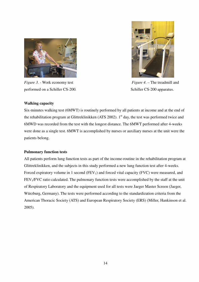
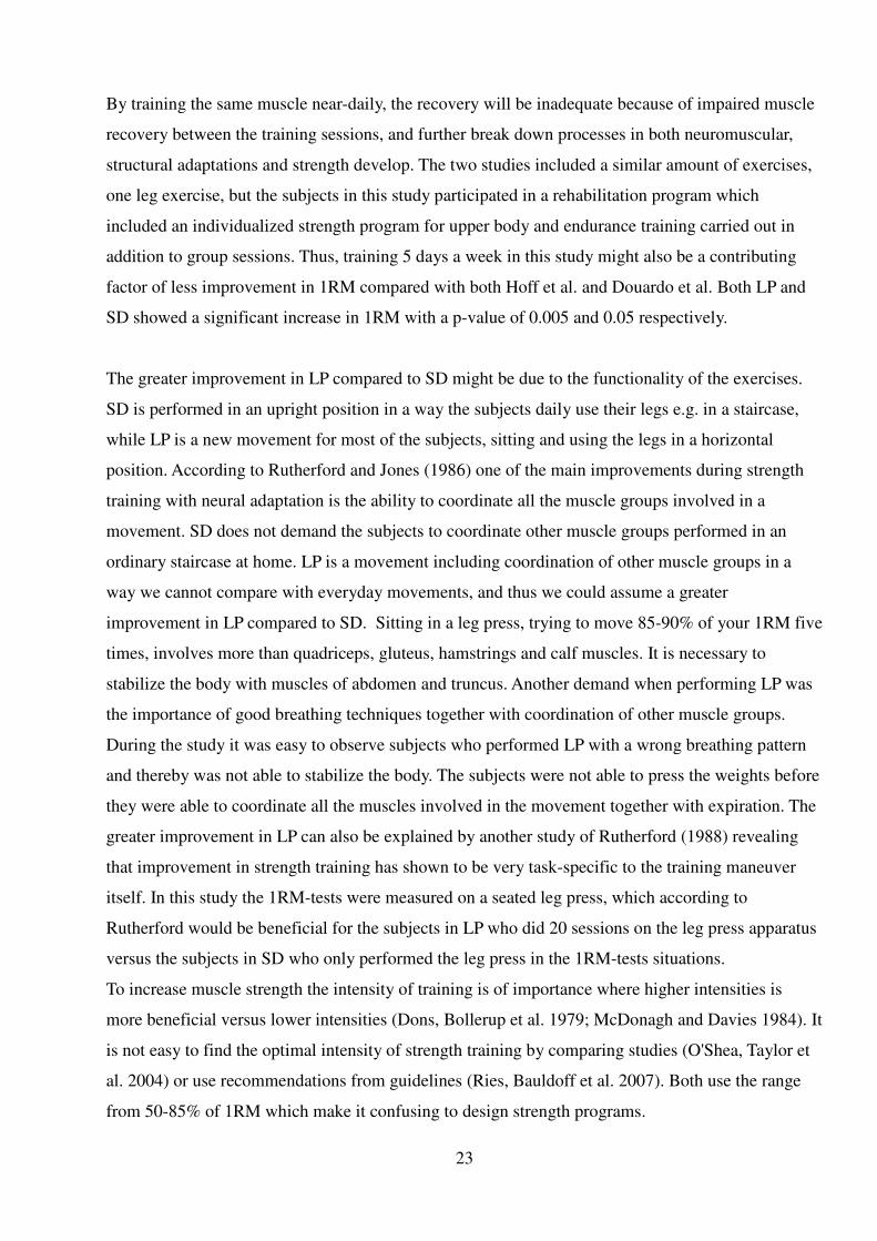
Work economy
Work economy was tested as a constant load test on a treadmill (Jaeger LE2000CE). From an initial
pilot work, a walking speed that was equivalent to 40 Watt (W) work rate was tested. Because 40 W
was corresponding to maximal work rate for some of the subjects, and a workload they could
maintain for 5 minutes was chosen. The mean W was 25 (±12). The work economy was determined
by measuring the steady state consumption of oxygen (VO2), using the mean VO2 -values noted after
4:40, 4:50 and 5:00 minutes measured as mL·min-1
. To measure the oxygen consumption, an Oxycon
Pro. Apparatus (Jaeger, Wûrtzburg, Germany) was validated initially in a pilot test. Unfortunately,
after testing the first six subjects, the Oxycon Pro had to be delivered for service. Therefore the rest
of the pre- and posttests had to be performed by using Schiller CS-200 (Schiller, Baar, Switzerland)
(Figure 3 and figure 4). To be aware of the differences between the two apparatus test-values from
measurements taken routinely of the staff working at the respiratory physiology laboratory were
used. Four tests were performed between January and Mai 2009. Standardized procedures with
randomization between the apparatus each time were followed and the same person walked at
different speeds for 5 minutes on both Oxycon and Schiller at the same day. The test at 4.8 km/t was
chosen because it corresponded closest to the tests performed by the subjects in this study, revealed
mL-values in mean (±SD) for Oxycon at 856.25 (± 29.83) and Schiller CS-200 1057.75 (± 146.15).
A non-parametric Wilcoxon Signed Rank test showed no significant difference between the
apparatus, p= 0.068. Prior to all tests, a manual of calibrating was followed from the producer.
14
Figure 3. - Work economy test Figure 4. – The treadmill and
performed on a Schiller CS-200. Schiller CS-200 apparatus.
Walking capacity
Six-minutes walking test (6MWT) is routinely performed by all patients at income and at the end of
the rehabilitation program at Glittreklinikken (ATS 2002). 1st day, the test was performed twice and
6MWD was recorded from the test with the longest distance. The 6MWT performed after 4-weeks
were done as a single test. 6MWT is accomplished by nurses or auxiliary nurses at the unit were the
patients belong.
Pulmonary function tests
All patients perform lung function tests as part of the income-routine in the rehabilitation program at
Glittreklinikken, and the subjects in this study performed a new lung function test after 4-weeks.
Forced expiratory volume in 1 second (FEV1) and forced vital capacity (FVC) were measured, and
FEV1/FVC ratio calculated. The pulmonary function tests were accomplished by the staff at the unit
of Respiratory Laboratory and the equipment used for all tests were Jaeger Master Screen (Jaeger,
Würzburg, Germany). The tests were performed according to the standardization criteria from the
American Thoracic Society (ATS) and European Respiratory Society (ERS) (Miller, Hankinson et al.
2005).

15
3.4 Scaling
Scaling is a mathematical procedure to establish a proper relationship between a body size variable
and factors like endurance capacity or muscle strength (McArdle, Katch et al. 2007, p. 517). When
comparing both muscle strength and work economy at submaximal exercise, heavier subjects will be
overestimated and the lighter subjects underestimated (Hoff 2002). A unit of mL·kg0.75
·min-1
has
shown to be convenient when comparing subjects with different body mass in endurance exercises
(Helgerud 1994), while body mass raised to the power of 0.67 is more indicative when comparing
strength performance, expressed kg· mb-0.67
(Wisloff, Helgerud et al. 1998).
3.5 Training intervention
Both LP and SD performed a four weeks training regime, a total of 20 training sessions. Each
session consisted of four sets of five repetitions with a focus on maximal mobilization in the
concentric movement. Both groups did five minutes light warm up on a treadmill before the training
session. The LP group performed all strength training on the same seated leg press machine they
used during 1RM testing with loads corresponding to 85-90% of 1RM. The subjects were instructed
to stop the eccentric movement at an angle of 90° in knees, a full stop before emphasis on maximal
mobilization to straight legs. When the subject was able to perform more than five repetitions in a
set, load was increased by 5 kg until five repetitions were the maximal repetitions being achieved.
Some subjects needed a rest between some of the repetitions, which was determined individually
dependent on ventilation limitations. All subjects rested for 2 minutes between each set. The SD
group performed a strength exercise by using a metal step device (Steens Industrier, Ski, Norway),
where the height of the step could be altered up and down (Figure 5). The training was performed
standing on a step with one leg, performing a backward step down until the tiptoe of the other leg
touched the floor. Then full stop without letting the tiptoe leg take any bodyweight before maximal
concentric movement to straight leg using the muscles of the leg standing on the step. The step
device was placed in front of a naked wall to avoid the possibility to use the arms to grab and drag
up (Figure 6). The height of the step was adjusted up until the subjects managed to perform
maximum five repetitions. Five repetitions with one minute rest before changing leg. Two minutes
rest between each set. All the strength training sessions in both groups were supervised to ensure
enough loads/height and correct execution. As participant in a rehabilitation program the subjects
took part in other activities during the four weeks, including gymnastics in groups, individual
training programs on treadmills and strength training programs for upper body.

16
Figure 5. – The step device. Figure 6. – Illustrating a backward step
in front of a naked wall.
3.6 Statistical analysis
The software program SPSS 16.0 was used to do the statistical analyses and construct the figures and
tables. The main outcomes; strength, walking economy and lung function were measured twice and
characterized as repeated measurements. There are several techniques that can be used to test the
difference between groups. Due to the low number of subjects, a nonparametric test was adopted for
the statistical analyses. A Wilcoxon Signed Rank test was used to analyze the changes within the
groups from pre- to posttest. Delta values (i.e. the difference from pre- to posttest) was calculated
and used in a Mann-Whitney U test to analyze the changes from pre- to posttest between groups.
Non-parametric tests use median and range values as measures of central tendency, but in order to
compare the results with other studies the parameters presented are as mean and standard deviation.
A p-value of less than 0.05 was considered to be statistically significant. Relationship between
variables were assessed with Spearman`s Rank Order Correlation in order of being a non-parametric.

17
4. RESULTS
Baseline values as age, physical characteristics and pulmonary function reveal no significant
difference between the LP group and SD group. Neither group experienced a significant change in
body weight during the 4 week period. Both groups completed the study protocol without any
adversity, and the LP and SD completed 97% and 97.5%, respectively of the planned 20 sessions.
4.1 Exercise data
Muscle strength
4 weeks of maximal strength training were significantly associated with increased muscle strength in
both LP (p< 0.005) and SD (p<0.05). 1RM increased by 26.5 kg (19%) in LP and 13.5 kg (10%) in
SD. There was no significant difference between the groups (p>0.05). (Table 2, Figure 7).
Table 2. – Alterations in strength parameters measured before and after 4 weeks of training.
Leg Press group Step Device group
n = 10 n = 10
_________________Pre Post Pre Post
Body mass (kg) 76.9 ± 18.6 77.3 ± 18.9 75.6 ± 12.5 5.6 ± 12.4
1RM (kg) 142.5 ± 56.4 169.0 ± 60.3 ** 127.0 ± 45.0 140.5 ± 50.8*
1RM (kg · mb-0.67
) 7.59 ± 2.05 9.00 ± 2.04 ** 6.95 ± 2.16 7.70 ± 2.51*
Data are presented as mean ± SD. 1RM, one-repetition maximum with a knee angle at 90°; mb, bodyweight..
Significant difference before and after training within group (* p<0.05, ** p< 0.005). No significant difference
between groups (p>0.05)
Work economy
Both groups significantly improved their work economy during a steady state treadmill test with a
reduction in oxygen consume in liters of 0.16 L·min-1
in both LP and SD, 17% and 18% reduction
respectively (p<0.005).There was no significant difference between groups (Table 3, Figure 7).

18
Table 3 – Physiological responses to a steady state treadmill exercise test after 4 weeks maximal
strength training
Leg Press Group Step Device group
n = 10 n = 10
_________________ Pre Post Pre Post__
VO2 (L·min-1
) 1.12 ± 0.34 0.96 ± 0.26** 1.03 ± 0.36 0.87 ± 0.20**
VO2 (mL·kg -1
·min-1
) 14.3 ± 1.8 12.5 ± 1.7** 13.6 ± 3.3 11.5 ± 2.0**
VO2 (mL·kg0.75
·min-1
) 42.6 ± 6.5 36.9 ± 5.5* 39.9 ± 11.2 34.0 ± 5.9*
Data are presented as mean (± SD). VO2, Oxygen uptake. Significantly difference before and after training for both
groups (* p<0.05; ** p< 0.005). No significantly difference between groups (p >0.05).
Walking capacity
Only 10 subjects were included in the results of the 6MWT, LP n = 6 and SD n = 4. Four subjects,
two from each group had to be excluded because they performed 6MWT with supplementary oxygen
at posttest. One in SD had an exacerbation at posttest which influenced the 6MWT by decreasing the
distance with 150 m. and was excluded, and three subjects in SD whilst two in LP did not perform
the posttest. LP had a significant improvement in 6MWD with 85 meters (19%) p<0.05, while SD
improved 6MWD with 14 meter (3%) which was not significant. No significant difference between
groups (Table 4, Figure 7).
Table 4. – Changes in distance walked in 6MWD after 4 weeks of maximal strength training.
Leg Press group Step Device group
n = 6 n= 4
____________ Pre Post Pre Post_____
6MWD, m 457 ± 97 542 ± 66* 444 ±74 457 ± 38___
Data are presented as mean (± SD). M, meter, Significant difference within LP after 4 weeks (*p<0.05).
Pulmonary function
There were no significant changes within the groups or between the groups. FEV1 increased with
0.12 L (8%) in LP and 0.01 L (0.7%) in SD. FVC increased with 0.13 L (5%) in LP and 0.09 L
(4%) in SD 2.9% of predicted (4%). FEV1/ FVC % increased by 5 % for both LP and SD with
2.2% and 2.7% respectively (Table 5, Figure 7).

19
Table 5 - Spirometric responses to 4 weeks training at the leg press machine and the step device.
Leg Press group Step Device group
n = 10 n = 10
_______________ Pre Post Pre Post
FEV1 (L) 1.37 ± 0.75 1.49 ± 0.75 1.31 ± 0.5 1.32 ± 0.5
Predicted % 45.6 ± 19.1 49.2 ±18.7 49.7 ± 15.9 50.1 ± 17.1
FVC (L) 2.76 ± 1.0 2.89 ± 1.02 2.58 ± 0.5 2.49 ± 0.7
Predicted % 74 ± 16.7 77.4 ± 20.5 79.3 ± 18.6 76.4 ± 23.4
FEV1/ FVC (%) 47.5 ± 10.9 49.7 ± 9.2 50.2 ± 15 52.9 ± 15.6
Data are presented as mean (± SD). FEV1, forced expiratory volume in one second; FVC, forced vital capacity.
No significant differences within groups or between groups before and after training (p>0.05).
Figure 7.- The percent change in maximal strength (1RM), work economy (Work E.), six minutes walking test
(6MWT) and pulmonary function (FEV1) after 4 weeks of maximal strength training for the leg press group (LP) and
the step device group (SD). *Significant difference between pre- and posttest (p < 0.05); ** (p < 0.005).
4.2 Correlation analyses
At baseline there was a significant correlation between FEV1 and 1RM (r = 0.66) and VO2 (r = 0.78),
and between 1RM and VO2 (r = 0.81) for all subjects (n=20). In LP the improvement in VO2
correlated significantly with both improvement in 1RM (r = 0.73) and FEV1 (r = 0.70). The
correlation was significant at the 0.05 level. No significant correlation of improvements in SD.

20
5. DISCUSSION
The aim of this study was to compare two different ways to perform a maximal strength training
exercise of the legs in COPD patients; one performed in a seated leg press machine using loads in
kg. as a resistance, and the other way by using the subjects own bodyweight on a step devise
combined with increasing the height of the step as increased intensity. Outcome were the effect on
1RM, walk economy and pulmonary function measured as FEV1. The major finding in this study is
that both strength training with loads in a seated leg press, and without load performed in a step
device significantly increased 1RM and walk economy during 4 weeks of training, components
which are related to increased exercise tolerance, quality of life and survival (Nici, Donner et al.
2006; Swallow, Reyes et al. 2007).
Leg press versus step device
In this study a comparison between maximal strength training performed in a seated leg press and on
a step device reveal that both exercises have significant improvement on maximal muscle strength
and walk economy. According to McArdle, (2007, p.518) it is the overload intensity and not the type
of exercise that applies the overload, that give strength improvements, and therefore it is not
surprising that both LP and SD improved muscle strength. Both LP and SD followed the same
design; strength training with maximal mobilization in the concentric movement and high intensity
with the ability to perform only five repetitions. This is a well known method to improve muscle
strength revealed by Behm and Sale (1993), while working against high loads and training the fastest
motor units will guarantee maximal voluntary contraction. Rapid movement is a method to increase
the rate of force development. The improvement in LP and SD is of clinical importance because
muscles of ambulation, and especially quadriceps, is the only evidenced based muscle group having
great impact on exercise capacity and in addition being a good predictor for both survival and
walking distance in COPD patients (Swallow, Reyes et al. 2007; Leite Rodrigues, Melo et al. 2009).
The improvement in LP is in line with other studies using a seated leg press to strengthen the
muscles of ambulation. Skumlien and Skogedal et al. (2007) showed significant improvement in a 4
weeks in-patient rehabilitation program, while two other out-patient studies have shown significant
improvement in muscle strength using a seated leg press (Simpson, Killian et al. 1992; Hoff, Tjonna
et al. 2007). The improvement in SD is difficult to compare with other studies because there are few
studies were other exercises than leg press has been used. It is more likely to compare with home-
based programs, but most of these studies do not include strength training, neither have muscle
strength as an outcome been measured.

21
One study by McMurdo and Johnstone (1995) had a trend toward improvement in exercises like “sit-
to-stand” and “Time-to-get-up” in a home-based exercises program for elderly people with poor
mobility. 86 subjects were allocated to a strength exercise group, a mobility exercise group or a
health education group. The subjects got verbal and written instruction and were visited for 30
minutes every 3-4 week in six month by a physiotherapist. They found no significant difference
between the groups and the trend towards improvement failed to attain statistical significance. The
significant improvement in SD in this study shows that this might be a convenient exercise to use as
an outcome in other home-based studies in the future.
In the present study both LP and SD showed a significant improvement in muscle strength,
revealing that both are well designed exercises for strengthening the leg muscles. The positive
effects from pulmonary rehabilitation are well documented and the challenge for COPD-patients is
to keep on training when arriving at home to maintain the effects gained during rehabilitation.
Emery and colleges (2003) followed COPD patients after one year of a 10-week intensive exercise
program. They were all given an individualized home exercise program and encouraged to continue
exercise at an exercise facility or on their own. Only 39% of the COPD patients had followed up the
exercise prescription and those were the only one who had maintained the physical effects gained
during the 10-weeks exercise program. COPD patients in Norway have a good chance to get routines
because they get financial support from the Norwegian social security system to do individual
training under supervision of a community physiotherapist. There are some problems;
physiotherapists do not have proper apparatus, like a seated leg press and do not give other strength
exercises for the legs instead, and it is well known that COPD patients often get out of training
routines due to exacerbations. The findings in this study show that SD is a good alternative exercise
to improve muscle strength in legs for COPD patients, giving the physiotherapists and COPD
patients the possibility to perform strength training independent of the available equipment.
According to Rutherford (1988) the SD should be preferred before LP. Rutherford showed that
improvement in strength training is very task-specific and training isolated muscle groups may not
be as effective to improve function capacity. Hence, an improvement in muscle strength gained from
SD is transferable in those activities which are required in daily life like rising up and walking steps.

22
Muscle strength
In the present study both LP and SD increased muscle strength significantly. LP showed a mean
increase of 26.5 kg (19%) p <0.005 and SD 13.5 kg (10%) p<0.05, but no significant difference
between the groups. Improved strength in the muscles of ambulation is important to prevent fall in
elderly subjects. This is special important for COPD patients because osteoporosis is a well known
side effect of drugs like glucocorticosteroids. The improved muscle strength in this study is less
compared with other studies. Hoff and colleagues (2007) showed an improvement of 27% in muscle
strength in COPD patients. They had approximately the same amount of sessions compared to this
study, 24 versus 20 respectively, and the same intensity of 85-90% of 1RM which could assume a
more similar improvement in muscle strength between these two studies. An important explanation
could be related to the initial extent of weakness of the subjects in the study by Hoff, were the mean
muscle strength was 118 kg compared to 142 kg. in this study. According to the dose-response
curve, subjects with initial low muscle strength would more easily improve muscle strength
compared to subjects with initial higher muscle strength. This could explain the impressive
improvement in leg muscle strength of 58 % in COPD patients in a study of Dourado and Tanni et al.
(2009), were the subjects had initial mean muscle strength of 98 kg.
Different body weight between the subjects will overestimate muscle strength in heavy athletes and
ought to be considered when comparing the different groups. By doing a dimensional scaling and
compare the relative strength in term of kg·mb-0.67
in these three studies, we can exclude body weight
to contribute the difference in muscle strength. A contributor that is of importance is duration of
training programs, which is supported by Green et al. (2001). They revealed that a rehabilitation
program of 4 weeks shows less benefit than similar training for longer duration of 7 weeks. This
might also be a factor to explain why Dourado et al. showed an improvement of 58% in 12 weeks
and Hoff et al. (2007) an improvement of 27% in 8 weeks while the present study only improved
19% after 4 weeks. But to compare weeks of training may not give us any answers without counting
the numbers of training sessions accomplished. Then we have to look for other explanations than
duration of the great improvement revealed by Hoff et al. compared to this study. Both Hoff et al.
and this study had approximately the same amount of training sessions, 24 versus 20 respectively.
The difference was the distribution of sessions with 5 sessions a week in this study and 3 sessions a
week in the study by Hoff et al. According to McArdle (2007, p. 523), training with multiple
exercises 4 or 5 days per week may produce less improvement than training 2 or 3 times per week.
23
By training the same muscle near-daily, the recovery will be inadequate because of impaired muscle
recovery between the training sessions, and further break down processes in both neuromuscular,
structural adaptations and strength develop. The two studies included a similar amount of exercises,
one leg exercise, but the subjects in this study participated in a rehabilitation program which
included an individualized strength program for upper body and endurance training carried out in
addition to group sessions. Thus, training 5 days a week in this study might also be a contributing
factor of less improvement in 1RM compared with both Hoff et al. and Douardo et al. Both LP and
SD showed a significant increase in 1RM with a p-value of 0.005 and 0.05 respectively.
The greater improvement in LP compared to SD might be due to the functionality of the exercises.
SD is performed in an upright position in a way the subjects daily use their legs e.g. in a staircase,
while LP is a new movement for most of the subjects, sitting and using the legs in a horizontal
position. According to Rutherford and Jones (1986) one of the main improvements during strength
training with neural adaptation is the ability to coordinate all the muscle groups involved in a
movement. SD does not demand the subjects to coordinate other muscle groups performed in an
ordinary staircase at home. LP is a movement including coordination of other muscle groups in a
way we cannot compare with everyday movements, and thus we could assume a greater
improvement in LP compared to SD. Sitting in a leg press, trying to move 85-90% of your 1RM five
times, involves more than quadriceps, gluteus, hamstrings and calf muscles. It is necessary to
stabilize the body with muscles of abdomen and truncus. Another demand when performing LP was
the importance of good breathing techniques together with coordination of other muscle groups.
During the study it was easy to observe subjects who performed LP with a wrong breathing pattern
and thereby was not able to stabilize the body. The subjects were not able to press the weights before
they were able to coordinate all the muscles involved in the movement together with expiration. The
greater improvement in LP can also be explained by another study of Rutherford (1988) revealing
that improvement in strength training has shown to be very task-specific to the training maneuver
itself. In this study the 1RM-tests were measured on a seated leg press, which according to
Rutherford would be beneficial for the subjects in LP who did 20 sessions on the leg press apparatus
versus the subjects in SD who only performed the leg press in the 1RM-tests situations.
To increase muscle strength the intensity of training is of importance where higher intensities is
more beneficial versus lower intensities (Dons, Bollerup et al. 1979; McDonagh and Davies 1984). It
is not easy to find the optimal intensity of strength training by comparing studies (O'Shea, Taylor et
al. 2004) or use recommendations from guidelines (Ries, Bauldoff et al. 2007). Both use the range
from 50-85% of 1RM which make it confusing to design strength programs.

24
According to McDonagh and Davies (1984) load less than 66% of 1RM give no increase in muscle
strength, and the discrepancies in studies may be attributable to the difference in the modalities and
the intensity of training employed. In this study 1RM in LP increased 19% while SD increased 11%.
The result may have been influenced by the lack of exact control of the resistance in SD which may
have lead to a lower intensity compared with LP. To prove the “66%- rule” of McDonagh and
Davies the subjects in SD had a mean 1RM at 127 kg., and to have significant improvement the
loads had to be at least 66% of 1RM – means 83 kg. The mean bodyweight in SD is 75.6 kg. which
is about 60% of 127 kg. and not enough to significantly increase strength. To increase the intensity
the step was adjusted higher up until 5 repetitions were maximal they could perform, and in that way
try to be at the same intensity level as the LP, 85-90% of 1RM. Anyway, this gives less control of the
intensity comparing to LP were exact kg. was adjusted.
Another component that could influence the greater improvement in LP compared to SD is the
strong motivation factor that accompanied the subjects in LP. In both groups most of the subjects
got very positively surprised over themselves at the 1RM test at baseline, and therefore it was very
easy for the subjects in LP to keep up the motivation and try to put new personal records in every
session. The same kind of motivation was not observed in SD.
Work economy
Results in work economy showed a significant improvement in both LP and SD with 17% and 18%
respectively (p< 0.005), but no significant difference between groups (p>0.05). It has been
demonstrated that maximal strength training with emphasis on maximal mobilization of force in the
concentric movement improve work economy and thus improve aerobic endurance performance
both in athletes and in COPD patients (Hoff, Gran et al. 2002; Hoff, Tjonna et al. 2007). Exercise
intolerance is a common hallmark in COPD and treatments which could improve endurance
performance among these patients would be of great benefit, studies have shown that COPD patients
have a low level of physical activity compared with other patient groups with a chronic disease
(Arne, Janson et al. 2009). Activity level decreases additionally during exacerbations giving the
COPD patients a mean walking time of 6-7 min·day -1
which fails to recover even after 1 month
(Pitta, Troosters et al. 2006).

25
According to greater improvement in 1RM for LP versus SD and a significant correlation between
changes in 1RM and work economy in LP, we could assume a greater difference in work economy
between LP and SD as a following response. The similar improvement in work economy in LP and
SD can be explained by the importance of RFD. Both groups performed their exercise with maximal
mobilization of force in the concentric movement which is, according to Schmidtbleicher (1992), a
method to increase RFD. RFD was not a measure outcome in this study, which is a weakness when
work economy was measured, but due to shortage of equipment.
Previous studies have shown that the improvement of RFD may be the main component of increased
work economy rather than improved 1RM after maximal strength training with emphasis on neural
adaptation (Hoff, Gran et al. 2002). Hoff and Gran et al. showed a great improvement in RFD
parallel with a small increase in 1RM, followed by improved work economy in cross-country skiers.
They indicated that increased RFD was a more important factor to influence work economy than
improved strength. Hoff and colleges (2002) explain the mechanisms behind improved work
economy after strength training with maximal mobilization on force by a change in the power-load
and load-velocity relationship, thereby making a standard workload relatively easier with a longer
muscle relaxation period. Further they discuss the reduced VO2 during a standard work load as a
result of reduced blood flow during muscle contraction due to increased RFD. The exact
mechanisms are not clear and should be addressed in future experiments.
The improvement in work economy in this study is not in line with Simpson and Killian et al (1992)
who revealed no changes in aerobe endurance after strength training in patients with chronic airflow
obstruction. The 1RM in leg press increased with 16%, which is in line with this study, but 6MWD-
test was unchanged. The lack of maximal mobilization in the concentric phase of the movement
might explain the unchanged 6MWT in that study. Simpson and colleges coached the subjects in
their study to obtain slow smooth movement with normal breathing during the concentric phase,
which increase strength via muscular hypertrophy, and not via neural adaptations and maximal
mobilization in concentric phase. These findings support the theory about RFD being a main
contributor to improved work economy, and theoretically an increased RFD may explain the similar
improvements in work economy for both LP and SD in this study.

26
COPD patients will not necessarily appreciate better muscle strength or decreased oxygen
consumption during a standard work load unless this leads to improved mobility. 6MWT is routinely
measured at income and departure for almost all patients at Glittreklinikken. 6MWT is related to
skeletal muscle dysfunction and gives an aspect of exercise performance. The problem to use the
results from 6MWT in this study is due to the small numbers of subjects. Four of the patients were
given supplementary oxygen during the stay, and conduct the last 6MWT with ambulatory oxygen
and therefore it could not be compared to the initial 6MWT. Furthermore, sometimes the 6MWT
have to be cancelled due to exacerbation or lack of nurses to accomplish the test, which happened to
six other subjects in this study, making the results weak.
Although the LP showed a significant improvement with a mean increased walking distance of 85
meter which is in line with Spruit and colleges (2002) who revealed a increase of 79 meters in
6MWD after 12 weeks of strength training in COPD patients. SD showed an improvement of 14
meters (3%) which is not significant, but exactly the same results as Skumlien and colleagues (2007)
showed in their study. The small changes of 14 meters in 6MWD might be a consequence of the
intensity; Skumlien et al. used 10 repetitions with intensities increasing from 62% to 70% of 15RM
and no focus on maximal mobilization of force in the concentric phase of the movement, while it has
been shown in several studies that high intensity and rapid movements is important factors to
influence work economy and thereby the endurance capacity (Hoff, Gran et al. 2002; Hoff, Tjonna et
al. 2007). The impact of intensity is supported by both Dourado et al. (2009) and Simpson et al.
(1992) both studies using 50-80% of 1RM, revealing small changes in 6MWD with 7% and 9%
respectively.
In the present study the intensity in SD was high enough to produce improvement according to the
significant improvement in work economy, so the small improvement in 6MWT for SD might be a
consequence of the small amount of subjects. Skumlien et al. (2007) had calculated a sample of n =
33 to be necessary to detect a change of 54 meters, which has been suggested as minimal important
clinical difference (Redelmeier, Bayoumi et al. 1997). Significant improvement in 6MWT has been
shown in studies with smaller samples than 33 subjects. Spruit, Gosselink et al. (2002) had 24
subjects with significant improvement and in this study LP increased 6MWT with 85 meters. So it is
difficult to interpret the lack of significance in SD from the small sample size alone. 6MWT is a self-
paced test and might be vulnerable to each subject`s daily condition.

27
The self-paced nature of 6MWT might explain the small change in SD, while two of the subjects in
SD were walking the last 6MWT the days preceding exacerbation and decreased their walking
distance from pre- to posttest by 25 and 30 meters. The combination of small samples and the
influence of other mechanisms like exacerbations make it difficult to discuss the results of 6MWT in
this study. Another way to evaluate the effects from maximal strength training into endurance
capacity is to calculate mechanical efficiency. Decreased mechanical efficiency is often accompany
COPD (Baarends, Schols et al. 1997) and a study has revealed increased mechanical efficiency after
maximal strength training by ca. 31% in COPD patients (Hoff, Tjonna et al. 2007). When calculating
mechanical efficiency according to McArdle, Katch et al. (2007, p. 211-212) in this study, LP
increased mechanical efficiency from 16% to 22% which correspond with the findings from Hoff et
al. with improvement from 16% to 21% in the maximal strength group. SD increased even more,
from 12% to 18% which is difficult to understand according to the similar improvement in work
economy between LP and SD and a lower increase in 1RM in SD. To investigate the mechanisms
behind improved mechanical efficiency, biopsies might be of great value. There are indications that
improved mechanical efficiency is due to increased muscle strength and coupled with an increased
reliance on type II fibers (Hoff, Tjonna et al. 2007). To evaluate mechanical efficiency has not been
the intension in this study but because of the increased focus on mechanical inefficiency in COPD
patients this should be a line of further research.
To involve patients in a study who participate in a rehabilitation program have some considerations.
It is well known that exercise intolerance is a common hallmark in COPD due to inactivity as a main
contributor (Rantanen, Guralnik et al. 1999; Watz, Waschki et al. 2008). Most of the patients coming
to Glittreklinikken will increase their activity level in 4-weeks compared to out-patients participating
in a rehabilitation program. The patients’ rooms at Glittreklinikken are 2 or 3 floors above and 200-
300 meters away from the canteen were the patients have to go four times a day for meals. So
without doing any additional exercising the patients have increased their activity level quite a lot by
walking in long halls and using the staircase compared to their activity level at home. This increased
activity level might very well influence on the findings of improved endurance capacity in this study.
Additionally the subjects get individualized endurance program on a treadmill and strength exercises
for upper body. This may also be a contributing factor for the similar improvement for both LP and
SD in work economy and is in line with Rutherford (1988) and the task-specificity of training
exercises and effects.
28
Both LP and SD did regular training endurance by walking on treadmills and thus given them a
better technique on that specific skill and therefore showing similar improvement for both groups.
An initial pilot validated the work economy test but it is difficult to avoid learning effects that can
influence the effects.
The findings of improved work economy in this study is in line with other studies showing that
maximal strength training with emphasis on rapid force in the concentric phase is an effective
method to improved work economy in both athletes and COPD patients. Other effects gained in an
in-patients rehabilitation program like increased activity level and learning effects, might have
influenced the results in this study. The mechanisms behind improved work economy after maximal
strength training could have been identified by including RFD and monitored the activity level of the
patients as outcome measures and should be addressed in future studies.
Pulmonal function
There was no significant change in pulmonary function, FEV1, in neither LP nor SD after 4 weeks of
maximal strength training. The mean improvement in LP and SD was 8 % and 0.7% respectively and
no significant difference between the groups. Spirometry and pulmonary function is essential for
diagnosis COPD and GOLD has developed a classification system with four stages were the stages
are defined from a combination of spirometric findings and symptoms (GOLD 2008 Updated).
Bernard and colleges (1998) showed a positive correlation between quadriceps strength and FEV1 %
of predicted, but it has been difficult to get evidence that strength training affect FEV1, and the lack
of significant changes in FEV1 in this study is in line with other studies. In a trial of 8 weeks with
weightlifting exercise in COPD patients Simpson and Killian et al. (1992) found significant
improvement in 1RM but no significant improvement in FEV1. The increase in 1RM was 16% and
FEV1 with 7% which is almost similar to the results found in LP in this study with 19% and 8%
respectively. These two findings are supported in a review of O`Shea (2004) were 13 articles related
to strength training in people with COPD are examined and no significant changes in respiratory
function appear after strength training.
FEV1 is a value of airflow limitation and seems to be hard to affect. Even medication like
bronchodilators have not shown significant changes in FEV1 (Rabe, Hurd et al. 2007), so how could
strength training affect FEV1 then? Literatures supporting the link between pulmonary function and
strength training have focused on improvement in respiratory muscles, which may be challenged
during maximal strength training, as a possible mechanism.

29
One study showed 22% increase in FEV1 after maximal strength training in COPD patients using a
seated leg press (Hoff, Tjonna et al. 2007). They explained the increased FEV1 with parallel training
adaptations of the abdominal muscles as a result of the biomechanics during the leg press machine.
The significant changes in FEV1 in their study may be explained by a greater increase in 1RM (27%)
compared to both this study (19%) and Simpson et al. (16%). The difference in biomechanics during
a leg press performance and a step device performance may explain the variance of FEV1 between
LP and SD in this study. LP increased more in both 1RM and FEV1 compared to SD, 19% versus
11% in 1RM and 8% versus 0.7% in FEV1 respectively. This reveal that performing strength training
in a step device gives less challenge to the abdominal muscles compared to leg press and therefore a
less change in FEV1. The strong correlation between 1RM and FEV1 is ratified by Bernard and
colleges (1998), and improvement in 1RM correlated positively with improvement in FEV1 in the
study by Hoff and Tjonna et al. (2007), but not in this study even if both studies used the same
intensity and apparatus. Before any conclusion is taken, more studies are needed.
It might be difficult to have FEV1 as an outcome measure when the subjects are participating in a
pulmonary rehabilitation program in our clinic as adjustments of the medicines are routinely done
during the 4 weeks. In this study seven subjects in LP and six in SD adjusted their medication during
the 4-weeks. Because nobody got new medication, it was mainly changing from one anticholinergic
into another new type. This might not influence the results of FEV1 because pharmacologic treatment
is used to prevent and control symptoms and reduce the frequency and severity of exacerbations.
Studies have shown that bronchodilator drugs have shown to increase exercise capacity without
necessarily producing significant changes in FEV1, while a combination of short-acting Beta-agonist
and an anticholinergic produces greater and more sustained improvements in FEV1 than either drug
alone (Rabe, Hurd et al. 2007)s. 541).
The small change in FEV1 in this study, which is supported in other studies, combined with few
studies with significant improvement in FEV1 after strength intervention, raise the question of using
FEV1 as an outcome measure after pulmonary rehabilitation of relevance. FEV1 is a good predictor
for diagnosing airway limitations, but even GOLD have seen the reduced importance of FEV1 and
added symptoms of the disease to classify the severity of COPD (2008 Updated).

30
To determine physical capacity studies have revealed FEV1 as a poor value compared to quadriceps
muscle strength to predict both survival and distance walked in COPD patients (Swallow, Reyes et
al. 2007; Leite Rodrigues, Melo et al. 2009). Other variables like VO2 max/peak and muscle strength are
highlighted in the pulmonary society to be more reliable and of greater clinical importance than
FEV1, and might replace FEV1 in further experiments.
Methodological limitations
No control group was added to this study of two reasons. First, in another study using the same
methodology as the present study, the control group showed no improvements without any
interventions except from continuing their normal daily living with modest regular activity (Hoff,
Tjonna et al. 2007). Second, it would be unethical to include COPD patients participating in the
rehabilitation program at Glittreklinikken in a control group and not giving them any exercises for
strengthening the legs while the importance of training the muscles of ambulation in COPD patients
is so well documented. The present study has some methodological limitations. Although reliable
equipment and apparatus were used, unfortunately both the Oxycon apparatus, measuring work
economy, and the Selection leg press apparatus was out of order for a longer or shorter period during
the study. This study was limitated by time, and more subjects could have been included with more
time available. Then the results from the six subjects measuring work economy by the Oxycon and
the four subjects using the leg press apparatus from Steens Industrier, would have been removed
from this study.

31
6. CONCLUSION
Peripheral muscle weakness is a common hallmark in COPD patients and affects exercise
performance negatively. Exercise training is widely regarded as the cornerstone of pulmonary
rehabilitation and later studies have shown that COPD patients are less ventilatory taxed during
strength training compared to endurance training, allowing them to increase the intensity and thereby
the effects. The results from this study is in line with other studies, revealing that maximal strength
training is well tolerated and give positive effects on muscle strength and work economy in COPD
patients. This is illustrated both through a significant increased maximal muscle strength and
improved work economy after 4 weeks of training. According to ATS/ERS guidelines, these
improvements are associated with increased exercise tolerance and quality of life in this group of
patients. To my knowledge, maximal strength training in a step device has never been used in a
strength study for COPD patients before, and the significant increase in muscle strength in this study
confirm that strength training is dependent on intensity and not the choice of exercise. This is of
clinical importance because it gives physiotherapists and COPD patients the possibility to perform
effective strength exercises independently of available equipment. Furthermore, performing one
exercise which gives improvement in both muscle strength and work economy might be manageable
for COPD patients to maintain or increase muscle strength non-supervised.

32
7. REFERENCE LIST
Aagaard, P., E. B. Simonsen, et al. (2002). "Increased rate of force development and neural drive of
human skeletal muscle following resistance training." J Appl Physiol 93(4): 1318-26.
Arne, M., C. Janson, et al. (2009). "Physical activity and quality of life in subjects with chronic
disease: Chronic obstructive pulmonary disease compared with rheumatoid arthritis and
diabetes mellitus." Scand J Prim Health Care: 1-7.
ATS, s. (2002). "Guidelines for the six-minute walk test." Am J Respir Crit Care Med 1;166(1): 111-
7.
Baarends, E. M., A. M. Schols, et al. (1997). "Decreased mechanical efficiency in clinically stable
patients with COPD." Thorax 52(11): 981-6.
Bassey, E. J., M. A. Fiatarone, et al. (1992). "Leg extensor power and functional performance in very
old men and women." Clin Sci (Lond) 82(3): 321-7.
Behm, D. G. and D. G. Sale (1993). "Velocity specificity of resistance training." Sports Med 15(6):
374-88.
Bernard, S., P. LeBlanc, et al. (1998). "Peripheral muscle weakness in patients with chronic
obstructive pulmonary disease." Am J Respir Crit Care Med 158(2): 629-34.
Bjorgen, S., J. Hoff, et al. (2009). "Aerobic high intensity one and two legs interval cycling in
chronic obstructive pulmonary disease: the sum of the parts is greater than the whole." Eur J
Appl Physiol.
Celli, B. R. and W. MacNee (2004). "Standards for the diagnosis and treatment of patients with
COPD: a summary of the ATS/ERS position paper." Eur Respir J 23(6): 932-46.
Dons, B., K. Bollerup, et al. (1979). "The effect of weight-lifting exercise related to muscle fiber
composition and muscle cross-sectional area in humans." Eur J Appl Physiol Occup Physiol
40(2): 95-106.
Dourado, V. Z., S. E. Tanni, et al. (2009). "Effect of three exercise programs on patients with chronic
obstructive pulmonary disease." Braz J Med Biol Res 42(3): 263-71.
Emery, C. F., R. L. Shermer, et al. (2003). "Cognitive and psychological outcomes of exercise in a 1-
year follow-up study of patients with chronic obstructive pulmonary disease." Health Psychol
22(6): 598-604.
Fleming, B. E., D. R. Wilson, et al. (1991). "A portable, easily performed muscle power test and its
association with falls by elderly persoms." Arch Phys Med Rehabil 72(11): 886-9.
GOLD, G. I. f. C. O. L. D. (2008 Updated). "Global strategy for diagnosis, management and
prevention of COPD." http://www.goldcopd.org.
Green, R. H., S. J. Singh, et al. (2001). "A randomised controlled trial of four weeks versus seven
weeks of pulmonary rehabilitation in chronic obstructive pulmonary disease." Thorax 56(2):
143-5.
Helgerud, J. (1994). "Maximal oxygen uptake, anaerobic threshold and running economy in women
and men with similar performances level in marathons." Eur J Appl Physiol Occup Physiol
68(2): 155-61.
Hoff, J., A. Gran, et al. (2002). "Maximal strength training improves aerobic endurance
performance." Scand J Med Sci Sports 12(5): 288-95.
Hoff, J., A. E. Tjonna, et al. (2007). "Maximal strength training of the legs in COPD: a therapy for
mechanical inefficiency." Med Sci Sports Exerc 39(2): 220-6.
Hoff, J. J. H., U. Wisløff (2002). "Endurance training into the next millenium; Muscular strength
training effects on aerobic endurance performance: A review.” " Am J Med Sports 4: 58-67.
Hoydal, K. L., J. Helgerud, et al. (2007). "Patients with coronary artery- or chronic obstructive
pulmonary disease walk with mechanical inefficiency." Scand Cardiovasc J 41(6): 405-10.
Leite Rodrigues, S., E. S. C. A. Melo, et al. (2009). "The influence of lung function and muscular
strength on the functional capacity of chronic obstructive pulmonary disease patients." Rev
Port Pneumol 15(2): 199-214.

33
McArdle, W. D., F. I. Katch, et al. (2007). Exercise physiology energy, nutrition, and human
performance. Philadelphia, Lippincott Williams & Wilkins.
McDonagh, M. J. and C. T. Davies (1984). "Adaptive response of mammalian skeletal muscle to
exercise with high loads." Eur J Appl Physiol Occup Physiol 52(2): 139-55.
McMurdo, M. E. and R. Johnstone (1995). "A randomized controlled trial of a home exercise
programme for elderly people with poor mobility." Age Ageing 24(5): 425-8.
Miller, M. R., J. Hankinson, et al. (2005). "Standardisation of spirometry." Eur Respir J 26(2): 319-
38.
Nici, L., C. Donner, et al. (2006). "American Thoracic Society/European Respiratory Society
statement on pulmonary rehabilitation." Am J Respir Crit Care Med 173(12): 1390-413.
O'Shea, S. D., N. F. Taylor, et al. (2004). "Peripheral muscle strength training in COPD: a systematic
review." Chest 126(3): 903-14.
Pate, R. R. and A. Kriska (1984). "Physiological basis of the sex difference in cardiorespiratory
endurance." Sports Med 1(2): 87-98.
Pellegrino, R., G. Viegi, et al. (2005). "Interpretative strategies for lung function tests." Eur Respir J
26(5): 948-68.
Pitta, F., T. Troosters, et al. (2006). "Physical activity and hospitalization for exacerbation of COPD."
Chest 129(3): 536-44.
Rabe, K. F., S. Hurd, et al. (2007). "Global strategy for the diagnosis, management, and prevention
of chronic obstructive pulmonary disease: GOLD executive summary." Am J Respir Crit
Care Med 176(6): 532-55.
Rantanen, T., J. M. Guralnik, et al. (1999). "Disability, physical activity, and muscle strength in older
women: the Women's Health and Aging Study." Arch Phys Med Rehabil 80(2): 130-5.
Redelmeier, D. A., A. M. Bayoumi, et al. (1997). "Interpreting small differences in functional status:
the Six Minute Walk test in chronic lung disease patients." Am J Respir Crit Care Med
155(4): 1278-82.
Ries, A. L., G. S. Bauldoff, et al. (2007). "Pulmonary Rehabilitation: Joint ACCP/AACVPR
Evidence-Based Clinical Practice Guidelines." Chest 131(5 Suppl): 4S-42S.
Rutherford, O. M. (1988). "Muscular coordination and strength training. Implications for injury
rehabilitation." Sports Med 5(3): 196-202.
Rutherford, O. M. and D. A. Jones (1986). "The role of learning and coordination in strength
training." Eur J Appl Physiol Occup Physiol 55(1): 100-5.
Schmidtbleicher, D. (1992). Training for power event. Strength and Power in Sport. P. V. Komi.
London, Blacwell Scientific Publications 3: 381-395.
Simpson, K., K. Killian, et al. (1992). "Randomised controlled trial of weightlifting exercise in
patients with chronic airflow limitation." Thorax 47(2): 70-5.
Skumlien, S., E. A. Skogedal, et al. (2007). "Four weeks' intensive rehabilitation generates
significant health effects in COPD patients." Chron Respir Dis 4(1): 5-13.
Spruit, M. A., R. Gosselink, et al. (2002). "Resistance versus endurance training in patients with
COPD and peripheral muscle weakness." Eur Respir J 19(6): 1072-8.
Swallow, E. B., D. Reyes, et al. (2007). "Quadriceps strength predicts mortality in patients with
moderate to severe chronic obstructive pulmonary disease." Thorax 62(2): 115-20.
Tesch, P. A. (1982). Short- and long-term histochemical and biological adaptions in muscle. Strength
and Power in Sport. P. V. Komi. London, Blackwell Scientific Publication: 381-395.
Troosters, T., R. Gosselink, et al. (2000). "Short- and long-term effects of outpatient rehabilitation in
patients with chronic obstructive pulmonary disease: a randomized trial." Am J Med 109(3):
207-12.
Watz, H., B. Waschki, et al. (2008). "Extrapulmonary effects of chronic obstructive pulmonary
disease on physical activity: a cross-sectional study." Am J Respir Crit Care Med 177(7):
743-51.
WHO (2007). "Chronic Obstructive pulmonary diseases (COPD)."

34
www.who.int/respiratory/copd/en/.
Wisloff, U., J. Helgerud, et al. (1998). "Strength and endurance of elite soccer players." Med Sci
Sports Exerc 30(3): 462-7.