
Left Ventricular Endocardial Pacing Techniques as an
23
5 Left Ventricular Endocardial Pacing Techniques as an Alternative for Ineffective Cardiac Resynchronization Therapy and the Role of Acute Hemodynamic Evaluation Berry M. van Gelder 1 , Patrick Houthuizen 1 , Mike G. Scheffer 2 , Lukas Dekker 1 and Frank A. Bracke 1 1 Catharina hospital, Eindhoven 2 Maasstad hospital, Rotterdam The Netherlands 1. Introduction Cardiac resynchronization therapy (CRT) has become an important treatment for patients with heart failure and left ventricular dyssynchrony (Cazeau et al., 2001; Abraham et al., 2002; Auricchio et al., 2002; Cleland et al., 2005). For left ventricular (LV) pacing in CRT, transvenous placement of a lead into one of the posterolateral tributaries of the coronary sinus (CS) is the first choice. However, even with growing experience and improvement of available materials and tools placement of a lead into the coronary venous system may fail. Failures can be due to inability to engage the coronary sinus, absence of suitable side branches in the posterolateral area, coronary vein stenosis, lead instability, high stimulation threshold, phrenic nerve stimulation or a combination (Auricchio et al., 2004; Bentkover et al., 2003; Gras et al., 2002). As alternative for surgical epicardial lead implantation, in case of failed coronary sinus implant, atrial transseptal approaches with endocardial LV lead placement have been described, using a modified transseptal puncture technique from either the right jugular vein (Jaïs et al., 1998; Jaïs et al., 2000; Leclercq et al., 1999; Jaïs et al., 2006) or from the left axillary vein (Sen et al., 2004). We modified the atrial transseptal technique for endocardial LV lead placement by using a standard transseptal puncture from the femoral vein, dilatation of the atrial septum and subsequent passage of the lead through the septum via the subclavian vein (Van Gelder et al., 2007). Shortly after, a second alternative technique was described for LV endocardial implantation, using a limited lateral thoracotomy with transapical insertion of the lead (Kassai et al., 2009). We also used this transapical technique in a number of patients in whom the trans-atrial septum implant was unsuccessful or contraindicated due to the presence of an artificial valve in mitral position. Besides being an alternative for failed coronary sinus implants, the flexibility of lead positioning by both endocardial techniques also offers the possibility of an endocardial implant at a different LV site in patients not responding to CRT. The efficacy of the www.intechopen.com
Transcript of Left Ventricular Endocardial Pacing Techniques as an

5
Left Ventricular Endocardial Pacing Techniques as an Alternative for Ineffective Cardiac
Resynchronization Therapy and the Role of Acute Hemodynamic Evaluation
Berry M. van Gelder1, Patrick Houthuizen1, Mike G. Scheffer2,
Lukas Dekker1 and Frank A. Bracke1 1Catharina hospital, Eindhoven 2Maasstad hospital, Rotterdam
The Netherlands
1. Introduction
Cardiac resynchronization therapy (CRT) has become an important treatment for patients
with heart failure and left ventricular dyssynchrony (Cazeau et al., 2001; Abraham et al.,
2002; Auricchio et al., 2002; Cleland et al., 2005). For left ventricular (LV) pacing in CRT,
transvenous placement of a lead into one of the posterolateral tributaries of the coronary
sinus (CS) is the first choice. However, even with growing experience and improvement of
available materials and tools placement of a lead into the coronary venous system may fail.
Failures can be due to inability to engage the coronary sinus, absence of suitable side
branches in the posterolateral area, coronary vein stenosis, lead instability, high stimulation
threshold, phrenic nerve stimulation or a combination (Auricchio et al., 2004; Bentkover et
al., 2003; Gras et al., 2002).
As alternative for surgical epicardial lead implantation, in case of failed coronary sinus
implant, atrial transseptal approaches with endocardial LV lead placement have been
described, using a modified transseptal puncture technique from either the right jugular
vein (Jaïs et al., 1998; Jaïs et al., 2000; Leclercq et al., 1999; Jaïs et al., 2006) or from the left
axillary vein (Sen et al., 2004).
We modified the atrial transseptal technique for endocardial LV lead placement by using a
standard transseptal puncture from the femoral vein, dilatation of the atrial septum and
subsequent passage of the lead through the septum via the subclavian vein (Van Gelder et
al., 2007). Shortly after, a second alternative technique was described for LV endocardial
implantation, using a limited lateral thoracotomy with transapical insertion of the lead
(Kassai et al., 2009). We also used this transapical technique in a number of patients in
whom the trans-atrial septum implant was unsuccessful or contraindicated due to the
presence of an artificial valve in mitral position.
Besides being an alternative for failed coronary sinus implants, the flexibility of lead
positioning by both endocardial techniques also offers the possibility of an endocardial
implant at a different LV site in patients not responding to CRT. The efficacy of the
www.intechopen.com

Modern Pacemakers - Present and Future
70
endocardial lead position at different locations can be evaluated in an acute hemodynamic
study using a sensor tipped pressure wire for invasive measurement of LVdP/ dtmax, prior
to permanent implantation (Van Gelder et al., 2004).
In this chapter we describe the technique and results of the transseptal and transapical
implant. The technical aspects and results of the temporary LV endocardial pacing and
hemodynamic studies in patients that were nonresponders are described in detail.
2. The atrial transseptal technique
2.1 Patients and methods In the period from January 2006 to May 2010 we attempted implantation of an endocardial LV
lead in 20 patients, (10 males; mean age 68±8.6 years). All patients had standard indications for
implantation of a CRT-P or CRT-D device. Clinical details are summarized in table 1.
Sex Age
(years) CM NYHA
EF
(%)
PQ
(ms)
QRS
(ms) ACT
Pt. 1 M 54 ICM III-IV 18 176 169 Yes
Pt. 2 F 71 DCM III-IV 20 182 200 Yes
Pt. 3 M 58 DCM III 23 188 142 Yes
Pt. 4 M 65 ICM IV 28 212 168 Yes
Pt. 5 F 78 DCM III 35 168 156 No
Pt. 6 F 68 DCM III 45 230 118 No
Pt. 7 M 82 ICM III 21 152 174 Yes
Pt. 8 F 80 ICM III 25 AF 200 Yes
Pt. 9 F 62 DCM III 30 190 170 Yes
Pt. 10 F 76 ICM III-IV 15 143 156 Yes
Pt. 11 M 74 ICM III-IV 18 VP 198 Yes
Pt. 12 M 70 DCM III-IV 22 240 280 Yes
Pt. 13 V 72 DCM III-IV 20 195 175 Yes
Pt. 14 M 74 ICM III 23 238 198 Yes
Pt. 15 M 55 DCM IV 13 AF 173 Yes
Pt. 16 V 55 ICM III 25 138 140 Yes
Pt. 17 V 62 DCM III 21 159 194 Yes
Pt. 18 M 66 ICM III 21 VP 220 Yes
Pt. 19 M 76 ICM III 27 VP 265 Yes
Pt. 20 V 64 ICM III 33 129 122 Yes
ACT = anti coagulant therapy; AF = Atrial fibrillation; CM = Cardiomyopathy DCM = Dilated
cardiomyopathy; EF = Ejection fraction; ICM = Ischemic cardiomyopathy; VP = Ventricular pacing
Table 1. Patient characteristics.
www.intechopen.com

Left Ventricular Endocardial Pacing Techniques as an Alternative for Ineffective Cardiac Resynchronization Therapy and the Role of Acute Hemodynamic Evaluation
71
Of notice, 18 patients were on anticoagulant therapy before the implantation. None of the
patients had echocardiographic evidence of left atrial or left ventricular thrombi. After the
initial implantation attempt of the CS lead, 11 out of the 20 patients had developed one or
more of the following complications. A high stimulation threshold (4 patients), phrenic
nerve stimulation (4 patients), and/ or one or more lead dislodgements (3 patients) during
short term follow-up with no alternative CS locations. The remaining 8 patients had an
unsuccessful implantation of a coronary sinus lead. In 6 of these patients, the right atrial and
ventricular lead together with the CRT devices were implanted during the first procedure
with the port of the LV lead sealed with an IS-1 plug. A transseptal procedure was
performed on a later date.
In 2 patients the transseptal procedure was performed during primo implantation, when
coronary sinus lead implantation was unsuccessful. In all patients we were convinced that
either persuading in the primo procedure or a new attempt in the failures at follow-up,
would not lead to a successful procedure. Under these conditions, the options of an
alternative implant route, either epicardial with a lateral thoracotomy, or a transseptal
endocardial approach were discussed with patient and/ or relatives.
2.2 Implantation technique The pacemaker or ICD pocket was made or reopened under local anesthesia. In primo
implants right ventricular and right atrial lead were implanted first. Subsequently the
subclavian vein was punctured and a 10 F sheath (Safe Sheath) was inserted, allowing
obtaining access to the right atrium with an Attain Deflectable 6226 or 6227 DEF Catheter
Delivery System (Medtronic Inc. Minneapolis, MN, USA). In 18 out of 20 patients implants
were performed from the left pectoral area, in 2 patients from the right.
In 19 patients a standard transseptal puncture employing a Adult Transseptal (Medtronic
Inc. Minneapolis, MN, USA) or a Preface 8F 307803M (Biosense Webster, Inc. Diamond Bar
CA 91765, USA) sheath was performed from the right femoral vein. After transseptal
puncture patients were anticoagulated with an intravenous injection of 5000 international
units of heparin. A 0.035” guide wire with a length of 260 cm was inserted into the left
atrium and advanced into one of the pulmonary veins or the left ventricle.
At that time the dilator of the transseptal sheath was removed from the system and a 4 cm
long 6mm angioplasty balloon (Cordis Corporation – Endovascular, Warren, New Jersey,
USA) inserted into the left atrium. The transseptal sheath was withdrawn into the right
atrium and the balloon positioned across the septal puncture site. The balloon was inflated
with contrast medium and the inflation was stored on X-ray in order to identify the
indentation of the balloon as a marker of the atrial septum puncture location (Fig.1-A). This
picture was used as a road map during the procedure. The balloon was deflated and
withdrawn into the transseptal sheath while the guide wire was kept in the left atrium.
The deflectable catheter inserted from the pectoral area was advanced in the right atrium
and the tip curved in a rigid J-shape pointing in a left-cranial direction. Using fluoroscopy
from a right and left anterior oblique view the tip of the deflectable catheter was positioned
towards the atrial septum puncture side guided by the transseptal guide wire and the
reference image of the balloon inflation. As a result, the deflectable catheter pointed towards
the atrial septum from an inferior direction. In the first 2 patients different catheters and
guide wire combinations were used to enter the left atrium through the deflectable catheter.
A standard 6F right Judkins or internal mammary artery angiography catheter together with
www.intechopen.com

Modern Pacemakers - Present and Future
72
a 0.035” hydrophilic stiff guide wire (Terumo Europe NV, Leuven, Belgium) inserted in the
deflectable catheter turned out to be the best combination to enter the left atrium and was
used as the first choice in the succeeding procedures (Fig. 1-B).
Fig. 1. Inflation of the 6 mm balloon at the level of the right atrial septum (arrow), panel A.
Atrial septum crossed with a 6F right Judkins catheter and Terumo guide wire, panel B.
Atrial septum crossed with deflectable catheter, panel C. Final position Medtronic 4076
screw-in lead position in left posterolateral area, panel D.
Different directions of the tip of the guide wire could be obtained by advancing and rotating
the right Judkins catheter just outside the deflectable catheter. When the hydrophilic guide
wire was in the left atrium an additional inflation of the balloon in the atrial septum was
performed to ensure adequate access to the left atrium and subsequently the balloon again
was withdrawn into the right atrium. Now the right Judkins catheter was advanced over the
hydrophilic guide wire into the left atrium. After passing the atrial septum, the tip of the
right Judkins catheter was turned in an inferior position, allowing advancement of the guide
wire and the right Judkins catheter into the left ventricle. Hereafter the deflectable sheath
was advanced through the atrial septum and positioned in the left ventricle at the
posterolateral area (Fig. 1-C).
Occasionally, after entrance of the guide wire into the left atrium, additional dilatation of the
septum with the balloon advanced from superior through the deflectable sheath was
necessary to create the right direction of the slit in the septum to allow passage of the sheath.
After removal of the right Judkins catheter, a standard bipolar screw-in lead Medtronic
4076-65 cm (Medtronic Inc. Minneapolis, MN, USA) was subsequently implanted through
the deflectable sheath in the posterolateral area (Fig.1-D).
www.intechopen.com

Left Ventricular Endocardial Pacing Techniques as an Alternative for Ineffective Cardiac Resynchronization Therapy and the Role of Acute Hemodynamic Evaluation
73
After testing for pacing and sensing thresholds and excluding phrenic nerve stimulation at
maximal output of the pacing system analyzer (10.0 V), the deflectable catheter was
removed using a longitudinal slitter tool, which is a standard device in removing coronary
sinus sheaths. The lead was sutured to the pectoral muscle and connected to the CRT device.
Anticoagulant therapy was (re)instituted immediately after the procedure: dalteparine in
therapeutic dose until adequate anticoagulation with an oral vitamin K antagonist (INR
between 3.0 and 4.0) was obtained.
In the last patient the right Judkins was exchanged for an Attain Select catheter (Medtronic
Inc, Minneapolis MN, USA), allowing insertion of a 4F Select Secure pacing lead.
2.3 Results Successful implant of a LV endocardial lead was obtained in 19 out of the 20 patients. In the
patient that failed (pt. 2) the atrial septum could not be traversed with the deflectable
catheter in combination with the right Judkins catheter and hydrophilic guide wire, even
after successful transseptal puncture and dilatation of the atrial septum with a 6 mm
balloon. In the remaining 19 patients it was relatively easy to find the atrial perforation with
the hydrophilic guide wire.
Fig. 2. Trans esophageal echocardiogram showing the position of the LV pacing lead
crossing the atrial septum and mitral valve (arrows). There is a low grade mitral
regurgitation at the site of the pacing lead.
After entering the left atrium with the hydrophilic guide wire it was sometimes difficult to
pass the right Judkins catheter and the deflectable guiding catheter into the left atrium.
When this problem was encountered, an additional inflation of the 6 mm balloon prior to
advancement of the deflectable catheter facilitated this maneuver. If advancement still
failed, inflation of a 6 mm balloon in the atrial septum, inserted from superior over the
hydrophilic guide wire through the deflectable catheter, was sufficient to enable passage of
www.intechopen.com

Modern Pacemakers - Present and Future
74
deflectable catheter. As indentation of the balloon was still visible at these additional
inflations, it became apparent that there was significant elastic recoil around the dilatation
site. After these maneuvers, the deflectable catheter could easily be passed into the LV
towards the posterolateral wall in all 19 patients, followed by positioning and fixation of the
lead. The mean stimulation threshold after fixation of the lead was 0.78±0.24 V, 1.2±0.5 mA
at 0.5 ms pulse width, and the amplitude of the intracardiac electrogram was 14.2±9.7 mV
with a slew rate of 3.1±1.0 V/ s. Stimulation with 10.0 V did not result in phrenic nerve
stimulation in any of the patients.
In 2 out of the first 4 patients dislodgement of the lead was observed within 24 hours after
implantation. In one patient the lead could be repositioned by insertion of a stylet, in the
second re-insertion of the deflectable catheter was necessary. Insufficient slack in the lead
was considered as the main cause of the dislodgements. After paying attention to this
phenomenon no further dislodgements were observed. Chronic stimulation threshold 2
months after implantation was 1.48±0.35V at 0.063±0.027 ms pulse width. Echocardiographic
studies after implant did not show a transseptal shunt in any of the patients. In 2 patients an
increase in mitral regurgitation was observed after implantation from grade I to II-III and
from grade II to III. Average mitral regurgitation for the first 10 patients was grade 1.6±0.8
pre implant and 2.0±0.9 post implant (p=0.19) (Fig.2).
There were no clinical signs of thrombo-embolic events in an average follow-up period of
32.8 ± 17.5 months. All patients improved clinically with at least 1 NYHA class. Three
patients died during follow-up.
2.4 Atrial septum perforation from the pectoral area In the previous paragraph access to the left atrium was obtained by a traditional transseptal
puncture followed by dilatation of the atrial septum with a 6 mm balloon.
Fig. 3. Intra Cardiac Echo (ICE) demonstrating tenting position (arrow) of the deflectable
catheter with dilator and guide wire against the fossa ovalis (A, C). Positioning of the
Medtronic 3830 lead in the posterolateral area through the Attain Select catheter (B, D).
www.intechopen.com

Left Ventricular Endocardial Pacing Techniques as an Alternative for Ineffective Cardiac Resynchronization Therapy and the Role of Acute Hemodynamic Evaluation
75
A more elegant approach, avoiding working in 2 separated sterile fields, would be
perforation of the septum from the device implant site. There is only one case report
describing atrial septum puncture from the left pectoral area using a modified
Brockenbrough needle (Sen et al., 2004). We recently performed a perforation with access to
the left atrium from the left pectoral area in a 64 year old female patient, NYHA class III,
with an ischemic cardiomyopathy, left bundle branch block, QRS width 132 ms, ejection
fraction 28%. She had a coronary sinus lead implant in the mid cardiac vein as only
accessible side branch. Unfortunately she developed untreatable phrenic nerve stimulation,
which necessitated a new intervention. Due to the lack of suitable coronary sinus side
branches, a transseptal approach was chosen.
After positioning a deflectable catheter (Medtronic DEF 6227) , with a dilator and guide wire
against the fossa ovalis using moderate pressure, tenting of the fossa ovalis was observed
on intra cardiac echo (ICE), (Fig. 3). In this position 40 Watt RF energy from a electrosurgical
unit ERBE ICC 200 (ERBE Elektromedizin GmbH, D-72072 Tuebingen, Germany) was
applied for 2 seconds, where after the guide smoothly passed the inter atrial septum.
Advancement of the guide wire was followed by introduction of the Attain Select catheter
(Medtronic Inc., Minneapolis, MN, USA) into the left atrium and subsequently into the left
ventricle. After positioning the Attain Select catheter against the posterolateral wall of the
left ventricle, a Medtronic SelectSecure lead was implanted in this area. After obtaining
satisfactory values for pacing and sensing, the Attain Select catheter and the deflectable
catheter were removed employing standard catheter slitting techniques.
Fig. 4. Tissue tracking traces of the patient with the septal perforation from the pectoral area,
showing restoration of synchrony after optimization.
Patient recovered quickly and the system was optimized in the referring hospital (Dr.MGS)
using echocardiographic techniques (Fig. 4). In the follow-up the patient was responder to
CRT demonstrated by improvement in clinical signs of heart failure, reduction in NYHA
class and reduction of LV dimensions on echocardiography.
www.intechopen.com

Modern Pacemakers - Present and Future
76
3. The transapical implantation technique
3.1 Introduction In our experience with transseptal LV endocardial implants, occasionally we failed to
implant the LV lead. In 2 cases where introduction of the guide wire and / or catheter into
the left atrium after dilatation of the atrial septum was unsuccessful; in a third patient with
failure of an epicardial LV lead and a persistent left superior vena cava and one patient with
a failed CS implant and an artificial valve in the mitral position, which is a contra indication
for the transseptal approach, we performed a transapical implant. In these 4 patients we
used a slight modification of the technique described by Kassaï et al. (Kassaï et al., 2009) to
perform a transapical approach.
3.2 Implantation technique The transapical implant is performed under total anesthesia with a left lateral thoracotomy
at the side of the left ventricular apex. When apical pulsations are not palpable the apex can
be located by transthoracic echocardiography. After opening of the thorax a small
pericardial incision is made over the LV apex. Next, LV apex is visualized and a purse string
suture is applied around the apex. LV is punctured though the purse string suture with an
18G needle allowing introduction of a 0.035” guide wire into the LV cavity. Over this guide
wire an 8.4 F Medtronic C304-59 SelectSite deflectable catheter with a dilator is introduced
into the left ventricle.
Fig. 5. Insertion place of the SelectSite deflectable lead after lateral thoracotomy. Catheter is
inserted through a previous placed purse string suture around the apex (arrow). Inset shows
the control mechanism of the deflectable catheter in detail. Distal curve of the deflectable
catheter can be controlled by rotation of the black handle.
www.intechopen.com

Left Ventricular Endocardial Pacing Techniques as an Alternative for Ineffective Cardiac Resynchronization Therapy and the Role of Acute Hemodynamic Evaluation
77
The external handle at the proximal end enables the operator to curve the distal end of the
catheter and together with torqability of the catheter all endocardial positions can be
reached. After positioning the deflectable catheter against the selected site of the LV wall, a
Medtronic SelectSecure 3830-74 was screwed to the endocardium.
After obtaining satisfactory values for pacing and sensing, the deflectable catheter was
removed employing standard catheter slitting techniques. Of note, the intracardiac EGM of
the LV lead should display an injury pattern already before screwing, which increases after
screwing and is maintained during removal of the deflectable catheter. During removal of
the deflectable catheter care should be taken to preserve enough slack of the lead inside the
LV cavity to prevent early dislocation.
Bleeding from the left ventricle insertion side can be controlled by the purse string suture
around the puncture hole. In case of continuous bleeding an extra purse string suture with
pledge material may be used to gain hemostasis.
After closure of pericardium and ribs, the lead is also fixed to muscular structures around
the ribs, employing the standard fixation sleeve, leaving some extra length between both
fixation points. From this place the lead is tunneled to a subcutaneous or muscular pocket
located in the left pectoral region in all patients. The postoperative anticoagulant regimen
was identical to the protocol for transseptal implants.
The postoperative hospitalization for these four patients was 5.8±1.0 days, which is
significantly longer than after a transseptal procedure. One patient needed a re-thoracotomy
at day 2 postoperative due bleeding at the site of the thoracotomy. There were no
hemodynamic complications during follow up. Radiographic control revealed a stable
position of the LV endocardial lead at 2 months follow-up. One example is displayed in
figure 6.
Fig. 6. Left anterior oblique view of one of the patients with a transapical implantation. The
left panel shows the lead position of the SelectSecure lead in the posterolateral area (arrow)
3 days after implant (left panel) and 2 months after implant in the right panel. Comparison
shows the stability of the lead inside the LV cavity and at both fixation points.
www.intechopen.com

Modern Pacemakers - Present and Future
78
3.3 Discussion and conclusion LV endocardial implantation LV endocardial pacing can be considered as an alternative for failed coronary sinus
implants, which still has a failure rate at implant of 8.4% (Leon et al., 2005). The combined
failure rate at implant and short term follow-up is estimated between 10 and 15%. Although
the current standard alternative for failed transvenous coronary sinus implants is a surgical
epicardial approach, LV endocardial lead placement is an alternative in selected patients,
either by transseptal or transapical technique.
The transseptal technique, first described by Jaïs et al., was later modified by Leclerq et al.
using a transseptal puncture from the right jugular vein (Jaïs et al. 1998; Leclercq et al.,
1999). Engagement of the fossa ovalis and the absence of classical anatomical landmarks
make a superior approach challenging. Further, in case of implant through the jugular vein
subcutaneous tunneling of the lead over the clavicle and sternum in left pectoral implants is
necessary, which increases the risk of skin erosion and lead damage.
Our technique resembles the initial idea of Jaïs et al. but is easier to perform and has the
advantage over the transseptal puncture from the jugular vein that entrance created by balloon
dilatation can be used from all upper thoracic veins that are used for implantation from the
right as well as the left side. Balloon dilatation of the atrial septum has been reported with a 8
mm balloon used during mitral balloon valvotomy, which did not result in a hemodynamic
relevant left to right shunt at follow-up (McKay et al., 1987; Cequier et al., 1988; Palacios et al.
1989) and would therefore be unlikely with the smaller 6 mm balloon.
The risk of thrombo-embolic complications from left ventricular endocardial lead placement
is a major concern for application of this technique. Not intended and unnoticed left
ventricular endocardial lead placement is a potential source for thromboembolism that
might result in neurological complications in patients not receiving anticoagulant therapy
(Sharifi et al., 1995; Van Gelder et al., 2000). Anticoagulant therapy with a recommended
target INR between 2.5 and 3.5 is mandatory in these patients, although the risk of thrombo-
embolic complications seems to diminish 3 years after implantation, suggesting complete
endotheliazation after this period (Sharifi et al., 1995). Long term follow-up showed no
thrombo-embolic complications as long as anticoagulant therapy was maintained (Pasquié et
al., 2007). In our series ninety percent (18 out of 20 patients) had already an indication for
anticoagulant therapy before the decision for CRT, as is common in this patient population.
During the follow-up of 32.8±17.5 months in this study no thrombo-embolic complications
with permanent damage have been noticed so far.
Mitral valve dysfunction due to the presence of the endocardial lead passing the mitral
valve was only observed in 2 of the first 10 patients, in whom conventional 7F screw-in
leads were used. Application of a very soft and flexible 4F lead (Medtronic SelectSecure
3830) gave a further reduction of mechanical effects on mitral valve function.
The generally accepted alternative for failed coronary sinus pacing is surgical epicardial
placement of the LV lead through a limited lateral thoracotomy or a thoracoscopic
approach. However, thoracotomy can be contraindicated due to previous cardiac surgery or
poor general condition. Further, complications of surgery can be anticipated as well. Joshi et
al. reported post operative complications after a robotically assisted left ventricular lead
placement in 7 out of 42 patients (Joshi et al., 2005). Koos et al. reported the comparison of
coronary venous lead placement versus a limited lateral thoracotomy, which was in favor of
the transvenous lead placement with respect to post implant hospitalization, increase in
functional capacity and mortality at 1 year follow-up (Koos et al., 2004).
www.intechopen.com

Left Ventricular Endocardial Pacing Techniques as an Alternative for Ineffective Cardiac Resynchronization Therapy and the Role of Acute Hemodynamic Evaluation
79
Apart from 2 dislodgement of the left ventricular lead in our early experience with
transseptal lead placement, there were no procedure related complications in this cohort.
Our preliminary results therefore suggest the feasibility and safety of LV transseptal
endocardial lead implantation. A main advantage is the possibility in case of a failed
coronary sinus lead positioning to implant the left ventricular lead during the same
procedure from the already created sub pectoral entry site, either from the right or left
pectoral area.
Considerations regarding anticoagulant regimen and freedom of selecting any pacing site
inside the LV of the transseptal approach can also be applied to the transapical approach.
There are 2 main differences between both techniques. First, the transapical approach has
the advantage that the LV lead is not crossing the mitral valve with the potential to cause
dysfunction, which was, however, almost completely eliminated by using very flexible 4F
leads.
The risk of mitral valve endocarditis as mentioned by Kassaï et al., due to close and
permanent contact between lead and the mitral valve, is not supported by the long-term
follow-up of LV endocardial leads as reported by Pasquié et al. (Kassaï et al., 2008; Kassaï et
al., 2009; Pasquié et al., 2007). It will be clear that in the presence of an artificial mitral valve
the transseptal approach is contra indicated. In our limited experience the transapical
approach has a 100% success rate and a predictable operation time of approximately 1.5
hour. A second difference is that the impact on hospitalization and morbidity is higher for
the transapical approach due to the lateral thoracotomy. The average postoperative
hospitalization was 5.8±1.0 days versus 1 day for transseptal approaches.
It can be concluded that both technique are safe and feasible as an alternative for failed CS
implants. The preference of the operator determines the choice of the technique; whereas
both methods have there specific advantages and disadvantages which may play a role in
the final choice.
4. Acute hemodynamic testing and ineffective CRT
4.1 Introduction Left ventricular endocardial pacing is initially considered for patients with failed coronary
sinus implants or preliminary lead failure during follow-up. However, nonresponders in
CRT can be related to patient selection, LV lead placement, presence of scar tissue, or
inadequate optimization of the atrio-ventricular (AV) and/ or interventricular (V-V) interval
(Yu et al., 2005).
The application of LV endocardial pacing either by transseptal or transapical approach
offered the operator the freedom of selection the optimal pacing site. A second argument for
LV endocardial pacing is the superior hemodynamic effect as compared to epicardial
pacing. (Garrigue et al., 2001; van Deursen et al., 2008).
After our initial experience with transseptal LV endocardial lead implantations for failed
implants or early lead failure, the following questions were raised:
1. Is there a role for LV endocardial pacing in nonresponders?
2. Can the hemodynamic effect of resynchronization be improved by endocardial pacing
and/ or an optimal lead position?
3. Can the hemodynamic effect be predicted prior to implantation from a temporary
hemodynamic pacing study?
4. Can all pacing modalities be performed in a temporary set up?
www.intechopen.com

Modern Pacemakers - Present and Future
80
The use of LVdP/ dtmax for selecting the optimal pacing site and evaluation of the optimal
setting of AV and V-V interval has been shown in the past (Butter C et al. 2001; Soliman et
al. 2009; van Gelder et al., 2007b). The results of the measurements, which are not depending
on the operator’s interpretation, are immediately available during the study. This implies
that the method is extremely suitable during decision making at implantation to determine
the hemodynamic effect of a left ventricular pacing site. The rapid hemodynamic
stabilization after changing pacing parameters also enables the operator to perform a high
number of measurements in a relative short period of time, which makes the method less
sensitive to changing baseline conditions as demonstrated in the majority of our studies
(Fig. 7).
Fig. 7. Recording of LV pressure (upper tracing) and LVdP/ dt (lower tracing) showing the
transition for intrinsic to biventricular pacing. The example shows the rapid hemodynamic
stabilization after extreme hemodynamic changes.
The procedure is minimal invasive and requires only 2 hours immobilization for the patient,
which makes it also suitable for use in an out-patient setting. Supported by this knowledge
we started hemodynamic evaluation of nonresponders with a temporary pacing study at
multiple endocardial LV locations.
4.2 Patients and methods Twelve patients, all males, age 75.4±7.4 years, 1 dilated and 11 ischemic cardiomyopathy,
NYHA class III-IV; ejection fraction 20.1±6.2% underwent a temporary hemodynamic pacing
study. Eight patients were nonresponders, 3 were failed CS implants and 1 was evaluated as
a candidate for CRT. The latter patient had a dual chamber ICD and progressive heart
failure associated with right bundle branch block (RBBB). There were 2 more patients with a
RBBB, 5 with a LBBB and 4 with RV pacing as baseline rhythm.
www.intechopen.com
Left Ventricular Endocardial Pacing Techniques as an Alternative for Ineffective Cardiac Resynchronization Therapy and the Role of Acute Hemodynamic Evaluation
81
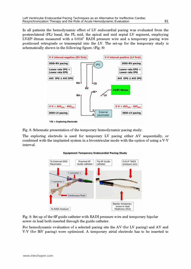
In all patients the hemodynamic effect of LV endocardial pacing was evaluated from the
posterolateral (PL) basal, the PL mid, the apical and mid septal LV segment, employing
LVdP/ dtmax measured with a 0.014” RADI pressure wire and a temporary pacing wire
positioned retrograde or transseptal into the LV. The set-up for the temporary study is
schematically shown in the following figure. (Fig. 8)
Fig. 8. Schematic presentation of the temporary hemodynamic pacing study.
The exploring electrode is used for temporary LV pacing either AV sequentially, or
combined with the implanted system in a biventricular mode with the option of using a V-V
interval.
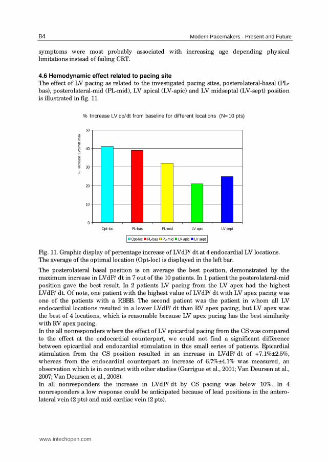
Fig. 9. Set up of the 6F guide catheter with RADI pressure wire and temporary bipolar
screw-in lead both inserted through the guide catheter.
For hemodynamic evaluation of a selected pacing site the AV (for LV pacing) and AV and
V-V (for BiV pacing) were optimized. A temporary atrial electrode has to be inserted to
www.intechopen.com

Modern Pacemakers - Present and Future
82
synchronize the external and implanted system. In the transseptal set-up a 6F guide
catheter, multipurpose or internal mammary catheter, are inserted through the 8F
transseptal sheath. For femoral or radial approach the same guide catheter can be used
employing a 6F sheath at the arterial entrance site, as shown in figure 9.
Fig. 10. Example of radiographic pictures in a left anterior oblique position of the 4 locations
that were hemodynamically tested during the temporary pacing study. The percentage
increase in LVdP/ dt compared to baseline is mentioned for every position.
4.3 Results 4.4 Failed coronary sinus implants (2 pts) Two patients developed a high stimulation threshold, approaching the level of exit block.
Both had a successful CS implant respectively 1 and 2 years before with placement of the
lead in the only accessible coronary sinus side branch. The latter was the reason for the
temporary study to justify implantation of an endocardial lead as a replacement of the
failing CS lead.
In the first patient, a 76 year old male with ICM, who was pacemaker dependent
LVdP/ dtmax decreased to 321 mmHg/ s when programmed to RV pacing, thus imitating
complete failure of the LV lead. Biventricular pacing from PL-basal, PL-mid, LV apex, LV
septum resulted in a LVdPdtmax of respectively 994, 933, 741, and 813 mmHg/ s, whereas
the CS lead, which was in a PL basal position resulted in 967 mmHg/ s. Patient underwent a
successful transseptal implant of an endocardial LV lead in the PL-basal area.
The second patient with an extreme high LV threshold, had a 17% increase in LVdP/ dt with
the CS lead, which was positioned in a PL basal position. Temporary pacing from other
www.intechopen.com

Left Ventricular Endocardial Pacing Techniques as an Alternative for Ineffective Cardiac Resynchronization Therapy and the Role of Acute Hemodynamic Evaluation
83
endocardial positions did not improve this increase. CS pacing could be maintained at
maximum LV output setting. A LV endocardial lead will be implanted at generator change
to avoid premature battery depletion of the new device.
4.5 Nonresponders (10 pts) In 2 patients 74 and 79 years old, both with ICM, and an ECG with right bundle branch
block (RBBB), QRS duration 170 and 175 ms did not respond to CRT with leads positioned
in the CS. The hemodynamic pacing study revealed no improvement in LVdP/ dtmax with
the implanted system. LV pacing from the 4 endocardial positions could not increase
LVdP/ dt above the intrinsic baseline value in both patients.
In one patient with a dual chamber ICD and increasing heart failure, but with a RBBB,
implantation of a CRT system was considered. Prior to implant a temporary pacing study
was performed to evaluate the hemodynamic effect of CRT. LV endocardial pacing showed
an increase in LVdP/ dtmax when pacing from the PL basal area of 17%. CS angiography
showed a suitable side branch in the target area and a successful implant was performed in
the preselected location.
Four patients, 1 with DCM and 3 with ICM, with LBBB in 3 patients and RV pacing in 1
patient, were all nonresponders. The mean ejection fraction prior to implant was 17.5±6.5%.
Hemodynamic pacing study revealed an increase of LVdP/ dtmax at the optimal
endocardial location of 25.2±4.8%. In 3 patients the optimal location was PL-basal, and PL-
mid in 1 patient. The patients with the lowest ejection fraction of 13 and 12% underwent a
successful transseptal and transapical implant respectively. One of these patients with DCM,
turned out to be superresponder with a reduction of NYHA class IV to II, an increase in
ejection fraction from 13 to 45% and a decrease in the end-diastolic and systolic diameter to
61 and 47 mm, respectively, with only minor mitral regurgitation (Bracke et al. 2010).
In one nonresponder (LBBB, QRS duration 206 ms), who was not optimized but
programmed to a standard setting with a V-V interval of 40 ms (LV pre-activation),
LVdP/ dtmax was 1017 mmHg/ s. During hemodynamic testing LVdP/ dtmax with RV
pacing was 1113 mmHg/ s, whereas CS pacing resulted in a LVdP/ dtmax of 870 mmHg/ s.
All endocardial positions except LV apex had a lower LVdP/ dtmax than with RV pacing.
LV apex had the same LVdP/ dt as RV pacing, which was not surprising, because of the
similarity in anatomical positions of bot pacing modalities. Changing the V-V timing to -40
ms (RV pre-activation), and selecting the proximal CS electrode increased LVdP/ dtmax for
biventricular pacing to 1193 mmHg/ s (+17.3%). Patient clinically improved after
optimization with 1 NYHA class.
In one patient who showed increasing heart failure after initial response to CRT, was
referred to our institution for hemodynamic evaluation. Hemodynamics at the current
setting showed a LVdP/ dtmax value of 720 mmHg/ s. After optimization of the AV and V-V
interval and selecting the proximal CS electrode of the dual unipolar lead instead of the
distal electrode LVdP/ dtmax could be increased with 11.5%, which resulted in a significant
clinical improvement with loss of weight and reduction of diuretics.
The last patient, male 87 years, NYHA class III, ejection fraction 25%, LBBB with QRS
duration of 154 ms presented as a clinical nonresponder. Baseline LVdP/ dt was 827
mmHg/ s and increased with 45% by optimized biventricular pacing employing the
implanted CS lead. In the best endocardial position (PL-basal) a gain of 51% could be
obtained. Although left ventricular remodeling with reduction of end-diastolic and systolic
diameter was significant, patient did subjectivally not improve. Presumably, patient’s
www.intechopen.com
Modern Pacemakers - Present and Future
84
symptoms were most probably associated with increasing age depending physical
limitations instead of failing CRT.
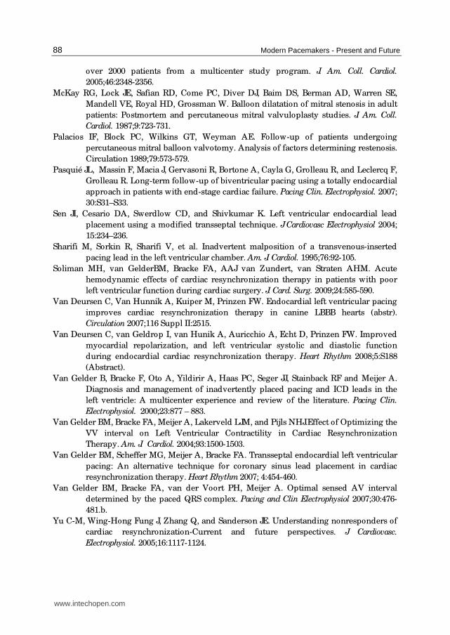
4.6 Hemodynamic effect related to pacing site The effect of LV pacing as related to the investigated pacing sites, posterolateral-basal (PL-
bas), posterolateral-mid (PL-mid), LV apical (LV-apic) and LV midseptal (LV-sept) position
is illustrated in fig. 11.
0
10
20
30
40
50
Opt-loc PL-bas PL-mid LV apic LV sept
Opt-loc PL-bas PL-mid LV apic LV sept
% Increase LV dp/dt from baseline for different locations (N= 10 pts)
% I
ncr
ease
LV
dP/d
t m
ax
Fig. 11. Graphic display of percentage increase of LVdP/ dt at 4 endocardial LV locations.
The average of the optimal location (Opt-loc) is displayed in the left bar.
The posterolateral basal position is on average the best position, demonstrated by the
maximum increase in LVdP/ dt in 7 out of the 10 patients. In 1 patient the posterolateral-mid
position gave the best result. In 2 patients LV pacing from the LV apex had the highest
LVdP/ dt. Of note, one patient with the highest value of LVdP/ dt with LV apex pacing was
one of the patients with a RBBB. The second patient was the patient in whom all LV
endocardial locations resulted in a lower LVdP/ dt than RV apex pacing, but LV apex was
the best of 4 locations, which is reasonable because LV apex pacing has the best similarity
with RV apex pacing.
In the all nonresponders where the effect of LV epicardial pacing from the CS was compared
to the effect at the endocardial counterpart, we could not find a significant difference
between epicardial and endocardial stimulation in this small series of patients. Epicardial
stimulation from the CS position resulted in an increase in LVdP/ dt of +7.1%±2.5%,
whereas from the endocardial counterpart an increase of 6.7%±4.1% was measured, an
observation which is in contrast with other studies (Garrigue et al., 2001; Van Deursen at al.,
2007; Van Deursen et al., 2008).
In all nonresponders the increase in LVdP/ dt by CS pacing was below 10%. In 4
nonresponders a low response could be anticipated because of lead positions in the antero-
lateral vein (2 pts) and mid cardiac vein (2 pts).
www.intechopen.com

Left Ventricular Endocardial Pacing Techniques as an Alternative for Ineffective Cardiac Resynchronization Therapy and the Role of Acute Hemodynamic Evaluation
85
5. Discussion
LV endocardial pacing is an alternative for failed coronary sinus implants as illustrated in
the first section of this chapter. The choice for application of endocardial LV pacing in case
of failed CS implant is based on a risk analysis in which factors like age, physical condition,
degree of heart failure and the presence of anticoagulant therapy play a role.
Remains the question, should hemodynamic testing for the evaluation of the effect of LV
pacing in patients with failed CS implants be performed prior to the LV endocardial
implant?
The answers that we get from the temporary hemodynamic studies are twofold. First, it is a
quantification of the improvement that can be obtained with LV endocardial pacing,
expressed as the increase in LVdP/ dtmax. Second, the pacing site where this optimal effect
is obtained is determined and is used as the target area for the permanent implant.
The latter justifies hemodynamic testing in failed implants with proper patient selection, or
short term failure in patients that are responding to CRT during this short follow-up, in
whom a transseptal implant is considered.
In patients not responding to CRT due to a suboptimal position of the CS lead, implant of an
endocardial LV lead can be considered. Hemodynamic evaluation in a temporary setting
seems to be mandatory prior to the implant procedure, in order to determine the effect on
contractility at the optimal endocardial site and the location of this site. This temporary
study prevents implants of a LV endocardial lead in patients in whom, based on the
outcome of the temporary study, with an increase in LVdP/ dtmax of less than 10% no
clinical improvement can be anticipated. On the other hand, patients that show an increase
in LVdP/ dt over 15% from an endocardial location are suitable candidates for an
endocardial implant and the optimal site in the temporary study determines the target area
for the permanent implant.
Implantation of an additional CS lead, thus creating triple site ventricular stimulation in
nonresponders is suggested by Leclercq et al., who demonstrated improvement in cardiac
volumes by echocardiographic techniques in this configuration over biventricular stimulation
(Leclercq et al., 2008). Although the effect and technique of triple ventricular stimulation needs
further investigation (Auricchio & Prinzen, 2008), the technique is not feasible in the patient
category with failed CS implants or short term failure of a single CS lead.
From our experience it became obvious that in patients with LBBB the postero-lateral basal
area is the optimal site with a significant lower hemodynamic response from different
locations like the septal and apical segment. From this experience it is difficult to
understand that in larger studies (MADIT CRT), with no specific focus on this item, is
concluded that the location of LV stimulation has no effect on clinical outcome.
In spite of the small number of patients and a non-homogeneous patient population
according to their electrocardiographic presentation, it is clear that in nonresponders an
increase in LVdP/ dt of less than 10% is objectivated. If pacing from a different LV location
with a rise in LVdP/ dt over 15% is initialized these patients became responders to CRT.
After measurement of the hemodynamic effect on LVdP/ dt in a nonresponder at the
programmed pacing parameters, optimization of AV and V-V interval should be performed
to exclude a suboptimal setting of AV and V-V interval as a contributor to non response. If
the patient also has a CS dual unipolar lead, the effect of selecting a different stimulation
electrode should also be evaluated. In 2 of our patients improvement in LVdP/ dtmax as
well as clinical status could be obtained by optimization of AV, V-V interval and selection of
www.intechopen.com

Modern Pacemakers - Present and Future
86
the CS pacing electrode. One can speculate that development of quadripolar CS leads with
programmable pacing configurations may contribute to a further optimization of CRT.
6. Conclusion
LV endocardial pacing from a transseptal or transapical approach can be considered after
failed CS implant or non responsiveness to CRT. The tentative benefit of a LV endocardial
implant can be hemodynamical evaluated by measurement of LVdP/ dt in a temporary set
up. The endocardial postero-lateral area proved to be significant better than apical and mid-
septal locations in patients with LBBB. In this small series the superior effect of endocardial
pacing compared to the epicardial counterpart could not be objectivated from the
hemodynamic evaluation.
7. References
Abraham WT, Fisher WG, Smith AL, Delurgio DB, Leon AR, Loh E, Kocovic DZ, Packer M,
Clavell AL, Hayes DL, Ellestad M, Messenger J, for the MIRACLE Study Group.
Cardiac resynchronization in chronic heart failure. N. Engl. J. Med. 2002;346:1845-
1853.
Auricchio A, Ding J, Spinelli JC, Kramer AP, Salo RW, Hoersch W, KenKnight BH, Klein
HU, for PATH-CHF Study Group. Cardiac resynchronization therapy restores
optimal atrioventricular mechanical timing in heart failure patients with ventricular
conduction delay. J. Am. Coll. Cardiol. 2002;39:1163–1169.
Auricchio A, Abraham WT. Cardiac resynchronization therapy: current state of the art. Cost
versus benefit. Circulation 2004;109:300–307.
Aurichio A, Prinzen FW. Cardiac Resynchronization Therapy: The more pacing sites, the
better the outcome ? J. Am. Coll. Cardiol. 2008;51;1463-1465.
Bentkover JD, Stewart EJ, Ignaszewski A, Lepage S, Liu P, Cooper J. New technologies and
potential cost savings related to morbidity and mortality reduction in Class III / IV
heart failure patients in Canada. Int. J. Cardiol. 2003;88:33-41.
Bracke FA, Houthuizen P, Rahel BM, and van Gelder BM. Left ventricular endocardial
pacing improves the clinical efficacy in a non-responder to cardiac
resynchronization therapy: role of acute haemodynamic testing. Europace 2010;
doi:10.1093/ europace/ euq043.
Butter C, Auricchio A, Stellbrink C, Fleck E, Ding J, Yu Y, Huvelle E., Spinelli J. Effect of
resynchronization therapy stimulation site on the systolic function of heart failure
patients. Circulation 2001;104:3026-3029.
Cazeau S, Leclercq C, Lavergne T, Walker S, Varma C, Linde C, Garrigue S, Kappenberger
L, Haywood G, Santini M, Bailleul C, Daubert JC, for the Multisite Stimulation in
Cardiomyopathies (MUSTIC) Study Investigators. Effects of multisite biventricular
pacing in patients with heart failure and intraventricular conduction delay. N. Eng.
J. Med. 2001;344: 873-880.
Cequier A, Bonan R, Dyrda I, Crépeau J, Dethy M, Petitclerc R, Waters D. Atrial shunting
after percutaneous mitral valvuloplasty. Circulation 1988;78: II-488 (Abstract).
www.intechopen.com

Left Ventricular Endocardial Pacing Techniques as an Alternative for Ineffective Cardiac Resynchronization Therapy and the Role of Acute Hemodynamic Evaluation
87
Cleland JGF, Daubert JC, Erdmann E, Freemantle N, Gras D, Kappenberger L, Tavazzi L,
for the cardiac resynchronization on morbidity and mortality in heart failure
(CARE-HF) study investigators. N. Eng. J. Med. 2005;352(15):1539-1549.
Garrigue S, Jaïs P, Espil G, Labeque J-N, Hocini M, Shah DC, Haïssaguerre M, Cleménty J.
Comparison of chronic biventricular pacing between epicardial and endocardial
left ventricular stimulation using Doppler tissue imaging in patients with heart
failure. Am. J. Cardiol. 2001; 88: 858-862.
Gras D, Leclercq C, Tang AS, Bucknall C, Luttikhuis HO, Kirstein-Pedersen A. Cardiac
Resynchronization Therapy in advanced heart failure; the multicenter InSync
clinical study. Eur. J. Heart Fail. 2002;4:311–320.
Jaïs P, Douard H, Shah DC, Barold S, Barat J-L, Clémenty J. Endocardial biventricular
pacing. Pacing Clin. Electrophysiol. 1998;21:2128-2131.
Jaïs P, Takabashi A, Garrigue S, Yamane T, Hocini M, Shah DC, Barold SS, Deisenhofer I,
Haïssaguerre, Clémenty J. Mid-term follow-up of endocardial biventricular pacing.
Pacing Clin. Electrophysiol. 2000;23:1744-1747.
Jaïs P, Sacher F, Laborderie J, Reuter S, Bordachar P, Hsu L-F, Sanders P, Hocini M, O’Neill
MD, Johnsson A, Takahasi, Y, Haissaguerre M, Clementy J. Tailored endocardial
left ventricular pacing is superior to coronary sinus in heart failure patients needing
cardiac resynchronization. Heart Rhythm 2006;3:S247.(abstract).
Joshi S, Steinberg JS, Ashton RC, Balaram S, Fischer A, and DeRose JJ. Follow-up of
robotically assisted left ventricular epicardial leads for cardiac resynchronization
therapy. J. Am. Coll. Cardiol. 2005;46:2358-2359.
Kassaï I, Szili-Torok . Concerns about the long-term outcome of transseptal cardiac
resynchronization therapy: what have we learned from surgical experience.
Europace 2008;10:121-122.
Kassaï I, Foldesi C, Szekely A,, Szili-Torok T. Alternative method for cardiac
resynchronization therapy. Transapical lead implantation. Ann. Thoracic. Surg.
2009;87(2):650-652.
Koos R, Sinha A-M, Markus K, Breithardt O-A, Mischke K, Zarse M, Scmid M, Autschbach
R, Hanrath P and Stellbrink C. Comparison of left ventricular lead placement via
the coronary venous approach versus lateral thoracotomy in patients receiving
cardiac resynchronization therapy. Am. J. Cardiol. 2004;94:59-63.
Leclercq C, Gadler F, Kranig W, Ellery S, Gras D, Lazarus A, Clementy J, Boulogne E,
Daubert J-C for the TRIP-HF (Triple Resynchronization in Paced Heart Failure
patients). A randomized comparison of triple-site versus dual site ventricular
stimulation in patients with congestive heart failure. J. Am. Coll. Cardiol.
2008;51:1455-1462.
Leclercq F, Hager FX, Macia JC, Mariottini CJ, Pasquié JL, Grolleau R. Left ventricular lead
insertion using a modified transseptal catheterization technique: a totally
endocardial approach for permanent biventricular pacing in end-stage heart
failure. Pacing Clin. Electrophysiol. 1999;22:1570-1575.
Leon AR, Abraham WT, Curtis AB, Daubert JP, Fisher WG, Gurley J, Hayes DL, Lieberman
R, Petersen-Stejskal S, Wheelan K. Safety of transvenous cardiac resynchronization
system implantation in patients with chronic heart hailure: combined results of
www.intechopen.com
Modern Pacemakers - Present and Future
88
over 2000 patients from a multicenter study program. J. Am. Coll. Cardiol.
2005;46:2348-2356.
McKay RG, Lock JE, Safian RD, Come PC, Diver DJ, Baim DS, Berman AD, Warren SE,
Mandell VE, Royal HD, Grossman W. Balloon dilatation of mitral stenosis in adult
patients: Postmortem and percutaneous mitral valvuloplasty studies. J. Am. Coll.
Cardiol. 1987;9:723-731.
Palacios IF, Block PC, Wilkins GT, Weyman AE. Follow-up of patients undergoing
percutaneous mitral balloon valvotomy. Analysis of factors determining restenosis.
Circulation 1989;79:573-579.
Pasquié JL, Massin F, Macia J, Gervasoni R, Bortone A, Cayla G, Grolleau R, and Leclercq F,
Grolleau R. Long-term follow-up of biventricular pacing using a totally endocardial
approach in patients with end-stage cardiac failure. Pacing Clin. Electrophysiol. 2007;
30:S31–S33.
Sen JI, Cesario DA, Swerdlow CD, and Shivkumar K. Left ventricular endocardial lead
placement using a modified transseptal technique. J Cardiovasc Electrophysiol 2004;
15:234–236.
Sharifi M, Sorkin R, Sharifi V, et al. Inadvertent malposition of a transvenous-inserted
pacing lead in the left ventricular chamber. Am. J. Cardiol. 1995;76:92-105.
Soliman MH, van GelderBM, Bracke FA, AAJ van Zundert, van Straten AHM. Acute
hemodynamic effects of cardiac resynchronization therapy in patients with poor
left ventricular function during cardiac surgery. J. Card. Surg. 2009;24:585-590.
Van Deursen C, Van Hunnik A, Kuiper M, Prinzen FW. Endocardial left ventricular pacing
improves cardiac resynchronization therapy in canine LBBB hearts (abstr).
Circulation 2007;116 Suppl II:2515.
Van Deursen C, van Geldrop I, van Hunik A, Auricchio A, Echt D, Prinzen FW. Improved
myocardial repolarization, and left ventricular systolic and diastolic function
during endocardial cardiac resynchronization therapy. Heart Rhythm 2008;5:S188
(Abstract).
Van Gelder B, Bracke F, Oto A, Yildirir A, Haas PC, Seger JJ, Stainback RF and Meijer A.
Diagnosis and management of inadvertently placed pacing and ICD leads in the
left ventricle: A multicenter experience and review of the literature. Pacing Clin.
Electrophysiol. 2000;23:877 – 883.
Van Gelder BM, Bracke FA, Meijer A, Lakerveld LJM, and Pijls NHJ.Effect of Optimizing the
VV interval on Left Ventricular Contractility in Cardiac Resynchronization
Therapy. Am. J. Cardiol. 2004;93:1500-1503.
Van Gelder BM, Scheffer MG, Meijer A, Bracke FA. Transseptal endocardial left ventricular
pacing: An alternative technique for coronary sinus lead placement in cardiac
resynchronization therapy. Heart Rhythm 2007; 4:454-460.
Van Gelder BM, Bracke FA, van der Voort PH, Meijer A. Optimal sensed AV interval
determined by the paced QRS complex. Pacing and Clin Electrophysiol 2007;30:476-
481.b.
Yu C-M, Wing-Hong Fung J, Zhang Q, and Sanderson JE. Understanding nonresponders of
cardiac resynchronization-Current and future perspectives. J Cardiovasc.
Electrophysiol. 2005;16:1117-1124.
www.intechopen.com
Modern Pacemakers - Present and FutureEdited by Prof. Mithilesh R Das
ISBN 978-953-307-214-2Hard cover, 610 pagesPublisher InTechPublished online 14, February, 2011Published in print edition February, 2011
InTech EuropeUniversity Campus STeP Ri Slavka Krautzeka 83/A 51000 Rijeka, Croatia
InTech ChinaUnit 405, Office Block, Hotel Equatorial Shanghai No.65, Yan An Road (West), Shanghai, 200040, China
Phone: +86-21-62489820
The book focuses upon clinical as well as engineering aspects of modern cardiac pacemakers. Modernpacemaker functions, implant techniques, various complications related to implant and complications duringfollow-up are covered. The issue of interaction between magnetic resonance imaging and pacemakers arewell discussed. Chapters are also included discussing the role of pacemakers in congenital and acquiredconduction disease. Apart from pacing for bradycardia, the role of pacemakers in cardiac resynchronizationtherapy has been an important aspect of management of advanced heart failure. The book provides anexcellent overview of implantation techniques as well as benefits and limitations of cardiac resynchronizationtherapy. Pacemaker follow-up with remote monitoring is getting more and more acceptance in clinical practice;therefore, chapters related to various aspects of remote monitoring are also incorporated in the book. Thecurrent aspect of cardiac pacemaker physiology and role of cardiac ion channels, as well as the present andfuture of biopacemakers are included to glimpse into the future management of conductions system diseases.We have also included chapters regarding gut pacemakers as well as pacemaker mechanisms of neuralnetworks. Therefore, the book covers the entire spectrum of modern pacemaker therapy including implanttechniques, device related complications, interactions, limitations, and benefits (including the role of pacingrole in heart failure), as well as future prospects of cardiac pacing.
How to referenceIn order to correctly reference this scholarly work, feel free to copy and paste the following:
Berry M. van Gelder, Patrick Houthuizen, Mike G. Scheffer, Lukas Dekker and Frank A. Bracke (2011). LeftVentricular Endocardial Pacing Techniques as an Alternative for Ineffective Cardiac ResynchronizationTherapy and the Role of Acute Hemodynamic Evaluation, Modern Pacemakers - Present and Future, Prof.Mithilesh R Das (Ed.), ISBN: 978-953-307-214-2, InTech, Available from:http://www.intechopen.com/books/modern-pacemakers-present-and-future/left-ventricular-endocardial-pacing-techniques-as-an-alternative-for-ineffective-cardiac-resynchroni
www.intechopen.com

Phone: +385 (51) 770 447 Fax: +385 (51) 686 166www.intechopen.com
Phone: +86-21-62489820 Fax: +86-21-62489821

© 2011 The Author(s). Licensee IntechOpen. This chapter is distributedunder the terms of the Creative Commons Attribution-NonCommercial-ShareAlike-3.0 License, which permits use, distribution and reproduction fornon-commercial purposes, provided the original is properly cited andderivative works building on this content are distributed under the samelicense.


![Tendril STS 2088 [OUS] - SJM€¦ · Tendril™ STS . Model 2088TC . Active-fixation Bipolar . Steroid-eluting . Endocardial . Pacing leads . User's Manual](https://static.fdocuments.in/doc/165x107/5f01af377e708231d4008cc7/tendril-sts-2088-ous-sjm-tendrila-sts-model-2088tc-active-fixation-bipolar.jpg)