Lecture 7 diseases of the vascular system - Pathology
27
Diseases of the vascular system Lecture 7 Introduction Normal blood flow through arteries and veins requires an intact system of blood vessels and adequate perfusion pressure to drive the blood through these vessels. A number of disease processes can affect normal function of the arteries and/or veins. Disease-induced changes may impair blood flow through arteries and disrupt delivery of oxygen and nutrients to tissues while disease processes affecting veins will disrupt removal of waste products from tissues and the return of blood to the heart. Arterial disease Arteries deliver oxygenated blood to the tissues and organs. Arteries can vary in size from the large aorta that transports blood from the heart to medium-sized arteries that deliver blood to organs and finally down to small arteries and arterioles that feed blood through capillary beds. Arterial diseases include conditions such as atherosclerosis, aneurysm, vasospastic conditions and various inflammatory disorders. One of the most common diseases affecting arteries is atherosclerosis. It is caused by deposition of lipid plaques in the walls of arteries. While the formation of atherosclerotic lesions can affect any artery, the coronary arteries of the heart are the mostly affected. General structure of Lipoproteins Because dietary lipids (such as fat and cholesterol which are oil-soluble substances) are insoluble in the plasma or lymph, they are transported as part of complex called a lipoprotein. Each type of lipoprotein consists of a large molecular complex of lipids combined with proteins called apoproteins. The major lipid constituents of lipoprotein are composed of : 1) Hydrophobic core Cholesterol esters & Triglycerides 2) Hydrophilic shell Unesterified cholesterol & Phospholipids 3) Apoproteins 170
-
Upload
areej-abu-hanieh -
Category
Health & Medicine
-
view
208 -
download
2
Transcript of Lecture 7 diseases of the vascular system - Pathology

Diseases of the vascular system Lecture 7
Introduction
Normal blood flow through arteries and veins requires an intact system of blood vessels and adequate perfusion pressure to drive the blood through these vessels.
A number of disease processes can affect normal function of the arteries and/or veins. Disease-induced changes may impair blood flow through arteries and disrupt delivery of
oxygen and nutrients to tissues while disease processes affecting veins will disrupt removal of waste products from
tissues and the return of blood to the heart.
Arterial disease
Arteries deliver oxygenated blood to the tissues and organs. Arteries can vary in size from the large aorta that transports blood from the heart to
medium-sized arteries that deliver blood to organs and finally down to small arteries and arterioles that feed blood through capillary beds.
Arterial diseases include conditions such as atherosclerosis, aneurysm, vasospastic conditions and various inflammatory disorders.
One of the most common diseases affecting arteries is atherosclerosis. It is caused by deposition of lipid plaques in the walls of arteries. While the formation of atherosclerotic lesions can affect any artery, the coronary arteries of the heart are the mostly affected.
General structure of Lipoproteins
Because dietary lipids (such as fat and cholesterol which are oil-soluble substances) are insoluble in the plasma or lymph, they are transported as part of complex called a lipoprotein.
Each type of lipoprotein consists of a large molecular complex of lipids combined with proteins called apoproteins.
The major lipid constituents of lipoprotein are composed of :
1) Hydrophobic core
Cholesterol esters &
Triglycerides
2) Hydrophilic shell
Unesterified cholesterol &
Phospholipids
3) Apoproteins
170

Hydrophobic: Repelling, tending not to combine with, or incapable of dissolving in water.
Hydrophilic: Having an affinity for water; readily absorbing or dissolving in water.
Classification of Lipoproteins
There are five types of lipoproteins, classified by their densities as measured by ultracentrifugation:
1) Chylomicrons
2) Very–low-density lipoprotein (VLDL)
3) Intermediate-density lipoprotein (IDL)
4) Low-density lipoprotein (LDL)
5) High-density lipoprotein (HDL)
171

Functions of cholesterol
Cholesterol & Vitamin D
7-dehydrocholesterol: A provitamin present in the skin that becomes vitamin D 3 when exposed to ultraviolet light.
Active form: Pro vitamin converted to cholecalciferol by ultraviolet light.( In skin )
Vitamin is hydroxylated in liver to: 25- hydroxy cholecalciferol.
Kidney forms-1,25 dihydrocholecalciferol ( Active)
172

Lipid Metabolism
173

Lipoprotein transport
There are two sites of lipoprotein synthesis: the small intestine and the liver.
The chylomicrons, which are the largest of the lipoprotein molecules, are synthesized in the wall of the small intestine.
Chylomicrons carry Triglycerides to the liver, skeletal muscles and adipose tissue.
Cholesterol content in liver is derived from:
Synthesizing it and secreted into systemic circulation
Take Cholesterol from blood from lipoproteins
Take Cholesterol that was absorbed from small intestine
Lipids are insoluble in water, the problem of transportation in the aqueous plasma is solved by associating the hydrophobic lipids (triglycerides & cholesterol esters) with the hydrophilic lipids (phospholipids & unesterified cholesterol) and Apoproteins to make water-mixable lipoproteins. and can thus be carried through water-based circulation (i.e. blood, lymph).
174

An overview of lipoprotein particle metabolism
175

176

Apolipoproteins
Apolipoproteins are proteins that bind lipids to form lipoproteins. They transport the lipids through the lymphatic and circulatory systems.
Apolipoprotein Functions
1) They are Part of the structure of the lipoprotein particles.
2) They are cofactors for enzymes of lipid metabolism (coenzymes), (examples: C-II for lipoprotein lipase and A-I for lecithin-cholesterol acyltransferase)
3) Lipid transport proteins
4) Receptor binding (ex: ligand that allows lipoprotein to bind to receptors on hepatocytes and peripheral cells which leads to transfer of Cholesterol from lipoprotein into cell), Examples:( apoB100 and apoE for LDL-receptors, apoA-I for HDL receptors)
Apolipoprotein Classes
There are six major classes of apolipoproteins: A, B, C, D, E and H.
The two important types of apolipoproteins:
o Apolipoprotein A (apo A)
o Apolipoprotein B (apo B)
177

Apolipoprotein A (apo A)
Apo A1 is the major protein component of high-density lipoprotein (HDL).
Deficiency of apo A1 is associated with HDL deficiencies, including Tangier disease (is an inherited disorder characterized by significantly reduced levels of high-density lipoprotein (HDL) in the blood) and systemic non-neuropathic amyloidosis (deposition of insoluble fibrillary proteins (amyloid) in extracellular spaces of organs and tissues, causing structural and functional organ damage).
Apo A1 may have a role in protection against Alzheimer's disease.
Apo A1 and apo E interact to modify triglyceride levels in coronary heart disease patients.
Apolipoprotein B (apo B)
Apo B is the main apolipoprotein of chylomicrons ( apo B-48) and low-density lipoproteins (LDLs) is (apo B-100).
High levels appear related to heart disease.
Apolipoprotein B and the non-fasting Apo B/Apo A1 ratios are thought to be:
a better marker for estimation of the risk of acute myocardial infarction and
a better guide to the adequacy of statin treatment than any cholesterol index.
to be predictive of ischemic stroke in patients with previous transient ischemic attack.
178

However, a previous study found that non-HDL-C and the ratio of total cholesterol to HDL-C were as good as, or better than, apolipoprotein fractions in the prediction of future cardiovascular events.
Non–HDL-C calculation
1) LDL-C + VLDL-C or
2) Total Cholesterol – HDL-C
HDL & Non-HDL Cholesterol
Since apoA1 is a component of anti-atherogenic HDL and apoB of atherogenic lipoproteins such as LDL, the apoB/apoA1 ratio can provide predictive risk information for the development of cardiovascular disease.
Hyperlipidemia
Definition: elevation of > 1 blood lipids (ex: TG, CHO, CHO esters, phospholipids)
Desirable serum cholesterol levels are those below 200 mg/dL.
179

Hyperlipidemia leads to an exponential increase in the risk of atherosclerosis and especially disease of the coronary arteries.
Function of CHO and TG: bile acid formation (made in liver and needed for nutrient absorption, digestion),as a component of cell membrane and formation of steroid hormones.
A number of genetic defects may lead to hyperlipoproteinemia.
Some, such as familial hypercholesterolemia, are associated with a greatly increased risk for atherosclerosis and arterial disease
Atherosclerosis
Background
Arteries are lined by a thin layer of cells called the endothelium.
The endothelium works to keep the inside of arteries toned and smooth, which keeps blood flowing.
Atherosclerosis is a syndrome affecting arterial blood vessels, a chronic inflammatory response in the walls of arteries starts when high blood pressure, smoking, or high cholesterol damage the endothelium.
At that point, cholesterol plaque formation begins.
Atherosclerosis tends to happen throughout the body.
Atherosclerosis usually causes no symptoms until middle or older age.
Steps in the Development of Atherosclerosis
1) Formation of lipid plaques may be precipitated by endothelial injury (examples: hypertension, immune response, toxins in cigarette smoke)
2) Infiltration of cholesterol molecules into blood vessel walls.
3) Monocytes enter area of injury and release growth factors that stimulate smooth muscle and endothelial cell proliferation.
4) The monocytes then become macrophages, engulf lipoproteins, and become lipid-filled “foam cells.”
5) Platelets adhere to the endothelial lesion; fibroblasts infiltrate area and cause progressive sclerosis or hardening of tissue.
6) Calcification of plaques may occur over time.
7) Significant narrowing of the blood vessel lumen can occur over time.
180

Initiation of atherosclerosis formation
Risk Factors for Atherosclerosis
181

Genetic Disorders of Lipid Metabolism
When to treat Hyperlipidemia
182

Aneurysm
An aneurysm is a localized, balloon-like swelling in the wall of an artery caused by weakening of the arterial wall.
Aneurysm may occur in any artery, but the aorta is most susceptible to aneurysm due to the constantly high pressure on the walls of that vessel. Cerebral aneurysms may also occur. They are most frequently located in the circle of Willis of the cerebral circulation.
A leading cause of aneurysm is atherosclerosis, as the formation of lipid plaques can erode arterial walls and compromise their integrity.
Aneurysms may also be associated with hypertension, vascular infections and the normal aging process.
Aneurysms that involve all three layers of the blood vessel wall are called true aneurysms to distinguish them from other types of arterial distention that may occur from external injury or trauma.
A dissecting aneurysm is a very severe condition in which there is a tear in the inner layers of the blood vessel (tunica intima and tunica media) and as a result bleeding occurs in the space below the adventitia of the vessel
Three forms of aneurysms
Berry aneurysm in the circle of Willis, fusiform-type aneurysm of the abdominal aorta, and aortic dissection.
183

Clinical manifestations of aneurysm
Depending upon size and location, aneurysms may be completely asymptomatic or may be associated with severe pain.
Aneurysms in the thoracic aorta most commonly present with back or neck pain, cough, difficulty swallowing or compression of the trachea.
Aneurysms in the abdominal aorta are often asymptomatic until they cause pain from compression on spinal nerves or abdominal organs.
Aneurysms of the cerebral arteries often present with symptoms that are characteristic of increased intracranial pressure. Stroke can result if the abnormal blood vessel ruptures and bleeding occurs into the brain tissue.
Treatment of aneurysms
Reduction of blood pressure and blood volume with appropriate drugs.
Surgical grafting to strengthen arterial walls.
Dissecting aneurysms are acutely life-threatening and require immediate surgical repair.
Vasospastic conditions
Two common vasospastic disorders are Raynaud’s disease and Raynaud’s phenomenon.
(Both had been discussed in Lecture 5 “Immunology” These disorders are characterized by vasospasm of small cutaneous blood vessels in the
fingers and toes.
Raynaud’s disease is a primary vasospastic disorder whose etiology is uncertain. It occurs most frequently in otherwise healthy young women and is often precipitated by strong emotions or exposure to cold.
Raynaud’s phenomenon, in contrast, is a vasospastic condition that occurs secondarily to other underlying diseases such as scleroderma, malignancy, or from blood vessel injury due to vibrations (jackhammers) or prolonged cold exposure (of butchers to low temperatures, frostbite). Localized vasospasm of arterial walls can acutely block blood flow to a particular region of tissue leading to numbness and discoloration. Severe or prolonged attacks of Raynaud’s disease or syndrome may result in cyanosis, ulceration and gangrene of fingers or toes.
Treatment of Raynaud’s disease: • involves avoidance of precipitating factors such as cold and strong emotions. • Vasodilator drugs or calcium channel blockers may be used to prevent vasoconstriction in
Raynaud’s phenomenon. • In severe cases the sympathetic nerves that innervate the local vasculature may be surgically
severed.
184

Scleroderma
Is a chronic systemic autoimmune disease (primarily of the skin -"derma") Characterized by fibrosis (or hardening -"sclero"), vascular alterations, and
autoantibodies.
Arterial inflammation
Thromboangiitis obliterans (Buerger’s disease) is an inflammatory disorder that can affect the aorta or peripheral arteries.
It occurs most often in young men who are heavy cigarette smokers. The exact etiology of this disorder is uncertain but it manifests with inflammatory lesions of the arteries that may result in vasospasm, occlusion and thrombus formation.
Treatment involves cessation of smoking and attempts to improve blood flow and reduce vasospasm through the use of vasodilator drugs.
185

Disease of the veins
1) Varicose veins 2) Chronic venous insufficiency 3) Venous thrombus 4) Embolism
Varicose veins
Varicose veins are veins that have become distended over time due to the pooling of blood in the lower extremities. This condition occurs most frequently in individuals who spend long periods of time standing (barbers, for example) or who have impaired return of blood from the lower extremities.
Veins are thin-walled vessels that are easily distended by the chronic pooling of blood in the lower extremities.
Chronic distention of veins can reduce effectiveness of one-way venous valves that are present in the lumen to prevent the back flow of blood and lead to a condition termed valvular incompetence.
These venous valves work in conjunction with skeletal muscle pumps in the legs to move blood back to the heart from the extremities.
The most common manifestations of varicose veins are aching and edema. Their appearance through the skin is also unsightly. Treatment often involves the use of support stockings to prevent venous pooling. Surgical interventions may also be used to improve appearance and reduce discomfort.
Chronic venous insufficiency
The presence of varicose veins and valvular incompetence can lead to a condition called chronic venous insufficiency.
As a result of chronically impaired blood flow, congestion, edema and poor tissue nutrition, pathologic changes may eventually occur in the lower extremities.
Manifestations of chronic venous insufficiency may include skin atrophy, dermatitis, ulceration and tissue necrosis. Infection or trauma of the lower extremities that occurs in a patient with chronic venous insufficiency may have serious consequences because poor blood flow reduces delivery of immune cells and impairs wound healing.
Treatment for chronic venous insufficiency involves interventions similar to those for varicose veins.
186

187

Venous thrombus
A thrombus is a blood clot that forms in the lumen of a blood vessel. A thrombus may form in an artery, but it is more common in veins due to the lower
pressure and reduced blood flow found in the venous circulation. Thrombi may form in superficial vessels of the skin and extremities or in deep veins of
circulation or tissues. Most superficial thrombi are benign and self-limiting, but deep vein thrombus (DVT) can
be much more dangerous.
Pathophysiology of DVT (Virchow’s Triad) The pathologist Rudolph Virchow was the first to describe the three main factors that may contribute to the formation of a thrombus:
1) Hypercoagulable State (Blood Clots are Likely) 2) Circulatory Stasis (Decreased Blood Flow) 3) Vascular Wall Injury (Endothelial Injury)
188

DVT Signs, Symptoms & Treatment Although a thrombus may present with pain, tenderness and swelling, it is estimated that
nearly half of all deep vein thrombi are asymptomatic. As most deep vein thrombi occur in the lower extremities, painful compression or
tenderness and swelling of the calf or thigh region might be used to diagnose a DVT in these areas.
DVT are associated with significant mortality and morbidity and require intensive treatment.
Treatment and prevention of venous thrombus Prevent blood stasis in susceptible patients through ambulation, use of elastic
stockings, exercise or elevation of legs Anticoagulation therapy (warfarin, heparin) Thrombolytic therapy to dissolve clots (streptokinase, TPA) Surgical removal of clots
189
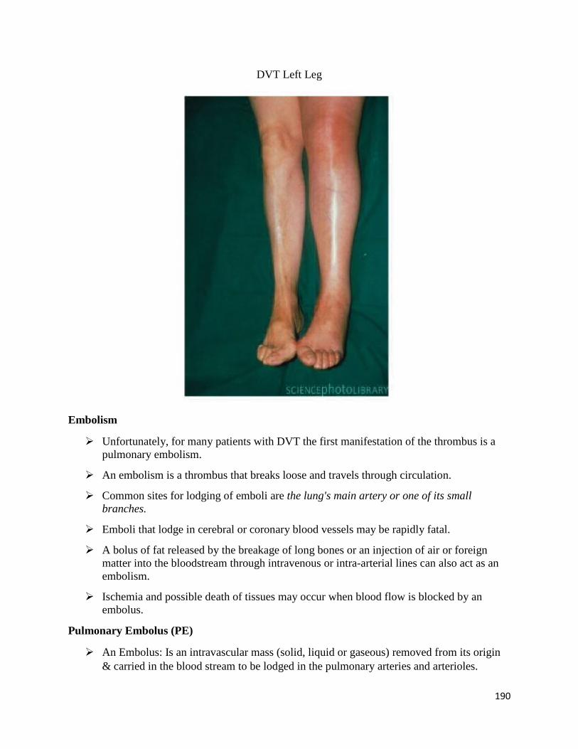
DVT Left Leg
Embolism
Unfortunately, for many patients with DVT the first manifestation of the thrombus is a pulmonary embolism.
An embolism is a thrombus that breaks loose and travels through circulation.
Common sites for lodging of emboli are the lung's main artery or one of its small branches.
Emboli that lodge in cerebral or coronary blood vessels may be rapidly fatal.
A bolus of fat released by the breakage of long bones or an injection of air or foreign matter into the bloodstream through intravenous or intra-arterial lines can also act as an embolism.
Ischemia and possible death of tissues may occur when blood flow is blocked by an embolus.
Pulmonary Embolus (PE)
An Embolus: Is an intravascular mass (solid, liquid or gaseous) removed from its origin & carried in the blood stream to be lodged in the pulmonary arteries and arterioles.
190

Pulmonary Embolus (PE): may be:
o Thrombotic (detached thrombus fragment)
o Non-thrombotic Septic embolism
o Fat embolism
o Air embolism
o Amniotic fluid embolism
o Tumor embolism
Assuming a normal circulation, an embolus formed in a systemic vein will always impact in the lungs, after passing through the right side of the heart. This will form a pulmonary embolism that will result in a blockage of the main artery of the lung and can be a complication of deep-vein thrombosis.
The most common sites of origin of pulmonary emboli are the femoral veins. The deep veins of the calf are the most common sites of actual thrombi.
191

Anticoagulant and thrombolytic drug therapy
Anticoagulant drugs prevent the formation of blood clots by interfering with distinct steps in the blood-clotting cascade.
Two of the most commonly used anticoagulants are warfarin (administered orally) and Heparin (administered intravenously).
Warfarin acts by inhibiting the synthesis of vitamin K-dependent clotting factors, which include Factors II, VII, IX, and X, and the anticoagulant proteins C and S. Warfarin prevents the reduction of vitamin K, which is a cofactor necessary for activity of a key carboxylase in the clotting cascade.
Heparin acts via an effect on antithrombin III.
As a result of its mechanism of action, warfarin does not exert an anticoagulant effect in vitro (i.e., blood in test tube) whereas heparin does.
192

Neither warfarin nor heparin has any action against clots that have already formed. Both drugs are bound to a significant extent to circulating plasma proteins that can alter their bioavailability.
A main potential adverse effect of both warfarin and heparin is unwanted bleeding and hemorrhage.
Antithrombotic drugs
193

Heparin Mechanism of Action
Activates antithrombin III (which is natural anticoagulant),this means inactivating thrombin and similar cofactors
Heparin binds with anti-thrombin forming (Heparin-AT III complex) to increase it’s efficacy of inhibiting by 1000x of the following:
Thrombin-mediated conversion of fibrinogen to fibrin
Platelet factor Xa
Platelet factor IXa
Platelet factor XIIa
Antiplatelet Drugs (Aspirin)
Aspirin is a potent inhibitor of platelet aggregation through its inhibition of the enzyme cyclo-oxygenase.
Inhibition of the cyclo-oxygenase enzyme reduces the formation of thromboxane A2, a substance that stimulates platelet aggregation.
Since platelet aggregation and activation appear to play a major role in thrombus formation, drugs like aspirin may be of significant therapeutic value in preventing their occurrence.
Aspirin is effective in preventing the tissue damage that accompanies blood vessel occlusion in arteriosclerosis and myocardial infarction.
194

Anticoagulant and thrombolytic drug therapy
Thrombolytic drugs are also known as fibrinolytic or clot-dissolving drugs.
Unlike anticoagulants that prevent the formation of blood clots, thrombolytic drugs cannot prevent their formation.
A number of thrombolytic drugs are now available for clinical use, including streptokinase, anistreplase, alteplase (tissue plasminogen activator) and urokinase.
These agents promote the formation of plasmin (from plasminogen), an enzyme that degrades the fibrin proteins that make up the framework of a thrombus.
The most common unwanted effects of these thrombolytic agents are unwanted bleeding and hemorrhage.
Thrombolytic drugs have proved to be of clinical benefit in reducing mortality in patients experiencing myocardial infarction.
195

Thrombolytic drugs fibrinolytic
196