Laparoscopic omentoplasty and split skin graft for deep ... · Sternal separation can either be the...
8
Case Report Malta Medical Journal Volume 28 Issue 01 2016 Abstract Treatment of sternotomy dehiscence secondary to infection is complex. We describe a case where following debridement and negative pressure therapy the greater omentum was harvested laparoscopically, pedicled on the right gastroepiploic artery and transposed through a subxiphoid window and laid into the chest wound. The omentum was covered with a split skin graft. The omental transposition provided a healthy vascular bed for the skin graft to be laid on top of. This technique allows for larger defects to be closed when due to the amount of bone loss the sternum cannot be brought together. Such procedures are normally performed when all other measures have failed and myocutaneous flaps cover the omentoplasty. Our case is novel in that the laparoscopic harvest and the use of direct skin grafting make this an option to be considered earlier as a single definitive procedure. Keywords chest wall reconstruction, deep sternal wound infection, omentoplasty, laparoscopic omentoplasty, sternum dehiscence, wound healing, Introduction Median sternotomies have been used for nearly a century and yet the management of its complications remains difficult. Deep sternal wound infection is a major cause of sternal dehiscence that is not secondary to technical reasons or wire failure. The incidence of deep sternal wound infection (DSWI) in cardiac patients ranges between 1 to 3 % 1-3 and carries a 30-day mortality of 7.3% compared to 1.6% in patients without infection. 4 The risk factors for sternal wound infection are diabetes, obesity, bilateral internal mammary harvest, prolonged operation time and blood transfusions perioperatively. Prevention of this serious complication is the first priority and this can be achieved by pre-operative chest hair shaving, perioperative antibiotics, meticulous midline sternotomy and its wire closure and sparing use of bone wax and diathermy. 5 The most common pathogens responsible for DSWI are gram-positive bacteria, namely Staphylococcus aureus and Staphylococcus epidermidis. Gram-negative organisms and fungi are rarely cultured. Sternal separation can either be the cause of DSWI by letting superficial infections penetrate deeper or it can be the result of already present Laparoscopic omentoplasty and split skin graft for deep sternal wound infection and dehiscence patient David Sladden, Francis. X. Darmanin, Benedict Axisa, Kevin Schembri, Joseph Galea David Sladden* Department of Cardiac Services, Mater Dei Hospital, Msida, Malta [email protected] Francis. X. Darmanin Department of Plastic Surgery, Mater Dei Hospital, Msida, Malta Benedict Axisa Department of Surgery, Mater Dei Hospital, Msida, Malta Kevin Schembri Department of Cardiac Services, Mater Dei Hospital, Msida, Malta Joseph Galea Department of Cardiac Services, Mater Dei Hospital, Msida, Malta *Corresponding Author 56
Transcript of Laparoscopic omentoplasty and split skin graft for deep ... · Sternal separation can either be the...

dRe
Case Report
Malta Medical Journal Volume 28 Issue 01 2016
Abstract
Treatment of sternotomy dehiscence secondary to
infection is complex. We describe a case where
following debridement and negative pressure
therapy the greater omentum was harvested
laparoscopically, pedicled on the right
gastroepiploic artery and transposed through a
subxiphoid window and laid into the chest wound.
The omentum was covered with a split skin graft.
The omental transposition provided a healthy
vascular bed for the skin graft to be laid on top of.
This technique allows for larger defects to be closed
when due to the amount of bone loss the sternum
cannot be brought together.
Such procedures are normally performed when
all other measures have failed and myocutaneous
flaps cover the omentoplasty. Our case is novel in
that the laparoscopic harvest and the use of direct
skin grafting make this an option to be considered
earlier as a single definitive procedure.
Keywords
chest wall reconstruction, deep sternal wound
infection, omentoplasty, laparoscopic omentoplasty,
sternum dehiscence, wound healing,
Introduction
Median sternotomies have been used for nearly
a century and yet the management of its
complications remains difficult. Deep sternal
wound infection is a major cause of sternal
dehiscence that is not secondary to technical
reasons or wire failure. The incidence of deep
sternal wound infection (DSWI) in cardiac patients
ranges between 1 to 3 %1-3 and carries a 30-day
mortality of 7.3% compared to 1.6% in patients
without infection.4
The risk factors for sternal wound infection are
diabetes, obesity, bilateral internal mammary
harvest, prolonged operation time and blood
transfusions perioperatively. Prevention of this
serious complication is the first priority and this can
be achieved by pre-operative chest hair shaving,
perioperative antibiotics, meticulous midline
sternotomy and its wire closure and sparing use of
bone wax and diathermy. 5
The most common pathogens responsible for
DSWI are gram-positive bacteria, namely
Staphylococcus aureus and Staphylococcus
epidermidis. Gram-negative organisms and fungi
are rarely cultured.
Sternal separation can either be the cause of
DSWI by letting superficial infections penetrate
deeper or it can be the result of already present
Laparoscopic omentoplasty and split skin graft
for deep sternal wound infection and
dehiscence patient
David Sladden, Francis. X. Darmanin, Benedict Axisa, Kevin
Schembri, Joseph Galea
David Sladden*
Department of Cardiac Services,
Mater Dei Hospital,
Msida, Malta
Francis. X. Darmanin
Department of Plastic Surgery,
Mater Dei Hospital,
Msida, Malta
Benedict Axisa
Department of Surgery,
Mater Dei Hospital,
Msida, Malta
Kevin Schembri Department of Cardiac Services,
Mater Dei Hospital,
Msida, Malta
Joseph Galea
Department of Cardiac Services,
Mater Dei Hospital,
Msida, Malta
*Corresponding Author
56

dRe
Case Report
Malta Medical Journal Volume 28 Issue 01 2016
infection causing sternal incompetence. Once this
happens the dead space between mediastinal
structures and skin is filled with a fibrinous matrix,
which harbours the pathogens and makes antibiotic
treatment much less effective. Collections form
behind the dehisced sternal edges and osteomyelitis
of the sternum becomes more significant after a few
weeks. This explains why these infections are
notoriously difficult to treat.
Traditionally these wounds are treated with
debridement, antibiotics and wound packing.
Eventual closure will require some sort of flap,
usually pectoral myocutaneous flaps. However, it
has been shown that obliteration of the dead space
offers improved outcomes following wound
debridement and prior to flap closure. The greater
omentum is an ideal candidate for this role as it is
resistant to infection due to plenty of
immunologically active cells, is very vascular and
absorbs wound secretions.6
The Case
A 52-year-old male, diabetic and ex-smoker
with a body mass index (BMI) of 42.3 underwent
coronary artery bypass grafting in January 2013. He
presented to casualty with a non-ST elevation
myocardial infarction (NSTEMI) and was found to
have left main stem stenosis with mid left anterior
descending (LAD) artery and mid circumflex artery
disease. The right coronary artery was blocked with
some retrograde filling. The ventriculogram gave an
ejection fraction of 36%. The left internal thoracic
artery (LITA) was used as a pedicled graft onto the
LAD and a saphenous vein graft was grafted onto
the first obtuse marginal branch. The antiplatelet
drugs clopidogrel and aspirin had been stopped one
week before surgery.
Post-operatively the patient suffered from
atelectasis and copious chest secretions resulting in
episodes of relative hypoxia. It was noted that the
patient was not compliant in adopting chest
protective manoeuvres while coughing. Glycaemic
control was poor.
On the tenth post-operative day he developed a
serosanguinous discharge from the wound that was
negative on swab culturing. Antibiotics were started
empirically but over the next few days this
discharge became purulent and the sternotomy
wound dehisced completely. The patient became
febrile and methicillin resistant Staphylococcus
aureus was cultured from both the wound and blood
cultures. He was started on teicoplanin and
gentamycin according to the sensitivity results.
Figure 1: The sternal wound one-month post-CABG before debridement
57

dRe
Case Report
Malta Medical Journal Volume 28 Issue 01 2016
Figure 2: CT thorax showing the infective sinus and sternal dehiscence with mediastinal collection
The wound was surgically debrided and all
wires were removed two weeks after the first
wound discharge was noticed. The patient spent the
following six weeks on negative pressure wound
therapy (NPWT) therapy, regular wound irrigation
and change of dressings. Antibiotics were continued
intravenously for four weeks and then orally for
another two weeks. By the end of this course of
antibiotics the wound was clean and clinically free
from infection and therefore omentoplasty and skin
grafting were organized.
Figure 3: The sternal wound after thorough debridement
58

dRe
Case Report
Malta Medical Journal Volume 28 Issue 01 2016
Figure 4: The laparoscopic harvest of the greater omentum
Ten weeks after CABG the patient underwent
wound debridement and laparoscopic omentoplasty
under general anaesthesia. The ulcer edges and bed
were thoroughly excised and debrided. A deep sinus
located at the cranial end of the wound was
identified and its depth defined using methylene
blue dye. The residual clean wound was packed and
covered with a sterile dressing. Four laparoscopy
ports were inserted and the greater omentum was
mobilized off the stomach and transverse colon,
ligating the small gastric arteries and the left
gastroepeploic artery. The omental flap was
pedicled on the right gastroepiploic artery.
The omentum was delivered through a small
subxiphoid midline incision and laid in the chest
wound. The omentum was well vascularised after
this transfer. The omentum was fixed with sutures
to the subcutaneous tissue.
The omental flap was covered with a meshed
split skin graft, which was harvested from the right
thigh, and was stapled in place.
Graft take at the cranial end of the repair was
incomplete and another split skin graft procedure
was performed under local anaesthetic to cover the
residual defect. Two weeks after this grafting the
patient was discharged home with a follow-up plan.
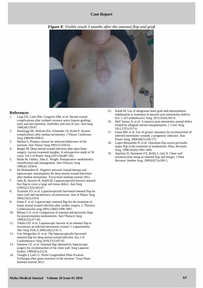
The wound healed well, and is covered by
healthy looking skin. The patient improved
steadily, his functional outcome was good and he
went back to work as a taxi driver. There was
eventual fibrous union that gave the patient rib cage
relative stability.
Discussion
The first laparoscopic omental harvest was
reported in 1993 by Salz et al. (7) Later, it was
reported as a flap for sternal wound closure with
many variations. Some report it without prior
NPWT and others use pectoral muscle flaps over
the omentum.6,8-11
In our case the combination of NPWT, omental
flap and skin graft was used. A case series by Van
Wingerden JJ et al reported 6 patients treated with
NPWT and omentoplasty, however 5 out of these 6
received local myocutaneous flap closure and only
1 had a skin graft to cover the omentoplasty. A few
points to note from this study were the use of large
amounts of foam in the wound when on NPWT
therapy, as done in our case and also the use of
fibrin glue to attach the omentum and skin graft
rather than sutures as in our case. They concluded
this three-pronged approach to be effective in
severe postoperative mediastinitis.12
The severity of mediastinitis is described using
the Oakley-Wright classification in table 1 below.5
59

dRe
Case Report
Malta Medical Journal Volume 28 Issue 01 2016
Figure 5: Retrieval of greater omentum flap through an opening in the diaphragm and out of the sternal wound
Figure 6: Omentum in the sternal defect
60

dRe
Case Report
Malta Medical Journal Volume 28 Issue 01 2016
Figure 7: Split skin graft overlying omentum and stapled in place
Table 1: Oakley-Wright classification of post sternotomy mediastinitis. A therapeutic trial involves a surgical
intervention such as prior grafting
Class Description
Type I Mediastinitis presenting within 2 weeks of operation in the absence of risk factors
Type II Mediastinitis presenting in 2-6 weeks of operation in the absence of risk factors
Type IIIa Type I plus one or more risk factors
Type IIIb Type II plus one or more risk factors
Type Iva Type I, II or III after one failed therapeutic trial.
Type IVb Type I, II or III after more than one failed therapeutic trial.
Type V Mediastinitis presenting more than 6 weeks after operation.
61

dRe
Case Report
Malta Medical Journal Volume 28 Issue 01 2016
The risk factors for post-operative infection are
mentioned in the introduction. Our patient has more
than one risk factor due to being diabetic and obese.
He also suffered from heavy bouts of coughing
post-op. Therefore, our patient classifies as a Type
IIIB mediastinitis.
De Brandere K. et al. used the same protocol as
Van Wingerden JJ, with negative pressure,
omentoplasty and myocutaneous flap advancement.
They also performed thorough wound debridement
and kept the patients on IV antibiotics and NPWT
therapy for several weeks prior to grafting. They
note the debate over which gastroepiploic artery is
the best pedicle for the omentum. They used the
right artery due to its larger size as seen in our case
too, however both have been shown to be equally
effective 6,12
Domene CE. et al13. report a case of a 62 year
old who underwent pectoralis muscle flap
reconstruction, which necrosed. The patient then
required re-debridement and omental flap harvested
laparoscopically and covered by a split skin graft.
The results were satisfactory.
There are other more modern techniques
reported in the literature such as plate fixation with
myocutaneous flaps following aggressive resection
for infection.14 However, there is significant risk
involved when introducing such a large amount of
foreign material such as this longitudinal plate and
numerous sternal wires. Another option is the use of
allogenic bone grafting or sternal transplantation. In
both cases the allograft was held in place by
titanium plates and results were excellent.15-16 The
problems of introducing foreign material and
allograft into a previously infected wound are still
present and the difficulties associated with
obtaining the allograft and performing the
procedure make this less applicable in most centres.
Today the availability of a made to measure 3D
printed sternum can offer structural stability and fill
the space that the omentum was filling. The
advantages as such are a better long-term result
however the need for plate fixation and the quantity
of foreign material makes it risky in the context of
infection.
There are potential complications associated
with the kind of procedure described here too. De
Brabendere et al reported an incisional hernia in
one patient and a partial dehiscence in another,
which settled conservatively 6 Rutger M. et al had a
27.3% wound dehiscence rate and an 18.2%
incisional hernia rate from the 11 cases treated with
omental flap reconstruction.4 Ghazi et al had an
overall recipient site morbidity of 23% and a donor
site complication rate of 27% from 52 patients
undergoing omental flap transposition.17 It is worth
mentioning that the majority of these patients had a
laparotomy for omental harvest and hence the
complication rate may need to be reviewed for
laparoscopic omental harvest. Lopez-Monjardin et
al conclude that using omental flaps for the
treatment of mediastinitis following open heart
surgery is more effective than simply using
myocutaneous flaps.18
Most studies seem to agree that the most
important factors are aggressive early local wound
debridement (to remove osteomyelitic bone),
NPWT therapy and multiple antibiotics for several
weeks. Then once infection free one proceeds to
laparoscopic omental harvest, transposition into
chest pedicled on either gastroepiploic artery and
covered by either pectoral myocutaneous flap or by
split skin graft. The case we report here followed
the above treatment bundle and the patient
recovered successfully. The literature concludes
that this treatment bundle is highly effective in
treating cases that have failed other attempts at
treatment, therefore class IVa or IVb. However,
here we describe it as a treatment option for a type
IIIb patient and in the light of data favouring
omental flap versus pectoral flap alone, this seems
justified in such patients who have multiple risk
factors for wound infection and dehiscence. The
stability of the sternum is good and our patient is
able to live a normal life with a cosmetic result that
is acceptable to him, although further surgery is on
offer by the plastic surgical team to refashion the
scar into a less visible one.
62
dRe
Case Report
Malta Medical Journal Volume 28 Issue 01 2016
Figure 8: Visible result 3 months after the omental flap and graft
References 1. Loop FD, Lytle BW, Cosgrove DM, et al. Sternal wound
complications after isolated coronary artery bypass grafting: early and late mortality, morbidity and cost of care. Ann Surg 1990;49:179-87.
2. Hazelrigg SR, Wellons HA, Schneider JA, Kolm P. Wound complications after median sternotomy. J Thorac Cardiovasc Surg 1989;98:1096-9.
3. Molina E. Primary closure for infected dehiscence of the sternum. Ann Thorac Surg 1993;55:459-63.
4. Rutger M. Deep sternal wound infection after open heart surgery: current treatment insights. A retrospective study of 36 cases. Eur J of Plastic Surg (2011)34;487-492.
5. Reida M. Oakley, John E. Wright. Postoperative mediastinitis: classification and management. Ann Thoracic Surg 1996;61:1030-6.
6. De Brabandere K. Negative pressure wound therapy and laparoscopic omentoplasty for deep sternal wound infections
after median sternotomy. Texas heart institute journal 2012. 7. Saltz R, Stowers R, Smith M. Laparoscopically harvest omental
free flap to cover a large soft tissue defect. Ann Surg (1993);217(5):542-67.
8. Acarturk TO. et al. Laparoscopically harvested omental flap for chest wall and intrathoracic reconstruction. Ann of Plastic Surg 2004;53(3):210-6
9. Puma F. et al. Laparoscopic omental flap for the treatment of
major sternal wound infection after cardiac surgery. J. Thoracic Cardiovascular surg 2003;126(6):1998-2002.
10. Milano CA. et al. Comparison of omental and pectoralis flaps for poststernotomy mediastinitis. Ann Thoracic Surg 1999;67(2):377-81.
11. Tebala GD. et al. Laparoscopic harvest of an omental flap to reconstruct an infected sternotomy wound. J. Laparoendosc Adv Surg Tech A 2006;16(2):141-5.
12. Van Wingerden JJ. et al. The laparoscopically harvested
omental flap for deep sternal wound infection. Eur J of Cardiothoracic Surg 2010;37(1):87-92.
13. Domene CE. et al. Omental flap obtained by laparoscopic surgery for reconstruction of the chest wall. Surg Laparosc Endosc 1998;8(3):215-8.
14. Tasoglu I, Lafci G. Novel Longitudinal Plate-Fixation Technique after gross resection of the sternum. Texas Heart Institute Journal 2012.
15. Kalab M. Use of allogenous bone graft and osteosynthetic stabilization in treatment of massive post-sternotomy defects. Eur. J. of Cardiothoracic Surg. 2012;41(6):182-4.
16. Dell’Amore A. et al. A massive post-sternotomy sternal defect treated by allograft sternal transplantation. J. Card. Surg. 2012;27(5):557-9.
17. Ghazi BH. et al. Use of greater omentum for reconstruction of infected sternotomy wounds: a prognostic indicator. Ann
Plastic Surg. 2008;60(2):169-173. 18. Lopez-Monjardin H. et al. Omentum flap versus pectoralis
major flap in the treatment of mediastinits. Plast. Reconstr. Surg. 1998;101(6):1481-1485.
19. Aquilina D, Darmanin FX, Briffa J, Gatt D. Chest wall reconstruction using an omental flap and Integra. J Plast Reconstr Aesthet Surg. 2009;62(7):e200-2.
63