

Lab Techniques: DNA/Genetics Doug Dluzen Lab Lecture 1 [email protected] Eom et al. Nature 1996.
58
-
Upload
eleanor-lynch -
Category
Documents
-
view
214 -
download
0
Transcript of Lab Techniques: DNA/Genetics Doug Dluzen Lab Lecture 1 [email protected] Eom et al. Nature 1996.

Overview• PCR
– Discovery– qRT-PCR– Genotyping
• Restriction Digests– Gel Electrophoresis– RFLP
• Western Blotting• Southern Blotting• Northern Blotting• Using Genomic Databases

The Polymerase Chain Reaction
http://elementy.ru/images/eltpub/kary-mullis.jpg Molecular Diagnostics: For the Clinical Laboratorian, Second Edition
-Allows one to replicate, study, and manipulate specific DNA sequences of interest
-PCR revolutionized molecular biology
-PCR technology is used in almost every lab and hospital

Amplifying DNA in Vitro: The Polymerase Chain Reaction (PCR)
• The polymerase chain reaction, PCR, can produce many copies of a specific target segment of DNA
• A three-step cycle—heating, cooling, and replication—brings about a chain reaction that produces an exponentially growing population of identical DNA molecules
• The key to PCR is an unusual, heat-stable DNA polymerase called Taq polymerase.
Adopted from Dr. Sairam Lecture Slides

Genomic DNA
Targetsequence
5
5
3
3
Adopted from Dr. Sairam Lecture Slides
The Steps of PCR

Denaturation
Annealing
Extension
Primers
Newnucleo-tides
Cycle 1yields
2molecules
5
5
3
3
2
3
1Figure 20.8b
Adopted from Dr. Sairam Lecture Slides

Cycle 2yields
4molecules
Adopted from Dr. Sairam Lecture Slides

Figure 20.8d
Cycle 3yields 8
molecules;2 molecules
(in white boxes)match target
sequence
Adopted from Dr. Sairam Lecture Slides

DNA technology allows us to study the sequence, expression, and function of a gene
• DNA cloning allows researchers to – Compare genes and alleles between individuals– Locate gene expression in a body– Determine the role of a gene in an organism
• Several techniques are used to analyze the DNA of genes – Already mentioned PCR
Adopted from Dr. Sairam Lecture Slides
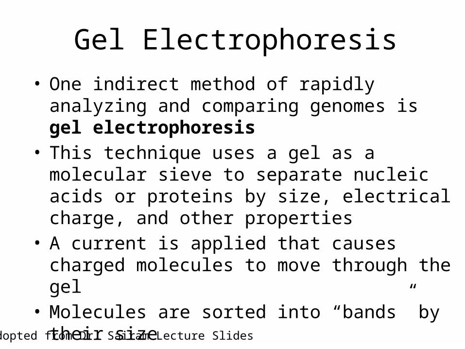
Gel Electrophoresis• One indirect method of rapidly analyzing and
comparing genomes is gel electrophoresis• This technique uses a gel as a molecular sieve to
separate nucleic acids or proteins by size, electrical charge, and other properties
• A current is applied that causes charged molecules to move through the gel
• Molecules are sorted into “bands” by their size
Adopted from Dr. Sairam Lecture Slides

Pulsed field gel electrophoresis
Adopted from Dr. Sairam Lecture Slides
http://academic.brooklyn.cuny.edu/biology/bio4fv/page/molecular%20biology/dsDNA.jpg
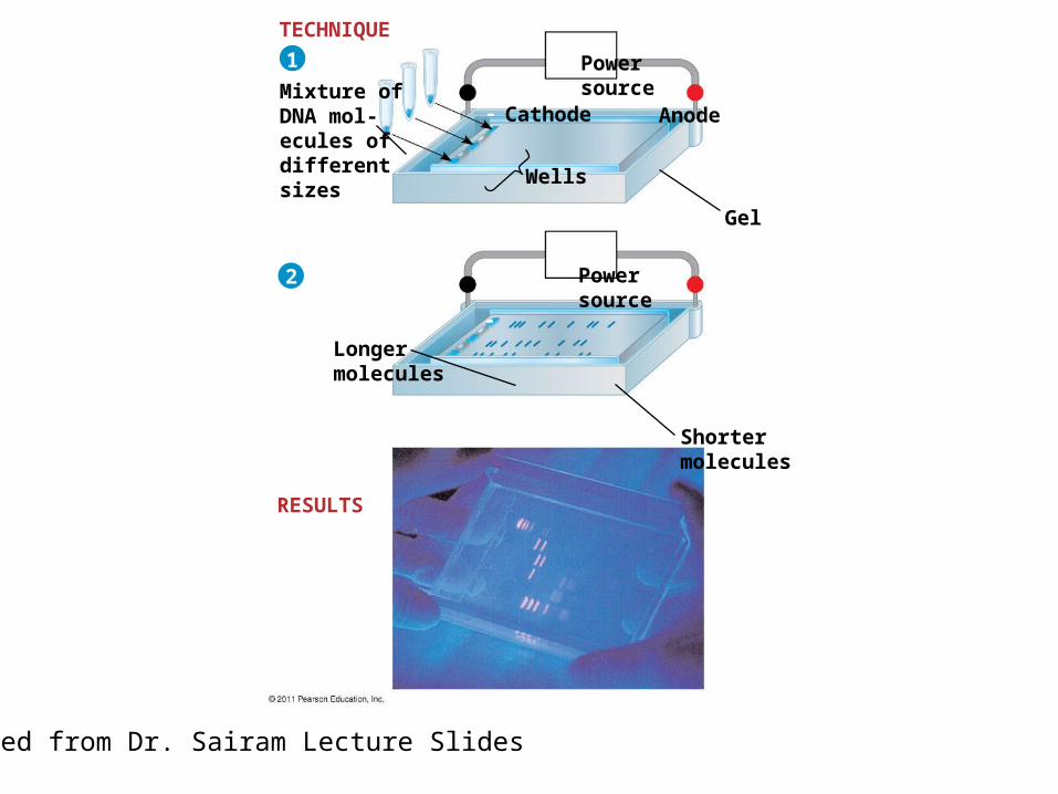
Mixture ofDNA mol-ecules ofdifferentsizes
Powersource
Powersource
Longermolecules
Cathode Anode
Wells
Gel
Shortermolecules
TECHNIQUE
RESULTS
1
2
Adopted from Dr. Sairam Lecture Slides

Oh the places You’ll Go
• Where have we been?– PCR, Electrophoresis
• Where are we going?– PCR Technologies

Technology #1: qRT-PCR
• Stands for quantitative real-time PCR• Allows one to:
– Assess gene expression levels– Measure and compare gene expression levels
• Uses PCR technology and resources with a fluorescent twist– Fluorescent light is used as a marker for gene
expression evaluation– The more light = more gene mRNA expression

Flourescent (qRT-PCR)
http://www.scripps.edu/florida/technologies/genomics/images/TaqManMethod.png
The MethodologyAn Amplifcation Curve: the visualization and data of qRT-PCR
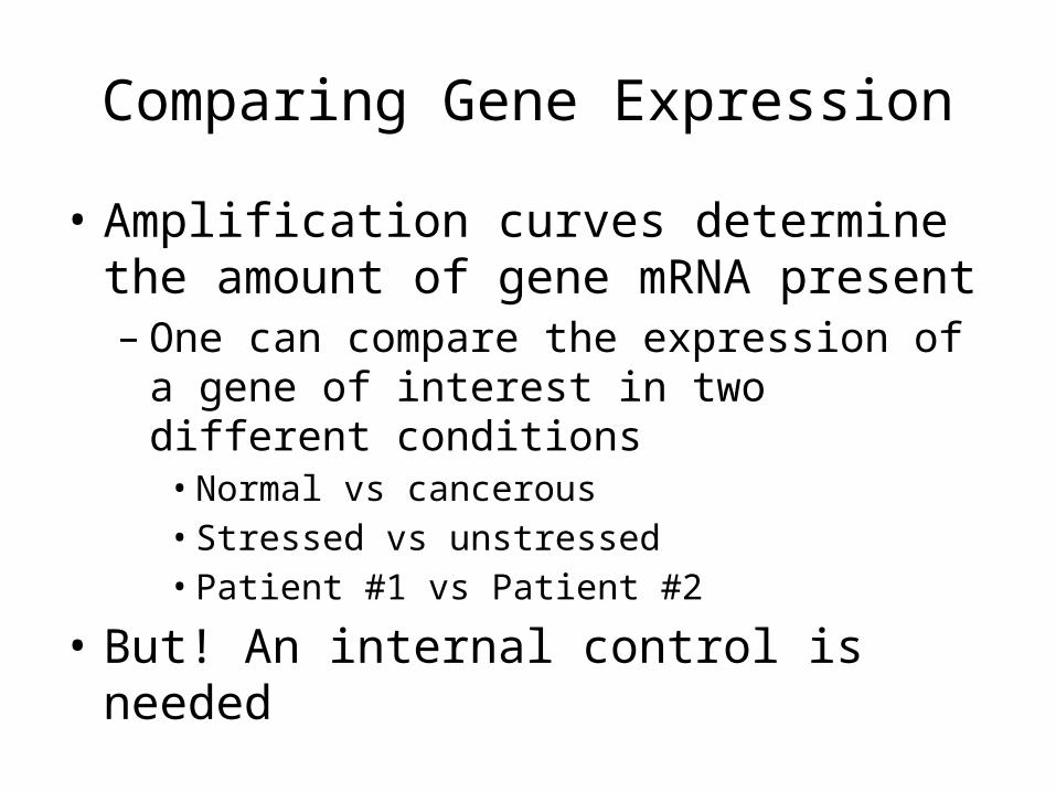
Comparing Gene Expression
• Amplification curves determine the amount of gene mRNA present– One can compare the expression of a gene of
interest in two different conditions• Normal vs cancerous• Stressed vs unstressed• Patient #1 vs Patient #2
• But! An internal control is needed

The Internal qRT-PCR Control
• Why do we need an internal control?– PCR gene expression analysis actually measures
the difference of a gene’s mRNA levels after that same gene’s expression was compared to a house-keeping gene ubiquitously expressed in all conditions

The internal qrt-pcr control
• How does this work?– 1. A gene’s (G.O.I.) mRNA is measured via PCR in
Tissue #1– 2. A control gene’s mRNA is measured via PCR in
Tissue #1– 3. A ratio of the GOI to the control is measured– 4. Steps 1 – 3 occur in Tissue #2 – 5. The ratios of Tissue 1 vs. Tissue 2 are then
compared to determine their relative expression • This is known as the ∆∆Ct method

The Ct value

• One can use PCR to determine an individual’s Single Nucleotide Polymorphisms (SNPs)
TeCh #2 - SNP Genotyping

Single Nucleotide Polymorphisms
• In humans, researchers analyze the genomes of many people with a certain genetic condition to try to find nucleotide changes specific to the condition
• Genetic markers called SNPs (single nucleotide polymorphisms) occur on average every 100–300 base pairs
• SNPs can be detected by PCR, and any SNP shared by people affected with a disorder but not among unaffected people may pinpoint the location of the disease-causing gene
© 2011 Pearson Educatio, Inc.
Adopted from Dr. Sairam Lecture Slides

DNA
SNP
Normal allele
Disease-causingallele
T
C
Single Nucleotide Polymorphisms
Adopted from Dr. Sairam Lecture Slides
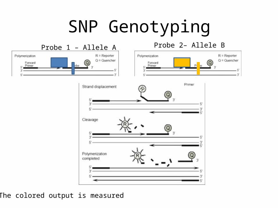
SNP GenotypingProbe 1 – Allele A Probe 2– Allele B
- The colored output is measured

TeCh #2 - SNP Genotyping
Wild Type Heterozygote Homozygous

SNP Genotyping

Tech #3 – Copy Number Variation
• In addition to SNPs, the human genome is widely variable with respect to gene copy number between any two individuals
He et al. Trends in Molecular Medicine. 2011.

Copy Number Variation

UGT2B17 CNV
• Taqman CNV assay designed to detect deletion polymorphism in pancreatic gDNA samples– 813 samples, Caucasian
• Each sample run as the following:– 2x Master mix – 5 uL– 20x CNV assay + 20 x reference assay (RNaseP) – 1 uL– 10 ng gDNA – 1 uL– Water – 3 uL
• 10 uL total

UGT2B17 Copy Number Variation – An Example

Analyzing DNA

Using Restriction Enzymes to Make Recombinant DNA
• Bacterial restriction enzymes cut DNA molecules at specific DNA sequences called restriction sites
• A restriction enzyme usually makes many cuts, yielding restriction fragments
• The most useful restriction enzymes cut DNA in a staggered way, producing fragments with “sticky ends.”
Adopted from Dr. Sairam Lecture Slides

Restriction Digest Sites
Adopted from Dr. Sairam Lecture Slides

DNA Ligase
• Sticky ends can bond with complementary sticky ends of other fragments
• DNA ligase is an enzyme that seals the bonds between restriction fragments
Adopted from Dr. Sairam Lecture Slides
Recombinant DNA molecule
One possible combinationDNA ligaseseals strands
DNA fragment addedfrom another moleculecut by same enzyme.Base pairing occurs.
Restriction enzymecuts sugar-phosphatebackbones.
Restriction site
DNA5
5
5
5
5
5
5
5
55
5
5
55
5
5
3
3
3
3
3
3
3
3
3
3
3
3
3
3
3
3
2
3
1
Sticky end
GAATTCCTTAAG
CTTAAG AATTC
G
GGAATTC
CTTAA
GG
GG
AATT CAATT CC TTAA C TTAA
Adopted from Dr. Sairam Lecture Slides

Using Restriction Enzymes
• In restriction fragment analysis, DNA fragments produced by restriction enzyme digestion of a DNA molecule are sorted by gel electrophoresis
• Restriction fragment analysis can be used to compare two different DNA molecules, such as two alleles for a gene if the nucleotide difference alters a restriction site
• Sequence changes that alter restriction sites are called RFLPs (restriction fragment length polymorphisms)
Adopted from Dr. Sairam Lecture Slides

Normal -globin allele
Sickle-cell mutant -globin allele
Largefragment
Normalallele
Sickle-cellallele
201 bp175 bp
376 bp
(a) DdeI restriction sites in normal andsickle-cell alleles of the -globin gene
(b) Electrophoresis of restrictionfragments from normal andsickle-cell alleles
201 bp175 bp
376 bp
Large fragment
Large fragment
DdeI DdeI DdeI DdeI
DdeI DdeI DdeI
RFLP Analysis
Adopted from Dr. Sairam Lecture Slides

Restriction Digests• Need to check if PCR fragment, generally
cloned into a useful reporter, has the correct orientation– DNA inserts can ligate into a plasmid in two
directions

Detection of Specific DNA and Protein Sequences - Blotting
• Whether you are analyzing DNA, RNA, or protein the underlying principle is the same
• Use of a probe that signals presence/absence of a gene or protein of interest
• Labeled (chemically, radioactively, etc.)probe is used to visualize your target

• A technique called Southern blotting combines gel electrophoresis of DNA fragments with nucleic acid hybridization
• Specific DNA fragments can be identified by Southern blotting, using labeled probes that hybridize to the DNA immobilized on a “blot” of gel
Southern Blotting
Adopted from Dr. Sairam Lecture Slides

Southern Blotting
Adopted from http://homepages.strath.ac.uk/~dfs99109/BB211/RecombDNAtechlect2.html#northerns

Southern Blotting
A southern blot can distinguish:1. The presence of a particular gene of interest2. Number of copies of that gene3. Genomic rearrangements4. Mutations of restriction digest sites
-Southern blots are very sensitive
Adopted from http://homepages.strath.ac.uk/~dfs99109/BB211/RecombDNAtechlect2.html#northerns

Northern Blotting
• Northern blotting combines gel electrophoresis of mRNA followed by hybridization with a probe on a membrane
• Identification of mRNA at a particular developmental stage suggests protein function at that stage
Adopted from http://homepages.strath.ac.uk/~dfs99109/BB211/RecombDNAtechlect2.html#northernsAdopted from Dr. Sairam Lecture Slides

Northern Blotting• Same principle as southern blotting, except
RNA is measured as opposed to DNA– RNA can also bind to nitrocellulose membrane– Uses formaldehyde as a denaturing reagent
• Used to identify tissue and temporal expression of a particular gene– Sensitive– Used to measure expression levels of particular
mRNA

Western Blotting
• Powerful tool to detect presence and expression levels of a particular protein– Use of an antibody – specific protein molecule will bind
to specific protein sequence on the protein of interest• This specific protein sequence is called an epitope
• As with northern and southern blotting, proteins are sorted by molecular weight, transferred to a membrane, and probed– Protein presence, expression, and quantity can be
measured

Western Blotting - Principles
Adopted from GE Healthcare: Western Blotting Principles

Western Blotting Methods
1. Electrophoresis – Denaturing Gel
Adopted from GE Healthcare: Western Blotting Principles

Western Blot Methods2. Transfer from gel to membrane
Adopted from GE Healthcare: Western Blotting Principles

Western Blot Methods
• Once protein transferred to membrane– Incubate in protein buffer (generally 5% milk
solution) to bind all regions of blot not bound by transferred protein
• Incubate with primary and secondary antibodies
• Visualize!

And now for something completely different…
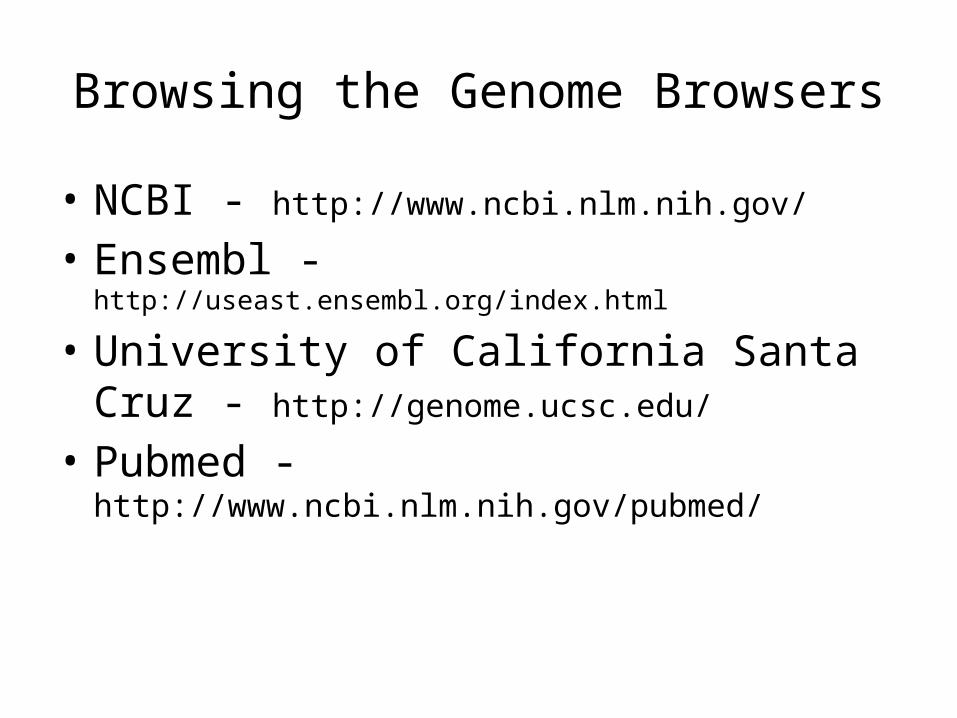
Browsing the Genome Browsers
• NCBI - http://www.ncbi.nlm.nih.gov/
• Ensembl - http://useast.ensembl.org/index.html
• University of California Santa Cruz - http://genome.ucsc.edu/
• Pubmed - http://www.ncbi.nlm.nih.gov/pubmed/
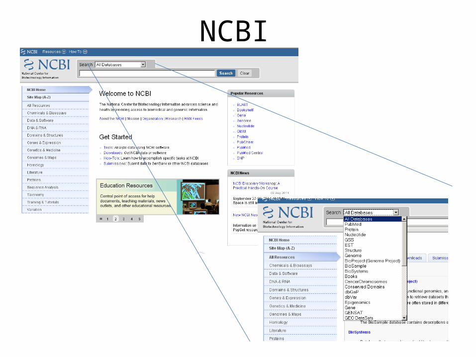
NCBI

BLAST!

Ensembl

Ensembl – THE MANY USES

I see U see UCSC

UCSC

Other Useful Odds and Ends
• HapMap Project – encyclopedia of human and mouse SNP variation and genotype frequencies www.hapmap.org
• TargetScan – microRNA prediction • Pubmed – uses NCBI database for literature
searches, protein and nucleotide sequences