
Lab on a Chip - Cornell...
12
Lab on a Chip PAPER Cite this: Lab Chip, 2014, 14, 3081 Received 25th March 2014, Accepted 20th May 2014 DOI: 10.1039/c4lc00371c www.rsc.org/loc Body-on-a-chip simulation with gastrointestinal tract and liver tissues suggests that ingested nanoparticles have the potential to cause liver injury Mandy B. Esch, a Gretchen J. Mahler, b Tracy Stokol c and Michael L. Shuler * a The use of nanoparticles in medical applications is highly anticipated, and at the same time little is known about how these nanoparticles affect human tissues. Here we have simulated the oral uptake of 50 nm carboxylated polystyrene nanoparticles with a microscale body-on-a-chip system (also referred to as multi-tissue microphysiological system or micro Cell Culture Analog). Using the ‘GI tract–liver–other tissues’ system allowed us to observe compounding effects and detect liver tissue injury at lower nanoparticle con- centrations than was expected from experiments with single tissues. To construct this system, we combined in vitro models of the human intestinal epithelium, represented by a co-culture of enterocytes (Caco-2) and mucin-producing cells (TH29-MTX), and the liver, represented by HepG2/C3A cells, within one microfluidic device. The device also contained chambers that together represented the liquid portions of all other organs of the human body. Measuring the transport of 50 nm carboxylated polystyrene nanoparticles across the Caco-2/HT29-MTX co-culture, we found that this multi-cell layer presents an effective barrier to 90.5 ± 2.9% of the nanoparticles. Further, our simulation suggests that a larger fraction of the 9.5 ± 2.9% nanoparticles that travelled across the Caco-2/HT29-MTX cell layer were not large nanoparticle aggregates, but primarily single nanoparticles and small aggregates. After crossing the GI tract epithelium, nanoparticles that were administered in high doses estimated in terms of possible daily human consumption (240 and 480 × 10 11 nanoparticles mL -1 ) induced the release of aspartate aminotransferase (AST), an intracellular enzyme of the liver that indicates liver cell injury. Our results indicate that body-on-a-chip devices are highly relevant in vitro models for evaluating nanoparticle interactions with human tissues. Introduction Because nanoparticles have the potential to change the way we treat and diagnose disease, studies that address nanoparticle effects on human tissues have become a prior- ity. In addition to medical uses, there are a number of com- mercial products that contain nanoparticles (Nanotechnology Consumer Product Inventory. Washington, DC: Project on Emerging Nanotechnologies, Woodrow Wilson International Center for Scholars. Available at http://www.nanotechproject. org/consumerproducts). Currently over 1030 products are available and their applications range from antibacterial coatings and paints to cosmetics such as suncreen. 1–5 However, little is known about the effects nanoparticles have on tissues of the human body. Recent studies have found that charged nanoparticles can affect phospholipid bilayers bearing phosphocholine headgroups, causing surface reconstruction, 6 and that carboxylated polystyrene nano- particles can alter the absorption of some nutrients through the intestines of poultry. 7 Such findings indicate that further evaluation of the implications of nanoparticle consumption through intended or accidental exposure is needed to estimate safe consumption levels. 8,9 Here we simulate non-life-threatening effects of ingested 50 nm carboxylated polystyrene nanoparticles on liver tissue using a ‘GI tract–liver–other tissues’ body-on-a-chip device. Previous studies of oral nanoparticle uptake have focussed on nanoparticle behavior directly in the intestine. One of these studies has shown that small drug delivery nano- particles (<670 nm) travel farther into the mucous layer of the intestine than do millimeter-sized nanoparticles, thus enhancing the bioavailability of orally administered drugs. 10,11 It is also known that both epithelial cells and microfold cells (M-cells) of the Peyer's patches in the intestine-associated lymphoid tissue facilitate nanoparticle uptake. 12–14 Small, charged nanoparticles (50 nm carboxylated nanoparticles) travel through the epithelial cell layer via Lab Chip, 2014, 14, 3081–3092 | 3081 This journal is © The Royal Society of Chemistry 2014 a Department of Biomedical Engineering, 305 Weill Hall, Cornell University, Ithaca, NY, 14853, USA. E-mail: [email protected]; Fax: +1 607 255 1136; Tel: +1 607 255 3293 b Department of Bioengineering, Binghamton University, Binghamton, NY 13902, USA c Department of Population Medicine and Diagnostic Sciences, College of Veterinary Medicine, S1-058 Schurman Hall, Cornell University, Ithaca, NY, 14853, USA Published on 27 June 2014. Downloaded by Cornell University Library on 11/09/2014 21:00:30. View Article Online View Journal | View Issue
Transcript of Lab on a Chip - Cornell...

Lab on a Chip
Publ
ishe
d on
27
June
201
4. D
ownl
oade
d by
Cor
nell
Uni
vers
ity L
ibra
ry o
n 11
/09/
2014
21:
00:3
0.
PAPER View Article OnlineView Journal | View Issue
Lab ChipThis journal is © The Royal Society of Chemistry 2014
aDepartment of Biomedical Engineering, 305 Weill Hall, Cornell University,
Ithaca, NY, 14853, USA. E-mail: [email protected]; Fax: +1 607 255 1136;
Tel: +1 607 255 3293bDepartment of Bioengineering, Binghamton University, Binghamton, NY 13902, USAc Department of Population Medicine and Diagnostic Sciences, College of
Veterinary Medicine, S1-058 Schurman Hall, Cornell University, Ithaca, NY,
14853, USA
Cite this: Lab Chip, 2014, 14, 3081
Received 25th March 2014,Accepted 20th May 2014
DOI: 10.1039/c4lc00371c
www.rsc.org/loc
Body-on-a-chip simulation with gastrointestinaltract and liver tissues suggests that ingestednanoparticles have the potential to cause liver injury
Mandy B. Esch,a Gretchen J. Mahler,b Tracy Stokolc and Michael L. Shuler*a
The use of nanoparticles in medical applications is highly anticipated, and at the same time little is known
about how these nanoparticles affect human tissues. Here we have simulated the oral uptake of 50 nm
carboxylated polystyrene nanoparticles with a microscale body-on-a-chip system (also referred to as
multi-tissue microphysiological system or micro Cell Culture Analog). Using the ‘GI tract–liver–other tissues’
system allowed us to observe compounding effects and detect liver tissue injury at lower nanoparticle con-
centrations than was expected from experiments with single tissues. To construct this system, we combined
in vitro models of the human intestinal epithelium, represented by a co-culture of enterocytes (Caco-2) and
mucin-producing cells (TH29-MTX), and the liver, represented by HepG2/C3A cells, within one microfluidic
device. The device also contained chambers that together represented the liquid portions of all other organs
of the human body. Measuring the transport of 50 nm carboxylated polystyrene nanoparticles across
the Caco-2/HT29-MTX co-culture, we found that this multi-cell layer presents an effective barrier to
90.5 ± 2.9% of the nanoparticles. Further, our simulation suggests that a larger fraction of the 9.5 ± 2.9%
nanoparticles that travelled across the Caco-2/HT29-MTX cell layer were not large nanoparticle aggregates,
but primarily single nanoparticles and small aggregates. After crossing the GI tract epithelium, nanoparticles
that were administered in high doses estimated in terms of possible daily human consumption (240 and
480 × 1011 nanoparticles mL−1) induced the release of aspartate aminotransferase (AST), an intracellular
enzyme of the liver that indicates liver cell injury. Our results indicate that body-on-a-chip devices are highly
relevant in vitro models for evaluating nanoparticle interactions with human tissues.
Introduction
Because nanoparticles have the potential to change theway we treat and diagnose disease, studies that addressnanoparticle effects on human tissues have become a prior-ity. In addition to medical uses, there are a number of com-mercial products that contain nanoparticles (NanotechnologyConsumer Product Inventory. Washington, DC: Project onEmerging Nanotechnologies, Woodrow Wilson InternationalCenter for Scholars. Available at http://www.nanotechproject.org/consumerproducts). Currently over 1030 products areavailable and their applications range from antibacterialcoatings and paints to cosmetics such as suncreen.1–5
However, little is known about the effects nanoparticleshave on tissues of the human body. Recent studies have
found that charged nanoparticles can affect phospholipidbilayers bearing phosphocholine headgroups, causing surfacereconstruction,6 and that carboxylated polystyrene nano-particles can alter the absorption of some nutrients throughthe intestines of poultry.7 Such findings indicate that furtherevaluation of the implications of nanoparticle consumptionthrough intended or accidental exposure is needed to estimatesafe consumption levels.8,9
Here we simulate non-life-threatening effects of ingested50 nm carboxylated polystyrene nanoparticles on liver tissueusing a ‘GI tract–liver–other tissues’ body-on-a-chip device.Previous studies of oral nanoparticle uptake have focussedon nanoparticle behavior directly in the intestine. One ofthese studies has shown that small drug delivery nano-particles (<670 nm) travel farther into the mucous layerof the intestine than do millimeter-sized nanoparticles,thus enhancing the bioavailability of orally administereddrugs.10,11 It is also known that both epithelial cells andmicrofold cells (M-cells) of the Peyer's patches in theintestine-associated lymphoid tissue facilitate nanoparticleuptake.12–14 Small, charged nanoparticles (50 nm carboxylatednanoparticles) travel through the epithelial cell layer via
, 2014, 14, 3081–3092 | 3081
Lab on a ChipPaper
Publ
ishe
d on
27
June
201
4. D
ownl
oade
d by
Cor
nell
Uni
vers
ity L
ibra
ry o
n 11
/09/
2014
21:
00:3
0.
View Article Online
para-cellular, energy-independent processes.7 A recentstudy by the authors has found that the uptake of 50 nm,carboxylated nanoparticles through the intestine changesthe absorption of iron as well as the sizes of macro-villi.7
Here we are interested in determining non-life-threateningeffects, if any, that may occur in tissues downstream of theintestine.
We use 50 nm, carboxylated polystyrene nanoparticles as amodel for inert, negatively charged nanoparticles and assessthe nanoparticle's potential to cause injury of in vitro livertissue. We choose 50 nm carboxylated polystyrene nano-particles because these nanoparticles had the most pro-nounced effects on iron uptake through the GI tractepithelium when compared to neutral and positively chargednanoparticles.7 In the study presented here we determinehow 50 nm carboxylated polystyrene nanoparticles thatcrossed the GI tract epithelium affect liver tissue. Testing thenanoparticle's effects on the liver is important since in vivothe blood stream coming from the GI tract transportsingested substances directly to the liver, exposing the liverto the highest nanoparticle concentrations and potentiallycausing damage.
To quantify liver damage due to ingested nanoparticles,we monitored changes in the integrity of the cell membranesof liver cells by measuring concentration of cytosolic enzymesin the cell culture medium. Cells whose membranes are atleast temporarily compromised, release cytosolic enzymes,which are routinely used as in vivo biomarkers of tissueinjury in animals and in humans.15 Thus the data obtainedwith our measurements are more relevant when correlatingin vitro and in vivo evaluations of tissue damage than thoseobtained with other methods of assessing cellular injury.
We hypothesized that the GI tract presents a significantbarrier to 50 nm carboxylated polystyrene nanoparticlesand that the limited nanoparticle travel across the GI tractepithelium would have the effect of limiting exposure ofthe liver to nanoparticles. To quantify nanoparticle travelacross the GI tract epithelium, we used fluorescently labelednanoparticles and measured the magnitude of fluorescencein the medium that was collected from the apical andbasolateral sides of the GI tract epithelium. We also quanti-fied changes in the level of nanoparticle aggregation withother nanoparticles and with macromolecules and changesin the magnitudes of zeta potentials of 50 nm carboxylatedpolystyrene nanoparticles that travelled across the GI tractepithelium.
Body-on-a-chip devices are well suited to simulate theuptake and circulation of therapeutics and environmentalcompounds in vitro.16–18 While both in vivo and in vitrostudies can uncover mechanisms that influence nanoparticleuptake and circulation, simulations with body-on-a-chipdevices can, inexpensively, direct our attention to effects thatshould be investigated further. Body-on-a-chip devicescontain several tissue analogs in the form of tissue culturesin physiologically scaled chambers that are arranged in physi-ologically correct order and with cell to liquid ratios that
3082 | Lab Chip, 2014, 14, 3081–3092
are close to in vivo values.19,20 The tissues that are explicitlyrepresented in the device are exposed to fluid flow ratesand shear stresses that are comparable to those observedin vivo. With these devices the combined response of severaltissues to nanoparticle exposure (or more generally, exposureto drugs, drug delivery systems, and environmental toxins)may be predicted. We have previously demonstrated a body-on-a-chip device that contained a multi-cellular modelof the GI tract epithelium in combination with a model ofthe liver. We have used this device to successfully simulatethe uptake, metabolism, and toxicity of acetaminophen.21
Here we use a similar system to mimic the oral uptake andfirst pass metabolism of 50 nm carboxylated polystyrenenanoparticles.
Materials and methodsMicrofabrication
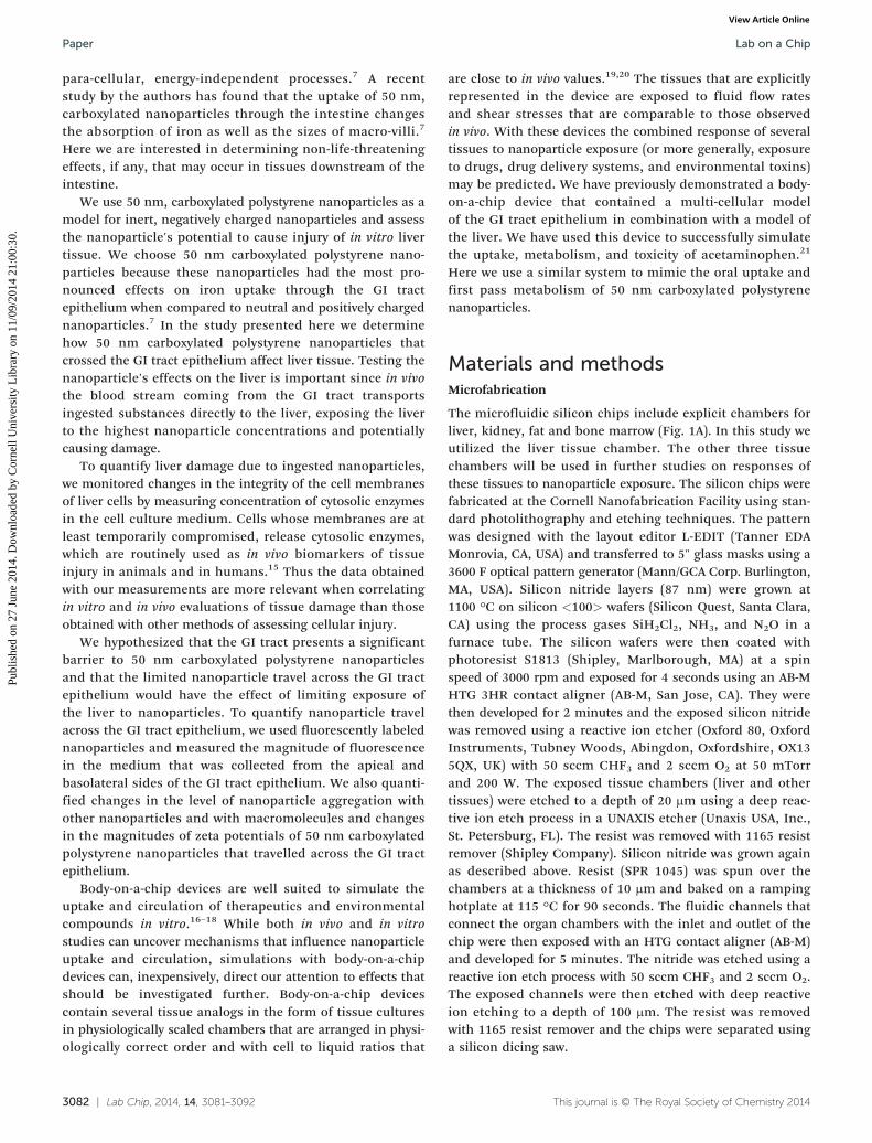
The microfluidic silicon chips include explicit chambers forliver, kidney, fat and bone marrow (Fig. 1A). In this study weutilized the liver tissue chamber. The other three tissuechambers will be used in further studies on responses ofthese tissues to nanoparticle exposure. The silicon chips werefabricated at the Cornell Nanofabrication Facility using stan-dard photolithography and etching techniques. The patternwas designed with the layout editor L-EDIT (Tanner EDAMonrovia, CA, USA) and transferred to 5" glass masks using a3600 F optical pattern generator (Mann/GCA Corp. Burlington,MA, USA). Silicon nitride layers (87 nm) were grown at1100 °C on silicon <100> wafers (Silicon Quest, Santa Clara,CA) using the process gases SiH2Cl2, NH3, and N2O in afurnace tube. The silicon wafers were then coated withphotoresist S1813 (Shipley, Marlborough, MA) at a spinspeed of 3000 rpm and exposed for 4 seconds using an AB-MHTG 3HR contact aligner (AB-M, San Jose, CA). They werethen developed for 2 minutes and the exposed silicon nitridewas removed using a reactive ion etcher (Oxford 80, OxfordInstruments, Tubney Woods, Abingdon, Oxfordshire, OX135QX, UK) with 50 sccm CHF3 and 2 sccm O2 at 50 mTorrand 200 W. The exposed tissue chambers (liver and othertissues) were etched to a depth of 20 μm using a deep reac-tive ion etch process in a UNAXIS etcher (Unaxis USA, Inc.,St. Petersburg, FL). The resist was removed with 1165 resistremover (Shipley Company). Silicon nitride was grown againas described above. Resist (SPR 1045) was spun over thechambers at a thickness of 10 μm and baked on a rampinghotplate at 115 °C for 90 seconds. The fluidic channels thatconnect the organ chambers with the inlet and outlet of thechip were then exposed with an HTG contact aligner (AB-M)and developed for 5 minutes. The nitride was etched using areactive ion etch process with 50 sccm CHF3 and 2 sccm O2.The exposed channels were then etched with deep reactiveion etching to a depth of 100 μm. The resist was removedwith 1165 resist remover and the chips were separated usinga silicon dicing saw.
This journal is © The Royal Society of Chemistry 2014

Fig. 1 Schematic of the silicon chip (A) and GI tract module (C) of the body-on-a-chip system, and the corresponding PBPK of the entire system(B). The device was operated with Caco-2/HT29-MTX co-cultures in the GI tract module and HepG2/C3A cells in the liver chamber. Cell culturemedium was recirculated through two fluidic circuits: the fluidic connections between the components of the system are represented as arrows in(B) (upper circuit in green = apical circulation and lower circuit in black = basolateral (systemic) circulation). The two fluidic circuits are separatedby Caco-2/HT29-MTX cell layers with developed barrier function. We added 50 nm carboxylated polystyrene nanoparticles to the apical circulationat varying concentrations and measured the amount of aspartate aminotransferase (AST) in the medium of the basolateral/systemic circulation.
Lab on a Chip Paper
Publ
ishe
d on
27
June
201
4. D
ownl
oade
d by
Cor
nell
Uni
vers
ity L
ibra
ry o
n 11
/09/
2014
21:
00:3
0.
View Article Online
The silicon chips were cleaned with a solution of sulfuricacid and hydrogen peroxide (3 : 1) at 70 °C before every use.This cleaning step is particularly important when the chip isre-used several times. The solution removes extracellularmatrix components that are deposited by cells grown on thechip in previous experiments.
The microfluidic GI tract module of the body-on-a-chipdevice was machined in plexiglass with round apical andbasolateral chambers so that transwell membrane inserts fitinto it (Fig. 1C). The resulting cell culture chambers (apicaland basolateral, separated by the transwell insert membrane)were 0.5 mm deep and 12 mm in diameter.
Body-on-a-chip systems operation
Two fluidic circuits were constructed (Fig. 1B): the firstrecirculated medium between a container that containedmedium with 50 nm carboxylated polystyrene nanoparticlesand the apical chamber of the GI tract module. The secondfluidic circuit represented the systemic circulation of thehuman body. This fluidic loop connected the basolateralside of the GI tract module with the silicon chip and the‘other organ’ container. The silicon chip contained the liverchamber as well as three other organ chambers (adipose,kidney, and bone marrow) to be used in future studies. The‘other organ’ container represents spaces for all other organsof the body (skin, muscle, brain, spleen, lung, heart, allglands, etc.). The containers and chambers that representthe liquid portions of organs other than those of interestin this study (gastrointestinal tract and liver) were filledwith medium. The medium in both circulation loops wasre-circulated with a peristaltic pump.
Body-on-a-chip systems design. We have previouslydescribed the design, operation and characterization ofthe ‘GI tract–liver–other organ’ system.21 Briefly, the human
This journal is © The Royal Society of Chemistry 2014
body (based on a 70 kg body) is scaled down by a factor of400 000 (considering that we are using cell monolayers of aheight between 3–5 μm), requiring an overall flow rate of3.59 μL min−1 through the entire systemic fluidic circuit, anda flow rate of 1.47 μL min−1 through the liver chamber. Thescaling was based on physiologic values obtained from datacollections by Davies et al. and Price et al.22,23 A flow rate of1.47 μL min−1 in the liver chamber allowed us to achieve anear-physiologic fluid residence time. The fluid residencetime in the liver compartment of a system that is scaled by afactor of 400 000 is 1.2 min and corresponds to the fluidresidence time in the liver in vivo.
To achieve this flow rate on a chip on which severalorgans are represented, the fluidic stream of the systemiccirculation is passively divided between the organ compart-ments on chip so that each fluidic stream experiences thesame pressure drop. Keeping the pressure drop across eachfluidic stream constant allows us to reach organ-specificfluid flow rates by adjusting the hydraulic resistance acrosseach fluidic branch. This goal can be achieved by choosingappropriate channel widths and lengths for the on chipfluidic channels that lead to and from each organ chamber.The resulting shear stress in the liver chamber wasestimated to be 1.01 dyn cm−2. Since the system wasdesigned to support several studies and we used it herewithout adipose, kidney and bone marrow cells, the fluidicflow was slightly biased on the chip. We measured the resi-dence time in the liver chamber for this configuration andfound that the fluid residence time in the liver chamberwith only liver cells and GI tract cells, is 2.1 ± 0.3 min.21
Cell culture
HepG2/C3A cells were obtained from the American TypeCulture Collection (Manassas, VA, USA) and cultured at 37 °C
Lab Chip, 2014, 14, 3081–3092 | 3083
Lab on a ChipPaper
Publ
ishe
d on
27
June
201
4. D
ownl
oade
d by
Cor
nell
Uni
vers
ity L
ibra
ry o
n 11
/09/
2014
21:
00:3
0.
View Article Online
in a 5% carbon dioxide atmosphere using Minimal EssentialMedium (MEM, Invitrogen, Carlsbad, CA, USA) with 1.0 mMsodium pyruvate and 10% FBS (Invitrogen). Intestinal cellcultures consisted of layers of human colon carcinoma Caco-2cells and HT29-MTX mucous producing cells in a ratio of 9 : 1.Caco-2 cells were obtained from Prof. Raymond Glahn'slaboratory at Cornell University at passage 17 and used atpassages 25 to 30. HT29-MTX cells were kindly provided byDr. Thécla Lesuffleur of INSERM U560 in Lille, France atpassage 11 and were used at passages 20 to 25. Both celllines were maintained at 37 °C in a 5% carbon dioxide atmo-sphere using Dulbecco's Modified Eagle Medium (DMEM,Invitrogen, Carlsbad, CA, USA) containing 4.5 g L−1 glucose,25 mM HEPES buffer and 10% heat inactivated fetal bovineserum (Invitrogen).
Single tissue experiments. For nanoparticle exposureexperiments with body-on-a-chip devices that contained onlyHepG2/C3A liver cells, HepG2/C3A cells were seeded ontosilicon chips at 62 500 cells (suspended in a volume of 80 μL)per liver chamber (0.68 cm2, 20 μm high). The remainingorgan chambers were left empty in the experimentsconducted for this study. Prior to use the chambers werecleaned with Piranha solution (H2O2 and H2SO4 mixed at aratio of 3 : 1). After cleaning, the chambers were pre-coatedwith poly-D-lysine for 5 minutes at a concentration of 4 μg cm−2
and with fibronectin for one hour at a concentration of8 μg cm−2. The cells were allowed to attach in the liverchamber of the silicon chip for 30 minutes and were thencovered with medium. On the next day the chips were placedinto Plexiglas housings and tubing for medium recirculationwas connected to a peristaltic pump.
Multi-organ experiments. For experiments with body-on-a-chip devices that contained both HepG2/C3A liver cells andCaco-2/HT29-MTX cell cultures, silicon chips were preparedwith HepG2/C3A cells as described above and transwells withCaco-2/HT29-MTX cells were prepared 16 days prior to siliconchip preparation as follows. Transwell culture plates (6-well)were treated with collagen at 8 μg cm−2 membrane surface,diluted in acetic acid (0.02 N) for one hour. After washingwith phosphate-buffered saline (PBS) Caco-2 and HT29-MTXcells were seeded into each well at 101 000 and 11 000 cellsper well respectively. The cells were cultured for 16 daysso that the transepithelial resistance was 200 Ω cm2 orhigher at the time of device assembly, indicating that tightjunctions between cells were fully developed. On day 17,the GI tract module and the silicon chip were connected toeach other and to a pump via tubing and medium was flownthrough the apical and basolateral fluidic loops at a flow rateof 3.59 μL per minute (Fig. 1).
Nanoparticle dose calculations
50 nm fluorescent, carboxylated polystyrene nanoparticles(Cat # 17149, YG-Fluoresbrite) were obtained from Poly-sciences Inc. (Warrington, PA). The doses of 50 nm nano-particles used in this study were in the mid and high range
3084 | Lab Chip, 2014, 14, 3081–3092
(16–480 × 1011 nanoparticles mL−1 cell culture medium) ofthose used previously. These concentrations were formulatedto mimic realistic human exposure.7
Nanoparticle exposure experiments
50 nm carboxylated polystyrene nanoparticles were diluted inmedium to yield various concentrations of 15–480 × 1011
nanoparticles mL−1. Vehicle-treated control medium andmedium containing nanoparticles were supplied to theassembled body-on-a-chip systems via the medium containerin the apical fluidic circuit (Fig. 1B). After 24 hours, mediumwas removed from the ‘other tissue’ chamber of thebasolateral/systemic fluidic circuit for enzyme measurementsand nanoparticle characterization.
On-chip cell viability
After 24 hours of exposure of cells within the body-on-a-chipdevice to nanoparticles or vehicle control, the medium withinthe tissue chambers was replaced with PBS containing fluo-rescent viability stains (Molecular Probes, Carlsbad, CA) inboth fluidic loops. The solution was circulated through theapical and basolateral/systemic circulation for 30 minutes.The solution was then replaced with PBS that washed out thedye. Pictures of the cell culture chambers were taken with afluorescence microscope and attached camera and the areacovered with viable cells was estimated with image process-ing software (ImageJ).
Enzyme quantitation in culture medium
To determine which cellular enzymes commonly used as bio-markers of liver injury in vivo would be useful for detectingtissue injury in our model, we measured concentrations ofalanine aminotransferase (ALT), aspartate aminotransferase(AST), glutamate dehydrogenase (GDH), and gamma-glutamyltranspeptidase (GGT) in the medium of cells cultured in12-well plates. Testing was performed at the clinical pathologylaboratory of the Animal Health Diagnostic Center at CornellUniversity, using an automated chemistry analyzer (HitachiModular P, Roche Diagnostics) with manufacturer's reagents.To compare the maximum amount of enzymes released fromeach cell type (Caco-2, HT29-MTX, and HepG2/C3A), 100 000cells were lysed with ethanol and enzyme concentrations weremeasured in the cell lysate that was diluted ten-fold, yieldingenzyme concentrations per 10 000 cells.
Quantification of nanoparticle passage acrossthe intestinal co-culture
To estimate the number of nanoparticles that traversedacross Caco-2/HT29-MTX co-cultures from the apical tothe basolateral chamber of the GI tract module, we usedfluorescent nanoparticles (Cat # 17149, YG-Fluoresbrite,Polysciences Inc., Warrington, PA, excitation: 441 nm,emission: 486 nm). After 24 hours of nanoparticle exposure,we collected 170 μL of medium from the ‘other organ’
This journal is © The Royal Society of Chemistry 2014

Lab on a Chip Paper
Publ
ishe
d on
27
June
201
4. D
ownl
oade
d by
Cor
nell
Uni
vers
ity L
ibra
ry o
n 11
/09/
2014
21:
00:3
0.
View Article Online
container and used a plate reader (Spectra Max Gemini EM,Molecular Devices, Sunnyvale, CA) to conduct fluorescencemeasurements.
Quantification of apparent permeability coefficientsfor 10 kDa dextran
The amount of transported 10 kDa Lucifer Yellow-conjugateddextran (Fisher Scientific Inc.) was measured with a fluores-cent plate reader using an excitation wavelength of 425 nmand an emission wavelength of 528 nm. The apparent perme-ability coefficient was calculated using the equation
P Qt A Capp
1
0
where ΔQ/Δt is the amount of lucifer yellow dextran trans-ported from the apical to the basolateral compartment pertime interval (t). C0 is the initial concentration in the apicalcompartment and A is the area of the membrane on whichCaco-2/HT29-MTX cells were cultured.
Measurement of pH
To determine if the cultured cells alter the pH of the mediumwithin the body-on-a-chip device, we collected 170 μL ofmedium from the apical and basolateral sides of the GI tractmodule and measured the pH with a pH meter equippedwith a micro pH electrode (DJ glass Ag/AgCl, Thermo Scientific,Beverly, MA).
Particle analysis with Zetasizer
To determine whether the surface charge of the nanoparticleschanges as a result of travelling through the GI tract epithe-lium, nanoparticles were collected from the apical andbasolateral sides of the GI tract module and diluted with830 μL medium. They were then analyzed with a Zetasizer(Malvern Instruments LTD, Worcestershire, UK) to determinetheir zeta potential and diameters. To yield nanoparticle sizedistributions, we measured 100 nanoparticles per sampleand measured every sample three times to minimize instru-ment errors.
Immunofluorescent staining of adherens junctions
To visually determine whether nanoparticles damage thetight junctions of the intestinal tissue cultures, we immuno-stained the epithelial co-culture for the tight junction proteinoccludin and imaged the cell layers with a confocal fluores-cence microscope. After 24 hours of exposure to nano-particles, the cells in the transwells were washed with PBS
This journal is © The Royal Society of Chemistry 2014
Table 1 Amounts of cytosolic enzymes released from cells after complete lys
HepG2/C3A
Aspartate aminotransferase (AST) 187.7 ± 11.2 U L−
Alanine aminotransferase (ALT) 1.33 ± 0.58 U L−
Gamma-glutamyl transpeptidase (GGT) 1.7 ± 1.53 U L−
three times and fixed in situ with 2% paraformaldehyde,rinsed with PBS containing 1% bovine serum albumin,permeabilized with 0.1% Triton X-100, and then immuno-stained with an antibody against occludin (rabbit anti-humanoccludin, 2 μg mL−1, Invitrogen Inc., Eugene, OR) for 40minutes at room temperature (at 0.04 μg mL−1). After washing,fluorescent secondary antibodies (Alexa-555-conjugated goatanti-rabbit antibody, 250 μg mL−1, Invitrogen Inc., Eugene, OR)were added at a concentration of 1.25 μg mL−1 for 40 minutesin the dark at room temperature. Cultures incubated withrabbit IgG (0.04 μg mL−1) and secondary goat anti-rabbit IgG(at a concentration of 1.25 μg mL−1, Invitrogen Inc., Eugene,OR) served as negative immunofluorescent control. Imageswere captured using a Leica SP2 confocal microscope (LeicaMicrosystems, Bannockburn, IL).
Transmission electron microscopy (TEM). Samples werecoated with carbon on a TEM grid and imaged with an FEIT12 spirit TEM system at the Cornell Center for MaterialsResearch.
Statistical analysis
Data represent mean ± the standard deviation of 3 to 6 exper-iments. Multiple means were compared with a one-wayANOVA, followed by a Bonferroni adjustment for the numberof pairwise comparisons, whereas comparisons of two meanswas performed with a paired T test (JMP software). For ASTmeasurements the one-way ANOVA was modeled on a log-transformed response. A p value of <0.05 was consideredsignificant.
ResultsQuantifying cellular damage to Caco-2, HT29-MTX andHepG2/C3A cells by measuring cytosolic enzyme release
Quantifying cellular damage in our in vitro system with amethod that allows us to later compare our data with in vivodata is important, considering that our results will need tobe confirmed with animal models. Since liver injury in ani-mals and humans is estimated by measuring concentrationsof intracellular enzymes in the blood, we first quantified theamounts of intracellular enzymes that are released from cellsin vitro due to cellular injury. For this purpose we preparedlysates of Caco-2, HT29-MTX, and HepG2/C3A cells, con-firmed complete lysis with fluorescent viability stains, andthen measured cytosolic enzyme concentrations in themedium. Our measurements show increased, but varyingconcentrations of AST in lysates of all three cell types(Table 1). HepG2/C3A cells released comparatively more ASTthan the other two cell types. Caco-2 and HT29-MTX cells
Lab Chip, 2014, 14, 3081–3092 | 3085
is. Concentrations are given per 10000 cells
Caco-2 HT29-MTX1 43.7 ± 6.66 U L−1 56.3 ± 15.5 U L−11 Not detected Not detected1 Not detected Not detected

Lab on a ChipPaper
Publ
ishe
d on
27
June
201
4. D
ownl
oade
d by
Cor
nell
Uni
vers
ity L
ibra
ry o
n 11
/09/
2014
21:
00:3
0.
View Article Online
contained only ~30% and ~23% of AST present in HepG2/C3Acells. HepG2/C3A cells, but not the two epithelial cell types,also released low amounts of ALT and GGT (Table 1). Glutamatedehydrogenase (GDH) was not released from any of the celltypes used here. Since ALT, GGT, and GDH were either notdetected or detected at very low amounts after cell lysis, wechose AST as a quantitative indicator of cellular damage in allsubsequent experiments. Besides providing a quantitative mea-sure for cellular injury, measuring concentrations of thisenzyme in plasma is used routinely to assess liver damagein vivo.15 Similarly the in vitro data generated with our devicescan be compared to data obtained with in vivo studies.
Since the goal of our study is to measure cellular injury inresponse to 50 nm carboxylated polystyrene nanoparticles, wetested whether these nanoparticles interfere with the assaywe used to measure AST concentrations. We added the maxi-mum amount of nanoparticles used in this study (480 × 1011
nanoparticles mL−1) to fresh cell culture medium that hadnot been exposed to cells and measured AST concentrations.In these samples, AST concentrations were undetectable,indicating that any AST measured in nanoparticle-exposedcells is not due to the interference of nanoparticles withthe assay and can be attributed to cellular release ofthis enzyme.
Exposure to 50 nm carboxylated polystyrene nanoparticlescauses cellular injury to HepG2/C3A cells
A) Experiments with ‘GI tract–liver–other tissues’ devices.To simulate the oral uptake of 50 nm carboxylated polystyrene
3086 | Lab Chip, 2014, 14, 3081–3092
Fig. 2 (A) Mean concentrations of aspartate aminotransferase (AST), measurchip devices that were operated with one tissue only (either GI tract or liver),of 50 nm carboxylated polystyrene nanoparticle exposure when HepG2/C3Acultures were combined with HepG2/C3A cells. Caco-2/HT29-MTX cell csignificant differences in measurements, a one-way ANOVA was modeled ondifferent according to the one-way ANOVA with Tukey's post-test are indstandard deviation of 3 to 6 experiments. (B) Percent area of on-chip liver chexposure to 50 nm carboxylated polystyrene nanoparticles at varying concsignificant differences were found according to a one-way ANOVA with Tuwhen P < 0.05). Data represent mean ± one standard deviation of 3 to 6 e
nanoparticles and their effects on liver tissue, we used amicrofluidic ‘GI tract–liver–other tissue’ body-on-a-chip devicein which we cultured Caco-2/HT29-MTX cells and HepG2/C3Acells and recirculated the medium through two closed fluidiccircuits, one that served the apical side of the Caco-2/HT29-MTXcultures and a second that served the basolateral side ofthe Caco-2/HT29-MTX culture as well as the HepG2/C3Aculture and the ‘other tissue’ compartment that were locateddownstream of it (Fig. 1B). We supplied nanoparticles to theapical side of the Caco-2/HT29-MTX culture and measured ASTconcentrations in the medium collected from the basolateralfluidic circuit after 24 hours.
When nanoparticles were added at a concentration of240 × 1011 or 480 × 1011 nanoparticles mL−1 medium, ASTconcentrations in the basolateral fluidic circuit rose signifi-cantly during the following 24 hours of medium recirculationcompared to controls (Fig. 2a). However, fluorescent live/deadstaining did not indicate a significant decrease in cell viabilityof HepG2/C3A cultures (Fig. 2b), indicating that cellular injurytook place at a level that was not detected with dye-basedoptical viability measurements. Optical viability assessment ofCaco-2/HT29-MTX cell layers did not reveal any differencesbetween treatments, however, these cell layers are denseand can be multilayered, making it difficult to detect smalldifferences in cell viability.
B) Experiments with ‘liver–other tissues’ and ‘GI tract–othertissues’ configurations. Since it is likely that the GI tract tissuecontributed to the rise in AST concentration measured in thedevice setup in which both tissue cultures (Caco-2/HT29-MTXand HepG2/C3A) were present, we subjected each of the tissues
This journal is © The Royal Society of Chemistry 2014
ed in medium collected from the systemic circulation side of body-on-a-or with both tissues. AST concentrations significantly increased as a resultcultures were present in the device, and when Caco-2/HT29-MTX cell
ultures alone did not respond to nanoparticle exposure. To determinea log-transformed response. Mean concentrations that were significantlyicated with an asterisk (P < 0.05, n = 3–6). Data represent mean ± oneambers that was covered with viable HepG2/C3A cells after 24 hours ofentrations. Measurements were conducted with viability stains, and nokey's post-test (n = 3–6, values were considered significantly differentxperiments.

Lab on a Chip Paper
Publ
ishe
d on
27
June
201
4. D
ownl
oade
d by
Cor
nell
Uni
vers
ity L
ibra
ry o
n 11
/09/
2014
21:
00:3
0.
View Article Online
alone to 50 nm carboxylated polystyrene nanoparticles. For thispurpose we operated the devices with one tissue at a time.This is accomplished by leaving one of the tissue chambersempty, meaning that medium still flows through thechamber, but no cells are present. When 50 nm carboxylatedpolystyrene nanoparticles were supplied to a system thatcontained HepG2/C3A cells only, they caused an increasein AST levels at a nanoparticle concentration of 480 × 1011
nanoparticles mL−1, but not at lower concentrations(Fig. 2a). The increase in AST levels in the medium during24 hours of device operation with only HepG2/C3A cellswas not significantly higher than that seen with devices inwhich both, Caco-2/HT29-MTX co-cultures and HepG2/C3Acultures were present. No significant change in HepG2/C3Acell viability was observed in response to any of the testedparticle concentrations (Fig. 2b), indicating that the amountof cellular injury was significant, but not detectable viadye-based viability measurements.
When Caco-2/HT29-MTX co-cultures were cultured alonewithin the body-on-a-chip devices, no significant changes inAST concentrations were observed on either side (apical andbasolateral) of the cell culture for any of the tested nanoparticleconcentrations (Fig. 2a). Similar to our earlier observations, nosignificant changes in Caco-2 or HT29-MTX cell viability wasobserved, keeping in mind that the Caco-2/HT29-MTX celllayers were dense.
GI tract tissue limits nanoparticle exposure to 50 nmcarboxylated nanoparticles
To estimate the number of nanoparticles that transferredfrom the apical to the basolateral side of the Caco-2/HT29-MTX cell cultures, we used our body-on-a-chip devices withCaco-2/HT29-MTX cell only. Fluorescently labeled 50 nmcarboxylated polystyrene nanoparticles were introduced intothe system at the apical side of the GI tract chamber andthe fluorescence was measured in the medium collected fromthe basolateral side. When supplied at a concentration of
This journal is © The Royal Society of Chemistry 2014
Fig. 3 Representative confocal images taken of nanoparticle accumulationsCaco-2/HT29-MTX co-cultures that were exposed to fluorescently labeleside. Tight junctions between cells were immunofluorescently labeled withobserved with confocal microscopy associated with the apical side of the ctions as shown here suggests that the nanoparticles could reside in or abov
480 × 1011 nanoparticles mL−1, 9.5% ± 2.9% of the nano-particles reached the basolateral side after 24 hours of deviceoperation. Nanoparticles that remained on the apical sideof the Caco-2/HT29-MTX cell layer were accumulated in highconcentration spots, in addition to being evenly distributedacross the cell layer at a low concentration. Confocal micros-copy images show that the nanoparticles in high concentra-tion spots resided mostly on the apical side of the Caco-2/HT29-MTX cell layer (Fig. 3a–c). The tight junction complexeswere stained immunofluorescently and, judging by confocalmicroscopy images, they appeared intact, indicating nogross damage to the junctions. The functionality of the tightjunctions was also confirmed with transport measurementsof fluorescently labeled 10 kDa dextran. We found that nano-particle exposure did not change the apparent permeabilitycoefficient exhibited by Caco-2/HT29-MTX cell layers for thismolecule (Table 2).
Nanoparticles and nanoparticle aggregates that cross the GItract barrier are smaller in size than those that remain onthe apical side
Size measurements of 50 nm carboxylated polystyrene nano-particles that were collected from the basolateral side of theGI tract chamber showed that nanoparticles and nanoparticleaggregates that crossed the Caco-2/HT29-MTX co-cultureswere on average smaller than those that remained on the api-cal side. The size distributions of nanoparticles that were col-lected from the basolateral side of the microfluidic GI tractmodule showed a peak at 55 ± 7 nm, indicating largely singlenanoparticles. Size distributions of nanoparticles collectedfrom the apical side exhibited a peak at 97 ± 7 nm (Fig. 4),consistent with aggregates of a small number of nano-particles. We confirmed the existence of such aggregates inthe nanoparticle population using transmission electronmicroscopy of nanoparticles (Fig. 6). Size distributions ofnanoparticles collected from the basolateral side alsocontained a particle population of smaller size (<15 nm).
Lab Chip, 2014, 14, 3081–3092 | 3087
at different focal planes (A: z = 0 μm, B: z = −1.5 μm, and C: z = −3 μm) ofd 50 nm carboxylated polystyrene nanoparticles (green) at their apicalanti-occludin antibodies (red). The majority of nanoparticles that wereell layer. Their slightly higher location and grouping in such accumula-e patches of mucous layers.

Table 2 Apparent permeability coefficients (Papp) of 10 kDa dextran in Caco-2/HT29-MTX cultures and Caco-2/HT29-MTX + HepG2/C3A cultures thatwere exposed to 50 nm carboxylated polystyrene nanoparticles at varying concentrations. The data suggest no significant differences in tight junctionfunctionality as a result of nanoparticle exposure
Concentration of 50 nm carboxylatedpolystyrene nanoparticles × 1011 in particles mL−1
Papp for 10 kDa dextran incm s−1 × 10⁻⁹ ± standard deviation
Papp for 10 kDa dextran incm s−1 × 10⁻⁹ ± standard deviation
Caco-2/HT29-MTX cells only Caco-2/HT29-MTX and HepG2/C3A cultures
Vehicle control 8.45 ± 2.74 11.16 ± 1.89120 8.27 ± 3.88 8.28 ± 1.84240 9.47 ± 3.64 9.94 ± 1.75480 10.5 ± 2.68 8.07 ± 0.61
Fig. 4 Size distributions of 50 nm carboxylated polystyrene nanoparticles that were stored at 37 °C in water or medium (A), or collected from theapical or basolateral/systemic circulation loop of body-on-a-chip devices that were operated with Caco-2/HT29-MTX and HepG2/C3A cellcultures (B). The data shown were obtained using a Zetasizer in intensity measurement mode. The peaks of the particle distributions of particlesstored in water and collected from the basolateral side of the body-on-a-chip devices were significantly different from those of nanoparticles thatwere stored in medium and those that were collected from the apical side of the body-on-a-chip device. Each distribution represents onehundred nanoparticles taken from each of the 3 to 6 samples. Data represent mean ± one standard deviation of 3 to 6 experiments. Peaks ofnanoparticle size distributions were significantly different from each other according to a one-way ANOVA with Tukey's post-test P < 0.05.
Lab on a ChipPaper
Publ
ishe
d on
27
June
201
4. D
ownl
oade
d by
Cor
nell
Uni
vers
ity L
ibra
ry o
n 11
/09/
2014
21:
00:3
0.
View Article Online
These nanoparticles were not present in any of the othersamples, indicating that they are either cellular debris, orsmall vesicles released due to Caco-2 cell transport activity.The size distributions of nanoparticles that were stored inmedium at 37 °C exhibited a peak at 97 ± 1 nm, and those ofnanoparticles stored in water had a peak at 39 ± 5 nm. Nano-particles that were collected from microfluidic devices thatwere operated with HepG2/C3A liver cells, but without intes-tinal epithelial cells were 99 ± 5 nm according to the peak ofthe distribution.
Changes in zeta potential
The zeta potential of 50 nm carboxylated polystyrene nano-particles that were collected from the basolateral side of themicrofluidic GI tract module after 24 hours of nanoparticleexposure was significantly smaller in magnitude (−11.7 ± 0.8)than that of nanoparticles that were stored in cell culturemedium for 24 hours (−17.5 ± 3.3), and that of nanoparticlesthat were stored in water (−36 ± 2.2) (Fig. 5). The pH ofmedium collected from the basolateral chambers wasmore basic than that of fresh cell culture medium, but not
3088 | Lab Chip, 2014, 14, 3081–3092
significantly different from that of medium collected fromapical chambers.
Discussion50 nm carboxylated polystyrene nanoparticles cause liverinjury in vitro
Simulations with ‘GI tract–liver–other tissues’ body-on-a-chip devices suggest that ingested 50 nm carboxylated poly-styrene nanoparticles cause cellular injury of in vitro livertissue. We estimated liver tissue injury by measuring therelease of the cytosolic enzyme AST into the cell culturemedium after 24 hours of nanoparticle exposure. CorrelatingAST concentrations with cellular injury is based on measure-ments with lysed cells, where lysates showed elevated ASTconcentrations as a result of cellular injury. Using the ‘GItract–liver–other tissues’ devices we found elevated ASTconcentrations (up to 8-fold compared to controls) in the cellculture medium as a result of adding 50 nm carboxylatedpolystyrene nanoparticles to the apical side of the GI tract tis-sue at concentrations that would constitute a relatively high
This journal is © The Royal Society of Chemistry 2014
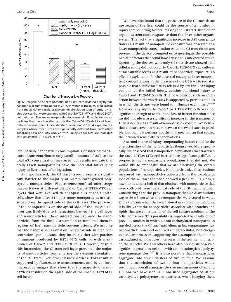
Fig. 5 Magnitude of zeta potential of 50 nm carboxylated polystyrenenanoparticles that were stored at 37 °C in water or medium, or collectedfrom the apical or basolateral/systemic circulation loop of body-on-a-chip devices that were operated with Caco-2/HT29-MTX and HepG2/C3Acell cultures. The mean magnitude decreases significantly for nano-particles that have travelled across the Caco-2/HT29-MTX cell layer.Data represent mean ± one standard deviation of 3 to 4 experiments.Samples whose mean sizes are significantly different from each otheraccording to a one-way ANOVA with Tukey's post-test are indicatedwith an asterisk (P < 0.05, n = 3–4).
Lab on a Chip Paper
Publ
ishe
d on
27
June
201
4. D
ownl
oade
d by
Cor
nell
Uni
vers
ity L
ibra
ry o
n 11
/09/
2014
21:
00:3
0.
View Article Online
level of daily nanoparticle consumption. Considering that GItract tissue contributes only small amounts of AST to thetotal AST concentration measured, our results indicate thatorally taken nanoparticles have the potential for causinginjury to liver tissue after ingestion.
As hypothesized, the GI tract tissue presents a signifi-cant barrier to the majority of 50 nm carboxylated poly-styrene nanoparticles. Fluorescence confocal microscopyimages (taken at different planes) of Caco-2/HT29-MTX celllayers that were exposed to nanoparticles at their apicalside, show that after 24 hours many nanoparticles are stillsituated on the apical side of the cell layer. The presenceof the nanoparticles on the apical side of the imaged celllayer was likely due to interactions between the cell layerand nanoparticles. These interactions captured the nano-particles from the fluidic stream and accumulated them inregions of high nanoparticle concentrations. We assumethat the nanoparticles arrest on the apical side in high con-centration spots because they interact either with patchesof mucous produced by HT29-MTX cells or with mem-branes of Caco-2 and HT29-MTX cells. However, despitethis interaction, the GI tract cell layer prevented the major-ity of nanoparticles from entering the systemic circulationof the ‘GI tract–liver–other tissues’ devices. This result issupported by fluorescence measurements and by confocalmicroscopy images that show that the majority of nano-particles resides on the apical side of the Caco-2/HT29-MTXcell layer.
This journal is © The Royal Society of Chemistry 2014
We have also found that the presence of the GI tract tissueupstream of the liver could be the source of a number ofinjury compounding factors, making the ‘GI tract–liver–otherorgans’ system more responsive than the ‘liver–other organs’system. The fact that a significant increase in AST concentra-tions as a result of nanoparticle exposure was observed at alower nanoparticle concentration when the GI tract tissue waspresent in the device prompted us to investigate the possiblenature of factors that could have caused this unexpected result.Operating the devices with only GI tract tissue showed thatcellular injury did not occur to Caco-2/HT29-MTX cell culturesat measurable levels as a result of nanoparticle exposure. Tooffer an explanation for the observed toxicity at lower nanopar-ticle concentrations in the presence of the GI tract tissue: it ispossible that soluble mediators released by low-level liver injurycompounds the initial injury, causing additional injury toCaco-2 and HT29-MTX cells. The possibility of such an inter-action between the two tissues is supported by previous studiesin which the tissues were found to influence each other.24–26
However, any injury to Caco-2 or HT29-MTX cells was notsignificant enough to result in the loss of barrier function sincewe did not observe a significant increase in the transport of10 kDa dextran as a result of nanoparticle exposure. We believethat a destructive interaction between the two tissues is possi-ble, but that it is perhaps not the only mechanism that causedthe increased sensitivity to nanoparticles.
A second source of injury compounding factors could be thecharacteristics of the nanoparticles themselves. More specifi-cally, we observed that nanoparticle populations that traversedthe Caco-2/HT29-MTX cell barrier have significantly differentproperties than nanoparticle populations that did not. Wewould like to emphasize that we are discussing the overallpopulations of nanoparticles. Nanoparticle size distributionsmeasured with nanoparticles collected from the basolateralside of the GI tract chamber, showed a peak at 55 ± 7 nm, asize that is almost half of that obtained with nanoparticles thatwere collected from the apical side of the GI tract chamber.Considering that the peak in nanoparticle size distributionswas at 39 ± 5 nm when the nanoparticles were stored in waterand 97 ± 1 nm when they were stored in cell culture medium,it is likely that the nanoparticles associate with proteins andlipids that are constituents of the cell culture medium or thecells themselves. This possibility is supported by results of ourprevious studies in which 50 nm carboxylated nanoparticlestraveled across the GI tract epithelium at low temperatures, i.e.nanoparticle transport occurred via paracellular, non-energy-dependent processes, supporting the assumption that 50 nmcarboxylated nanoparticles interact with the cell membranes ofepithelial cells. We and others have also previously observedsignificant protein association with 50 nm carboxylated polysty-rene nanoparticles.7,27 It is also possible that nanoparticlesaggregate into small clusters of two to four. We assumethat the association of two to four nanoparticles wouldresult in an overall nanoparticle size measurement of around100 nm. We have seen ~100 nm sized aggregates of 50 nmcarboxylated polystyrene nanoparticles when imaging them
Lab Chip, 2014, 14, 3081–3092 | 3089

Fig. 6 Transmission electron microscopy image of 50 nm carboxylatedpolystyrene nanoparticles that were collected from the apical side of theCaco-2/HT29 MTX co-culture. The image shows that nanoparticles inthe solution associate with each other to form larger aggregates.
Lab on a ChipPaper
Publ
ishe
d on
27
June
201
4. D
ownl
oade
d by
Cor
nell
Uni
vers
ity L
ibra
ry o
n 11
/09/
2014
21:
00:3
0.
View Article Online
with transmission electron microscopy (Fig. 6). Given theseassumptions, the data suggest that the nanoparticle populationthat arrived at the basolateral side of the Caco-2/HT29-MTX celllayer consisted of a larger fraction of single nanoparticles andsmaller nanoparticle aggregates (nominal diameter of 55 nmwith low-level protein associations) than the nanoparticlepopulation that did not cross the Caco-2/HT29-MTX celllayer. The data suggest that the Caco-2/HT29-MTX cell layerpresents a higher level barrier to the transport of 50 nmcarboxylated polystyrene nanoparticles that associate with asignificant amount of proteins or that aggregate into largerclusters of a nominal diameter of 97 nm. It is not clear, how-ever, how this fact impacts the observed increased sensitivityof the ‘GI tract–liver–other tissues’ device to nanoparticleexposure, since the original nanoparticle population maycontain a higher fraction of aggregates, but likely also containssingle nanoparticles at a concentration that is not lower thanthe single nanoparticle concentration in the filtrate. Perhapsthe constitution of the macromolecular corona around thenanoparticles plays a role in changing the toxic potential ofsingle nanoparticles.
Nanoparticles collected from the basolateral side of theCaco-2/HT29-MTX cell layer also showed a decreased magni-tude of their zeta potential compared to those that werestored in cell culture medium. This decrease in magnitude isprobably due to association with and masking of the chargeby ions, proteins and lipids that occurs during the 24 hoursof exposure to cell cultures. This assumption is supported bythe fact that the magnitude of zeta potential is significantlyhigher when the nanoparticles are stored in water. Nano-particles that crossed the GI tract barrier via the paracellularroute likely experience the greatest exposure to lipids. Thefact that the trend of decrease in zeta potential magnitude asa result of exposure to cell cultures becomes significant whenthe nanoparticles have crossed the GI tract cell layer indicatesthat nanoparticles that were in close contact with cells and
3090 | Lab Chip, 2014, 14, 3081–3092
cellular membranes associate with more ions, proteins, orlipids than those that were not. As mentioned above, perhapsthe macromolecular corona of these nanoparticles differs inits constitution from that of nanoparticles that were not inclose contact with cell cultures. However, a detailed analysisof the macromolecules that are associated with the nano-particles would be necessary to make more conclusivestatements.
These changed nanoparticle properties, namely the levelof aggregation with other nanoparticles or macromolecules,and the magnitude of zeta potential of nanoparticles thattraversed the GI tract barrier could reflect an increased toxicpotential of the nanoparticles that reached the liver tissue inthe device configuration in which both the GI tract and theliver tissues were present.
Since the viability data measured here do not suggest asignificant decrease in cell viability as a result of exposure to50 nm carboxylated polystyrene nanoparticles at the testedconcentrations, it is possible that the injury that occurred asa result of nanoparticle exposure is either too small to bedetected with viability stains, or that the injury is of transientand sublethal nature. AST normally resides in the cytoplasmand the mitochondria of cells and is released into the culturemedium when cells undergo cell lysis or membrane damage.28
The enzyme is also released when membrane damage is oftransient nature. We have previously shown that 50 nm carbox-ylated polystyrene nanoparticles at the same dose affect ironuptake through Caco-2/HT29-MTX cell layers and cause adecrease in transepithelial resistance (TER).7 An interactionbetween nanoparticles and phospholipid bilayers has alsobeen suggested by Wang et al. who showed that negativelycharged nanoparticles induce local gelation in otherwise fluidmembranes.6 These findings support our data, which suggesttransient or low-level membrane damage as a result of nano-particle exposure at the concentrations used here.
In addition to the advantage of being a more sensitivequantitative measure for cellular damage than cell viabilitydyes, measurement of enzyme concentrations in body-on-a-chip in vitro models can be more directly correlated to tissueinjury in future in vivo studies of nanotoxicity than cell viabil-ity measurements, which are restricted to in vitro use. AST isa recognized plasma biomarker of liver injury in animals andhumans and is thus suitable for such measurements.15,28
The percentage of transported 50 nm carboxylated poly-styrene nanoparticles measured here with the body-on-a-chipdevice is slightly higher than that reported earlier from exper-iments in static culture (4.55%).7 This difference may be dueto the fact that the cells within microfluidic body-on-a-chipdevices are cultured under shear stress, which has beenshown to affect cell morphology and function in other celltypes.29–31
System design and operation
To reflect the multi-cell type composition of GI tract tissue,we utilized a co-culture of Caco-2 cells and mucous
This journal is © The Royal Society of Chemistry 2014

Lab on a Chip Paper
Publ
ishe
d on
27
June
201
4. D
ownl
oade
d by
Cor
nell
Uni
vers
ity L
ibra
ry o
n 11
/09/
2014
21:
00:3
0.
View Article Online
producing HT29-MTX cells. In a previous study we conductedwith seeding ratios of 10 : 1, 5 : 5, 7.5 : 2.5 and 9 : 1 we foundthat, when evaluated after 16 days of cell culture, seedingratios of 7.5 : 2.5 and 9 : 1 resulted inmucous-covered cell layersthat simulated the uptake of iron with physiologic relevance.32
Here we selected a seeding ratio of 9 : 1 to conduct experiments.Since HT/29-MTX cell overgrowth can lead to a decrease inbarrier function, we selected only those cultures for experi-ments that exhibited a transepithelial resistance of 200 Ohm× cm2 or higher. This practice ensured that nanoparticletransport did not occur through large gaps in the cell layercaused by HT/29-MTX cell overgrowth.
We designed the body-on-a-chip devices so that alltissues of the human body are represented in the devices.Representing the total volume of fluid in the human bodyusing a body-on-a-chip system requires a reservoir for fluid oforgans (we call this the ‘other tissue’ reservoir) that are notexplicitly expressed. This fluid represents the blood and inter-stitial fluid volume in the body. The use of an ‘other tissue’chamber provides a crude mimic of fluid volume, which willdilute the concentration of any excreted metabolites or nano-particles to a value representative of that in the circulation ofthe human body. An important assumption in such a modelis that none of the metabolites and nanoparticles are seques-tered or modified chemically in any tissues other than the GIor liver compartments. Clearly, this system is idealized, butwe believe it is a useful model.
A more sophisticated model would break the ‘other tissues’compartments into various organ compartments. The chipwe have fabricated contains compartments for kidney, adi-pose tissue, and bone marrow. For this initial study we havenot populated these compartments with tissue constructs.The details of nanoparticle distributions in this system withthese empty compartments plus an ‘other tissue’ compart-ment (now reduced in size by the volume of the fluidretained in the kidney, adipose, and bone marrow compart-ments) is not significantly different than a system with achip without these empty compartments. To eliminate smallchanges to the fluid flow distribution that occurs as a resultof leaving organ chambers empty, it is also possible to add anon-absorptive hydrogel to the fraction of these chambersthat would otherwise have been occupied by the solid portionof the organ analogs. This strategy allows us to utilize body-on-a-chip systems for many different studies.
In our devices we used monolayer cell cultures, which areeasily observed. However, the use of 3D tissues in the futurewill make our simulations more realistic. In particular, suchtissues may allow for more authentic cellular behavior, as wellas more physiologic liquid to cell ratios than those we wereable to achieve with the current device. Such 3D constructscould consist of cells that were entrapped in hydrogels,cultured in a polymeric matrix, or grown as organoids.
Since nanoparticle uptake and transport in the humanbody has been of interest to the research community, therehave been several techniques and systems that were usedfor the study or oral nanoparticle uptake.33 Some of these
This journal is © The Royal Society of Chemistry 2014
systems allow for the study of nanoparticle uptake underconditions of peristalsis,34 and in the presence of gastricfluids.35,36
Conclusions
Simulations with ‘GI tract–liver–other tissues’ body-on-a-chip devices suggest that ingested 50 nm carboxylated poly-styrene nanoparticles cause sublethal cellular injury toin vitro liver tissue. The injury occurred at concentrations of240–480 × 1011 nanoparticles mL−1, a high concentrationestimated in terms of possible daily consumption. Although,the GI tract tissue presents a significant barrier to nano-particles, our results suggest that the presence of GI tracttissue upstream of the liver adds a number of injurycompounding factors, making the device more sensitivewhen evaluating nanoparticle-induced injury. It is possiblethat the higher sensitivity comes from soluble mediators thatwere released because of low-level cellular injury of liver cells,compounding the initial injury by causing additional lowlevel damage to Caco-2 and HT29-MTX cells. It is alsopossible that the changed properties of nanoparticles thatcrossed the Caco-2/HT29-MTX cell layer could reflect anincreased toxic potential. The level of aggregation with othernanoparticles was significantly reduced in nanoparticlepopulations that traversed the GI tract barrier. These nano-particles also exhibited a decreased zeta potential comparedto those that were stored in cell culture medium, a findingthat is probably due to the combined effects of a more basicpH in cell cultures as well as associations with macromole-cules that has occurred as a result of close contact of thenanoparticles with Caco-2/HT29-MTX cell cultures. The two-organ systems allowed us to observe compounding effects oftissue-tissue interactions between the GI tract and the liverthat caused a higher level of liver injury than was expectedfrom experiments with liver tissue alone. Our experimentssuggest that multi-organ in vitro devices are useful andimportant tools for assessing toxicities of environmental toxi-cants, drugs, and engineered nanoparticles.
Acknowledgements
Financial support for this work was provided by the NationalInstitutes of Health/National Center for Advancing Transla-tional Sciences (grant no. is UH2 TR000516-01), the NationalScience Foundation under grant no. CBET-1106153, the ArmyCorps of Engineers under Agreement ID W9132T-07-2-0010.This work was performed in part at the Cornell NanoScaleFacility, a member of the National Nanotechnology Infra-structure Network, which is supported by the National ScienceFoundation (grant ECS-0335765). The HT29-MTX cell line waskindly contributed by Dr. Thecla Lesuffleur of INSERM U560 inLille, France. We would also like to acknowledge John Grazulof the CCMR of Cornell University for imaging nanoparticlesamples using transmission electron microscopy (NSF grantno. DMR 1120296).
Lab Chip, 2014, 14, 3081–3092 | 3091
Lab on a ChipPaper
Publ
ishe
d on
27
June
201
4. D
ownl
oade
d by
Cor
nell
Uni
vers
ity L
ibra
ry o
n 11
/09/
2014
21:
00:3
0.
View Article Online
References
1 S. Kaida, H. Cabral, M. Kumagai, A. Kishimura, Y. Terada,
M. Sekino, I. Aoki, N. Nishiyama, T. Tani and K. Kataoka,Cancer Res., 2010, 70, 7031–7041.2 N. W. S. Kam and H. Dai, Phys. Status Solidi B, 2006, 243,
3561–3566.3 R. G. Ellis-Behnke, Y.-X. Liang, D. K. C. Tay, P. W. F. Kau,
G. E. Schneider, S. Zhang, W. Wu and K.-F. So, Nanomed.:Nanotechnol., Biol. Med., 2006, 2, 207–215.4 A. Schroeder, D. A. Heller, M. M. Winslow, J. E. Dahlman,
G. W. Pratt, R. Langer, T. Jacks and D. G. Anderson, Nat.Rev. Cancer, 2011, 12, 39–50.5 E. Lavik and J. Ustin, Science, 2012, 337, 658–659.
6 B. Wang, L. Zhang, S. C. Bae and S. Granick, Proc. Natl.Acad. Sci. U. S. A., 2008, 105, 18171–18175.7 G. J. Mahler, M. B. Esch, E. Tako, T. L. Southard,
S. D. Archer, R. P. Glahn and M. L. Shuler, Nat.Nanotechnol., 2012, 7, 264–271.
8 M. A. Dobrovolskaia, D. R. Germolec and J. L. Weaver, Nat.
Nanotechnol., 2009, 4, 411–414.9 R. F. Service, Science, 2004, 304, 1732–1734.
10 C. Durrer, J. M. Irache, F. Puisieux, D. Duchêne andG. Ponchel, Pharm. Res., 1994, 11, 674–679.11 C. Durrer, J. M. Irache, F. Puisieux, D. Duchêne and
G. Ponchel, Pharm. Res., 1994, 11, 680–683.12 A. Hillery, P. Jani and A. Florence, J. Drug Targeting, 1994, 2,
151–156.13 G. J. Russell-Jones, Adv. Drug Delivery Rev., 2001, 46, 59–73.
14 M. Shakweh, G. Ponchel and E. Fattal, Expert Opin. DrugDelivery, 2004, 1, 141–163.15 N. Enomoto, S. Yamashina, H. Kono, P. Schemmer, C. A. Rivera,
A. Enomoto, T. Nishiura, T. Nishimura, D. A. Brenner andR. G. Thurman, Hepatology, 1999, 29, 1680–1689.
16 M. Baker, Nature, 2011, 471, 661–665.
17 P. Neuži, S. Giselbrecht, K. Länge, T. J. Huang and A. Manz,Nat. Rev. Drug Discovery, 2012, 11, 620–632.
3092 | Lab Chip, 2014, 14, 3081–3092
18 R. Khamsi, Nature, 2005, 435, 12–13.
19 M. B. Esch, T. L. King and M. L. Shuler, Annu. Rev. Biomed.Eng., 2011, 13, 55–72.20 J. H. Sung, M. B. Esch and M. L. Shuler, Expert Opin. Drug
Metab. Toxicol., 2010, 6, 1063–1081.21 G. J. Mahler, M. B. Esch, R. P. Glahn and M. L. Shuler,
Biotechnol. Bioeng., 2009, 104, 193–205.22 P. S. Price, R. B. Conolly, C. F. Chaisson, E. A. Gross,
J. S. Young, E. T. Mathis and D. R. Tedder, Crit. Rev.Toxicol., 2003, 33, 469–503.
23 B. Davies and T. Morris, Pharm. Res., 1993, 10, 1093–1095.
24 S. Hyung Choi, M. Nishikawa, A. Sakoda and Y. Sakai,Toxicol. In Vitro, 2004, 18, 393–402.25 P. M. van Midwoud, M. T. Merema, E. Verpoorte and
G. M. M. Groothuis, Lab Chip, 2010, 10, 2778–2786.26 S. H. Choi, O. Fukuda, A. Sakoda and Y. Sakai, Mater. Sci.
Eng., C, 2004, 24, 333–339.27 M. Lundqvist, J. Stigler, G. Elia, I. Lynch, T. Cedervall and
K. A. Dawson, Proc. Natl. Acad. Sci. U. S. A., 2008, 105,14265–14270.
28 S. K. Ramaiah, Food Chem. Toxicol., 2007, 45, 1551–1557.
29 E. Tzima, EMBO J., 2001, 20, 4639–4647. 30 M. B. Esch, D. J. Post, M. L. Shuler and T. Stokol, TissueEng., Part A, 2011, 17, 2965–2971.31 P. Fernandez, C. Bourget, R. Bareille, R. Daculsi and
L. Bordenave, Tissue Eng., 2007, 13, 1607–1614.32 G. J. Mahler, M. L. Shuler and R. P. Glahn, J. Nutr. Biochem.,
2009, 20, 494–502.33 J. M. Gamboa and K. W. Leong, Adv. Drug Delivery Rev.,
2013, 65, 800–810.34 H. J. Kim, D. Huh, G. Hamilton and D. E. Ingber, Lab Chip,
2012, 12, 2165–2174.35 F. Ingels, S. Deferme, E. Destexhe, M. Oth, G. Van den
Mooter and P. Augustijns, Int. J. Pharm., 2002, 232,183–192.
36 N. Patel, B. Forbes, S. Eskola and J. Murray, Drug Dev. Ind.
Pharm., 2006, 32, 151–161.This journal is © The Royal Society of Chemistry 2014


