L-sepiapterin restores SLE serum-induced ... - lupus.bmj.com · ones Buien etflal Lupus Science...
11
1 Jones Buie JN, et al. Lupus Science & Medicine 2019;0:e000294. doi:10.1136/lupus-2018-000294 L-sepiapterin restores SLE serum- induced markers of endothelial function in endothelial cells Joy N Jones Buie, 1,2 Dorea Pleasant Jenkins, 3 Robin Muise-Helmericks, 3 Jim C Oates 2,4 To cite: Jones Buie JN,Pleasant Jenkins D, Muise-Helmericks R, et al. L-sepiapterin restores SLE serum-induced markers of endothelial function in endothelial cells. Lupus Science & Medicine 2019;0:e000294. doi:10.1136/ lupus-2018-000294 Received 13 July 2018 Revised 18 October 2018 Accepted 14 November 2018 1 Department of Neurology, Medical University of South Carolina, Charleston, South Carolina, USA 2 Department of Medicine, Division of Rheumatology, Medical University of South Carolina, Charleston, South Carolina, USA 3 Department of Regenerative Medicine and Cell Biology, Medical University of South Carolina, Charleston, South Carolina, USA 4 Medical Service, Rheumatology Section, Ralph H. Johnson VA Medical Center, Charleston, South Carolina, USA Correspondence to Dr Jim C Oates; oatesjc@musc. edu Immunology and inflammation © Author(s) (or their employer(s)) 2019. Re-use permitted under CC BY-NC. No commercial re-use. See rights and permissions. Published by BMJ. ABSTRACT Objective SLE serves as an independent risk factor` for endothelial dysfunction (ED) not explained by Framingham risk factors. We sought to understand the development of SLE-induced ED on a cellular level in order to develop strategies aimed at reversing cellular abnormalities. This study assessed the impact of SLE patient serum on endothelial nitric oxide synthase (eNOS), nitric oxide (NO) production and functional changes in the cell. Methods Human umbilical vein endothelial cells (HUVECs) cultured in serum of either SLE (n=25) or healthy patients (n=14) or endothelial basal medium 2 (EBM-2) culture media supplemented with fetal bovine serum with or without L-sepiapterin were used for our studies. We applied the fluorescent probe DAF-FM diacetate for intracellular NO detection using flow cytometry. Total RNA isolates were analysed using reverse transcription PCR for eNOS mRNA expression. Oxygen consumption rate was determined using seahorse analysis. Neutrophil adhesion and migration were determined using a calcein AM microscopy assay. Results The mRNA expression of eNOS was increased in SLE cultured HUVECs compared with healthy control (p<0.05). The SLE eNOS mRNA level correlated with SLE patient age (p=0.008); however, this trend was not observed with healthy patients. SLE serum reduced NO production in HUVECs compared with EBM-2 cultured cells (p<0.05). Co-treatment of endothelial cells with L-sepiapterin preserved HUVEC capacity to produce NO in SLE conditions (p<0.01). SLE serum enhanced neutrophil migration (p<0.01) but not neutrophil adhesion compared with healthy controls. The bioenergetic health index was not different. Conclusions SLE likely causes disruption of endothelial cell eNOS function and NO modulated pathways. INTRODUCTION SLE is a heterogeneous chronic autoim- mune inflammatory syndrome that predom- inately impacts women of childbearing age. Although specific clinical and immunological criteria have been defined, its clinical course is highly variable with some patients experi- encing life-threatening cardiovascular disease complications which account for one-third of deaths in patients with SLE. 1 2 A significant proportion of patients with SLE display accelerated endothelial dysfunction which precedes cardiovascular disease (CVD). The endothelium plays a pivotal role in governing vascular function and, thus, prevents the development of vascular abnormalities. 3–5 Chronic inflammation promotes endothelial cell injury leading to generation of superoxide and expression of cell adhesion markers. 6 7 These perturbations in endothelial cell func- tion promote oedema, leucocyte trafficking and organ damage. While causes of endothe- lial dysfunction in SLE are multifactorial, the specific biological underpinnings governing the development of endothelial dysfunction in SLE are incompletely understood and represent an important area of research. Nitric oxide (NO) is a membrane-perme- able free radical, continuously synthesised by the endothelial nitric oxide synthase (eNOS) dimeric enzyme. 8 Diffusion of NO across the cellular membrane as a paracrine mediator has impacts on cellular function critical for blood vessel dilation and unobstructed blood flow. 9–11 Diminished release of NO and loss of eNOS expression have been consistently linked to endothelial dysfunction. 12 13 Para- doxically, uncoupled eNOS, likely resulting from oxidation of tetrahydrobiopterin (BH 4 ), leads to eNOS enzymatic dysfunction. 14–16 Biopsies from patients with severe lupus nephritis show diminished eNOS expres- sion. 17 18 Accordingly, our previous work demonstrated that genetic ablation of eNOS in lupus-prone MRL/lpr mice resulted in accelerated, more severe disease with signif- icant declines in survival. 19 MRL/lpr mice lacking eNOS display increased superoxide production associated with increased MCP1 production, increased glomerular crescentic and necrotic lesions, and reduced levels of the anti-inflammatory cytokine interleukin (IL)-10. 20 Accordingly, a positive correla- tion between H 2 O 2 and eNOS levels in vitro AUTHOR PROOF on 2 May 2019 by guest. Protected by copyright. http://lupus.bmj.com/ Lupus Sci Med: first published as 10.1136/lupus-2018-000294 on 19 February 2019. Downloaded from
Transcript of L-sepiapterin restores SLE serum-induced ... - lupus.bmj.com · ones Buien etflal Lupus Science...

1Jones Buie JN, et al. Lupus Science & Medicine 2019;0:e000294. doi:10.1136/lupus-2018-000294
L-sepiapterin restores SLE serum-induced markers of endothelial function in endothelial cells
Joy N Jones Buie,1,2 Dorea Pleasant Jenkins,3 Robin Muise-Helmericks,3 Jim C Oates2,4
To cite: Jones Buie JN, Pleasant Jenkins D, Muise-Helmericks R, et al. L-sepiapterin restores SLE serum-induced markers of endothelial function in endothelial cells. Lupus Science & Medicine 2019;0:e000294. doi:10.1136/lupus-2018-000294
Received 13 July 2018Revised 18 October 2018Accepted 14 November 2018
1Department of Neurology, Medical University of South Carolina, Charleston, South Carolina, USA2Department of Medicine, Division of Rheumatology, Medical University of South Carolina, Charleston, South Carolina, USA3Department of Regenerative Medicine and Cell Biology, Medical University of South Carolina, Charleston, South Carolina, USA4Medical Service, Rheumatology Section, Ralph H. Johnson VA Medical Center, Charleston, South Carolina, USA
Correspondence toDr Jim C Oates; oatesjc@ musc. edu
Immunology and inflammation
© Author(s) (or their employer(s)) 2019. Re-use permitted under CC BY-NC. No commercial re-use. See rights and permissions. Published by BMJ.
AbstrActObjective SLE serves as an independent risk factor` for endothelial dysfunction (ED) not explained by Framingham risk factors. We sought to understand the development of SLE-induced ED on a cellular level in order to develop strategies aimed at reversing cellular abnormalities. This study assessed the impact of SLE patient serum on endothelial nitric oxide synthase (eNOS), nitric oxide (NO) production and functional changes in the cell.Methods Human umbilical vein endothelial cells (HUVECs) cultured in serum of either SLE (n=25) or healthy patients (n=14) or endothelial basal medium 2 (EBM-2) culture media supplemented with fetal bovine serum with or without L-sepiapterin were used for our studies. We applied the fluorescent probe DAF-FM diacetate for intracellular NO detection using flow cytometry. Total RNA isolates were analysed using reverse transcription PCR for eNOS mRNA expression. Oxygen consumption rate was determined using seahorse analysis. Neutrophil adhesion and migration were determined using a calcein AM microscopy assay.Results The mRNA expression of eNOS was increased in SLE cultured HUVECs compared with healthy control (p<0.05). The SLE eNOS mRNA level correlated with SLE patient age (p=0.008); however, this trend was not observed with healthy patients. SLE serum reduced NO production in HUVECs compared with EBM-2 cultured cells (p<0.05). Co-treatment of endothelial cells with L-sepiapterin preserved HUVEC capacity to produce NO in SLE conditions (p<0.01). SLE serum enhanced neutrophil migration (p<0.01) but not neutrophil adhesion compared with healthy controls. The bioenergetic health index was not different.Conclusions SLE likely causes disruption of endothelial cell eNOS function and NO modulated pathways.
IntROduCtIOnSLE is a heterogeneous chronic autoim-mune inflammatory syndrome that predom-inately impacts women of childbearing age. Although specific clinical and immunological criteria have been defined, its clinical course is highly variable with some patients experi-encing life-threatening cardiovascular disease complications which account for one-third of deaths in patients with SLE.1 2 A significant
proportion of patients with SLE display accelerated endothelial dysfunction which precedes cardiovascular disease (CVD). The endothelium plays a pivotal role in governing vascular function and, thus, prevents the development of vascular abnormalities.3–5 Chronic inflammation promotes endothelial cell injury leading to generation of superoxide and expression of cell adhesion markers.6 7 These perturbations in endothelial cell func-tion promote oedema, leucocyte trafficking and organ damage. While causes of endothe-lial dysfunction in SLE are multifactorial, the specific biological underpinnings governing the development of endothelial dysfunction in SLE are incompletely understood and represent an important area of research.
Nitric oxide (NO) is a membrane-perme-able free radical, continuously synthesised by the endothelial nitric oxide synthase (eNOS) dimeric enzyme.8 Diffusion of NO across the cellular membrane as a paracrine mediator has impacts on cellular function critical for blood vessel dilation and unobstructed blood flow.9–11 Diminished release of NO and loss of eNOS expression have been consistently linked to endothelial dysfunction.12 13 Para-doxically, uncoupled eNOS, likely resulting from oxidation of tetrahydrobiopterin (BH4), leads to eNOS enzymatic dysfunction.14–16 Biopsies from patients with severe lupus nephritis show diminished eNOS expres-sion.17 18 Accordingly, our previous work demonstrated that genetic ablation of eNOS in lupus-prone MRL/lpr mice resulted in accelerated, more severe disease with signif-icant declines in survival.19 MRL/lpr mice lacking eNOS display increased superoxide production associated with increased MCP1 production, increased glomerular crescentic and necrotic lesions, and reduced levels of the anti-inflammatory cytokine interleukin (IL)-10.20 Accordingly, a positive correla-tion between H2O2 and eNOS levels in vitro
AU
THO
R P
RO
OF
on 2 May 2019 by guest. P
rotected by copyright.http://lupus.bm
j.com/
Lupus Sci M
ed: first published as 10.1136/lupus-2018-000294 on 19 February 2019. D
ownloaded from

Jones Buie JN, et al. Lupus Science & Medicine 2019;0:e000294. doi:10.1136/lupus-2018-0002942
Lupus Science & Medicine
exists.21 NO produced in the presence of O2.- yields
peroxynitrite (ONOO-) which oxidises the essential eNOS co-factor BH4 to dihydrobiopterin (BH2) and biop-terin. This oxidation of BH4 leads to uncoupling of the eNOS homodimer to monomers, resulting in reactive oxygen species (ROS) rather than NO production.22 23 ROS production itself leads to endothelial dysfunction. L-sepiapterin (L-sep) is a precursor for tetrahydrobiop-terin (BH4) synthesis, and previous studies have shown its efficacy in restoring eNOS function, possibly through a recoupling mechanism.24 Thus, L-sep may serve as a viable therapeutic option when eNOS uncoupling is the predominant mechanism of endothelial dysfunction in an inflammatory microenvironment, such is present in patients with SLE.
A hallmark of endothelial inflammatory responses which coincide with diminished endothelial-NO is increased neutrophil migration and adhesion to the endothelial cell surface.25 26 Neutrophil migration and adhesion are processes that promote progression of atherosclerosis through the release of myeloperoxidase, promoting ROS production, and sequestering monocytes to the intravascular space.27 Recent studies link NETosis, a process of neutrophil death emerging as a potential pathogenic process in SLE, to atherosclerosis.28 In the present study, we used human umbilical vein endothe-lial cells (HUVECs) to study the impact of SLE serum on eNOS expression and NO production in vitro. We hypothesised that SLE serum would negatively impact NO production in HUVECs, leading to enhanced leuco-cyte adhesion and migration.
PatIents and MethOdsPatient populationSpecimens for this study were stored and collected from study visits that were part of a longitudinal observational cohort study known as the SLE Gullah Health or SLEIGH study initiated in 2002 by Dr. Diane Kamen and has been previously described.29 All patients classified as having SLE met 4 of the 11 classification criteria as specified by the 1997 American College of Rheumatology criteria.30
Clinical characteristics of the study populationPatients with SLE were evaluated during regular clinical visits by clinicians trained in SLE disease activity measures. The clinical and laboratory elements of the SLE Disease Activity Index (SLEDAI) were recorded if they were attrib-uted to SLE disease activity. The scores were recorded for each blood collection visit. Control volunteers were eval-uated for autoimmune disease and the presence of cardi-ovascular disease or risk factors (hypertension, smoking, hypercholesterolemia or previous myocardial infarction, cardiac or brain revascularisation, or stroke).
Blood collectionBlood from healthy and participants with SLE were collected in a sterile vacutainer blood collection tube and whole blood was allowed to clot at 25°C for 10
min. Samples were centrifuged to remove the clot and remaining serum was stored in aliquots at −80°C for future use.
neutrophil isolationNeutrophils were isolated as previously described.31 Briefly, 20 mL of human blood was acquired from healthy volunteers and cells were isolated using Lymphocyte Separation Medium (Cellgro, Manassas, Virginia, USA). The assay was validated based on forward scatter and side scatter using flow cytometry (data not shown).
endothelial cell culture and serum culturingPrimary human umbilical vein endothelial cells from pooled donors were purchased from Lonza (Walkersville, Maryland, USA) and cultured according to the manufac-turer’s instructions in 5% CO2 at 37°C in humidified air. Cells were cultured in endothelial cell basal medium-2 (EBM-2) supplemented with EBM-2 SingleQuot (Lonza), pH 7.6–8.0. Media was changed every other day until cells were 70%–80% confluent. HUVECs were subcultured using TryPLE Express, pH 8.0 (ThermoFisher Scientific, Waltham, Massachusetts, USA). Cell growth was limited to 12 population doublings, and all experiments were carried out using cells between passages 3–5. For serum experiments, cells were cultured with 20%–50% serum for 6 or 24 hours prior to further analysis.
Real-time reverse transcription-PCR (Rt2PCR)To detect changes in mRNA levels, HUVECs were treated with 20% SLE or healthy sera for 6 or 24 hours as spec-ified. Cells were harvested following treatment and total RNA was extracted using a Trizol (ThermoFisher Scien-tific)-RNeasy kit (Qiagen, Frederick, Maryland, USA) hybrid protocol as previously described.32 RNA integrity was assessed using a NanoDrop 2000c UV-Vis spectropho-tometer (ThermoScientific, Wilmington, Delaware, USA) and samples with A260/280 ratios of 1.8–2.1 were used. Single strand cDNA was synthesised from 1 µg of RNA using an iScript cDNA synthesis kit (Bio-Rad, Hercules, California, USA). For each reaction, 1 µL of cDNA product was used for signal amplification with SsoAdvanced universal SYBR (BioRad). A CFX96 Real Time PCR Detection System (Bio-Rad) was used to assess changes in NOS3 and GAPDH, using commercially available primers (Qiagen). The relative expression was calculated using the equation 2-ΔΔCt (Δ; experimental gene cycle threshold (Ct) – house-keeping gene (Ct)). The fold change gene expression of interest was calculated based on normalisation to GAPDH . PCR was performed ≥3 independent experiments with at least three replicates.
Measurement of nitric oxide productionFor real-time detection of NO production in HUVECs, 1.2×10⁵ cells were seeded in a 12-well tissue culture plate. Following adherence, cells were serum starved for 6 hours in endothelial basal media (EBM) containing 0.2% fetal bovine serum (FBS). Cells were stimulated with either 50% healthy or SLE sera ± L-sep (5 µM; 6 hours), the
AU
THO
R P
RO
OF
on 2 May 2019 by guest. P
rotected by copyright.http://lupus.bm
j.com/
Lupus Sci M
ed: first published as 10.1136/lupus-2018-000294 on 19 February 2019. D
ownloaded from

Jones Buie JN, et al. Lupus Science & Medicine 2019;0:e000294. doi:10.1136/lupus-2018-000294 3
Immunology and inflammation
eNOS-specific inhibitor, Nω-Nitro-L-arginine (L-NNA, 10 µM; 30 min pre-incubation (Tocris; Bristol, UK)) or the NO donor 3,3ʹ-diamino-4ʹ-methoxyflavone (DD1, 10 µM, Tocris). Following stimulation, cells were washed twice with phosphate buffered saline (PBS) and loaded with 1 µM DAF-FM diacetate (4-amino-5-methylamino-2’,7’-dif-luorofluorescein diacetate, 1 µM) (ThermoFisher Scien-tific) in phenol red-free EBM for 30–45 min. Cells were washed twice with PBS and dissociated from plates using phenol-red free TryPLE Express (ThermoFisher Scien-tific) and fixed with 2% paraformaldehyde for 3 min. A population of 2000–10 000 cells were gated to remove doublets and controls and analysed based on their fluo-rescence intensities using a FACS Calibur flow cytometer (Becton Dickenson, San Diego, USA). The mean fluo-rescence intensity (MFI) was normalised to respective populations in unstimulated cells. In order to discrimi-nate between NO and other gaseous molecules previously shown to augment DAF-FM fluorescence, we performed a urate assay to optimise our assay (data not shown).
Oxygen consumptionEndothelial cells were seeded at 20 000 cells/well on a Seahorse 96-well XF Cell Culture Microplate as detailed by the manufacturer (Seashore Bioscience/Agilent Tech-nologies, Santa Clara, California, USA) and allowed to adhere overnight in complete EBM-2 (EBM-2 basal media plus EBM-2 SingleQuots, Lonza, Basel, Switzerland). The following day cells were rinsed with 1× PBS and 50% control or SLE patient serum was added to wells and allowed to incubate for 24 hours (six samples per group with five replicates per patient sample). The Seahorse XF Analyzer (Seashore Bioscience/Agilent Technologies) was used to determine basal oxygen consumption rate (OCR). Four basal rate measurements were followed by four measurement cycles following each injection (1 µM oligomycin, 1 µM carbonyl cyanide 4-(trifluoromethoxy) phenylhydrazone and 2 µM AA rotenone). Consump-tion rates were calculated as previously described.33 The bioenergetic health index (BHI) was calculated using the following formula: BHI= (ATP-linked × Reserve Capcity)/(Proton Leak × Non-mitochondrial OCR).34
neutrophil adhesion assayHUVECs were plated at 5.0×10⁴ cells/mL in a 24-well plate (Costar) and allowed to adhere overnight. HUVECs were serum starved for 3 hours in phenol-red free 0.2% FBS EBM media (Lonza) prior to activation with 10% sera for 4 hours. Tumour necrosis factor-α (100 ng/mL) was used as the positive control. Neutrophils isolated from healthy human blood as outlined previously were labelled with Calcein AM (Life Technologies) at 5×10⁵ cells/mL. Neutrophils were washed gently four times in warm serum-free EBM culture media prior to co-culturing with HUVECs for 60 min after which non-adherent cells were removed by repeated gentle washing (four times) with EBM culture media. Fluorescence intensity was measured at 520 nM with a FLUOStar Omega microplate reader
(Cary, North Carolina, USA), and images were captured using confocal microscopy. Data are reported as ratios of the number of neutrophils to the number of endothelial cells as averages from three different visual fields.
neutrophil migration assayTranswell migration assays were performed as described elsewhere.35 Briefly, transwell inserts (3 µm pore) were pre-coated with fibrinogen and allowed to incubate over-night for 24 hours. HUVECs were seeded in 24-well plates at 1.0×10⁴ cells per well and allowed to adhere overnight. Cells were activated with 50% sera from healthy and SLE controls for 6 hours and washed once in PBS. Neutrophils were added to chambers and inserted into media ± IL-8 (1.25 nM, Cell Signaling). After 60 min, the number of neutrophils in the lower chambers was visualised using 4× magnification and quantified using confocal microscopy. All values were normalised to untreated controls.
statistical analysis and data handlingDescriptive statistics are reported as mean±SD or IQR for continuous variables. Gaussian distribution was deter-mined using the D’Agostino-Pearson omnibus normality test and the Shapiro-Wilk normality test. Paired and unpaired (where appropriate) two-tailed Student’s t-test and non-parametric Mann-Whitney test were used on non-parametric data analysis on lupus and controls. Corre-lations were determined using Pearson’s or Spearman’s correlation analysis and are reported accordingly. Stand-ardised univariate regression analysis was performed to adjust for lupus-associated indicators of disease activity and β-coefficients and p values were reported. Analysis of variance test with Tukey’ Fisher’s probable least signifi-cance post-test was used to analyse NO and neutrophil adhesion data. No mathematical correction was made for multiple comparisons. Data are presented as mean±SEM. Differences were considered significant if the p value was ≤0.05. Statistical analysis was performed using IBM SPSS Software V.25 or GraphPad Prism V.6.0f (San Diego, Cali-fornia, USA).
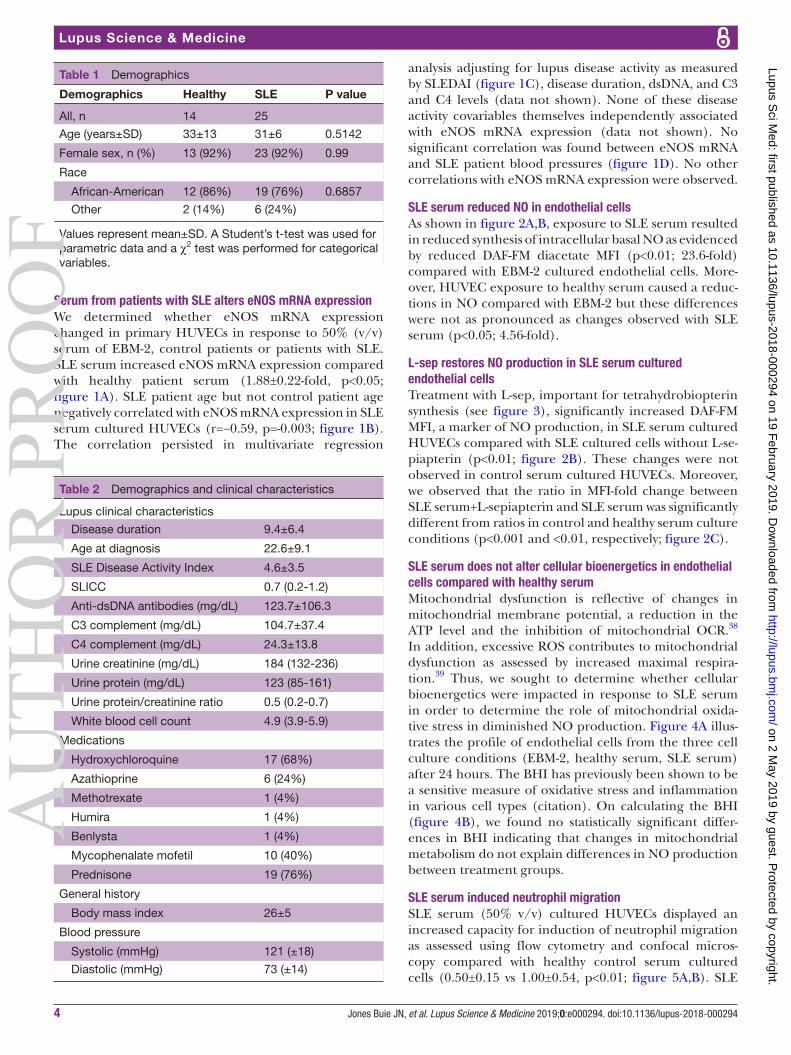
Resultsdemographic and clinical characteristics of study participantsSLE (n=25) and healthy controls (n=14) did not differ in sex, race or age. Mean±SD disease duration for patients with SLE was 9.4±6.4 and the age at diagnosis was 22.6±9.1. SLEDAI36 37 was 4.6±3.5, reflecting overall mild lupus disease activity. Complement C3 levels were 104.7±37.4 while C4 levels were 24.3±13.8 further supporting the notion that patients with lupus in this study had mild disease activity. Patients with SLE had normal blood pres-sures and an overall body mass index of 26±5, indicating a group of participants that were slightly overweight. Most patients with SLE were taking prednisone and/or antima-larial medications (76%, 68%, respectively) at the time of the study visit (tables 1 and 2).
AU
THO
R P
RO
OF
on 2 May 2019 by guest. P
rotected by copyright.http://lupus.bm
j.com/
Lupus Sci M
ed: first published as 10.1136/lupus-2018-000294 on 19 February 2019. D
ownloaded from
Jones Buie JN, et al. Lupus Science & Medicine 2019;0:e000294. doi:10.1136/lupus-2018-0002944
Lupus Science & Medicine
Table 1 Demographics
Demographics Healthy SLE P value
All, n 14 25
Age (years±SD) 33±13 31±6 0.5142
Female sex, n (%) 13 (92%) 23 (92%) 0.99
Race
African-American 12 (86%) 19 (76%) 0.6857
Other 2 (14%) 6 (24%)
Values represent mean±SD. A Student’s t-test was used for parametric data and a χ2 test was performed for categorical variables.
Table 2 Demographics and clinical characteristics
Lupus clinical characteristics
Disease duration 9.4±6.4
Age at diagnosis 22.6±9.1
SLE Disease Activity Index 4.6±3.5
SLICC 0.7 (0.2-1.2)
Anti-dsDNA antibodies (mg/dL) 123.7±106.3
C3 complement (mg/dL) 104.7±37.4
C4 complement (mg/dL) 24.3±13.8
Urine creatinine (mg/dL) 184 (132-236)
Urine protein (mg/dL) 123 (85-161)
Urine protein/creatinine ratio 0.5 (0.2-0.7)
White blood cell count 4.9 (3.9-5.9)
Medications
Hydroxychloroquine 17 (68%)
Azathioprine 6 (24%)
Methotrexate 1 (4%)
Humira 1 (4%)
Benlysta 1 (4%)
Mycophenalate mofetil 10 (40%)
Prednisone 19 (76%)
General history
Body mass index 26±5
Blood pressure
Systolic (mmHg) 121 (±18)
Diastolic (mmHg) 73 (±14)
serum from patients with sle alters enOs mRna expressionWe determined whether eNOS mRNA expression changed in primary HUVECs in response to 50% (v/v) serum of EBM-2, control patients or patients with SLE. SLE serum increased eNOS mRNA expression compared with healthy patient serum (1.88±0.22-fold, p<0.05; figure 1A). SLE patient age but not control patient age negatively correlated with eNOS mRNA expression in SLE serum cultured HUVECs (r=−0.59, p=-0.003; figure 1B). The correlation persisted in multivariate regression
analysis adjusting for lupus disease activity as measured by SLEDAI (figure 1C), disease duration, dsDNA, and C3 and C4 levels (data not shown). None of these disease activity covariables themselves independently associated with eNOS mRNA expression (data not shown). No significant correlation was found between eNOS mRNA and SLE patient blood pressures (figure 1D). No other correlations with eNOS mRNA expression were observed.
sle serum reduced nO in endothelial cellsAs shown in figure 2A,B, exposure to SLE serum resulted in reduced synthesis of intracellular basal NO as evidenced by reduced DAF-FM diacetate MFI (p<0.01; 23.6-fold) compared with EBM-2 cultured endothelial cells. More-over, HUVEC exposure to healthy serum caused a reduc-tions in NO compared with EBM-2 but these differences were not as pronounced as changes observed with SLE serum (p<0.05; 4.56-fold).
l-sep restores nO production in sle serum cultured endothelial cellsTreatment with L-sep, important for tetrahydrobiopterin synthesis (see figure 3), significantly increased DAF-FM MFI, a marker of NO production, in SLE serum cultured HUVECs compared with SLE cultured cells without L-se-piapterin (p<0.01; figure 2B). These changes were not observed in control serum cultured HUVECs. Moreover, we observed that the ratio in MFI-fold change between SLE serum+L-sepiapterin and SLE serum was significantly different from ratios in control and healthy serum culture conditions (p<0.001 and <0.01, respectively; figure 2C).
sle serum does not alter cellular bioenergetics in endothelial cells compared with healthy serumMitochondrial dysfunction is reflective of changes in mitochondrial membrane potential, a reduction in the ATP level and the inhibition of mitochondrial OCR.38 In addition, excessive ROS contributes to mitochondrial dysfunction as assessed by increased maximal respira-tion.39 Thus, we sought to determine whether cellular bioenergetics were impacted in response to SLE serum in order to determine the role of mitochondrial oxida-tive stress in diminished NO production. Figure 4A illus-trates the profile of endothelial cells from the three cell culture conditions (EBM-2, healthy serum, SLE serum) after 24 hours. The BHI has previously been shown to be a sensitive measure of oxidative stress and inflammation in various cell types (citation). On calculating the BHI (figure 4B), we found no statistically significant differ-ences in BHI indicating that changes in mitochondrial metabolism do not explain differences in NO production between treatment groups.
sle serum induced neutrophil migrationSLE serum (50% v/v) cultured HUVECs displayed an increased capacity for induction of neutrophil migration as assessed using flow cytometry and confocal micros-copy compared with healthy control serum cultured cells (0.50±0.15 vs 1.00±0.54, p<0.01; figure 5A,B). SLE
AU
THO
R P
RO
OF
on 2 May 2019 by guest. P
rotected by copyright.http://lupus.bm
j.com/
Lupus Sci M
ed: first published as 10.1136/lupus-2018-000294 on 19 February 2019. D
ownloaded from

Jones Buie JN, et al. Lupus Science & Medicine 2019;0:e000294. doi:10.1136/lupus-2018-000294 5
Immunology and inflammation
Figure 1 SLE sera-induced endothelial nitric oxide synthase (eNOS) mRNA expression in human umbilical vein endothelial cells. (A) eNOS mRNA levels from human umbilical vein endothelial cells (HUVECs) treated with buffer 20% control or SLE sera were as follows: for buffer controls (9), 1±0.06918; for healthy controls (14),1.032±0.3294; for SLE (22) 1.888±0.2229 (mean±SEM). (B) Lupus-induced eNOS mRNA levels correlate with patient age but not control derived eNOS mRNA, p<0.01. (C) Lack of association between eNOS mRNA and SLE Disease Activity Index (SLEDAI) scores. (D) Lack of association between NOS3 expression and systolic blood pressure (BP) or diastolic BP measured in millimetre of mercury. *p<0.05, **p<0.001, ***p<0.0001. Samples were analysed using a Kruskal-Wallis non-parametric multiple comparisons test and a Dunn’s post-test and Spearman’s correlation. r=rho value.
cultured cells also enhanced neutrophil adhesion to the endothelial cell surface compared with cells cultured in EBM-2 media but not healthy control serum. However, these differences were not statistically significant (p=0.07; figure 6A,B). It should be noted that 83% of the SLE patient serum samples used for these experiments were derived from patients who were taking stable doses of prednisone and at least one additional immunosuppres-sive therapy medication which may have impacted the robustness of neutrophil adherence to the endothelial cell surface under SLE culture conditions.
To determine whether the observed changes in neutro-phil adhesion were due to eNOS uncoupling and subse-quent declines in NO production, we supplemented cell cultures with L-sep to further understand the role of NO in the observed paradigm. L-sep reversed neutrophil adhe-sion in cell cultured in EBM-2 media (p<0.05); however, L-sep did not reverse neutrophil adhesion induced by SLE serum (figure 6A,B). These results suggest that other
factors beyond NO might be responsible for changes in neutrophil adhesion to the endothelial cell surface.
dIsCussIOnIn this study, we observed that SLE serum greatly enhanced eNOS mRNA expression, a phenomenon negatively associated with age. In addition, we observed that SLE serum diminished basal NO in endothelial cells and that L-sep restored levels to normal. While cellular bioenergetics contribute to changes in cell metabolism and signalling pathways, we did not observe differences between the impact of SLE and healthy serum on cellular bioenergetics. Increased levels of neutrophil migration were observed under SLE serum versus healthy serum culture conditions. Moreover, neutrophil adhesion also increased; however, L-sep did not reverse the impacts of SLE on neutrophil adhesion to the endothelial cell surface. Together, these data provide the rationale for the hypothesis that preservation of eNOS function and
AU
THO
R P
RO
OF
on 2 May 2019 by guest. P
rotected by copyright.http://lupus.bm
j.com/
Lupus Sci M
ed: first published as 10.1136/lupus-2018-000294 on 19 February 2019. D
ownloaded from

Jones Buie JN, et al. Lupus Science & Medicine 2019;0:e000294. doi:10.1136/lupus-2018-0002946
Lupus Science & Medicine
Figure 2 L-sepiapterin (L-sep) restores nitric oxide (NO) production in SLE sera cultured humanumbilical vein endothelial cells (HUVECs). (A) Representative histograms of DAF-FM fluorescence intensity measured in HUVECs cultured in control conditions (grey) or control conditions+L-sepiapterin (black) in the following order endothelial basal medium 2 (EBM-2) buffer (far left), healthy control serum (middle) and SLE serum (far right). (B) A column graph representing the mean fold change of the median fluorescence intensity normalised to buffer controls±SEM. Analysis was conducted using a two-way analysis of variance (ANOVA) with Fisher’s least significance difference post-hoc test. *p<0.05, **p<0.01. (C) A graph representing the mean fold change of L-sep/no L-sep for each serum treatment group. Analysis was conducted using a one-way ANOVA. ***p<0.001, compared with buffer control, ##p<0.01 compared with healthy control.
subsequent NO production may play an important role in protecting against SLE-mediated endothelial dysfunc-tion.
Multiple studies have shown that conventional cardio-vascular disease risk factors cause an increase rather than decrease in eNOS expression.40 41 Moreover, while mech-anisms involved in SLE serum induced changes in eNOS gene expression are unknown, SLE serum contains large quantities of Crib receptor ligands, including anti-endo-thelial cell and double-stranded DNA autoantibodies.42 Previous studies demonstrated that FcγRIIB receptor engagement yields disruption in eNOS enzymatic activity and NO production.43 44 Whether these factors trigger activation of pathways that lead to compensatory increases eNOS expression is unknown.
Interestingly, SLE patient age negatively correlated with eNOS mRNA expression. Chronological age is an important non-modifiable cardiovascular risk factor in
SLE and non-SLE populations. The relationship between SLE patient age and in vitro endothelial cell eNOS mRNA expression is paradoxical due to conflicting results showing an overall increase in eNOS mRNA expression in SLE. Age is associated with rises in oxidative stress which contributes to progressive declines in endothe-lium-dependent vasodilation associated with dimin-ished NO bioavailability. Moreover, with age, traditional risk factors including hypertension and dyslipidaemia increase in prevalence.45 We postulate that inflamma-tory cytokines such as IFN-α, the presence of oxidative stress and the prevalence of conventional cardiovascular disease risk factors could work synergistically to suppress eNOS mRNA expression with age in patients with SLE. However, this hypothesis was not tested in this study.
The role of NO in lupus is multifaceted and opposi-tional in specific clinical manifestations of the disease. Recent work has suggested that the imbalance between
AU
THO
R P
RO
OF
on 2 May 2019 by guest. P
rotected by copyright.http://lupus.bm
j.com/
Lupus Sci M
ed: first published as 10.1136/lupus-2018-000294 on 19 February 2019. D
ownloaded from

Jones Buie JN, et al. Lupus Science & Medicine 2019;0:e000294. doi:10.1136/lupus-2018-000294 7
Immunology and inflammation
Figure 3 Postulated impact of SLE on endothelial nitric oxide (NO) synthase activity and endothelial nitric oxide synthase (eNOS) uncoupling. (A) In normal conditions, basal eNOS oxidises L-arginine to L-citrulline and produces NO. However, in the presence of components found in lupus serum, NAPDH oxidase is activated leading to reactive oxygen species and subsequent production of peroxynitrite. (B) Supplementation of low-dose L-sepiapterin in cell cultures raises intracellular BH4 levels leading to improvements in NO production in lupus serum cultured cells.
Figure 4 Serum does not cause mitochondrial dysfunction. (A) The cellular mitochondrial profile of humanumbilical vein endothelial cells in different cell culture conditions defined by the use of the inhibitors, oligomycin (oligo), FCCP and antimycin A (AntiA). (B) The bioenergetic health index of endothelial cells in buffer (2% fetal bovine serum), healthy control serum (50% v/v), or SLE serum (50% v/v), calculated based on the cellular mitochondrial profile. Analysis was conducted using a one-way analysis of variance and Fisher’s least significance difference post-test. BHI, bioenergetic health index; FCCP, trifluoromethoxy carbonylcyanide phenylhydrazone; OCR, oxygen consumption rate.
[NO]/[ONOO-] significantly contributes to the develop-ment of endothelial dysfunction.46 In SLE, it is postulated that this imbalance stems from a non-resolving inflamma-tory response42–44 47 that leads to impaired eNOS protein expression and activation, enhanced ROS generation,
conversion of BH4 to BH2 and subsequent perpetu-ating of eNOS uncoupling.48 In the current study, L-sep preserved NO production in response to SLE serum. The preservation of NO production after L-sep treatment may have resulted from increased de novo BH4 synthesis;
AU
THO
R P
RO
OF
on 2 May 2019 by guest. P
rotected by copyright.http://lupus.bm
j.com/
Lupus Sci M
ed: first published as 10.1136/lupus-2018-000294 on 19 February 2019. D
ownloaded from

Jones Buie JN, et al. Lupus Science & Medicine 2019;0:e000294. doi:10.1136/lupus-2018-0002948
Lupus Science & Medicine
Figure 5 Lupus serum induces neutrophil chemotaxis. (A) Humanumbilical vein endothelial cells (HUVECs) were stimulated for 6 hours with 50% serum and transwell inserts containing 50 000 calcein AM neutrophils/insert were placed in the well as outlined in the ‘Materials and methods’ section. Images represent calcein am (green) stained neutrophils (first column), bright field +calcein AM stained (second column) of HUVEC cells exposed to endothelial basal medium 2 (EBM-2) (control), Interleukin (IL)-8 (positive control) healthy serum, or SLE serum (B) The graph represents the mean ratio of endothelial cells/neutrophils±SEM n=5 SLE, n=5 SLE +LN, n=5 healthy, n=3 buffer controls. All data were analysed using Kruskal-Wallis analysis of variance and Dunn’s post-test.++p<0.01 compared with healthy control.
still, future studies are needed to assess L-sep-mediated preservation of NO. However, a rise in endogenous NO production failed to prevent neutrophil adhesion. These findings contradict the established concept that NO can reduce adhesion molecule expression.49 The reason for this discrepancy is likely multifactorial including the fact that mechanisms mediated by NO are concentration dependent and we did not assess NO production quan-titatively but rather qualitatively. Moreover, our studies relied on the use of endogenous rather than exogenous NO previously shown to prevent oxidation of LDL,50 smooth muscle cell migration51 and adhesion molecule expression.52 Still, these studies did not assess the effec-tiveness of exogenous NO under opposing inflamma-tory conditions. Our findings are more consistent with in vitro data showing increases in eNOS gene expres-sion coupled with losses in NO production are indica-tive of dysfunctional eNOS which produces higher levels of superoxide.53 Peroxynitrite, a molecule generated through the interaction of NO and superoxide, oxidises BH4 to BH2 which is not adequate for eNOS enzymatic activity or production of biologically effective concentra-tions of NO. Thus, oxidation of de novo BH4 could also serve as a potential mechanism of impaired NO bioavail-ability; however, we did not assess BH4 concentrations in
this study. Alternatively, mitochondrial dysfunction may contribute to losses in viable BH4.
Endothelial cells have a very low, mitochondrial content and predominately rely on glycolysis for glucose oxidation and fatty acid oxidation flux.54 As a result, endothelial cells produce lower levels of oxphos-generated ROS and adjust more readily to hypoxic environments.55 Under certain conditions, however, mitochondria become dysfunc-tional and produce higher levels of ROS. In our studies, the basal OCR increased in response to human serum (both control and SLE). We would postulate that this is expected given the increase in fatty acids and glucose introduced into the cell culture environment. Moreover, we did not observe differences in bioenergetic health indices between human serum groups, which suggests that SLE serum-specific contents do not induce mito-chondrial dysfunction and that mitochondrial oxidative stress was not likely the cause of reductions in bioavailable NO in the SLE group.
Our study demonstrated that SLE serum cultured endothelial cells have an increased capacity to induce migration of neutrophils to the endothelial cell surface and bind neutrophils compared with control serum or buffer cultured cells. These findings suggest that factors present in SLE serum may induce adhesion molecule
AU
THO
R P
RO
OF
on 2 May 2019 by guest. P
rotected by copyright.http://lupus.bm
j.com/
Lupus Sci M
ed: first published as 10.1136/lupus-2018-000294 on 19 February 2019. D
ownloaded from

Jones Buie JN, et al. Lupus Science & Medicine 2019;0:e000294. doi:10.1136/lupus-2018-000294 9
Immunology and inflammation
Figure 6 Lupus serum induces neutrophil adhesion to the endothelial cell surface. (A) Humanumbilical vein endothelial cells (HUVECs) were stimulated for 6 hours with 10% serum for 6 hours and stimulated cells were exposed to neutrophils isolated from fresh human blood as outlined in the ‘Materials and methods’ section. images are representative of 6/8 SLE sera and 4/5 control samples. Images represent calcein AM (green) stained neutrophils (first column), bright field +calcein AM stained (second column) of HUVEC cells exposed to endothelial basal medium 2 (EBM-2) (control), healthy serum, or SLE serum in the presence or absence of L-sepiapterin, 40× magnification. (B) Graph represents fluorescence intensity ±SEM. Quantification of neutrophils are reported as means±SEM n=9 SLE, five controls. *p<0.05 compared with EBM-2,++p<0.01 and+++p<0.001 compared with EBM-2 +L-sepiapterin. All data were analysed using two-way analysis of variance and Fisher’s least significant difference post-test.
expression, possibly through diminished NO. Previous studies suggest that diminished NO bioavailability further promotes leucocyte adhesion to the endothelial cell surface as NO is known to be a modulator of adhesion and emigration.49 56 Surprisingly, supplementation of cultures with L-sep did not reverse neutrophil adhesion to the endothelial cell surface induced by SLE serum. This suggests that other factors present in SLE serum, including cytokines and chemokines, may promote adhe-sion molecule expression that cannot be overcome by endogenous NO production in an in vitro cell culture model. Future studies will need to specifically target the NO pathway to differentiate between the role of dimin-ished NO and the inflammatory burden in promoting neutrophil adhesion in SLE serum cell cultures. More-over, dose–response experiments examining therapeutic concentrations of BH4 in vitro are needed.
This study has several limitations. First, our sample size for these analysis were small; thus, results must be
interpreted with caution. Moreover, the clinical profile of healthy controls was incomplete. Thus, we cannot rule out the possibility that they may have had cardiovascular disease risk factors present that attributed to observed alterations in NO production. In addition, we did not examine changes in protein expression or phosphory-lation of eNOS in response to healthy control and SLE serum. However, based on our preliminary data, we did observe changes in eNOS dimerisation in response to SLE serum which may account for altered NO produc-tion (data not shown). Previous studies assert that these changes rather than changes in mRNA expression are more important contributors to endothelial dysfunction. One caveat to this study is the lack of investigation into the impact of higher concentrations of BH4 on these pathways independent of its effect on eNOS coupling. For instance, use of higher concentrations could have resulted in non-specific superoxide scavenging, which may have impeded our ability to truly assess the isolated role of
AU
THO
R P
RO
OF
on 2 May 2019 by guest. P
rotected by copyright.http://lupus.bm
j.com/
Lupus Sci M
ed: first published as 10.1136/lupus-2018-000294 on 19 February 2019. D
ownloaded from

Jones Buie JN, et al. Lupus Science & Medicine 2019;0:e000294. doi:10.1136/lupus-2018-00029410
Lupus Science & Medicine
eNOS coupling in our experiments. Another limitation is our incomplete understanding of specific factors present in SLE serum that may be responsible for the observed effects on endothelial cell function. However, these data provide a proof of concept that SLE serum interferes with endothelial cell function by disrupting eNOS activity and NO bioavailability and that L-sep might be a viable ther-apeutic option for preventing or restoring the effects of SLE on the endothelium if the dose–response relation-ship is thoroughly understood.
In summary, endothelial dysfunction is due, in part, to dysfunctional eNOS and diminished NO bioavailability. Lupus serves as an independent risk factor for endothe-lial dysfunction and contributes to accelerated athero-sclerosis.57 Our data provide evidence for dysfunctional eNOS in vitro in the presence of SLE serum.
acknowledgements The authors thank Dr. Diane Kamen and Jackie Eudaly for providing and processing SLE patient samples.
Contributors JNJB designed and conducted experiments and wrote and edited the manuscript. DPJ conducted experiments and edited the manuscript. RM-H edited the manuscript. JCO wrote and edited the manscript.
Funding The authors are supported by the following: DPJ by National Institute of Diabetes and Digestive and Kidney Diseases T32DK083262 and National Institute of Dental and Craniofacial Research T32DE017551, JNJB by National Institute of Arthritis and Musculoskeletal Diseases 5F31AR06415002, JCO by R01 AR045476, P30 AR072582, and VA Merit Award I01CX001248, JCO and clinical cohort by P60 AR062755, and National Center for Advancing Translational Sciences UL1 TR001450.
Competing interests None declared.Patient consent for publication Obtained.ethics approval Institutional Review Board at the Medical University of South Carolina.Provenance and peer review Not commissioned; externally peer reviewed.data sharing statement Data, protocols and materials will be made available to researchers upon request and disclosed where restrictions apply.
Open access This is an open access article distributed in accordance with the Creative Commons Attribution Non Commercial (CC BY-NC 4.0) license, which permits others to distribute, remix, adapt, build upon this work non-commercially, and license their derivative works on different terms, provided the original work is properly cited, appropriate credit is given, any changes made indicated, and the use is non-commercial. See: http:// creativecommons. org/ licenses/ by- nc/ 4. 0/.
RefeRences 1. Lerang K, Gilboe I-M, Steinar Thelle D, et al. Mortality and years of
potential life loss in systemic lupus erythematosus: a population-based cohort study. Lupus 2014;23:1546–52.
2. Björnådal L, Yin L, Granath F, et al. Cardiovascular disease a hazard despite improved prognosis in patients with systemic lupus erythematosus: results from a Swedish population based study 1964-95. J Rheumatol 2004;31:713–9.
3. van Paassen P, Duijvestijn A, Debrus-Palmans L, et al. Induction of endothelial cell apoptosis by IgG antibodies from SLE patients with nephropathy: a potential role for anti-endothelial cell antibodies. Ann N Y Acad Sci 2007;1108:147–56.
4. Bruce IN, Gladman DD, Ibañez D, et al. Single photon emission computed tomography dual isotope myocardial perfusion imaging in women with systemic lupus erythematosus. II. Predictive factors for perfusion abnormalities. J Rheumatol 2003;30:288–91.
5. Bruce IN, Burns RJ, Gladman DD, et al. Single photon emission computed tomography dual isotope myocardial perfusion imaging in women with systemic lupus erythematosus. I. Prevalence and distribution of abnormalities. J Rheumatol 2000;27:2372–7.
6. Castellon X, Bogdanova V. Chronic inflammatory diseases and endothelial dysfunction. Aging Dis 2016;7:81–9.
7. Gremmel T, Perkmann T, Kopp CW, et al. Interleukin-6 and asymmetric dimethylarginine are associated with platelet activation after percutaneous angioplasty with stent implantation. PLoS One 2015;10:e0122586.
8. Alderton WK, Cooper CE, Knowles RG. Nitric oxide synthases: structure, function and inhibition. Biochem J 2001;357:593–615.
9. Miersch S, Espey MG, Chaube R, et al. Plasma membrane cholesterol content affects nitric oxide diffusion dynamics and signaling. J Biol Chem 2008;283:18513–21.
10. Förstermann U. Properties and mechanisms of production and action of endothelium-derived relaxing factor. J Cardiovasc Pharmacol 1986;8(Suppl 10):S45–51.
11. Ignarro LJ, Harbison RG, Wood KS, et al. Activation of purified soluble guanylate cyclase by endothelium-derived relaxing factor from intrapulmonary artery and vein: stimulation by acetylcholine, bradykinin and arachidonic acid. J Pharmacol Exp Ther 1986;237:893–900.
12. Napoli C, Ignarro LJ. Nitric oxide and atherosclerosis. Nitric Oxide 2001;5:88–97.
13. Napoli C, de Nigris F, Williams-Ignarro S, et al. Nitric oxide and atherosclerosis: an update. Nitric Oxide 2006;15:265–79.
14. Shinozaki K, Kashiwagi A, Nishio Y, et al. Abnormal biopterin metabolism is a major cause of impaired endothelium-dependent relaxation through nitric oxide/O2- imbalance in insulin-resistant rat aorta. Diabetes 1999;48:2437–45.
15. Hong HJ, Hsiao G, Cheng TH, et al. Supplemention with tetrahydrobiopterin suppresses the development of hypertension in spontaneously hypertensive rats. Hypertension 2001;38:1044–8.
16. Schumacher M, Halwachs G, Tatzber F, et al. Increased neopterin in patients with chronic and acute coronary syndromes. J Am Coll Cardiol 1997;30:703–7.
17. Furusu A, Miyazaki M, Abe K, et al. Expression of endothelial and inducible nitric oxide synthase in human glomerulonephritis. Kidney Int 1998;53:1760–8.
18. Bollain-y-Goytia JJ, Ramírez-Sandoval R, Daza L, et al. Widespread expression of inducible NOS and citrulline in lupus nephritis tissues. Inflamm Res 2009;58:61–6.
19. Gilkeson GS, Mashmoushi AK, Ruiz P, et al. Endothelial nitric oxide synthase reduces crescentic and necrotic glomerular lesions, reactive oxygen production, and MCP1 production in murine lupus nephritis. PLoS One 2013;8:e64650.
20. Al Gadban MM, German J, Truman JP, et al. Lack of nitric oxide synthases increases lipoprotein immune complex deposition in the aorta and elevates plasma sphingolipid levels in lupus. Cell Immunol 2012;276:42–51.
21. Drummond GR, Cai H, Davis ME, et al. Transcriptional and posttranscriptional regulation of endothelial nitric oxide synthase expression by hydrogen peroxide. Circ Res 2000;86:347–54.
22. Karbach S, Wenzel P, Waisman A, et al. eNOS uncoupling in cardiovascular diseases--the role of oxidative stress and inflammation. Curr Pharm Des 2014;20:3579–94.
23. Szelényi Z, Fazakas Á, Szénási G, et al. Inflammation and oxidative stress caused by nitric oxide synthase uncoupling might lead to left ventricular diastolic and systolic dysfunction in patients with hypertension. J Geriatr Cardiol 2015;12:1–10.
24. Mata-Greenwood E, Jenkins C, Farrow KN, et al. eNOS function is developmentally regulated: uncoupling of eNOS occurs postnatally. Am J Physiol Lung Cell Mol Physiol 2006;290:L232–L241.
25. Dal Secco D, Paron JA, de Oliveira SH, et al. Neutrophil migration in inflammation: nitric oxide inhibits rolling, adhesion and induces apoptosis. Nitric Oxide 2003;9:153–64.
26. Hossain M, Qadri SM, Liu L. Inhibition of nitric oxide synthesis enhances leukocyte rolling and adhesion in human microvasculature. J Inflamm 2012;9:28.
27. Odobasic D, Kitching AR, Holdsworth SR. Neutrophil-mediated regulation of innate and adaptive immunity: the role of myeloperoxidase. J Immunol Res 2016;2016:2349817:1–11.
28. Mozzini C, Garbin U, Fratta Pasini AM, et al. An exploratory look at NETosis in atherosclerosis. Intern Emerg Med 2017;12:13–22.
29. Kamen DL, Barron M, Parker TM, et al. Autoantibody prevalence and lupus characteristics in a unique African American population. Arthritis Rheum 2008;58:1237–47.
30. Hochberg MC. Updating the American College of Rheumatology revised criteria for the classification of systemic lupus erythematosus. Arthritis Rheum 1997;40:1725.
31. Oh H, Siano B, Diamond S. Neutrophil isolation protocol. Journal of Visualized Experiments 2008.
32. Haimov-Kochman R, Fisher SJ, Winn VD. Modification of the standard Trizol-based technique improves the integrity of RNA isolated from RNase-rich placental tissue. Clin Chem 2006;52:159–60.
AU
THO
R P
RO
OF
on 2 May 2019 by guest. P
rotected by copyright.http://lupus.bm
j.com/
Lupus Sci M
ed: first published as 10.1136/lupus-2018-000294 on 19 February 2019. D
ownloaded from

Jones Buie JN, et al. Lupus Science & Medicine 2019;0:e000294. doi:10.1136/lupus-2018-000294 11
Immunology and inflammation
33. Beeson CC, Beeson GC, Schnellmann RG. A high-throughput respirometric assay for mitochondrial biogenesis and toxicity. Anal Biochem 2010;404:75–81.
34. National Center for Health Statistics (US). Health, United States, 2015: with special feature on racial and ethnic health disparities. Hyattsville (MD): National Center for Health Statistics (US), 2016.
35. Justus CR, Leffler N, Ruiz-Echevarria M, et al. In vitro cell migration and invasion assays. J Vis Exp 2014.
36. Tiao J, Feng R, Carr K, et al. Using the American College of Rheumatology (ACR) and Systemic Lupus International Collaborating Clinics (SLICC) criteria to determine the diagnosis of systemic lupus erythematosus (SLE) in patients with subacute cutaneous lupus erythematosus (SCLE). J Am Acad Dermatol 2016;74:862–9.
37. Romero-Diaz J, Isenberg D, Ramsey-Goldman R. Measures of adult systemic lupus erythematosus: updated version of British Isles Lupus Assessment Group (BILAG 2004), European Consensus Lupus Activity Measurements (ECLAM), Systemic Lupus Activity Measure, Revised (SLAM-R), Systemic Lupus Activity Questionnaire for Population Studies (SLAQ), Systemic Lupus Erythematosus Disease Activity Index 2000 (SLEDAI-2K), and Systemic Lupus International Collaborating Clinics/American College of Rheumatology Damage Index (SDI). Arthritis Care Res 2011;63(Suppl 11):S37–46.
38. Ren J, Pulakat L, Whaley-Connell A, et al. Mitochondrial biogenesis in the metabolic syndrome and cardiovascular disease. J Mol Med 2010;88:993–1001.
39. Zorov DB, Juhaszova M, Sollott SJ. Mitochondrial ROS-induced ROS release: An update and review. Biochimica et Biophysica Acta (BBA) - Bioenergetics 2006;1757:509–17.
40. Guzik TJ, Mussa S, Gastaldi D, et al. Mechanisms of increased vascular superoxide production in human diabetes mellitus: role of NAD(P)H oxidase and endothelial nitric oxide synthase. Circulation 2002;105:1656–62.
41. Hink U, Li H, Mollnau H, et al. Mechanisms underlying endothelial dysfunction in diabetes mellitus. Circ Res 2001;88:E14–22.
42. van der Zee JM, Siegert CE, de Vreede TA, et al. Characterization of anti-endothelial cell antibodies in systemic lupus erythematosus (SLE). Clin Exp Immunol 1991;84:238–44.
43. Mineo C, Gormley AK, Yuhanna IS, et al. FcgammaRIIB mediates C-reactive protein inhibition of endothelial NO synthase. Circ Res 2005;97:1124–31.
44. Venugopal SK, Devaraj S, Yuhanna I, et al. Demonstration that C-reactive protein decreases eNOS expression and bioactivity in human aortic endothelial cells. Circulation 2002;106:1439–41.
45. Erdozain JG, Villar I, Nieto J, et al. Predictors of peripheral arterial disease in SLE change with patient's age. Lupus Sci Med 2017;4:e000190.
46. Huang J, Lin SC, Nadershahi A, et al. Role of redox signaling and poly (adenosine diphosphate-ribose) polymerase activation in vascular smooth muscle cell growth inhibition by nitric oxide and peroxynitrite. J Vasc Surg 2008;47:599–607.
47. Denny MF, Thacker S, Mehta H, et al. Interferon-alpha promotes abnormal vasculogenesis in lupus: a potential pathway for premature atherosclerosis. Blood 2007;110:2907–15.
48. Kuzkaya N, Weissmann N, Harrison DG, et al. Interactions of peroxynitrite, tetrahydrobiopterin, ascorbic acid, and thiols: implications for uncoupling endothelial nitric-oxide synthase. J Biol Chem 2003;278:22546–54.
49. Kubes P, Suzuki M, Granger DN. Nitric oxide: an endogenous modulator of leukocyte adhesion. Proceedings of the National Academy of Sciences 1991;88:4651–5.
50. Maggi MF, Cristol JP, Guérin MC, et al. [Protection of oxidation of LDL by nitric oxide: implication in atherogenesis]. C R Seances Soc Biol Fil 1995;189:375–87.
51. Gorog P, Kovacs IB. Inhibition of vascular smooth muscle cell migration by intact endothelium is nitric oxide-mediated: interference by oxidised low density lipoproteins. J Vasc Res 1998;35:165–9.
52. Spiecker M, Peng HB, Liao JK. Inhibition of endothelial vascular cell adhesion molecule-1 expression by nitric oxide involves the induction and nuclear translocation of IkappaBalpha. J Biol Chem 1997;272:30969–74.
53. Kawashima S. The two faces of endothelial nitric oxide synthase in the pathophysiology of atherosclerosis. Endothelium 2004;11:99–107.
54. Groschner LN, Waldeck-Weiermair M, Malli R, et al. Endothelial mitochondria—less respiration, more integration. Pflügers Archiv - European Journal of Physiology 2012;464:63–76.
55. Ghesquière B, Wong BW, Kuchnio A, et al. Metabolism of stromal and immune cells in health and disease. Nature 2014;511:167–76.
56. Tiefenbacher CP, Lee C-H, Kapitza J, et al. Sepiapterin reduces postischemic injury in the rat heart. Pflugers Arch 2003;447:1–7.
57. El-Magadmi M, Bodill H, Ahmad Y, et al. Systemic lupus erythematosus: an independent risk factor for endothelial dysfunction in women. Circulation 2004;110:399–404.
AU
THO
R P
RO
OF
on 2 May 2019 by guest. P
rotected by copyright.http://lupus.bm
j.com/
Lupus Sci M
ed: first published as 10.1136/lupus-2018-000294 on 19 February 2019. D
ownloaded from