CYSTOID MACULAR OEDEMA. ETIOLOGY. DIAGNOSIS. EVOLUTION … heredia-macularoedema.pdf · CYSTOID...
30
221 CYSTOID MACULAR OEDEMA. ETIOLOGY. DIAGNOSIS. EVOLUTION AND TREATMENT CARLOS DANTE HEREDIA GARCÍA. SUMMARY Cystoid macular oedema (CMO), also known as Irvine-Gass syndrome, is the most common cause of decreased central visual acuity (CVA) in different clinical and postoperative eye conditions. Clinical conditions include certain predominantly vascular alterations (diabetic retinopathy, arterial hypertension, retinal vessel obstructions, etc.). Clinical CMO also occurs in various inflammatory conditions (Behçet’s disease, chronic uveitis, etc.), drug-induced, iatrogenic or toxic disorders (topical epinephrine, systemic nicotinic acid, talc retinopathy, etc.), hereditary degenerative diseases (retinitis pigmentosa and related conditions) and tumours (malignant choroid melanoma, choroid hemangioma, etc.). The postsurgical forms of CMO mainly comprise intracapsular aphakia, posterior capsulotomy with YAG laser in implantology, refractive surgery, penetrating kerato- plasty in aphakic eyes, suprascleral implants or exoplants plus cryotherapy in the surgical management of retinal detachment, pars plana vitrectomy, peripheral iridec- tomy, etc. A retrospective study has been made of 29 cases subjected to intracapsular cata- ract extraction (ICCE) in the Centro de Oftalmología Barraquer (COB) (Barcelona, Spain) between 1981 and 1994, with a presentation of our results and suggestions following strict patient follow-up over 13 years. KEY WORDS Cystoid macular oedema. Photocoagulation. Visual acuity. Fluorescein angiography. Anales de la Real Academia de Doctores de España Volumen 7, pp. 221-250, 2003
Transcript of CYSTOID MACULAR OEDEMA. ETIOLOGY. DIAGNOSIS. EVOLUTION … heredia-macularoedema.pdf · CYSTOID...

221
CYSTOID MACULAR OEDEMA. ETIOLOGY.DIAGNOSIS. EVOLUTION
AND TREATMENT
CARLOS DANTE HEREDIA GARCÍA.
SUMMARY
Cystoid macular oedema (CMO), also known as Irvine-Gass syndrome, is themost common cause of decreased central visual acuity (CVA) in different clinical andpostoperative eye conditions.
Clinical conditions include certain predominantly vascular alterations (diabeticretinopathy, arterial hypertension, retinal vessel obstructions, etc.). Clinical CMO alsooccurs in various inflammatory conditions (Behçet’s disease, chronic uveitis, etc.),drug-induced, iatrogenic or toxic disorders (topical epinephrine, systemic nicotinicacid, talc retinopathy, etc.), hereditary degenerative diseases (retinitis pigmentosa andrelated conditions) and tumours (malignant choroid melanoma, choroid hemangioma,etc.).
The postsurgical forms of CMO mainly comprise intracapsular aphakia, posteriorcapsulotomy with YAG laser in implantology, refractive surgery, penetrating kerato-plasty in aphakic eyes, suprascleral implants or exoplants plus cryotherapy in thesurgical management of retinal detachment, pars plana vitrectomy, peripheral iridec-tomy, etc.
A retrospective study has been made of 29 cases subjected to intracapsular cata-ract extraction (ICCE) in the Centro de Oftalmología Barraquer (COB) (Barcelona,Spain) between 1981 and 1994, with a presentation of our results and suggestionsfollowing strict patient follow-up over 13 years.
KEY WORDS
Cystoid macular oedema.Photocoagulation.Visual acuity.Fluorescein angiography.
Anales de la Real Academia de Doctores de EspañaVolumen 7, pp. 221-250, 2003

222
INTRODUCTION
Cystoid macular oedema (CMO) has always been closely associated to conventio-nal intracapsular cataract extraction (ICCE) surgery for, as will be seen below, thefirst findings of the syndrome were reported precisely in patients undergoing thissurgical procedure.
Nevertheless, CMO is not exclusively related to ICCE, since it is also diagnosedin routine practice in various ocular conditions, both surgical and clinical; the latterparticularly include some inflammatory, degenerative, vascular and other disorders(Tables 1-6).
Surgical procedures— Intracapsular cataracts.— Extracapsular cataract extraction + intraocular lens (IOL).— Following posterior capsulotomy with YAG laser in implantology post-phacoemulsifica-
tion.— Penetrating keratoplasty in aphakic eyes.— Refractive surgery.— Peripheral iridectomy.— Filtering surgery for glaucoma.— Hypotony.— Cyclocryotherapy.— Suprascleral implants in retinal detachment surgery.— Cryotherapy.— Pars plana vitrectomy.— Use of hyaluronidase in surgery under local anaesthesia... C.D.H.G.
TABLE 1. Causes of cystoid macular oedema (CMO).
Hereditary degenerative diseases— Sex-linked (chromosome X) hereditary retinoschisis*.— Dominant autosomal macular dystrophy*.— Retinitis pigmentosa.— Wagner’s disease.— Goldmann and Favre disease.— Familial hereditary vitreoretinopathy, etc.* Without dye leakage. C.D.H.G.
TABLE 2. Causes of cystoid macular oedema (CMO).
Drug induced, iatrogenic or toxic disorders— Epinephrine instillation.— Nicotinic acid*.— Griseofulvin*.— Talc retinopathy.— Phototoxicity (induced by coaxial illumination of the surgical microscope).— Idiopathic...* Without dye leakage from the perifoveal retinal capillaries. C.D.H.G.
TABLE 3. Causes of cystoid macular oedema (CMO).

223
Tumours— Choroid hemangioma.— Malignant choroid melanoma (primary and secondary).— Postoperative following melanoma excision.— Ecto- and mesodermal phakomatosis.— Coats’ disease.— Choroidoretinal angiomatosis... C.D.H.G.
TABLE 4. Causes of cystoid macular oedema (CMO).
Vascular disorders— Retinal vessel obstructions.— Diabetic retinopathy.— Panretinal photocoagulation for diabetic retinopathy.— Retinal aneurysms and telangiectasias.— Vascular collagenosis.— Arterial hypertension.— Arteriosclerosis... C.D.H.G.
TABLE 5. Causes of cystoid macular oedema (CMO).
Inflammatory conditions— Chronic uveitis.— Peripheral uveitis or pars planitis.— Toxoplasmosis.— Histoplasmosis.— Nematode-induced endophthalmitis.— Sarcoidosis.— Behçet’s syndrome.— Birdshot choroidoretinopathy.— Neurosyphilis.— Accidental ocular contusion.— Sympathetic ophthalmia... C.D.H.G.
TABLE 6. Causes of cystoid macular oedema (CMO).
From the historical perspective, the first biomicroscopic observation of changes inthe macular area in patients undergoing ICCE was made by Hruby in 1950.
In 1953, Irvine confirmed the findings reported by Hruby in 1000 cases subjectedto the same surgical procedure (22).
This author described the symptoms of the clinical condition, i.e., photophobia,loss of central visual acuity (CVA), metamorphopsia, conjunctival hyperaemia,aqueous humour turbidity due to the accumulation of cells (leukocytes) with flare orthe presence of proteins, papillitis, vitreitis, retinal phlebitis, miosis, increased intrao-cular pressure, vitreous prolapse in the anterior chamber due to delayed breakage ofanterior hyaloid membrane, etc.
Irvine added that the condition occurs during the late postoperative period, i.e.,after one to three months, and never or only rarely earlier, particularly in eyes pre-

224
viously having an excellent anatomic and functional outcome, as confirmed by otherauthors (5,8,21).
In 1966, Gass and Norton reported that the macular changes described by Hruby,Irvine and later Chandler (absence of the foveolar reflex and a peculiar blurriness inthe macular region) are difficult to visualize ophthalmoscopically but can be easilyseen by slit lamp examination using the Goldmann lens (retroillumination with a fineoblique slit, with visualization of the thickening and alteration of the anterior profileline of Busacca’s parallelepiped) as the clinically significant swelling or oedematypical of the condition described (36).
These latter authors subsequently reported the first results of fluorescein angio-gram (FA) studies that allowed a more careful and detailed observation of the syndro-me and constituted a valuable contribution to the establishment of a precise diagnosis(5,8,13,14,37).
Thanks to FA, the incidence of CMO diagnosed in patients undergoing ICCEincreased from 3% to 54%, with an average of 40% (23).
The fluorescein angiographic image is very typical and suggestive, since it shows apetal-shaped accumulation of dye in late phase angiograms, corresponding to contrastleakage due to the hyperpermeability of the dilated perifoveal capillaries (Figure 1).
This gradual fluorescein oozing eventually results in dye accumulation in thecircumfoveal cystoid spaces. The retinal cystoid spaces are pathognomonic of CMOand, according to some authors, originate from the degenerating Müller cell layer(intracellular theory); according to others, however, the origin is to be found betweenthe external plexiform layer (or Henle’s fibre layer) and the internal granulosa (extra-
FIGURE 1. Late phase fluorescein angiogram. Cystoid macular oedema. Typical petal-shapedor stellate image. The former is the generally used term. Right eye (R.E.).

225
cellular theory). In any case, there is general agreement on the existence of breakageof the blood-aqueous and blood-retinal barriers and/or local anoxia (1).
Extravasation or loss of dye from the capillary network of the second cranialnerve (papillary hyperfluorescence) is sometimes seen, as well as the presence offluorescein in the anterior chamber (aqueous humour) arising from the anterior andmiddle uvea (12,14)(Figure 2).
CMO can also be confirmed by optical coherence tomography (OCT) and A-Bultrasonography (33)(Figure 2A).
FIGURE 2. Papillary hyperfluorescence. R.E.
FIGURE 2A. A-B ultrasonography. Cystoid macular oedema.

226
The intracellular theory proposes the development of cystic or pseudocystic reti-nal cavities from degenerated Müller cells; histopathologically, an initial oedema ofthese cells would subsequently lead to the formation of vacuoles that would fill thecell cytoplasm and give rise to a burst secondary to cell wall rupture. Cysts or cystoidformations would then result, particularly following effraction of the neighbouringcells (42).
The extracellular theory is in turn based on the findings of electron microscopicstudies. According to this theory, cyst formation would be the result of serum exu-dation distending the extracellular space between the internal nuclear and externalplexiform or Henle retinal layers (15).
Other pathogenetic hypotheses have also been proposed, of which only a few aredescribed below for the sake of brevity. Nevertheless, no single all encompassingetiological theory has been developed to date.
The tractional, vascular and inflammatory theories will be discussed.
The hypothesis of vitreous traction associated to surgery is based on the presenceof webs of the gel incarcerated within the corneal wound following ICCE, at a timewhen no alternative technique was favoured (3).
In addition to the above described symptoms, these fibres would be responsiblefor the discoria (pearl shaped pupil) resulting from wedging in the corneoscleralincision with traction upon the base of the vitreous and the macula. This latter eventwould be the cause of CMO, due to mechanical traction by the fibres in the adhesionareas, particularly during pupil dilatation and contraction, and in endophthalmodone-sis or iris movement on the vitreous adhered to the macula, or trembling motion ofthe eye (ophthalmodonesis)(Figures 3, 4 and 5).
FIGURE 3. Cystoid macular oedema. Biomicroscopy of the anterior segment. Vitreous wedgedin corneoscleral incision. ICCE. Tractional theory R.E.

227
In addition to the inclusion of vitreous tufts in the scar, changes in secondarybiochemical activity occur, as well as the presence according to some authors of apremacular or posterior precortical vitreous bursa where different substances withtoxic effects upon the macula, such as oxygen free radicals, cytokines, etc., are depo-sited. In this context, age-related posterior vitreous detachment (PVD) results in theformation of a hole in the posterior cortex of the gel through which these toxicsubstances —previously stored and retained within the bursa premacularis— are nowable to diffuse to the macula (25,38,41).
FIGURE 4. Lateral view. Photograph of the same case (discoria). Tractional theory.
FIGURE 5. Narrow slit, anterior plane. Same case as in Figures 3 and 4.

228
The vascular theory in turn attributes the pathogenesis of CMO to cardiovasculardisease, systemic arterial hypertension, diabetes mellitus, arteriosclerosis and otherorganic conditions involving marked instability of the chorio-retinal vascularization,leading to increased perimacular capillary permeability (16,28) Table 5.
The inflammatory effect of surgery must be summed to the above considerations.Thus, according to some authors a mixed inflammatory and vascular theory should beproposed, while others (as shown below) advocate the pure inflammatory hypothesis.
Support of the inflammatory theory comes from the clinical signs that typify thesyndrome: conjunctival hyperaemia, the Tyndall effect or cells and flare in the aqueoushumour (the Tyndall effect being the result of cell concentrates —mainly leuko-cytes— that cross the vascular endothelium, causing active inflammation; the flareeffect is caused by proteins resulting from the vascular hyperpermeability secondaryto wall alteration), vitreous turbidity, photophobia, CVA impairment, micropsia, anddysmegalopsia - in sum, anterior and posterior polaritis (Figure 6).
FIGURE 6. Anterior polaritis secondary to ICCE with CMO.R.E. (Flare).
Substances causing ocular inflammation have a predominant role in the postope-rative inflammatory mechanism leading to CMO. These include prostaglandins (PGEand PGF2), leukotrienes, neuropeptides (particularly substance P), lysosomal enzy-mes, histamine, endotoxins, interleukins, immune complexes, bradykinin, serotonin,acetylcholine and other less important vasoactive substances that nevertheless influen-ce the changes in biochemical activity (2,29).
These biotoxic elements from the anterior uvea migrate as a result of the surgicalinsult towards the macular area via two pathways: (a) Rupture of the blood-aqueousbarrier and anterior aspect of the vitreous or anterior hyaloid membrane (which actsas a physiological-mechanical barrier when intact), reaching the perifoveal capillaries

229
to cause local dilatation and hyperpermeability by altering the zonulae occludens, orjunctional complexes of the capillary endothelial cells (internal blood-retinal barrier),and the resulting transudate leakage into the retinal thickness (17,18,40) (Diagrams 1and 2).
(b) Passage of the inflammatory mediators towards the macula as a result of PVD,which causes dehiscence of the posterior vitreous cortex (25,38,41).
The inflammatory theory, hypothesis or doctrine has been confirmed by a numberof authors using fluorophotometric methods (7,30,35).
DIAGRAM 1. Anteroposterior passage of inflammatory mediators (prostaglandins, immunecomplexes, lysosomes, endotoxins, interleukins, etc.).
CYSTOID
MACULAR
OEDEMA
Postoperative damaging, inflammatory action.—
Causes—
release of prostaglandins, leukotrienes, toxins, immune complexes, etc.,within the anterior chamber (aqueous humour) due to rupture
of the blood-aqueous barrier (shown by fluorophotometry).—
These cross the vitreous to reach—
the perifoveal capillaries (leading to rupture of the blood-retinal barriers)—
thus resulting in—
cystoid macular oedema, (shown by FA).
DIAGRAM 2. Schematic representation of the inflammatory-vascular hypothesis of CMO pa-thogenesis following ICCE, (relationship between prostaglandins, other inflammatory media-
tors and blood-eye barriers).

230
Prostaglandins (PG) are synthesized in all tissues from arachidonic acid which,under the action of two enzymes (endoperoxide isomerase and prostacyclin synthase),forms PGH2 - a biologically active substance that in turn yields PGG2 and thus alsothe forms PGE2, PGF2 alfa, and PGD2, which are released locally and act in situ (31).
In turn, leukotrienes are derived from arachidonic acid contained within leuko-cytes by the metabolic pathway of lipooxygenases 5, 12 and 15 (4,6).
PGE2 and PGF2 are involved, along with leukotrienes B4 and E4, in the patho-genesis of CMO (according to the inflammatory theory)(32).
Recently, the release of other inflammatory mediators such as interleukins hasalso been suspected to be involved in the development of CMO (10).
MATERIAL AND METHODS
A total of 29 cases corresponding to the period 1981-1994 were retrospectivelystudied in the Centro de Oftalmología Barraquer (COB)(Barcelona, Spain).
This patient group consisted of 18 men and 11 women (Graph 1).
The mean patient age was 65.5 years, with a range of 51-85 years.
The development of CMO spanned an average of 3 months (range 1-5.5 months).
All cases but one were subjected to ICCE under general anaesthesia (GA), in theawareness that CMO is less frequent with GA than when locoregional anaesthesia(LRA) is given, since administration of hyaluronidase at retrobulbar level when LRAis used explains the pathogenesis and occurrence of some CMO (37).
Actually concepts and behaviour have been widely changed.
A group of 22 patients (76%) received medical treatment only, in the form oftopical indometacin (10 mg/ml eyedrops) in fractionated doses: one drop every 8hours.
GRAPH 1. Cystoid macular oedema. Sex distribution.
MALES FEMALES
11
18
C.D.H.G.

231
Three patients (10%) were treated in the same way though adding laser photocoa-gulation, while the remaining four patients (14%) received only laser therapy. One ofthese four individuals showed marked hypersensitivity to both systemic and topicalindometacin, thus warranting increased emphasize on laser therapy (Graph 2).
GRAPH 2. Cystoid macular oedema. Percentage distribution by treatment.
INDOMETACIN
INDOMETACIN + LASER
LASER C.D.H.G
3 = 10%
4 = 14%22 = 76%
In all cases an accurate diagnosis was basically established by decreased CVA,metamorphosia with Amsler´s test, posterior pole biomocroscopy, ophthalmoscopy,FA, OCT…
Where necessary, when no angiogram is available, it is possible to perform an-gioscopy using indirect ophthalmoscopy, or also by biomicroscopy with cobalt bluefilter, after oral administration or sodium fluorescein (27,34).
All patients were followed-up on by the same examiner physician over 2, 4 and6 months of treatment. All cases that failed to comply with the established protocolwere discarded (Figures 7 to 30).
All cases that failed to comply with the established protocol were discarded.
Photocoagulation was carried out using a monochromatic green wavelength argondevice in four cases, delivering between 150 and 155 shots to a 200 µm spot (time0.2 s, power 130 mW, approximately). In the remaining three cases the yellow dyewavelength was used, delivering 130 to 140 shots to a 200 µm spot (time 0.2 s, power200 mW). (Figures 7, 8, 9 and 10).

232
FIGURE 7. CMO secondary to ICCE. FA. R.E.
FIGURE 8. CMO secondary to ICCE. FA. R.E.

233
FIGURES 9 AND 10. FA. Late phases (R.E.). Six months after atypical laser photocoagula-tion of the posterior pole, without NSAID’s. CVA. Initial. R.E. = 0.3 No. 2 with refractive
correction(C). Without metamorphopsia (WM). CVA. Final. R.E. = 0.9 No. 1 C. WM.
234
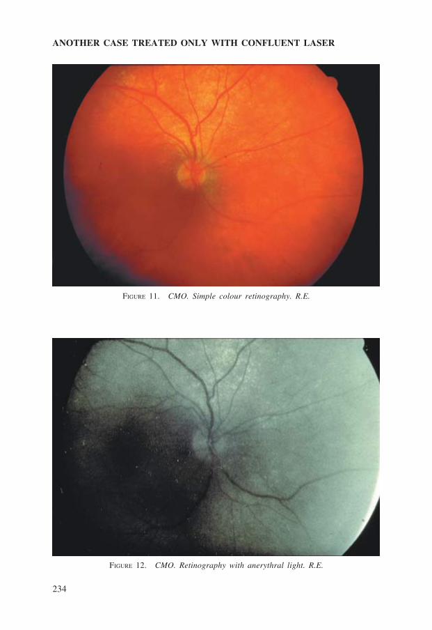
ANOTHER CASE TREATED ONLY WITH CONFLUENT LASER
FIGURE 11. CMO. Simple colour retinography. R.E.
FIGURE 12. CMO. Retinography with anerythral light. R.E.

235
FIGURE 13. CMO. Arteriovenous phase. FA. R.E.
FIGURE 14. CMO. Typical. Late phases. FA. R.E.

236
FIGURE 15. CMO. Laser photocoagulation. Four months later. Same case as before. FA.Late phases. R.E.
FIGURE 16. CMO. Simple colour retinography. Previous case. R.E.CVA. Initial. R.E. = 0.5 No. 1 C. WM.CVA. Final. R.E. = 0.65 No. 1 C. WM.
237
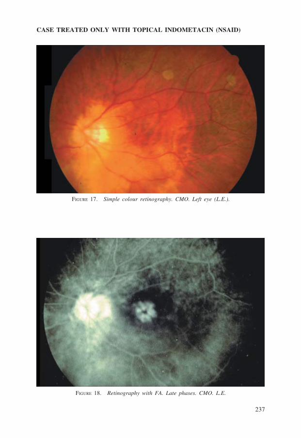
CASE TREATED ONLY WITH TOPICAL INDOMETACIN (NSAID)
FIGURE 17. Simple colour retinography. CMO. Left eye (L.E.).
FIGURE 18. Retinography with FA. Late phases. CMO. L.E.

238
FIGURE 19. Simple colour retinography. Previous case. Four months after starting localtreatment. L.E.
FIGURE 20. Retinography with FA of same case. L.E.CVA. Initial. L.E. = 0.4 No. 1 C. WM.CVA. Final. L.E. = 0.95 No. 1 C. WM.

239
ANOTHER CASE TREATED ONLY WITH TOPICAL NSAID’S
FIGURE 21. Simple colour retinography. CMO. L.E. Macula with typical yellowishappearance.
FIGURE 22. Retinography with FA. Late phases. CMO. L.E.

240
FIGURE 23. FA. Late phases. Previous case. Four months after starting local treatment.CMO. L.E.
FIGURE 24. Simple colour retinography in the same case. CMO cured by topical indome-tacin. L.E. CVA. Initial. L.E. = 0.4 No. 2 C. WM. CVA. Final. L.E. = 0.9 No. 1 C. WM.

241
ANOTHER CASE TREATED ONLY WITH TOPICAL NSAID’S
FIGURE 25. FA. Late phases. Typical CMO. R.E.
FIGURE 26. Late phases. Typical CMO. R.E.

242
FIGURE 27. Late phases. Typical CMO. R.E.
FIGURE 28. Simple colour retinography of the same case, healed after three months withdaily instillation of NSAID’s (one drop every 8 hours). R.E.
243
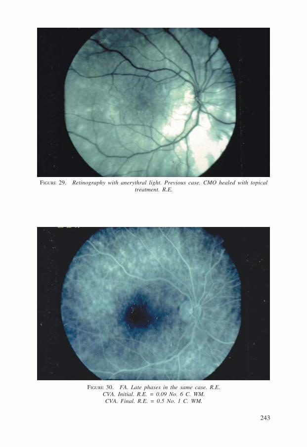
FIGURE 29. Retinography with anerythral light. Previous case. CMO healed with topicaltreatment. R.E.
FIGURE 30. FA. Late phases in the same case. R.E.CVA. Initial. R.E. = 0.09 No. 6 C. WM.CVA. Final. R.E. = 0.5 No. 1 C. WM.

244
RESULTS
The biostatistical tests applied to the CVA variables were only performed in themost numerous group, i.e., the one treated with topical indometacin (22 cases = 76%).For this, Pearson’s test was used (chi-squared = 0.5074) for N = 1 and K = 2, witha significance level α = 0.10, and a theoretical N distribution (mean = 0.330; σ =0.1475).
Improvement in CVA in under 6 months after topical NSAID’s instillation was0.330 ± 0.1 - yielding a value of 0.13, which is clinically and statistically significantbased on the test used. The 95% confidence interval corresponded to an upper andlower limit of 0.3878 and 0.221, respectively (Graph 3).
GRAPH 3. Indometacin treatment (according to Pearson’s test).ICVA: Initial central visual acuity. FCVA: Final central visual acuity. C: With refractive
correction.
0
0,2
0,4
0,6
0,8
1
1,2
I.C.V.A. (C) F.C.V.A. (C) RESULTS
C.D.H.G.
It should be added that in this first group, CVA continued to improve in subse-quent controls.
The second group, treated with indometacin plus laser photocoagulation, showedan improvement of 0.05 (Graph 4).
GRAPH 4. Treatment with indometacin and laser.
-0,2
-0,1
0
0,1
0,2
0,3
0,4
0,5
0,6
I.C.V.A. (C) F.C.V.A. (C) RESULTS
C.D.H.G.

245
The third group, given only confluent laser therapy, showed an improvement of0.10 (Graphs 5 and 6).
GRAPH 5. Laser treatment.
GRAPH 6. Cystoid macular oedema. Total cases and treatments.
-0,6
-0,4
-0,2
0
0,2
0,4
0,6
0,8
1
2 3 4
I.C.V.A. (C) F.C.V.A. (C) RESULTS
C.D.H.G.
0,12
0,14
0
0,02
0,04
0,06
0,08
0,1
INDOMETACIN INDOMETACIN +
LASER
LASER
C.D.H.G.
DISCUSSION
No single aetiology is able to fully account for the development of cystic orcystoid macular oedema; consequently, the condition should be regarded as having amultifactorial origin.
Despite spontaneous resolution of the clinical picture in many cases (in up to 80%according to some authors), in a large and significant number of cases the course ofthe disease does not lead to ad integrum restitution, and a foveal perforation, macularepiretinal membrane or disc-shaped scar may eventually result in some cases. The twolatter conditions occur when the syndrome becomes chronic, and require immediateand selective treatment (9,39).
Transudate accumulation within the Henle fibre or external plexiform layer su-rrounding the fovea is explained in histopathological terms by the fact that this thick

246
retinal membrane may collect relatively large amounts of fluid within its multiplecavities - with edematization occurring in response to minimal stimuli.
The avascular nature of the macular area substantially limits fluid and oedemaabsorption by the Henle and internal nuclear layers. This condition explains the appli-cation of physical therapy (laser photocoagulation) in certain specific cases, based onthe concept that laser therapy stimulates the release of inhibitory factors from thepigmentary epithelial cells to increase the reabsorption of fluid deposited within theretinal layers after pumping towards the choroid; this is an adjuvant mechanism forthe resolution of CMO. However, not all authors readily accept this latter postulate,and some reject the idea.
Preventive drug treatment (particularly topical) is widely used, since the blood-retinal barriers (particularly the external) hinder passage into the posterior segmentwhen drugs are administered systemically (19,26,32).
Nevertheless, oral carbonic anhydrase inhibitors (specifically, acetazolamide 250mg every 12 hours) are commonly prescribed, for they also increase the absorptioncapacity of the choroid and pigmentary epithelial cells, thus initially contributing toremove the intra-retinal fluid by stimulating active ion transport pump. In theory, thisis the way in which all transudates typically found in CMO are removed.
The use of conventional topical corticoids was once very widespread. However,although such drugs do exert potent anti-inflammatory effects, their side effects (im-mune suppression, delayed healing, secondary glaucoma, enhancement of bacterial andfungal overinfection, etc.) advise against their use. Furthermore, the conventional cor-ticoids only inhibit the leukotrienes, regardless of the administration route employed.
Non-steroidal anti-inflammatory drugs (NSAIDs) are very effective, as reflectedby the results of the present study.
In this context, both indometacin and sodium diclofenac eyedrops readily penetra-te inside the eyeball, causing marked inhibition of prostaglandin synthesis without theuntoward effects elicited by these same drugs when administered systemically. EarlyCMO remission is thus achieved by blocking of the inflammatory reaction mediators.Moreover, sodium diclofenac, which is also analgesic and acts only upon the anteriorsegment, can be combined with indometacin, since the latter acts upon the posteriorpole - thereby avoiding possible incompatibilities between both two drugs.
The formulation of the indometacin preparation used in the present study includestwo alcoholic preservatives (benzyl and phenylethyl alcohol), which in addition to thealkaline nature (pH 9) of the solution account for the burning sensation referred bysome patients after instillation.
This very mild symptom has been little reported in our series, for we recommen-ded rubbing the eyedrop bottle before use, and storage in the home refrigerator doorcompartment between local treatments.
We likewise observed no cases of intraocular hypertension, bullous keratitis oriatrogenic miosis. Surgery was not required (pars plana vitrectomy), since among

247
other parameters patient response to medical treatment was excellent and the durationof the process was under 6 months in all cases. Moreover, significant improvementsin CVA have been recorded over time (after 1-2 years).
In pseudophakic eyes even by extracapsular cataract extraction (ECCE), the inci-dence of CMO is much lower - as also occurs when the intraocular lens (IOL) isplaced into the capsular sac (CS) instead of the anterior chamber (AC). However, thedevelopment of CMO has occasionally been reported in patients subjected to cataractextraction with IOL implantation in the CS (24,39).
In our opinion, this latter observation is due to age-related PVD, which cau-ses the hole in the central posterior hyaloid (bursa premacularis) through whichthe vasoactive substances described by Kishi, Shimizu and Worst diffuse towardsthe macula. (25,41).
Authors such as Hitchings suggest that in cases of ICCE the incidence of CMOcan reach 50%, both when the conventional technique is used and when the IOL isimplanted in the AC (20).
One aspect that remains to be addressed is why the fundamentally inflammatorycondition develops in late phases rather than suddenly or during the early postopera-tive period. This supports the immune hypothesis of the syndrome, for it accounts forthe presence of autoantigens which could induce an immune reaction when slowlyreleased or deposited in the vitreous (10).
CONCLUSIONS
Although the natural history of CMO tends towards spontaneous resolution in80% of cases (particularly in subclinical presentations), immediate therapeutic mea-sures relieves the troublesome symptoms, prevents potential chronic complicationsand contributes to ensure prompt vision recovery.
The intact posterior capsule in phacoemulsification or ECCE plus IOL preventsthe inflammatory mediators from reaching the posterior pole, however, if in thesepseudoaphakic eyes capsule photocoagulation is performed (e.g., capsulotomy withYAG laser) due to opacification of the latter, with the subsequent reduction in visualacuity, the incidence of CMO increases for obvious reasons - reaching the proportionobserved for ICCE.
We have no long experience with the administration of other inhibitors such asaspirin, carbonic anhydrase inhibitors or with the instillation of eyedrops composedof flurbiprofen or ketorolac tromethamol in CMO (11). Nor with intravitreal and sub-tenon‘s injections of steroids (triamcinolone acetonide), neither by sustained releaseof intraocular implants.
The high frequency of CMO has decreased with the increasing performance ofECCE and phacoemulsification, particularly when the IOL is implanted into the CS,because of the reasons given above.

248
OCT is supremely useful as diagnostic method.
In some cases, laser photocoagulation is quite successful.
In selected cases a surgical approach ought to be done.
Recently intravitreal injection of triamcinolone acetonide is being used as aadjunct therapeutic option for long-standing cystoid macular oedema and severalmacular diseases including age-related macular degeneration. Our clinical re-sults should be soon published.
As stressed in this study, for the moment we consider topical medical treat-ment with indometacin to be superior to the other management options in full-blown postoperative CMO. On the other hand, attention is drawn to the impor-tance of prophylaxis with this same drug, in compliance with the recommendationsof many authors who consider the topical route to afford global stabilization ofthe blood-aqueous barrier.
REFERENCES
1. Alió, Jorge. Ruiz Moreno, J.Mª., y To-más, Santiago. Edema macular quísticopost-quirúrgico. Inflamaciones oculares.71 Ponencia Oficial de la Sociedad Es-pañola de Oftalmología. EDIKA. MED.1995; 435-447. Barcelona.
2. Ambache, N. Kavanagh, L., and Whi-ting, J. Effect of mechanical stimulatio-ns on rabbits eyes release of active subs-tances in anterior chamber perfusates. J.Physiol. 1965; 176: 378-408.
3. Barraquer, J. Troutman, Richard C.Rutllán, J., y Binkhorst, Richard D. Ci-rugía del segmento anterior del ojo. Ins-tituto Barraquer. 1964; Vol 1. 414-416.Barcelona.
4. Bernstrom, K., and Hammarstrom, S. Anovel leukotriene formed by transpepti-dation of LTE. Biochem. Biophys. Res.Common. 1982; 109: 800-804.
5. Berrocal, J. A. R. Incidence of cystoidmacular oedema after different cataractoperations. Mod. Probl. Ophthal. 1977;18: 518-520. Basel.
6. Borgeat, P. Samuelson, B. Transforma-tion of arachidonic acid by rabbit poly-morphonuclear leukocytes. Formation
of a novel dihydroxy eicosanoic acid. J.Biol. Am. 1979; 254: 2643-2646.
7. Cunha-Vaz, J.G. Travessos, A.Breakdown of the blood retinal barriersand cystoid macular oedema. Surv.Ophthal. 1984; 28 (suppl.): 485-492.
8. Chandler, P.A. Complications after ca-taract extraction. Clinical aspects.Trans. Amer. Acad. Ophthal. Otolaryng.1954; 58: 382-392.
9. Drews, R.C. The present understandingof cystoid macular oedema. Trans.Ophthal. Soc. U.K. 1985; 104 (7): 744-747.
10. Faure, J.P. Bloch-Michel, E. Le Hoang,P., et Vadot, E. Immunopathologie del’oeil. Société française d’ophtalmologieet Masson. 1988. Paris.
11. Flach A.J. Jampol L.M. Weinberg D., etal. Improvement in visual acuity inchronic aphakic and pseudoaphakic cys-toid macular oedema after treatmentwith topical 0.5% ketorolac trometha-mine. Am. J. Ophtal. 1995; 92: 1102-1111.
12. García Arumí, J., y Villalonga, P. Ede-ma macular quístico post-operatorio.

249
Curso de avances en el tratamiento delas uveítis y las inflamaciones post-ope-ratorias. Centro de Retina-Vítreo. 1994;373-378. Barcelona.
13. Gass, J.D.M., and Norton, E.W.D. Cys-toid macular oedema and papilloedemafollowing cataract extraction. Arch.Ophthal. 1966; 76: 646-661.
14. Gass, J.D.M., and Norton, E.W.D. Fluo-rescein studies of patients with macularoedema and papilloedema followingcataract extraction. Trans. Amer.Ophthal. Soc. 1967; 64: 232-249.
15. Gass, J.D.M. Anderson, D.R., and Da-vis, E.B. A clinical fluorescein angio-graphic and electron microscopic corre-lation of cystoid macular oedema. Am.J. Ophthal. 1985; 100: 82-86.
16. Hawkins, W.R. Cystoid maculopathy.108 Irvine-Gass syndrome. In CurrentConcepts in cataract surgery. J.M.Emery and D. Paton (eds.). St. Louis.C.V. Mosby Co. 1976; 300-302.
17. Heredia García, Carlos Dante. Estudiode las inmunoglobulinas en el líquidosubretiniano. Revista d’or de oftalmolo-gia. 1983; 2 (1-6): 47.55. Barcelona.
18. Heredia García, Carlos Dante. Microci-rugía de algunas afecciones de fondo deojo. Instituto de España. Real Academiade Medicina de Zaragoza. Conferenciasy Comunicaciones. Curso 1989. Vol.LIV. 1990; 37-64. Zaragoza.
19. Heredia García, Carlos Dante. BallarínMilán, Martín. Sabater García, Ginés.Ventajas de la corticoterapia tópica enafecciones quirúrgicas vitreo-retinianas.Revista d’or de oftalmologia. 1993; 1(1): 29-42. Barcelona.
20. Hitchings, R.A. Chisholm, I.H., andBird, A.C. Aphakic macular oedema:incidence and pathogenesis. Invest.Ophthal. 1975; 14: 68-72.
21. Hitchings, R.A., and Chisholm, I.H. In-cidence of aphakic macular oedema a
prospective study. Brit. J. Ophthal.1975; 59: 444-450.
22. Irvine, S.R. A newly defined vitreoussyndrome following cataract surgery.Interpreted according to recent conceptsof the structure of the vitreous. Amer. J.Ophthal. 1953; 36: 599-619.
23. Irvine, S.R., et al. Macular oedema aftercataract extraction. Ann. Ophthal. 1971;3: 1234-1240.
24. Jurjo, C. Sánchez, C. Asenjo, J., y Huer-va, V. Edema macular cistoide tras ciru-gía de catarata en pacientes con retino-patía diabética. Annals d’oftalmologia.1996; 6 (3): 18-19. Barcelona.
25. Kishi, S., and Shimizu, K. Posteriorprecortical vitreous pocket. Arch.Ophthal. 1990; 108: 979-982.
26. Klein, R.M. Katzin, H.M., and Yannuzi,L.A. The effect of indomethacin pre-treatment on aphakic cystoid macularoedema. Amer. J. Ophthal. 1979; 87:487-489.
27. Lim, Li, and Chee, Caroline, K.L. Oralfundal fluorescein angiography in dia-betic retinopathy. Comunicación oralpresentada durante el XXVII CongresoInternacional de Oftalmología. Toronto.Ontario. Canadá. Junio 1994; 26-30.
28. Meredith, T.A., et al. Perifoveal vascu-lar leakage and macular oedema afterintracapsular cataract extraction. Brit. J.Ophthal. 1976; 60: 765-769.
29. Miyake, K., et al. Prostaglandins as acausative factor of the cystoid macularoedema after lens extraction. (IV). FoliaOphthal. Jpn. 1978; 29: 447-450.
30. Miyake, K. Vitreous fluorphotometry inaphakic or pseudophakic eye with per-sistant cystoid oedema. Jpn. J. Ophthal.1985; 29: 146-152.
31. Oliw, W. Granstrom, E., and Anggalrd,E. The prostaglandins and essential fattyacids. In: C. Pace-Asciak and E. Grans-trom: Prostaglandins and related subs-

250
tances. Elseiver. 1983; 5: 1-44. Amster-dam.
32. Peterson, Michael. Yoshizumi, Marc O.Hepler, Robert. Mondino Bartly andKreiger Allan. Topical indomethacin inthe treatment of chronic cystoid macu-lar oedema. Graefe’s Arch. Clin. Exp.Ophthal. 1992; 230: 401-405.
33. Puliafito, C.A. Hee, R.H. Schuman, J.S.Fujimoto, J.G. Optical coherence tomo-graphy of ocular diseases. Slack incor-porated. 1996. Thorofare. N.J.
34. Quentel, G. Attali, Ph., et Coscas, G.Angiographie a la fluoresceine par voieorale. Interets et limites. Bull. Soc.Opht. France. 1984; 5: 567-568. Paris.
35. Quentel, G. Zerah, I. Coscas, G. Vi-treous fluorphotometry studies in apha-kic. Graefe’s Arch. Clin. Exp. Ophthal.1985; 222: 248-249.
36. Rodríguez Barrios, Raúl. Massera Lere-na, María Julia. Fondo de ojo. EditorialInter-Médica. S.R.L. 1959; 22-24. Bue-nos Aires.
37. Roque, A. Sellarés, M.A. Maseras, M.,y Duch, F. Efecto de la técnica anesté-
sica en la cirugía de la catarata sobre laaparición del edema macular quísticodel afáquico. Arch. Soc. Esp. Oftalmol.1991; 61: 453-458.
38. Sebag, J., and Balazs, E.A. Pathogene-sis of cystoid macular oedema: and ana-tomic consideration of vitreoretinal ad-hesions. Surv. Ophthal. 1984; 28:493-498.
39. Stark, Walter J. Terry, Arlo C., andMaumenee, A. Edward. Anterior seg-ment surgery. Iols, lasers and refractivekeratoplasty. Edited by Williams &Wilkins. 1987. U.S.A.
40. Worst, J.G.F. Biotoxizitat des Kam-merwassers. Klin. Monatsbl. Augenkei-lk. 1975; 167: 376-384.
41. Worst, J.G.F. The bursa intravitrealespremacularis: new developments inophthalmology. In: Documenta ophthal-mology procceding series. 1976; 275-279. Nijmegen. Holanda.
42. Yanoff, M. Fine, B.S. Brucker, A.J., andEagle, R.C. Jr. Pathology of human cys-toid macular oedema. Surv. Ophthal.1984; suppl: 505-551.

![Assessment report - ema.europa.eu › en › documents › variation... · (ie, cystoid ME [CME]). ME and cystoid macular oedema (CME) are common causes of poor visual outcome following](https://static.fdocuments.in/doc/165x107/5f1f96e2bf8ab064bd6ec5c9/assessment-report-ema-a-en-a-documents-a-variation-ie-cystoid-me.jpg)