Kunwardeep Sohal, R3 2 ways to destroy RBC: issues within the RBC and/or its membrane...
15
Transcript of Kunwardeep Sohal, R3 2 ways to destroy RBC: issues within the RBC and/or its membrane...

Kunwardeep Sohal, R3
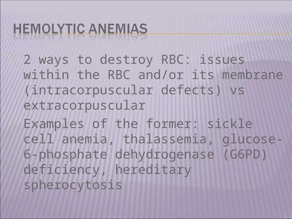
2 ways to destroy RBC: issues within the RBC and/or its membrane (intracorpuscular defects) vs extracorpuscular
Examples of the former: sickle cell anemia, thalassemia, glucose-6-phosphate dehydrogenase (G6PD) deficiency, hereditary spherocytosis
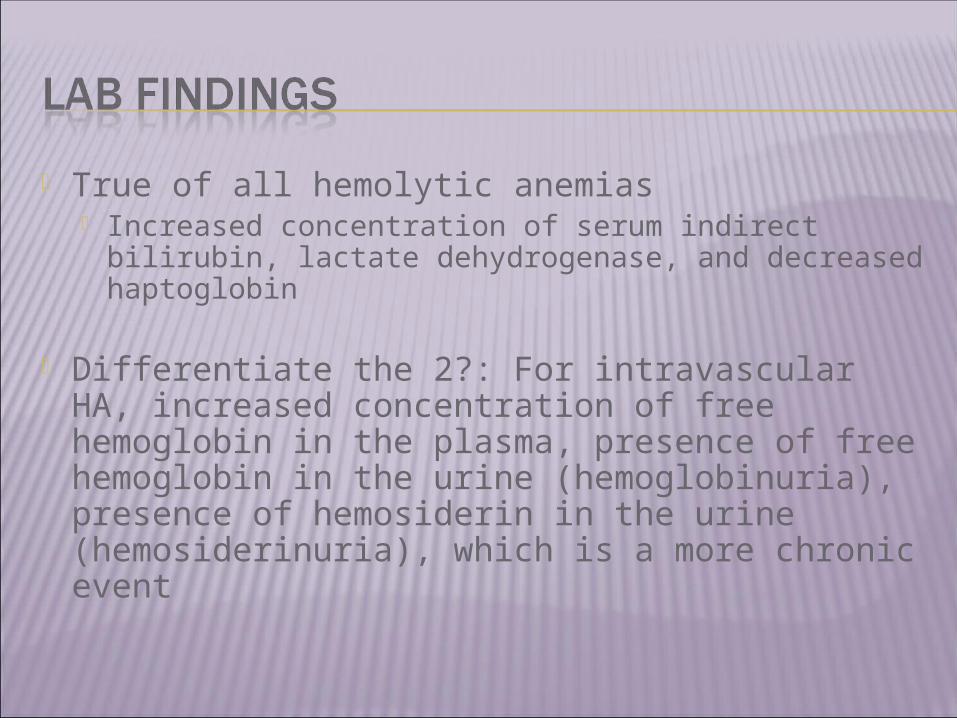
True of all hemolytic anemias Increased concentration of serum indirect
bilirubin, lactate dehydrogenase, and decreased haptoglobin
Differentiate the 2?: For intravascular HA, increased concentration of free hemoglobin in the plasma, presence of free hemoglobin in the urine (hemoglobinuria), presence of hemosiderin in the urine (hemosiderinuria), which is a more chronic event

Acquired G6PD deficiency in essence Red blood cells contain relatively high
concentrations of reduced glutathione (GSH), a sulfhydryl-containing tripeptide that functions as an intracellular reducing agent
Under normal circumstances, oxidant accumulation does not occur, since these compounds are rapidly inactivated by GSH in conjunction with glutathione peroxidase
GSHGSSG (oxidized), GSH levels are restored by glutathione reductase (this reaction requires the NADPH generated by G6PD)

What else is NADPH required for? Major physiologically important pathway for reducing methemoglobin back to hemoglobin is the NADH-dependent reaction catalyzed by cytochrome b5 reductase (b5R)oxidative stress leads to buildup of methemoglobin and oxygen carrying capacity drops
The oxygen dissociation curve is "left-shifted" and oxygen delivery to the tissues is impaired
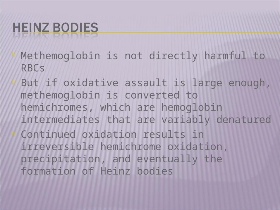
Methemoglobin is not directly harmful to RBCs
But if oxidative assault is large enough, methemoglobin is converted to hemichromes, which are hemoglobin intermediates that are variably denatured
Continued oxidation results in irreversible hemichrome oxidation, precipitation, and eventually the formation of Heinz bodies
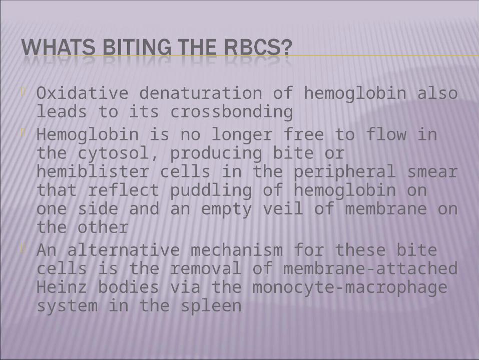
Oxidative denaturation of hemoglobin also leads to its crossbonding
Hemoglobin is no longer free to flow in the cytosol, producing bite or hemiblister cells in the peripheral smear that reflect puddling of hemoglobin on one side and an empty veil of membrane on the other
An alternative mechanism for these bite cells is the removal of membrane-attached Heinz bodies via the monocyte-macrophage system in the spleen
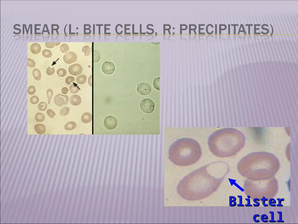
Blister Blister cellcell

As a result, patients with G6PD deficiency are susceptible to oxidative hemolysis by a number of drugs, however, those with nml amounts of G6PD are still open to this event if quantity is high enough
Common ones we see: Dapsone, Nitrofurantoin, Phenazopyridine, Primaquine, Sulfamethoxazole, Ribavirin
Pyridium (bladder analgesic): The recommended maximum duration of therapy is two days. However, it is not uncommon to see patients who have been given a prescription for one to four weeks. In some patients, the hemolysis is sufficiently severe to induce acute renal failure, presumably due to hemoglobinuria
Signs of anemia (vitals, physical exam) CBCs: irregularly shaped cells, since these
undeformable cells are unable to undergo elastic recoil after fighting their way through the sinus wall of the spleen
Blood Gas: presence of clinical "cyanosis" in the face of a normal arterial PO2 (PaO2) as obtained by arterial blood gases
Methemoglobinemia has been variously described as dark-red, chocolate, or brownish to blue in color
Pulse oximetry is inaccurate in monitoring oxygen saturation in the presence of methemoglobinemia

Avoid the causative agent Methemoglobin levels in excess of 30
percent of hemoglobin can be dangerous and values above 50 percent fatal
Blood transfusion or exchange transfusion may be helpful in patients who are in shock, and dialysis may be necessary for those with renal failure
Methylene blue IV provides an artificial electron acceptor for the reduction of methemoglobin to hemoglobin (can’t use in G6PD, ascorbic acid used instead)
Important to check levels after event has occurred (several weeks)
Immediately after a hemolytic episode, G6PD levels in pts with A- (one of the variants) may be normal, since the mature cells have been lysed, and only younger cells with normal G6PD levels, are present (A- retic faster while taking drug)

www.uptodate.com, “Extrinsic nonautoimmune hemolytic anemia due to drugs”
Dr Ma’s brain “Diagnostic Approach to
Hemoglobinopathies” Kutlar F. Hemoglobin. 2007;31(2):243-50


![FIS for the RBC/RBC Handover...4.2.1.1 The RBC/RBC communication shall be established according to the rules of the underlying RBC-RBC Safe Communication Interface [Subset-098]. Further](https://static.fdocuments.in/doc/165x107/5e331307d520b57b5677b3fa/fis-for-the-rbcrbc-handover-4211-the-rbcrbc-communication-shall-be-established.jpg)