Kidney Disease and Hypertension in Pregnancy intact renal function is necessary for the physiologic...
20
10 Kidney Disease and Hypertension in Pregnancy K idney disease and hypertensive disorders in pregnancy are dis- cussed. Pregnancy in women with kidney disease is associated with significant complications when renal function is impaired and hypertension predates pregnancy. When renal function is well preserved and hypertension absent, the outlook for both mother and fetus is excellent. The basis for the close interrelationship between reproductive function and renal function is intriguing and suggests that intact renal function is necessary for the physiologic adjustments to pregnancy, such as vasodilation, lower blood pressure, increased plasma volume, and increased cardiac output. The renal physiologic adjustments to pregnancy are reviewed, including hemodynamic and metabolic alterations. The common primary and secondary renal diseases that may occur in pregnant women also are discussed. Some considerations for the management of end-stage renal disease in pregnancy are given. Hypertensive disorders in pregnancy are far more common than is renal disease. Almost 10% of all pregnancies are complicated by either preeclampsia, chronic hypertension, or transient hypertension. Preeclampsia is of particular interest because it is associated with life-threatening manifestations, including seizures (eclampsia), renal failure, coagulopathy, and rarely, stroke. Significant progress has been made in our understanding of some of the pathophysiologic manifes- tations of preeclampsia; however, the cause of this disease remains unknown. The diagnostic categories of hypertension in pregnancy, pathophysiology of preeclampsia, and important principles of preven- tion and treatment also are reviewed. Phyllis August CHAPTER
Transcript of Kidney Disease and Hypertension in Pregnancy intact renal function is necessary for the physiologic...
10
Kidney Disease andHypertension in Pregnancy
Kidney disease and hypertensive disorders in pregnancy are dis-cussed. Pregnancy in women with kidney disease is associatedwith significant complications when renal function is impaired
and hypertension predates pregnancy. When renal function is well preserved and hypertension absent, the outlook for both mother andfetus is excellent. The basis for the close interrelationship betweenreproductive function and renal function is intriguing and suggeststhat intact renal function is necessary for the physiologic adjustmentsto pregnancy, such as vasodilation, lower blood pressure, increasedplasma volume, and increased cardiac output.
The renal physiologic adjustments to pregnancy are reviewed,including hemodynamic and metabolic alterations. The common primary and secondary renal diseases that may occur in pregnantwomen also are discussed. Some considerations for the management ofend-stage renal disease in pregnancy are given.
Hypertensive disorders in pregnancy are far more common than isrenal disease. Almost 10% of all pregnancies are complicated byeither preeclampsia, chronic hypertension, or transient hypertension.Preeclampsia is of particular interest because it is associated with life-threatening manifestations, including seizures (eclampsia), renal failure, coagulopathy, and rarely, stroke. Significant progress has beenmade in our understanding of some of the pathophysiologic manifes-tations of preeclampsia; however, the cause of this disease remainsunknown. The diagnostic categories of hypertension in pregnancy,pathophysiology of preeclampsia, and important principles of preven-tion and treatment also are reviewed.
Phyllis August
C H A P T E R

10.2 Systemic Diseases and the Kidney
Anatomic Changes in the Kidney During Pregnancy
Increased kidney size
Increased renal blood flow
Increased glomerular filtration rate
Dilation of urinary tract
FIGURE 10-1
Anatomic changes in the kidney during pregnancy. During pregnancy,kidney size increases by about 1 cm. More striking are the changes inthe urinary tract. The calyces, renal pelvis, and ureters dilate. Thedilation is more marked on the right side than the left and is apparentas early as the first trimester. Hormonal mechanisms and mechanicalobstruction are responsible. Intravenous pyelography may demon-strate the iliac sign in which ureteral dilation terminates at the level ofthe pelvic brim where the ureter crosses the iliac artery. Ureteral dila-tion and urinary stasis contribute to the increased incidence of asymp-tomatic bacteriuria and pyelonephritis in pregnancy.
Changes in Renal Function During Pregnancy
↓ Uric acid reabsorption
↑ Urinary calcium
↑ Renin
↑ Urinary protein
↑ Aldosterone
↑ Sodium reabsorption
↑ Water reabsorption
↑ Glucosuria↑ Aminoaciduria
↑ Renal blood flow
Renal vasodilation
↑ Glomerular filtration rate
↓ Serum creatinine
FIGURE 10-2
Changes in renal function during pregnancy. Marked renal hemo-dynamic changes are apparent by the end of the first trimester.Both the glomerular filtration rate (GFR) and effective renal plas-ma flow (ERPF) increase by 50%. ERPF probably increases to agreater extent, and thus, the filtration fraction is decreased duringearly and mid pregnancy. Micropuncture studies performed in ani-mals suggest the basis for the increase in GFR is primarily theincrease in glomerular plasma flow [1]. The average creatinine leveland urea nitrogen concentration are slightly lower than in pregnantwomen than in those who are not pregnant (0.5 mg/d and 9mg/dL, respectively). The increased filtered load also results inincreased urinary protein excretion, glucosuria, and aminoaciduria.The uric acid clearance rates increase to a greater extent than doesthe GFR. Hypercalciuria is a result of increased GFR and ofincreases in circulating 1,25-dihydroxy-vitamin D3 in pregnancy(absorptive hypercalciuria). The renin-angiotensin system is stimu-lated during gestation, and cumulative retention of approximately950 mEq of sodium occurs. This sodium retention results from acomplex interplay between natriuretic and antinatriuretic stimulipresent during gestation [2].

10.3Kidney Disease and Hypertension in Pregnancy
A Altered osmoregulation:↓ Serum sodium and ↓ Posmwith ↓ Osmotic Thresholdfor the argenine vasopressin release and thirst
C Mild hypokalemia may beobserved due to ↑ glomerular filtration rate, ↑ urine flow, and ↑ aldosterone
B Serum chloride levels areunchanged compared with women who are not pregnant
D Mild respiratory alkalosis isassociated with small decreases in plasma bicarbonate
Na+ 136 mEq/L
Cl-
104 mEq/L
3.7 mEq/LK+
20 mEq/LHCO
3
50
120
60
70
80
90
100
110
4 8 12 16 20 24 28 32 36 40 PP
SittingStanding
Gestation, wk
Blo
od
pre
ssu
re, m
mH
g
A
*
* *
***
**
*
(7) (16) (18) (18) (18) (19) (18) (18)(15) (19)
4 8 12 16 20 24 28 32 36 38 PP
0
2
4
6
8
10
12
14
PRAPostpartum angiotensinogen values
(N)
Gestation, wk
PRA
, ng/
mL/
h
B
Serum Electrolytes in Pregnancy
FIGURE 10-3
Serum electrolytes in pregnancy. A, During normal gestation, serumosmolality decreases by 10 mosm/L and serum sodium (Na+) decreasesby 5 mEq/L. A resetting of the osmoreceptor system occurs, withdecreased osmotic thresholds for both thirst and vasopressin release[3]. B, Serum chloride (Cl-) levels essentially are unchanged duringpregnancy. C, Despite significant increases in aldosterone levelsduring pregnancy, in most women serum potassium (K+) levels areeither normal or, on average, 0.3 mEq/L lower than are values inwomen who are not pregnant [4]. The ability to conserve potassi-um may be a result of the elevated progesterone in pregnancy [5].D, Arterial pH is slightly increased in pregnancy owing to mild respiratory alkalosis. The hyperventilation is believed to be aneffect of progesterone. Plasma bicarbonate (HCO-
3) concentrationsdecrease by about 4 mEq/L [6].
Blood Pressure and the Renin-Aldosterone System in Pregnancy
FIGURE 10-4
Blood pressure and the renin-aldosterone system in pregnancy.Normal pregnancy is associated with profound alterations incardiovascular and renal physiology. These alterations areaccompanied by striking adjustments of the renin-angiotensin-aldosterone system. A, Blood pressure and peripheral vascularresistance decrease during normal gestation. The decrease inblood pressure is apparent by the end of the first trimester of
pregnancy and often approaches prepregnancy levels at term. B, Despite the decrease in blood pressure, plasma renin activity(PRA) increases during the first few weeks of pregnancy; onaverage, close to a fourfold increase in PRA occurs by the end ofthe first trimester, with additional increases until at least 20weeks. The source of the increased renin is thought to be thematernal renal release of renin.
(Continued on next page)

10.4 Systemic Diseases and the Kidney
0 0
20
40
60
80
100
20
40
60
80
100
120
Uri
ne
ald
ost
ero
ne,
µg/
d
Plasma ald
ostero
ne, ng/100m
L
Urine aldosteronePlasma aldosterone
C
0
8 12 16 20 24 28 32 36 38 PP
50
100
150
200
Gestation, wk
24-h
r N
a+ a
nd
K+, m
Eq
Urine sodium
Urine potassium
D
FIGURE 10-4 (Continued)
C, Changes in renin are associated with commensurate changes inthe secretory rate of aldosterone. Although a correlation existsbetween the increase in renin and that of aldosterone, the latterincreases to a greater degree in late pregnancy. This observationsuggests that other factors may regulate secretion to a greater degreethan does angiotensin II in late gestation. Urinary aldosterone
increases in late gestation to a greater degree than does plasmaaldosterone, which may reflect an increased production of the 3-oxo conjugate measured in urine. D, Despite the marked increas-es in aldosterone during pregnancy, 24-hour urinary sodium andpotassium excretion remain in the normal range. PP— postpartum.(From Wilson and coworkers [7]; with permission.)
Functional Significance of the Stimulated Renin-Angiotensin System in Pregnancy
60
65
70
75
80
85
0
5
10
15
20
25
T = 0 T = 60 T = 0 T = 60
P < 0.005
P < .05
*
*
MA
P, m
m H
g
PRA
, mg/
mL/
h
A B
Pregnant (n = 9)Nonpregnant (n = 8)
FIGURE 10-5
Functional significance of the stimulated renin-angiotensin system(RAS) in pregnancy. We determine whether changes in the RAS inpregnancy are primary, and the cause of the increase in plasma vol-ume, or whether these changes are secondary to the vasodilationand changes in blood pressure. To do so, we administered a singledose of captopril to normotensive pregnant women in their firstand second trimesters and age-matched normotensive women whowere not pregnant. We then measured mean arterial pressure (MAP)and plasma renin activity (PRA) before and 60 minutes after the dose.
A, Despite similar baseline blood pressures, blood pressure decreasedmore in pregnant women compared with those who were not preg-nant in response to captopril. This observation suggests that theRAS plays a greater role in supporting blood pressure in pregnan-cy. B, Baseline PRA was higher in pregnant women compared withthose who were not pregnant, and pregnant women had a greaterincrease in renin after captopril compared with those who were notpregnant. T—time. (From August and coworkers [8]; with permission.)

10.5Kidney Disease and Hypertension in Pregnancy
Pregnancy and the Course of Renal Disease
INTERRELATIONSHIPS BETWEEN PREGNANCY AND RENAL DISEASE
Impact of pregnancy on renal disease
Hemodynamic changes → hyperfiltration
Increased proteinuria
Intercurrent pregnancy-related illness, eg, preeclampsia
Possibility of permanent loss of renal function
Impact of renal disease on pregnancy
Increased risk of preeclampsia
Increased incidence of prematurity,intrauterine growth retardation
FIGURE 10-6
Pregnancy may influence the course of renal disease. Some womenwith intrinsic renal disease, particularly those with baseline azotemiaand hypertension, suffer more rapid deterioration in renal functionafter gestation. In general, as kidney disease progresses and functiondeteriorates, the ability to sustain a healthy pregnancy decreases. Thepresence of hypertension greatly increases the likelihood of renaldeterioration [2]. Although hyperfiltration (increased glomerularfiltration rate) is a feature of normal pregnancy, increased intra-glomerular pressure is not a major concern because the filtrationfraction decreases. Possible factors related to the pregnancy-relateddeterioration in renal function include the gestational increase inproteinuria and intercurrent pregnancy-related illnesses, such aspreeclampsia, that might cause irreversible loss of renal function.Women with renal disease are at greater risk for complicationsrelated to pregnancy such as preeclampsia, premature delivery, and intrauterine growth retardation.
Diabetes Mellitus and Pregnancy
RENAL DISEASE CAUSED BY SYSTEMIC ILLNESS
Gestation in pregnant women with diabetic nephropathy is complicated by the following:
Increased proteinuria, 70%
Decreased creatinine clearance, 40%
Increased blood pressure, 70%
Preeclampsia, 35%
Fetal developmental problems, 20%
Fetal demise, 6%
FIGURE 10-7
Diabetes mellitus is a common disorder in pregnant women. Patients with overt nephropathyare likely to develop increased proteinuria and mild but usually reversible deteriorations inrenal function during pregnancy. Hypertension is common, and preeclampsia occurs in35% of women. (From Reece and coworkers [9]; with permission.)

10.6 Systemic Diseases and the Kidney
Pregnancy and Systemic Lupus Erythematosus
FIGURE 10-8
Patients with systemic lupus erythematosus(SLE) often are women in their childbearingyears. Pregnancies in women with evidenceof nephritis are potentially hazardous, partic-ularly if active disease is present at the timeof conception or if the disease first developsduring pregnancy. When hypertension andazotemia are present at the time of concep-tion the risk of complications increases, as itdoes with other nephropathies [10–14]. Thepresence of high titers of antiphospholipidantibodies also is associated with poor preg-nancy outcome [15]. The presence of anti-phospholipid antibodies or the lupus anti-coagulant is associated with increased fetalloss, particularly in the second trimester;increased risk of arterial and venous throm-bosis; manifestations of vasculitis such asthrombotic microangiopathy; and anincreased risk of preeclampsia. Treatmentconsists of anticoagulation with heparinand aspirin.
RENAL DISEASE ASSOCIATED WITH SYSTEMIC ILLNESS
Pregnancy and SLE*
Poor outcome is associated with the following:
Active disease at conception
Disease first appearing during pregnancy
Hypertension, azotemia in the first trimester
High titers of antiphospholipid antibodies orlupus anticoagulant
Antiphospholipid antibody syndrome in pregnancy
Increased fetal loss
Arterial and venous thromboses
Renal vasculitis, thrombotic microangiopathy
Preeclampsia
Treatment: heparin and aspirin?
*Systemic lupus erythematosus (SLE) is unpredictable during pregnancy.
Lupus Versus Preeclampsia
PE
+
+
-
+
-
+/-
+/-
-
SLE
+
+
+
+
+
-
+
+
Proteinuria
Hypertension
Erythrocyte casts
Azotemia
Low C3, C4
Abnormal liver function test results
Low platelet count
Low leukocyte count
C—complement; minus sign—absent; plus sign—present; PE—preeclampsia; SLE—systemic lupus erythematosus.
FIGURE 10-9
In the second or third trimester of pregnancy a clinical flare-up oflupus may be difficult to distinguish from preeclampsia. Treatmentof a lupus flare-up might involve increased immunosuppression,whereas the appropriate treatment of preeclampsia is delivery. Thus,it is important to accurately distinguish these entities. Preeclampsiais rare before 24 weeks’ gestation. Erythrocyte casts and hypocom-plementemia are more likely to be a manifestation of lupus, whereasabnormal liver function test results are seen in preeclampsia and notusually in lupus.
LUPUS FLARE-UP VERSUS PREECLAMPSIA

10.7Kidney Disease and Hypertension in Pregnancy
Chronic Primary Renal Disease in Pregnancy
CAUSES OF CHRONIC PRIMARYRENAL DISEASE IN PREGNANCY
Anatomic, congenital
Glomerulonephritis
Interstitial nephritis
Polycystic kidney disease
FIGURE 10-10
Primary renal disease in pregnancy that is chronic (ie, preceded pregnancy) may resultfrom any of the causes of renal disease in premenopausal women. Overall, the outcome inpregnancy is favorable when the serum creatinine level is less than 1.5 mg/dL and bloodpressure levels are normal in early pregnancy.
Advanced Renal Disease Caused by Polycystic Kidney Disease
POLYCYSTIC KIDNEY DISEASEAND PREGNANCY
Increased incidence of urinary tract infection
Maternal hypertension associated with poor outcome
Extrarenal complications: subarachnoid hemorrhage,liver cysts
FIGURE 10-11
Although advanced renal disease caused by polycystic kidney disease (PKD) usually devel-ops after childbearing, women with this condition may have hypertension or mildazotemia. Certain considerations are relevant to pregnancy. Pregnancy is associated withan increased incidence of asymptomatic bacteriuria and urinary infection that may bemore severe in women with PKD. The presence of maternal hypertension has been shownto be associated with adverse pregnancy outcomes [16]. Pregnancy has been reported to beassociated with increased size and number of liver cysts owing to estrogen stimulation.Women with intracranial aneurysms may be at increased risk of subarachnoid hemorrhageduring labor.
Management of Chronic Renal Disease During Pregnancy
MANAGEMENT OF CHRONIC RENAL DISEASE DURING PREGNANCY
Preconception counseling
Multidisciplinary approach
Frequent monitoring of blood pressure (every 1–2 wk) and renal function (every mo)
Balanced diet (moderate sodium, protein)
Maintain blood pressure at 120–140/80–90 mm Hg
Monitor for signs of preeclampsia
FIGURE 10-12
Management of chronic renal disease during pregnancy is bestaccomplished with a multidisciplinary team of specialists.Preconception counseling permits the explanation of risks involvedwith pregnancy. Patients should understand the need for frequentmonitoring of blood pressure and renal function. Protein restrictionis not advisable during gestation. Salt intake should not be severelyrestricted. When renal function is impaired, modest salt restrictionmay help control blood pressure. Blood pressure should be main-tained at a level at which the risk of maternal complications owingto elevated blood pressure is low. Patients should be monitoredclosely for signs of preeclampsia, particularly in the third trimester.

10.8 Systemic Diseases and the Kidney
Renal Disease During Pregnancy
MOST COMMON CAUSES OF DE NOVORENAL DISEASE IN PREGNANCY
Glomerulonephritis
Lupus nephritis
Acute renal failure
Interstitial nephritis
Obstructive uropathy
FIGURE 10-13
Renal disease may develop de novo during pregnancy. The usualcauses are new-onset glomerulonephritis or interstitial nephritis,lupus nephritis, or acute renal failure. Rarely, obstructive uropathydevelops as a result of stone disease or large myomas that haveincreased in size during pregnancy.
RENAL EVALUATION DURING PREGNANCY
Serology
Function
Ultrasonography
Biopsy: <32 wk
Deteriorating function
Morbid nephrotic syndrome
FIGURE 10-14
Investigation of the cause of renal disease during pregnancy can be conducted with serolog-ic, functional, and ultrasonographic testing. Renal biopsy is rarely performed during gesta-tion. Renal biopsy usually is reserved for situations in which renal function suddenly deteri-orates without apparent cause or when symptomatic nephrotic syndrome occurs, particular-ly when azotemia is present. Almost no role exists for renal biopsy after gestational week32 because at this stage the fetus will likely be delivered, independent of biopsy results [17].
New-Onset Azotemia, Proteinuria, and Hypertension Occurring in the Second Half of Pregnancy
INTRINSIC RENAL DISEASE VERSUS PREECLAMPSIA
Serum creatinine
Urinary protein
Uric acid
Blood pressure
Liver function test results
Platelet count
Urine analysis
Renal disease
>1.0 mg/dL
Variable
Variable
Variable
Normal
Normal
Variable
Preeclampsia
0.8–1.2 mg/dL
>300 mg/d
>5.5 mg/dL
>140/90 mm Hg
May be increased
May be decreased
Protein, with or without erythrocytes, leukocytes
Investigation of the Cause of Renal Disease During Pregnancy
FIGURE 10-15
New-onset azotemia, proteinuria, and hypertension occurring inthe second half of pregnancy should be distinguished from pre-eclampsia. Most cases of preeclampsia are associated with onlymild azotemia; significant azotemia is more suggestive of renal dis-ease. Azotemia in the absence of proteinuria or hypertension wouldbe unusual in preeclampsia, and thus, would be more suggestive ofintrinsic renal disease. Thrombocytopenia, elevated liver functiontest results, and significant anemia are not typical features of renaldisease (except for thrombotic microangiopathic syndromes) andare features of the variant of preeclampsia known as the hemolysis,elevated liver enzymes, and low platelet count (HELLP) syndrome.
10.9Kidney Disease and Hypertension in Pregnancy
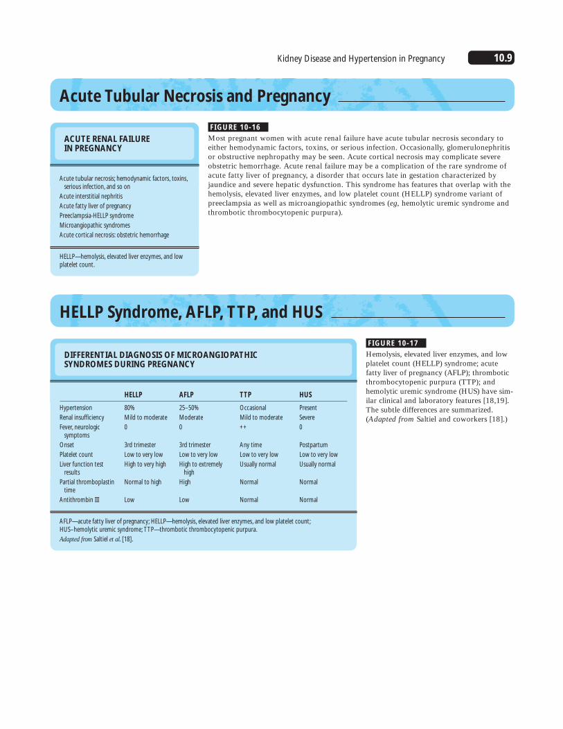
Acute Tubular Necrosis and Pregnancy
ACUTE RENAL FAILURE IN PREGNANCY
Acute tubular necrosis; hemodynamic factors, toxins,serious infection, and so on
Acute interstitial nephritis
Acute fatty liver of pregnancy
Preeclampsia-HELLP syndrome
Microangiopathic syndromes
Acute cortical necrosis: obstetric hemorrhage
HELLP—hemolysis, elevated liver enzymes, and lowplatelet count.
FIGURE 10-16
Most pregnant women with acute renal failure have acute tubular necrosis secondary toeither hemodynamic factors, toxins, or serious infection. Occasionally, glomerulonephritisor obstructive nephropathy may be seen. Acute cortical necrosis may complicate severeobstetric hemorrhage. Acute renal failure may be a complication of the rare syndrome ofacute fatty liver of pregnancy, a disorder that occurs late in gestation characterized byjaundice and severe hepatic dysfunction. This syndrome has features that overlap with thehemolysis, elevated liver enzymes, and low platelet count (HELLP) syndrome variant ofpreeclampsia as well as microangiopathic syndromes (eg, hemolytic uremic syndrome andthrombotic thrombocytopenic purpura).
HELLP Syndrome, AFLP, TTP, and HUS
FIGURE 10-17
Hemolysis, elevated liver enzymes, and lowplatelet count (HELLP) syndrome; acutefatty liver of pregnancy (AFLP); thromboticthrombocytopenic purpura (TTP); andhemolytic uremic syndrome (HUS) have sim-ilar clinical and laboratory features [18,19].The subtle differences are summarized.(Adapted from Saltiel and coworkers [18].)
DIFFERENTIAL DIAGNOSIS OF MICROANGIOPATHIC SYNDROMES DURING PREGNANCY
Hypertension
Renal insufficiency
Fever, neurologicsymptoms
Onset
Platelet count
Liver function testresults
Partial thromboplastintime
Antithrombin III
HELLP
80%
Mild to moderate
0
3rd trimester
Low to very low
High to very high
Normal to high
Low
TTP
Occasional
Mild to moderate
++
Any time
Low to very low
Usually normal
Normal
Normal
AFLP
25–50%
Moderate
0
3rd trimester
Low to very low
High to extremelyhigh
High
Low
HUS
Present
Severe
0
Postpartum
Low to very low
Usually normal
Normal
Normal
AFLP—acute fatty liver of pregnancy; HELLP—hemolysis, elevated liver enzymes, and low platelet count;HUS–hemolytic uremic syndrome; TTP—thrombotic thrombocytopenic purpura.
Adapted from Saltiel et al. [18].
10.10 Systemic Diseases and the Kidney
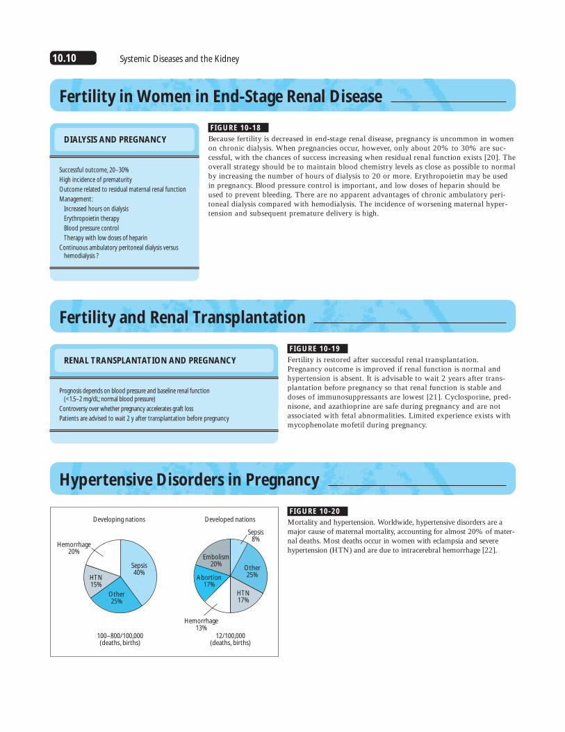
Fertility in Women in End-Stage Renal Disease
DIALYSIS AND PREGNANCY
Successful outcome, 20–30%
High incidence of prematurity
Outcome related to residual maternal renal function
Management:
Increased hours on dialysis
Erythropoietin therapy
Blood pressure control
Therapy with low doses of heparin
Continuous ambulatory peritoneal dialysis versushemodialysis ?
FIGURE 10-18
Because fertility is decreased in end-stage renal disease, pregnancy is uncommon in womenon chronic dialysis. When pregnancies occur, however, only about 20% to 30% are suc-cessful, with the chances of success increasing when residual renal function exists [20]. Theoverall strategy should be to maintain blood chemistry levels as close as possible to normalby increasing the number of hours of dialysis to 20 or more. Erythropoietin may be usedin pregnancy. Blood pressure control is important, and low doses of heparin should beused to prevent bleeding. There are no apparent advantages of chronic ambulatory peri-toneal dialysis compared with hemodialysis. The incidence of worsening maternal hyper-tension and subsequent premature delivery is high.
Fertility and Renal Transplantation
RENAL TRANSPLANTATION AND PREGNANCY
Prognosis depends on blood pressure and baseline renal function (<1.5–2 mg/dL; normal blood pressure)
Controversy over whether pregnancy accelerates graft loss
Patients are advised to wait 2 y after transplantation before pregnancy
FIGURE 10-19
Fertility is restored after successful renal transplantation.Pregnancy outcome is improved if renal function is normal andhypertension is absent. It is advisable to wait 2 years after trans-plantation before pregnancy so that renal function is stable anddoses of immunosuppressants are lowest [21]. Cyclosporine, pred-nisone, and azathioprine are safe during pregnancy and are notassociated with fetal abnormalities. Limited experience exists withmycophenolate mofetil during pregnancy.
Hypertensive Disorders in Pregnancy
FIGURE 10-20
Mortality and hypertension. Worldwide, hypertensive disorders are amajor cause of maternal mortality, accounting for almost 20% of mater-nal deaths. Most deaths occur in women with eclampsia and severehypertension (HTN) and are due to intracerebral hemorrhage [22].
Developing nations Developed nations
Sepsis40%
Other25%
HTN15%
Hemorrhage20%
Sepsis8%
Other25%
HTN17%
Hemorrhage13%
Abortion17%
Embolism20%
100–800/100,000(deaths, births)
12/100,000(deaths, births)

10.11Kidney Disease and Hypertension in Pregnancy
FETAL CONSEQUENCES OFMATERNAL HYPERTENSIONDURING PREGNANCY
3- to 6-fold increase in stillbirths
5- to 15-fold increase in intrauterine growth restriction
Premature delivery
Long-term developmental and neurologic problems
FIGURE 10-21
Hypertensive disorders in pregnancy areassociated with increased incidences of still-birth, fetal growth restriction, prematuredelivery, and long-term developmental prob-lems secondary to prematurity. These com-plications are more frequent when hyperten-sion is due to preeclampsia.
CLASSIFICATION OF HYPERTENSIVE DISORDERS IN PREGNANCY
Preeclampsia, eclampsia
Chronic hypertension
Chronic hypertension with superimposed preeclampsia
Transient hypertension
FIGURE 10-22
Several classification systems exist for hyper-tensive disorders of pregnancy. The one usedmost commonly in the United States is thatproposed in 1972 by the American Collegeof Obstetricians and Gynecologists andendorsed by the National High BloodPressure Education Program. The distinctionis made between the pregnancy-specifichypertensive disorder (preeclampsia, and theconvulsive form, eclampsia) and chronichypertension that precedes pregnancy, whichusually is due to essential hypertension.Women with chronic hypertension are atgreater risk for preeclampsia (20–25%).Transient hypertension refers to late preg-nancy elevations in blood pressure, withoutany of the laboratory or clinical features ofpreeclampsia. This disorder may recur witheach pregnancy (in contrast to preeclampsia,which usually is a disease of first pregnancy)and usually indicates a genetic predispositionto essential hypertension.
CLINICAL FEATURES OF PREECLAMPSIA
Historical:NulliparityMultiple gestationsFamily historyPreexisting renal or vascular decrease
Hypertension:140/90 mm Hg after 20 wk or30 mm Hg increase in systolic pressure or15 mm Hg increase in diastolic pressure
Sudden appearance of edema, especially in hands and face
Rapid weight gain
Headache, visual disturbances, abdominal or chest pain
FIGURE 10-23
The diagnosis of preeclampsia is strength-ened when one or more of the risk factorsare present. Hypertension develops after 20weeks, with normal blood pressures in thefirst half of pregnancy. Although edema is afeature of many normal pregnancies, itssudden appearance in the face and hands inassociation with a rapid weight gain, is sug-gestive of preeclampsia. Headache, visualdisturbances, and abdominal or chest painare signs of impending eclampsia.
CLINICAL FEATURES OF CHRONIC HYPERTENSION IN PREGNANCY
Women are older, more likely to be multiparous
Hypertension: present before 20 wk, or documented previous pregnancy
Blood pressure may be significantly lower or normal in mid pregnancy
Risk of superimposed preeclampsia of 15–30%
FIGURE 10-24
Women with chronic hypertension are usually older and may bemultiparous. Although hypertension often is detectable before 20 weeks, in some women the pregnancy-mediated vasodilation is sufficient to normalize blood pressure so that women withstage 1 or 2 hypertension may have normal blood pressures bythe time of their first antepartum visit. The risk of preeclampsiais substantially increased in women with chronic hypertension.

10.12 Systemic Diseases and the Kidney
LABORATORY ABNORMALITIES IN PREECLAMPSIA AND CHRONIC HYPERTENSION
FIGURE 10-25
Laboratory tests are helpful in making the diagnosis of preeclampsia.In addition to proteinuria, which may occur late in the course of thedisease, hyperuricemia, mild azotemia, hemoconcentration, and hypo-calciuria are observed commonly. Some women with preeclampsiamay develop a microangiopathic syndrome with hemolysis, elevatedliver enzymes, and low platelet counts (HELLP). The presence of theHELLP syndrome usually reflects severe disease and is considered anindication for delivery. Women with uncomplicated chronic hyperten-sion have normal laboratory test results unless superimposedpreeclampsia or underlying renal disease exists.
Renal:
Creatinine
Uric acid
Urinary protein
Urinary calcium
Heme:
Hematocrit
Platelets
Liver function tests:
Aspartate aminotransferase
Alanine aminotransferase
Albumin
Preeclampsia
Increased; increasedblood urea nitrogen,creatinine
Increased (>5.5 mg/dL)
>300 mg/d
<150 mg/d
Increased (>38%)
Decreased
Increased
Increased
Decreased
Chronic hypertension
Normal
Normal
<300 mg/d
>200 mg/d
Normal
Normal
Normal
Normal
Normal
Pathophysiology of preeclampsia
Maternal diseaseVasoplasm
Intravascular coagulationEndothelial dysfunction
Placental diseaseAbdominal implantationPlacental vascular lesions
Maternalsyndrome
(HTN, renal, CNS)
Fetalsyndrome
(IUGR, IUD, prematurity)
Genetic susceptibility(maternal x fetal)
FIGURE 10-26
Preeclampsia is a syndrome with both maternal and fetal manifes-tations. Current evidence suggests that an underlying genetic pre-disposition leads to abnormalities in placental adaptation to thematernal spiral arteries that supply blood to the developing feto-placental unit. These abnormalities in the maternal spiral arterieslead to inadequate perfusion of the placenta and may be the earli-est changes responsible for the maternal disease. The maternal dis-ease is characterized by widespread vascular endothelial cell dys-function, resulting in vasospasm and intravascular coagulation and,ultimately, in hypertension (HTN), renal, hepatic, and central ner-vous system (CNS) abnormalities. The fetal syndrome is a conse-quence of inadequate placental circulation and is characterized bygrowth restriction and, rarely, demise. Premature delivery mayoccur in an attempt to ameliorate the maternal condition. IUD—intrauterine death; IUGR—intrauterine growth retardation.

10.13Kidney Disease and Hypertension in Pregnancy
GENETICS OF PREECLAMPSIA
Increased incidence observed in mothers, daughters,granddaughters of probands
Mode of inheritance unknown:
Single recessive gene ?
Shared maternal-fetal recessive gene ?
Dominant gene with incomplete penetrance ?
FIGURE 10-27
A positive family history is a risk factor for preeclampsia, and the incidence is approxi-mately 4 times greater in first-degree relatives of index cases [23]. Cooper and coworkers[24] also noted an increased incidence in relatives by marriage (eg, daughter-in-laws), and10 instances in which the disease occurred in one but not the other monozygotic twin.These data raise the possibility of paternal or fetal genetic influence [24]. The mode ofinheritance of preeclampsia is not known. Several possibilities have been suggested, includ-ing a recessive gene with the possibility of a maternal-fetal genotype-by-genotype interac-tion or a dominant maternal gene with incomplete penetrance.
Villus(containing fetal
arteriole and venule)
Intervillus space(maternal blood)
Umbilical vein
Umbilical artery
Spiral arteries
Normal pregnancy Preeclampsia
Myometrium
Decidua
A B
FIGURE 10-28
Uteroplacental circulation in normal pregnancy and preeclampsia. A, Normal placentation involves the transformation of the branchesof the maternal uterine arteries—the spiral arteries—from thick-walled muscular arteries into saclike flaccid vessels that permitdelivery of greater volumes of blood to the uteroplacental unit. B, Evidence exits that in women with preeclampsia this process isincomplete, resulting in relatively narrowed spiral arteries anddecreased perfusion of the placenta [25].
Zone I Zone II and III Zone IV Zone V
Invasion
FV
AV
Maternal bloodspace
Uterineblood vessels
Mother(uterus)
Fetus(placenta)
Cell column of anchoring villus
Cytotrophoblaststem cells
Basement membrane
Fetalstroma
Syncytiotrophoblast
A
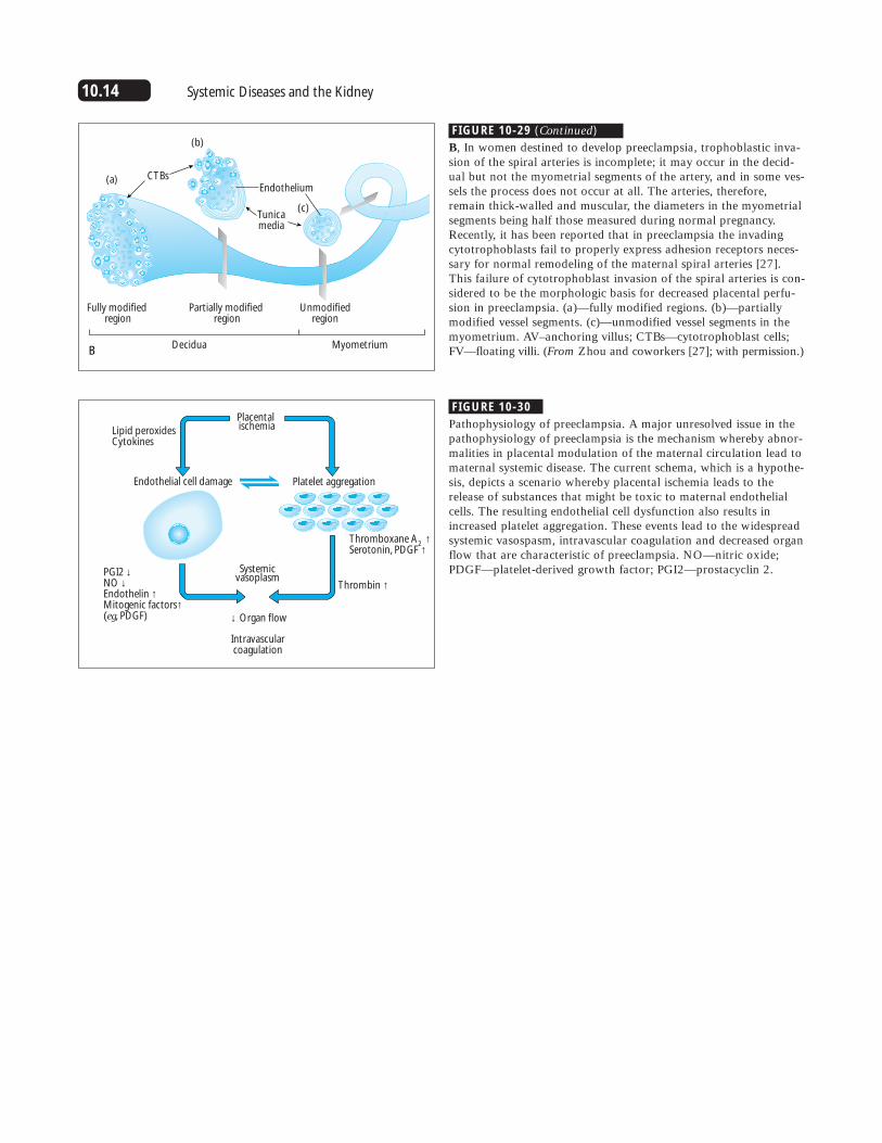
FIGURE 10-29
Transformation of the spiral arteries. A, The process by which thematernal spiral arteries are transformed into dilated vessels in preg-nancy is believed to involve invasion of the spiral arterial walls byendovascular trophoblastic cells. These cells migrate in retrogradefashion, involving first the decidual and then the myometrial seg-ments of the arteries and then causing considerable disruption atall layers of the vessel wall. The mechanisms involved in this com-plex process are only beginning to be elucidated. These mecha-nisms involve alterations in the adhesion molecules of the invadingtrophoblast cells, such that they acquire an invasive phenotype andmimic vascular endothelial cells [26].
(Continued on next page)
10.14 Systemic Diseases and the Kidney
Decidua Myometrium
Fully modified region
Partially modified region
Unmodifiedregion
CTBsEndothelium
Tunicamedia
(a)
(b)
(c)
B
FIGURE 10-29 (Continued)
B, In women destined to develop preeclampsia, trophoblastic inva-sion of the spiral arteries is incomplete; it may occur in the decid-ual but not the myometrial segments of the artery, and in some ves-sels the process does not occur at all. The arteries, therefore,remain thick-walled and muscular, the diameters in the myometrialsegments being half those measured during normal pregnancy.Recently, it has been reported that in preeclampsia the invadingcytotrophoblasts fail to properly express adhesion receptors neces-sary for normal remodeling of the maternal spiral arteries [27].This failure of cytotrophoblast invasion of the spiral arteries is con-sidered to be the morphologic basis for decreased placental perfu-sion in preeclampsia. (a)—fully modified regions. (b)—partiallymodified vessel segments. (c)—unmodified vessel segments in themyometrium. AV–anchoring villus; CTBs—cytotrophoblast cells;FV—floating villi. (From Zhou and coworkers [27]; with permission.)
Placental ischemia
Systemicvasoplasm
Lipid peroxidesCytokines
Endothelial cell damage
PGI2 ↓NO ↓Endothelin ↑Mitogenic factors↑(eg, PDGF) ↓ Organ flow
Intravascularcoagulation
Thrombin ↑
Thromboxane A2
↑Serotonin, PDGF ↑
Platelet aggregation
FIGURE 10-30
Pathophysiology of preeclampsia. A major unresolved issue in thepathophysiology of preeclampsia is the mechanism whereby abnor-malities in placental modulation of the maternal circulation lead tomaternal systemic disease. The current schema, which is a hypothe-sis, depicts a scenario whereby placental ischemia leads to therelease of substances that might be toxic to maternal endothelialcells. The resulting endothelial cell dysfunction also results inincreased platelet aggregation. These events lead to the widespreadsystemic vasospasm, intravascular coagulation and decreased organflow that are characteristic of preeclampsia. NO—nitric oxide;PDGF—platelet-derived growth factor; PGI2—prostacyclin 2.

10.15Kidney Disease and Hypertension in Pregnancy
Central nervous sytem
Hepatic
Renal
Vascular
Cardiac
VasospasmReduced flowIntravascular coagulation
Visual disturbancesSeizuresHyperemia, focal anemiaThrombosis, hemorrhage
↓ Cardiac output↓ Plasma volume↑ Atrial natriuretic factorPulmonary edema
↑ Systemic vascular resistance↑ Blood pressure↑ Angiotensin II sensitivity
EndotheliosisProteinuria↓ Glomerular filtration rate↓ Renal blood flow↓ Urinary sodium, uric acid, and calcium excretion↓ Plasma renin activity
Periportal hemorrhagic necrosisSubcapsular hematoma↑ Aspartate aminotransferase↑ Alanine aminotransferase
FIGURE 10-31
Maternal manifestations of preeclampsia. Preeclampsia is a multisystem maternal disorder, with dramatic alterations in heart, kidney, circulation, liver, and brain. Interestingly, all of these abnormalities resolve within a few weeks of delivery.

10.16 Systemic Diseases and the Kidney
The endotheliumand platelet-vessel
wall interaction
Endothelialcells
Vascular smoothmuscle cells
cGMP/cAMP
RelaxationAntiproliferation
NO/PGl2
PThr
Thr
5-HT 5-HTAII
TXA2
cGMP
Platelets
Endothelin
↓ Placental hormones(eg, estrogen, progesterone)
↑ Circulating endothelial toxins
↑ Sympathetic nervous system
Compensatoryresponses:↓ Plasma renin↓ Aldosterone
++
S1
A
TX ET
S2
−
ContractionProliferation
FIGURE 10-32
Hypertension in preeclampsia. Although the mechanism of the increased blood pressure in preeclampsia is not established, evidence suggests it may involve multiple processes. A possible scenario involves the following:decreased placental production of estrogen and progesterone, both of which have hemodynamic effects;increased circulating endothelial toxins, possibly released from a poorly perfused placenta; and increased activity of the sympathetic nervous system. These processes may then result in alterations in platelet– vascularendothelial cell function, with decrease in vasodilators such as nitric oxide and prostacyclin and increased production
FIGURE 10-33
Light microscopy of the renal lesion of preeclampsia: glomerularendotheliosis. On light microscopy, the glomeruli from preeclamp-tic women are characterized by swelling of the endothelial andmesangial cells. This swelling results in obliteration of the capillarylumina, giving the appearance of a bloodless glomerulus. On occa-sion, the mesangium, severely affected, may expand. Thrombosisand fibrinlike material and foam cells may be present, and epithe-lial crescents have been described in rare instances [2].
↓ Urinary calcium Hypocalciuria
↓ Urate excretion
↓ Renin
↑ Proteinuria
↓ Renal vasodilation
↓ Glomerular filtration rate
↓ Renal blood flow
FIGURE 10-34
Functional renal alterations in preeclampsia. The functional conse-quences of glomerular endotheliosis and of the hormonal alter-ations in preeclampsia are summarized in this schematic diagramof the nephron in preeclampsia. Suppression of the renin-angiotensin system occurs, probably in response to vasoconstric-tion and elevated blood pressure. The glomerular lesion leads toproteinuria, which may be heavy. Renal hemodynamic changesinclude modest decreases in the glomerular filtration rate (GFR)and renal blood flow (RBF). Decreased sodium and uric acid excre-tion may be caused by increased proximal tubular reabsorption.The mechanism for the marked hypocalciuria is not known.
of vasoconstrictors such as endothelin (ET).Compensatory suppres-sion of the renin-angiotensin systemoccurs, suggesting thatexcess angiotensin II(AII) does not play amajor role in preeclamptichypertension (HT).Finally, sodium retentionowing to renal vasocon-striction may furtherincrease blood pressure.cAMP—cyclic adenosinemonophosphate; cGMP—cyclic guanosinemonophosphate; 5-HT—serotonin; PThr—parathyroid hormone;S2—serotonergic receptors;Thr—thombin TX—thromboxane; TXA2—thromboxane A2.(Adapted from Lüscherand Dubey [28]; with permission.)

10.17Kidney Disease and Hypertension in Pregnancy
Trial
Smaller studies(<200 women)
Larger studies:EPHREDA (1990)Hauth (1993)Italian (1993)Sibai (1993)Viinikka (1993)CLASP (1994)
All larger trials
All trials
Antiplatelettherapy
10/319(3.1%)
5/1565/303
12/56569/15709/103)
313/4659
413/7356
423/7675(5.5%)
Controltherapy
50/284(17.6%)
Odds ratio and 95% Cl (horizontal line)(antiplatelet: placebo)
8/7417/3039/477
94/156511/105)
352/4650
491/7174
541/7458(7.3%)
Number of trials
11
6
17
0 0.5 1.0 1.5
25% SD 6odds reduction(2p = 0.00002)
Odds ratioOverall results
Antiplatelettherapybetter
Antiplatelettherapyworse
Favors calcium Favors controlStudy
Marya et al.,1987Villar et al.,1987
Lopez-Jaramillo et al.,1989Lopez-Jaramillo et al.,1990
Montanaro et al.,1990Villar and Repke,1990
Belizan et al.,1991Cong et al.,1993
Sanchez-Ramos et al.,1994Pooled estimate
0.65 (0.31–1.38)0.43 (0.06–3.14)0.03 (0.002–0.49)0.07 (0.004–1.27)0.25 (0.06–1.03)0.13 (0.007–2.65)0.66 (0.34–1.27)0.19 (0.009–4.10)0.22 (0.07–0.74)0.38 (0.22–0.65)
0.001 0.01 0.1 1.0 10.0
OR
FIGURE 10-35
Prevention of preeclampsia with low-doseaspirin. Investigators have sought methodsto prevent preeclampsia (eg, salt restriction,prophylactic diuretics, and high-proteindiets). One approach that has been exten-sively investigated in the last 10 years istherapy with low-dose aspirin. It washypothesized that such therapy reversed theimbalance between prostacyclin and throm-boxane that may be responsible for some ofthe manifestations of the disease. Severallarge trials now have been completed, andmost have had negative results. Shown hereis an overview of the effects of aspirin onproteinuric preeclampsia reported from alltrials of antiplatelet therapy (through 1994)as analyzed by the Collaborative Low-doseAspirin in Pregnancy (CLASP) CollaborativeGroup [28]. Odds ratios (area proportionalto amount of information contributed) and99% confidence interval (CI) are plotted forvarious trials. A black square to the left ofthe solid vertical line suggests a benefit (how-ever, this indication is significant at 2p >0.01only if the entire CI is to the left of solid ver-tical line). (From CLASP CollaborativeGroup [29]; with permission.)
FIGURE 10-36
Prevention of preeclampsia using calciumsupplementation. Another preventive strategythat has been extensively investigated, withconflicting outcomes, is calcium supplemen-tation. The rationale for this approach isbased on the observations that low dietarycalcium intake may increase the risk forpreeclampsia, and that preeclampsia is charac-terized by abnormalities in calcium metabolismthat suggest a calcium deficit, eg, decreasedvitamin D and hypocalciuria [31]. A recentmeta-analysis of 14 trials of calcium supple-mentation in pregnancy concluded that calci-um supplementation during pregnancy leadsto reductions in blood pressure and a lowerincidence of preeclampsia. In contrast, alarge randomized trial of calcium supple-mentation in 4589 low-risk women failed todemonstrate a benefit of calcium therapy[31]. CI—confidence interval; OR—oddsratio. (From Bucher and coworkers [30];with permission.)

10.18 Systemic Diseases and the Kidney
TREATMENT OF PREECLAMPSIA
Close monitoring of maternal and fetal conditions
Hospitalization in most cases
Lower blood pressure for maternal safety
Seizure prophylaxis with magnesium sulfate
Timely delivery
ANTIHYPERTENSIVE THERAPYIN PREECLAMPSIA
Decreased uteroplacental blood flow and placentalischemia are central to the pathogenesis ofpreeclampsia.
Lowering blood pressure does not prevent or curepreeclampsia and does not benefit the fetus unlessdelivery can be safely postponed.
Lowering blood pressure is appropriate for maternal safety:maintain blood pressure at 130–150/85–100 mm Hg.
ANTIHYPERTENSIVE THERAPY IN PREECLAMPSIA
Imminent delivery
Hydralazine (intravenous, intramuscular)
Labetalol (intravenous)
Calcium channel blockers
Diazoxide (intravenous)
Delivery postponed
Methyldopa
Labetalol, other � blockers
Calcium channel blockers
Hydralazine
� blockers
Clonidine
FIGURE 10-37
Treatment of preeclampsia requires close monitoring of both the maternal and fetal condi-tion to maximize chances of avoiding catastrophes such as seizures, renal failure, and fetaldemise. Close surveillance is best accomplished in the hospital in all but the mildest cases.Maternal hypertension should be treated to avoid cerebrovascular and cardiovascularcomplications. Magnesium sulfate is the treatment of choice for seizure prophylaxis andusually is instituted immediately after delivery. When the fetus is mature, delivery is indi-cated in all cases. When the fetus is immature, the decision to deliver is made after careful-ly assessing both the maternal and fetal condition. When maternal health is in jeopardy,delivery is necessary, even with a premature fetus.
FIGURE 10-38
Some controversy exists regarding when to institute antihypertensive therapy in womenwith preeclampsia. The basis for this controversy is that decreased uteroplacental perfusionis believed to be important in the pathophysiology of this disorder, and concern exists thatlowering maternal blood pressure may compromise uteroplacental blood flow and lead tofetal distress. Furthermore, lowering maternal blood pressure does not cure preeclampsia.Thus, antihypertensive therapy is instituted when the blood pressure reaches a level atwhich the physician considers the maternal condition to be in danger from hypertension.For most physicians, this treatment threshold is at approximately 150/100 mm Hg.Aggressive lowering of blood pressure is not advisable.
FIGURE 10-39
When blood pressure increases acutely and delivery is likely withinthe next 24 hours, use of a parenteral antihypertensive agent ispreferable. Intravenous hydralazine or labetalol are acceptableagents for pregnant women, and both have been used successfullyin preeclampsia. Calcium channel blockers should be used withcaution because they may act synergistically with magnesium sul-fate, resulting in precipitous decreases in blood pressure. Rarely,agents such as diazoxide may be needed; however, when hyperten-sion is severe, maternal safety takes priority over pregnancy status. When delivery can be postponed safely for several days, an oralagent is indicated. Methyldopa is one of the safest drugs in preg-nancy and has been used extensively with excellent maternal andfetal outcome. Labetalol and other � blockers have been used suc-cessfully in preeclampsia. Calcium channel blockers also may beused as either second- or third-line agents. Oral hydralazine is safein pregnancy. Limited experience exists with � blockers or cloni-dine, although anecdotal reports suggest these agents are safe.
10.19Kidney Disease and Hypertension in Pregnancy
Pre-pregnancy
10 20 28 32 38
60
70
80
90
100
110
120
130
140
150
Gestation, wk
Blo
od
pre
ssu
re, m
m H
g
Dia
sto
licSy
sto
lic
Preconception
First trimester
Second trimester
Third trimester
Screen for secondary hypertension (pheo, renovascular hypertension)Counseling: Increased risk of preeclampsia (25%) Lifestyle adjustments: increase rest, decrease exercise
Adjust medications: discontinue ACE inhibitors
Diastolic BP, mm Hg
Diastolic BP, mm Hg
<90
Consider careful decrease in
BP medication
<90
Consider careful decrease in
BP medication
≥ 100
Increase medication
90–100
Adjust medications:Stop ACE and angiotensin II β blockersDecrease diuretic dose
90–100
Continue treatment
≥ 100
Indicates significant hypertension:
consider stopping work; close surveillance for preeclampsia
Baseline evaluation for secondary hypertension if clinically suspected
Nonpharmacologic treatment ❑ Home BP monitoring ❑ Adequate rest
Increased surveillance for preeclampsiaCheck BP every 2 weeks
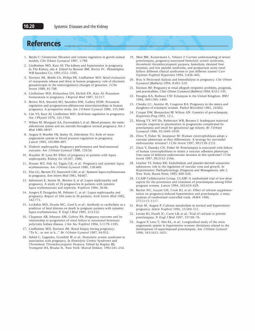
Treatment alogrithm for chronic hypertension
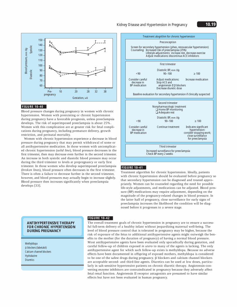
FIGURE 10-40
Blood pressure changes during pregnancy in women with chronichypertension. Women with preexisting or chronic hypertensionduring pregnancy have a favorable prognosis, unless preeclampsiadevelops. The risk of superimposed preeclampsia is about 25%.Women with this complication are at greater risk for fetal compli-cations during pregnancy, including premature delivery, growthrestriction, and perinatal mortality.
Women with chronic hypertension experience a decrease in bloodpressure during pregnancy that may permit withdrawal of some orall antihypertensive medication. In those women with uncomplicat-ed chronic hypertension (solid line), blood pressure decreases in thefirst trimester, then may decrease even further in the second trimester.An increase in both systolic and diastolic blood pressure may occurduring the third trimester to levels at prepregnancy or early firsttrimester. In those women who develop superimposed preeclampsia(broken lines), blood pressure often decreases in the first trimester.There is often a failure to decrease further in the second trimester,however, and blood pressures may actually begin to increase slightly.Blood pressure then increases significantly when preeclampsiadevelops [33].
FIGURE 10-41
Treatment algorithm for chronic hypertension. Ideally, patientswith chronic hypertension should be evaluated before pregnancy sothat secondary hypertension can be diagnosed and treated appro-priately. Women can be counseled regarding the need for possiblelife-style adjustments, and medications can be adjusted. Blood pres-sure (BP) medications may require adjustment, depending on themagnitude of the pregnancy-related changes in blood pressure. Inthe latter half of pregnancy, close surveillance for early signs ofpreeclampsia increases the likelihood the condition will be diag-nosed before it progresses to a severe stage.
ANTIHYPERTENSIVE THERAPYFOR CHRONIC HYPERTENSIONDURING PREGNANCY
Methyldopa
� blockers (labetalol)
Calcium channel blockers
Hydralazine
Diuretics
FIGURE 10-42
The overall treatment goals of chronic hypertension in pregnancy are to ensure a success-ful full-term delivery of a healthy infant without jeopardizing maternal well-being. Thelevel of blood pressure control that is tolerated in pregnancy may be higher, because therisk of exposure of the fetus to additional antihypertensive agents might outweigh the ben-efits to the mother (for the duration of pregnancy) of having a normal blood pressure.Most antihypertensive agents have been evaluated only sporadically during gestation, andcareful follow-up of children exposed in utero to many of the agents is lacking. The onlyantihypertensive agent for which such follow-up exists is methyldopa. Because no adverseeffects have been documented in offspring of exposed mothers, methyldopa is consideredto be one of the safest drugs during pregnancy. � blockers and calcium channel blockersare acceptable second- and third-line agents. Diuretics can be used at low doses, particu-larly in salt-sensitive hypertensive patients on chronic diuretic therapy. Angiotensin-con-verting enzyme inhibitors are contraindicated in pregnancy because they adversely affectfetal renal function. Angiotensin II receptor antagonists are presumed to have similareffects but have not been evaluated in human pregnancy.
10.20 Systemic Diseases and the Kidney
References
1. Baylis C: Glomerular filtration and volume regulation in gravid animalmodels. Clin Obstet Gynaecol 1987, 1:789.
2. Lindheimer MD, Katz AI: The kidney and hypertension in pregnancy.In The Kidney, edn 4. Edited by Brenner BM, Rector FC. Philadelphia:WB Saunders Co; 1991:1551–1595.
3. Davison JM, Shiells EA, Philips PR, Lindheimer MD: Serial evaluationof vasopressin release and thirst in human pregnancy: role of chorionicgonadotropin in the osmoregulatory changes of gestation. J ClinInvest 1988, 81:798.
4. Lindheimer MD, Richardson DA, Ehrlich EN, Katz AI: Potassiumhomeostasis in pregnancy. J Reprod Med 1987, 32:517.
5. Brown MA, Sinosich MJ, Saunders DM, Gallery EDM: Potassiumregulation and progesterone-aldosterone interrelationships in humanpregnancy. A prospective study. Am J Obstet Gynecol 1986, 155:349.
6. Lim VS, Katz AI, Lindheimer MD: Acid-base regulation in pregnancy.Am J Physiol 1976, 231:1764.
7. Wilson M, Morganti AA, Zervoudakis I, et al.: Blood pressure, the renin-aldosterone system and sex steroids throughout normal pregnancy. Am JMed 1980, 68:97.
8. August P, Mueller FB, Sealey JE, Edersheim TG: Role of renin-angiotensin system in blood pressure regulation in pregnancy. Lancet 1995, 345:896–897.
9. Diabetic nephropathy. Pregnancy performance and fetal-maternal outcome. Am J Obstet Gynecol 1988, 159:56.
10. Hayslett JP, Lynn RI: Effect of pregnancy in patients with lupusnephropathy. Kidney Int 18:207, 1980.
11. Houser MT, Fish AJ, Tagatz GE, et al.: Pregnancy and systemic lupuserythematosus. Am J Obstet Gynecol 1980, 138:409.
12. Fine LG, Barnett EV, Danovitch GM, et al.: Systemic lupus erythematosusin pregnancy. Ann Intern Med 1981, 94:667.
13. Imbasciati E, Surian M, Bottino S, et al: Lupus nephropathy and pregnancy. A study of 26 pregnancies in patients with systemic lupus erythematosus and nephritis. Nephron 1984, 36:46.
14. Jungers P, Dougodos M, Pelissier C, et al.: Lupus nephropathy andpregnancy. Report of 104 cases in 36 patients. Arch Intern Med 1982,142:771.
15. Lockshin MD, Druzin MC, Goel S, et al.: Antibody to cardiolipin as apredictor of fetal distress on death in pregnant patients with systemiclupus erythematosus. N Engl J Med 1985, 313:152.
16. Chapman AB, Johnson AM, Gabow PA: Pregnancy outcome and itsrelationship to progression of renal failure in autosomal dominantpolycystic kidney disease. J Am Soc Nephrol 1994, 5:1178–1185.
17: Lindheimer MD, Davison JM. Renal biopsy during pregnancy: “To b... or not to b...” Br J Obstet Gynecol 1987, 94:932.
18. Saltiel C, Legendre, Grunfeld JP, et al.: Hemolytic uremic syndrome inassociation with pregnancy. In Hemolytic Uremic Syndrome andThrombotic Thrombocytopenic Purpura. Edited by Kaplan BS,Trompeter RS, Moake JL. New York: Marcel Dekker; 1992:241–254.
19. Sibai BM, Kustermann L, Velasco J: Current understanding of severepreeclampsia, pregnancy-associated hemolytic uremic syndrome,thrombotic thrombocytopenic purpura, hemolysis, elevated liverenzymes, and low platelet syndrome, and postpartum acute renal failure: different clinical syndromes or just different names? CurrOpinion Nephrol Hypertens 1994, 3:436–445.
20. Hou S: Peritoneal dialysis and hemodialysis in pregnancy. Clin ObstetGynaecol (Balliere) 1994, 8:491–510.
21. Davison JM: Pregnancy in renal allograft recipients: problems, prognosis,and practicalities. Clinc Obstet Gynaecol (Balliere) 1994, 8:511–535.
22. Douglas KA, Redman CW: Eclampsia in the United Kingdom. BMJ1994, 309:1395–1400.
23. Chesley LC, Annitto JE, Cosgrove RA: Pregnancy in the sisters anddaughters of eclamptic women. Pathol Microbiol 1961, 24:662.
24. Cooper DW, Brenneckes SP, Wilton AN: Genetics of pre-eclampsia.Hypertens Preg 1993, 12:1.
25. Khong TY, WF De, Robertson WB, Brosens I: Inadequate maternalvascular response to placentation in pregnancies complicated bypreeclampsia and small for gestational age infants. Br J ObstetGynaecol 1986, 93:1049–1059.
26. Zhou Y, Fisher SJ, Janatpour M: Human cytotrophoblasts adopt avascular phenotype as they differentiate. A strategy for successfulendovascular invasion? J Clin Invest 1997, 99:2139–2151.
27. Zhou Y, Damsky CH, Fisher SJ: Preeclampsia is associated with failureof human cytotrophoblasts to mimic a vascular adhesion phenotype.One cause of defective endovascular invasion in this syndrome? J ClinInvest 1997, 99:2152–2164.
28. Lüscher TF, Dubey RK: Endothelium and platelet=derived vasoactivesubstances: role in the regulation of vascular tone and growth. InHypertension: Pathophysiology, Diagnosis and Management, edn 2.New York: Raven Press; 1995: 609–630.
29. CLASP Collaborative Group. CLASP: A randomized trial of low-doseaspirin for the prevention and treatment of preeclampsia among 9364pregnant women. Lancet 1994, 343:619–629.
30. Bucher HC, Guyatt GH, Cook RJ, et al.: Effect of calcium supplemen-tation on pregnancy-induced hypertension and preeclampsia: a meta-analysis of randomized controlled trials. JAMA 1996,275:1113–1117.
31. Hojo M, August P: Calcium metabolism in normal and hypertensivepregnancy. Semin Nephrol 1995, 15:504–511.
32. Levine RJ, Hauth JC, Curet LB, et al.: Trial of calcium to preventpreeclampsia. N Engl J Med 1997, 337:69–76.
33. August P, Lenz T, Ales KL, et al.: Longitudinal study of the reninangiotensin system in hypertensive women: deviations related to thedevelopment of superimposed preeclampsia. Am J Obstet Gynecol1990, 163:1612–1621.