Journal of Molecular Structure - Kocaeli...
11
The vibrational studies and theoretical investigation of structure, electronic and non-linear optical properties of Sudan III [1-{[4-(phenylazo) phenyl]azo}-2-naphthalenol] Aslı Esme a , Seda Gunesdogdu Sagdinc b,⇑ a Department of Elementary Science Education, Kocaeli University, Umuttepe Campus, 41380 Kocaeli, Turkey b Department of Physics, Science and Art Faculty, Kocaeli University, Umuttepe Campus, 41380 Kocaeli, Turkey highlights Structure, electronic and NLO properties, NBO analysis were studied for Sudan III. The FT-IR and FT-Raman spectra of Sudan III were recorded. The calculated vibrational frequencies are in good accordance with experimentally measured values. article info Article history: Received 8 March 2013 Received in revised form 9 May 2013 Accepted 13 May 2013 Available online 21 May 2013 Keywords: Sudan III DFT Vibrational studies NBO NLO properties abstract Sudan III [1-{[4-(phenylazo) phenyl]azo}-2-naphthalenol] is non-ionic fat-soluable dye used as an addi- tive in gasoline, oils and plastics. The molecular structure, molecular electrostatic potential, NBO analysis, linear and non-linear optical properties of Sudan III have been investigated using density functional the- ory (DFT) calculation with 6-311G(d,p) basis set. To investigate the tautomeric stability, optimization cal- culations at B3LYP/6-311G(d,p) level were performed for the azo (OH) and hydrazo (NH) forms of the title compound. The calculated first-order hyperpolarizability value is comparable with the reported values and attractive object for future studies of non-linear optics. FT-IR (4000–400 cm 1 ) and FT-Raman (3500–50 cm 1 ) spectra of Sudan III have been recorded. The vibrational frequencies determined exper- imentally are compared with those obtained theoretically and a vibrational assignment and analysis of the fundamental modes of the compound is performed. The scaled frequencies resulted in good agree- ment with the observed spectral patterns. Ó 2013 Elsevier B.V. All rights reserved. 1. Introduction Azo-compounds are widely used as synthetic organic colorants. Generally, synthetic colorants can be classified as water-soluble or fat-soluble colorants based on their solubility. Most fat-soluble synthetic colorants present in the market are azo compounds, such as Sudan III (SIII) [1]. Belonging to the azo-dye class, sudan dyes are non-ionic fat-soluble dyes used in gasoline, diesel, lubricating grease and polymer dye production, and as dye for food (chilli) and tattoos. SIII [1-{[4-(phenylazo) phenyl]azo}-2-naphthalenol] is fat-soluble dye predominantly used for demonstrating the pres- ence of triglycerides in frozen sections. In addition, SIII is com- monly used for coloring waxes, oils and spirit varnishes [2]. About 50% of the total world colorant production belongs to the so-called azo dyes compounds. The main feature of this dye family is the presence of the azo group (AN@NA) which gives the possi- bility of providing a more extended electronic conjugation of p electrons, and consequently allowing for a strong light absorption in the visible region of the electromagnetic spectrum [3]. Recently, the structural parameters of SIII have been calculated by Hartree–Fock (HF) and density functional theory (DFT) methods for investigation of the tautomerism in it [4]. In that study, the azo (OH) tautomer was found to be preferred in gas phase at the HF le- vel, while the NH isomer was found to be more favorable in the gas phase according to the B3LYP results. Experimental assignment of fundamental frequencies of Raman spectrum for SIII reported in previous work [5] was incomplete and rather tentative due to com- plexity of vibrational spectrum and without wavenumbers of cal- culated spectrum. Also, Dou et al. [5] was reported IR and Raman spectra of OH isomer of SIII between 1700 and 400 cm 1 region. Unfortunately, high-frequency modes (between 4000 and 2000 cm 1 ) were not assigned for SIII, whereas their values are of particular importance for determining tautomer form of SIII. Therefore, the present investigation was undertaken to study the vibrational spectra completely. 0022-2860/$ - see front matter Ó 2013 Elsevier B.V. All rights reserved. http://dx.doi.org/10.1016/j.molstruc.2013.05.022 ⇑ Corresponding author. Tel.: +90 2623032046; fax: +90 2623032003. E-mail address: [email protected] (S.G. Sagdinc). Journal of Molecular Structure 1048 (2013) 185–195 Contents lists available at SciVerse ScienceDirect Journal of Molecular Structure journal homepage: www.elsevier.com/locate/molstruc
Transcript of Journal of Molecular Structure - Kocaeli...

Journal of Molecular Structure 1048 (2013) 185–195
Contents lists available at SciVerse ScienceDirect
Journal of Molecular Structure
journal homepage: www.elsevier .com/ locate /molst ruc
The vibrational studies and theoretical investigation of structure,electronic and non-linear optical properties of Sudan III[1-{[4-(phenylazo) phenyl]azo}-2-naphthalenol]
0022-2860/$ - see front matter � 2013 Elsevier B.V. All rights reserved.http://dx.doi.org/10.1016/j.molstruc.2013.05.022
⇑ Corresponding author. Tel.: +90 2623032046; fax: +90 2623032003.E-mail address: [email protected] (S.G. Sagdinc).
Aslı Esme a, Seda Gunesdogdu Sagdinc b,⇑a Department of Elementary Science Education, Kocaeli University, Umuttepe Campus, 41380 Kocaeli, Turkeyb Department of Physics, Science and Art Faculty, Kocaeli University, Umuttepe Campus, 41380 Kocaeli, Turkey
h i g h l i g h t s
� Structure, electronic and NLO properties, NBO analysis were studied for Sudan III.� The FT-IR and FT-Raman spectra of Sudan III were recorded.� The calculated vibrational frequencies are in good accordance with experimentally measured values.
a r t i c l e i n f o
Article history:Received 8 March 2013Received in revised form 9 May 2013Accepted 13 May 2013Available online 21 May 2013
Keywords:Sudan IIIDFTVibrational studiesNBONLO properties
a b s t r a c t
Sudan III [1-{[4-(phenylazo) phenyl]azo}-2-naphthalenol] is non-ionic fat-soluable dye used as an addi-tive in gasoline, oils and plastics. The molecular structure, molecular electrostatic potential, NBO analysis,linear and non-linear optical properties of Sudan III have been investigated using density functional the-ory (DFT) calculation with 6-311G(d,p) basis set. To investigate the tautomeric stability, optimization cal-culations at B3LYP/6-311G(d,p) level were performed for the azo (OH) and hydrazo (NH) forms of the titlecompound. The calculated first-order hyperpolarizability value is comparable with the reported valuesand attractive object for future studies of non-linear optics. FT-IR (4000–400 cm�1) and FT-Raman(3500–50 cm�1) spectra of Sudan III have been recorded. The vibrational frequencies determined exper-imentally are compared with those obtained theoretically and a vibrational assignment and analysis ofthe fundamental modes of the compound is performed. The scaled frequencies resulted in good agree-ment with the observed spectral patterns.
� 2013 Elsevier B.V. All rights reserved.
1. Introduction
Azo-compounds are widely used as synthetic organic colorants.Generally, synthetic colorants can be classified as water-soluble orfat-soluble colorants based on their solubility. Most fat-solublesynthetic colorants present in the market are azo compounds, suchas Sudan III (SIII) [1]. Belonging to the azo-dye class, sudan dyes arenon-ionic fat-soluble dyes used in gasoline, diesel, lubricatinggrease and polymer dye production, and as dye for food (chilli)and tattoos. SIII [1-{[4-(phenylazo) phenyl]azo}-2-naphthalenol]is fat-soluble dye predominantly used for demonstrating the pres-ence of triglycerides in frozen sections. In addition, SIII is com-monly used for coloring waxes, oils and spirit varnishes [2].
About 50% of the total world colorant production belongs to theso-called azo dyes compounds. The main feature of this dye familyis the presence of the azo group (AN@NA) which gives the possi-
bility of providing a more extended electronic conjugation of pelectrons, and consequently allowing for a strong light absorptionin the visible region of the electromagnetic spectrum [3].
Recently, the structural parameters of SIII have been calculatedby Hartree–Fock (HF) and density functional theory (DFT) methodsfor investigation of the tautomerism in it [4]. In that study, the azo(OH) tautomer was found to be preferred in gas phase at the HF le-vel, while the NH isomer was found to be more favorable in the gasphase according to the B3LYP results. Experimental assignment offundamental frequencies of Raman spectrum for SIII reported inprevious work [5] was incomplete and rather tentative due to com-plexity of vibrational spectrum and without wavenumbers of cal-culated spectrum. Also, Dou et al. [5] was reported IR and Ramanspectra of OH isomer of SIII between 1700 and 400 cm�1 region.Unfortunately, high-frequency modes (between 4000 and2000 cm�1) were not assigned for SIII, whereas their values are ofparticular importance for determining tautomer form of SIII.Therefore, the present investigation was undertaken to study thevibrational spectra completely.

Fig. 1. (a) Tautomer equilibrium for the SIII, (b) OH form and (c) NH form of the optimized structures obtained using B3LYP/6-311G(d,p) level of SIII.
186 A. Esme, S.G. Sagdinc / Journal of Molecular Structure 1048 (2013) 185–195
It is known that organic molecules formed by a donor–acceptorpair connected to a p-delocalized framework present attractivenon-linear optics (NLO) characteristics, which can be estimatedfrom their hyperpolarizabilities [6–9]. Recently, there has been agrowing interest in the nonlinear optical (NLO) properties of azomaterials with donor–acceptor groups for their large nonlinearrefraction [10], which are interesting for the application in opticalstorage, optical-limiting and optical switching application [11,12].Their nonlinear optical response may result from an electronicand/or nonelectronic process. Electronic nonlinearities occur asthe result of the nonlinear response of bound electrons on an ap-plied optical field. Furthermore, the usually good planarity of theazo bridge plays an important role in larger p electron transmis-sion effects [13–16]. The first-order hyperpolarizability (b), givesinformation about the material capability to generate second ordernon-linear effects. Also, the experimental and theoretical studieshave been expanded to understand many aspects of molecularhyperpolarizabilities [13].
The use of quantum chemical method as DFT for molecularhyperpolarizability is expected to supply a guidance and accelaratesubsequent experimental studies [17,18]. Thus, in the presentwork, the molecular structure, EHOMO (the highest occupied molec-ular orbital energy), ELUMO (the lowest unoccupied molecular orbi-tal energy), LUMO–HOMO energy gap (DE), electron affinity (A),ionization potential (I), global hardness (g), softness (r),electronegativity (v), dipole moment (l), polarizabilities (hai),the anisotropy of the polarizabilities (hDai) and the first-orderhyperpolarizabilities (b) have been investigated using B3LYP meth-od with 6-311G(d,p) basis set on the azo (OH) and hydrazo (NH)forms of SIII azo dye (Fig. 1a). Moreover, vibrational frequenciesof SIII have been studied by FT-IR and FT-Raman spectroscopiesand the vibrational spectra of the solid state SIII has been assignedbased on B3LYP/6-311G(d,p) method.
2. Experimental
SIII was purchased from Sigma–Aldrich Company and was usedwithout any further purification. The Fourier transform infrared(FT-IR) spectrum of SIII have been recorded using Shimadzu FT-IR 8201PC spectrometer with the KBr technique, in the region4000–400 cm�1 which was calibrated by polystyrene. The FT-Ra-man spectrum of SIII has been measured and have been recordedusing a Nd:YAG laser (9394.8 cm�1) for excitation in the regionof 3500–50 cm�1 on a Bruker-FRA-106 spectrophotometer with ascanning speed of 200 cm�1 min�1 and a spectral width of4.0 cm�1.
3. Method of calculations
All calculations were performed using the Gaussian 09W [19]software package and GaussView Rev 5.0.9 [20] visualizationprograms. The DFT calculations were performed using Becke’sthree-parameter hybrid functional [21] with the Lee–Yang–Parrcorrelation functional [22], a combination that gives rise to thewell-known B3LYP method. In addition, B3LYP results using 6-311G(d,p) basis set was also used to obtain the NLO properties,energies (EHOMO, ELUMO and DE = ELUMO � EHOMO) and other quan-tum chemical parameters of SIII. After geometry optimization,the vibrational spectra have been calculated with the B3LYP/6-311G(d,p) method. The frequencies obtained from the B3LYP calcu-lations have been scaled by a factor of 0.967, which is a typicalcorrection factor for B3LYP frequencies [23].
In this study, the natural bond orbital (NBO) calculations havebeen performed using NBO 3.1 program [24] as implemented inthe Gaussian 09W package at the DFT/B3LYP level. In the contextof the HF theorem, the EHOMO and ELUMO are used to approximatethe ionization potential (I) and electron affinity (A) given by Koop-mans’ theorem [25], respectively. Although no formal proof of thistheorem exists within DFT, its validity is generally accepted. I and Aare related to
I ¼ �EHOMO A ¼ �ELUMO ð1Þ
If we assume that these relations are valid within the DFTframe, the electronegativity (v) and hardness (g) can be estimatedwith
v ¼ I þ A2
� �; g ¼ I � A
2
� �ð2Þ
softness (r) is also given by [26]
r ¼ 1=g ð3Þ
Polarizabilities were calculated at the same level of theory usingthe standard Gaussian 09W keyword ‘Polar’ [27]. This keywordmeans that the polarizabilities were obtained analytically ratherthan by numerical differentiation. The energy of an unchargedmolecule under a weak, general electric field can be expressed byBuckingham type expansion [28–30]
E ¼ E0 � liFi � ð1=2ÞaijFiFj � ð1=6ÞbijkFiFjFk þ � � � ð4Þ
where E is the energy of a molecule under the electric field F, E0 isthe unperturbed energy of a free molecule, Fi is the vector compo-nent of the electric field in the i direction, and liaij;bijk are thedipole moment, the polarizability and the first-order

Tabl
e1
Som
eex
peri
men
tal
and
calc
ulat
edbo
ndle
ngth
s(Å
)an
ddi
hedr
alan
gles
(�)
atB3
LYP
leve
lof
taut
omer
icfo
rms
for
SIII
.
Bon
dle
ngt
hs
(Å)
C2A
N1
N1A
N2
N2A
C1
1C
14A
N3
N3A
N4
N4A
C1
7C 2
AC 3
C3A
OO
AH
HA
N2
6-31
1G(d
,p)
OH���
N1.
379
1.27
51.
406
1.41
21.
256
1.41
61.
416
1.32
91.
002
1.65
0c
NH���
O1.
323
1.30
81.
394
1.41
01.
256
1.41
61.
478
1.24
81.
717c
1.03
4
6-31
Ga
OH���
N1.
388
1.27
51.
414
1.42
01.
259
1.42
21.
417
1.35
21.
002
1.67
9c
NH���
O1.
332
1.30
91.
402
1.41
71.
259
1.42
21.
476
1.26
91.
714c
1.03
7
X-r
ayb
1.42
81.
247
1.42
81.
428
1.24
71.
428
––
––
Bon
dan
gles
(�)
Dih
edra
lan
gles
(�)
C2A
N1A
N2
N1A
C2A
C 1N
1A
C2A
C3
C3A
C2A
C1
O1A
C3A
C2
O1A
C 3A
C4
N1A
N2A
C 11A
C 12
N2A
N1A
C2A
C 1N
3A
N4A
C1
7A
C1
8N
4A
N3A
C1
4A
C1
3
6-31
1G(d
,p)
OH���
N11
7.15
311
6.83
112
3.76
311
9.40
612
2.22
711
7.64
30.
000
179.
992
0.00
0�
179.
995
NH���
O11
9.94
511
6.95
412
2.99
012
0.05
612
1.69
512
1.19
4�
0.00
2�
179.
990
0.00
117
9.99
2
X-r
ayb
114.
100
115.
600
123.
700
120.
700
––
–�
164.
110
––
aR
ef.[
4].
bR
ef.[
36].
cH
ydro
gen
bon
ds.
A. Esme, S.G. Sagdinc / Journal of Molecular Structure 1048 (2013) 185–195 187
hyperpolarizability, respectively. Here, each subscript of i, j and kdenotes the indices of the Cartesian axes x, y, z, and a repeated sub-script means a summation over the Cartesian indices x, y, z. Theground state dipole moment l, the polarizability hai and the anisot-ropy of the polarizability hDai using the x, y, z components they aredefined as [31,32]
l ¼ ðl2x þ l2
y þ l2z Þ
1=2 ð5Þ
hai ¼ axx þ ayy þ azz
3ð6Þ
hDai ¼ðaxx � ayyÞ2 þ ðayy � azzÞ2 þ ðazz � axxÞ2 þ 6ða2
xy þ a2xz þ a2
yzÞ2
" #1=2
ð7Þ
The first-order hyperpolarizability b is a third rank tensor thatcan be described by 3 � 3 � 3 matrix. The 27 components of 3Dmatrix can be reduced to 10 components due to the Kleinman sym-metry [33]. The output from Gaussian 09 provides 10 componentsof this matrix as bxxx, bxxy, bxyy, byyy, bxxz, bxyz, byyz, bxzz, byzz, bzzz,respectively. The components of the first-order hyperpolarizabilitycan be calculated using the following equation:
b ¼ffiffiffiffiffiffiffiffiffiffiffiffiffiffiffiffiffiffiffiffiffiffiffiffiffiffiffiffiffiffiffiffiffiffiffiffiffiffiffiffiffiffiffiffiffiffiffiffiffiffiffiffiffiffiffiffiffiffiffiffiffiffiffiffiffiffiffiffiffiffiffiffiffiffiffiffiffiffiffiffiffiffiffiffiffiffiffiffiffiffiffiffiffiffiffiffiffiffiffiffiffiffiffiffiffiffiffiffiffiffiffiffiffiffiffiffiffiffiffiffiffiffiffiffiffiffiffiffiffiffiffiffiðbxxx þ bxyy þ bxzzÞ
2 þ ðbyyy þ byzz þ byxxÞ2 þ ðbzzz þ bzxx þ bzyyÞ
2q
ð8Þ
Since the values of the polarizabilities (hai and hDai) and thefirst-order hyperpolarizability (b) of Gaussian 09W output [19]are reported in atomic units (a.u.), the calculated values have beenconverted into electrostatic units (esu) (a: 1 a.u. = 0.1482 � 10�24
esu; b: 1 a.u. = 8.6393 � 10�33 esu) [34].
4. Results and discussion
4.1. Geometrical structure
In general, sudan dyes display two possible tautomeric forms,the azo (OAH) and hydrazo (NAH) forms, as shown in Fig. 1a forSIII [4]. Depending on the tautomers, two types of intramolecularhydrogen bonds are observed in sudan dyes: OAH���N in azo andNAH���O in hydrazo tautomers. Several researchers have studiedthe azo/hydrazo forms of sudan dyes [4,35]. The position and nat-ure of the equilibrium depends on the solvent utilized and temper-ature being two of the most impotant. In this study, the optimizemolecular structures for the azo (OH) and hydrazo (NH) tautomersof the azo dye SIII using the DFT/B3LYP level with 6-311G(d,p) ba-sis set are depicted in Fig. 1b and c). The comparative optimizedstructural parameters such as bond lengths, bond angles and dihe-dral angles for the azo (OH) and hydrazo (NH) tautomers of SIII byperforming B3LYP with 6-311G(d,p) basis set are presented inTable 1. The experimental structure of SIII has not been reporteduntil now; therefore we have compared the calculated bondlengths of SIII with its experimentally available parent compoundtrans-azobenzene from the X-ray study [36] and B3LYP/6-31G val-ues obtained by Dos Santos et al. [4].
Analyses of the conformation of individual rings for azo dyeshave been important [37,38], thus the dihedral angles aroundNAN moiety of studied molecule have been investigated. Thevalues of N1AN2AC11AC12, N2AN1AC2AC1, N3AN4AC17AC18 andN4AN3AC14AC13 dihedral angles are given in Table 1. It can be seenthat all the rings for both forms were found planar (�180�). Theplanarity of molecules favors a strong intramolecular hydrogenbond. The predicted OAH� � �N (OH) and NAH� � �O (NH) distancesfor the compound SIII are, respectively, 1.650 Å for the OH tauto-mer, and 1.717 Å for the NH tautomer (with B3LYP/6-311G(d,p)).
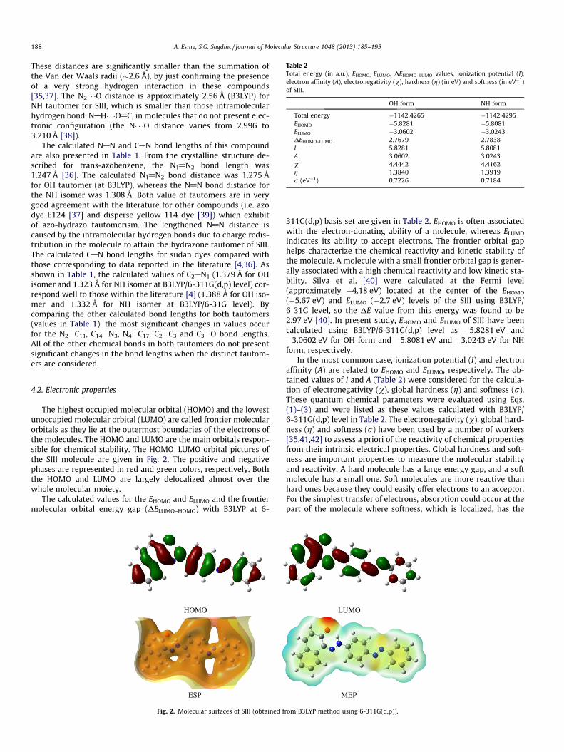
Table 2Total energy (in a.u.), EHOMO, ELUMO, DEHOMO–LUMO values, ionization potential (I),electron affinity (A), electronegativity (v), hardness (g) (in eV) and softness (in eV�1)of SIII.
OH form NH form
Total energy �1142.4265 �1142.4295EHOMO �5.8281 �5.8081ELUMO �3.0602 �3.0243DEHOMO–LUMO 2.7679 2.7838I 5.8281 5.8081A 3.0602 3.0243v 4.4442 4.4162g 1.3840 1.3919r (eV�1) 0.7226 0.7184
188 A. Esme, S.G. Sagdinc / Journal of Molecular Structure 1048 (2013) 185–195
These distances are significantly smaller than the summation ofthe Van der Waals radii (�2.6 Å), by just confirming the presenceof a very strong hydrogen interaction in these compounds[35,37]. The N2� � �O distance is approximately 2.56 Å (B3LYP) forNH tautomer for SIII, which is smaller than those intramolecularhydrogen bond, NAH� � �O@C, in molecules that do not present elec-tronic configuration (the N� � �O distance varies from 2.996 to3.210 Å [38]).
The calculated NAN and CAN bond lengths of this compoundare also presented in Table 1. From the crystalline structure de-scribed for trans-azobenzene, the N1@N2 bond length was1.247 Å [36]. The calculated N1@N2 bond distance was 1.275 Åfor OH tautomer (at B3LYP), whereas the N@N bond distance forthe NH isomer was 1.308 Å. Both value of tautomers are in verygood agreement with the literature for other compounds (i.e. azodye E124 [37] and disperse yellow 114 dye [39]) which exhibitof azo-hydrazo tautomerism. The lengthened N@N distance iscaused by the intramolecular hydrogen bonds due to charge redis-tribution in the molecule to attain the hydrazone tautomer of SIII.The calculated CAN bond lengths for sudan dyes compared withthose corresponding to data reported in the literature [4,36]. Asshown in Table 1, the calculated values of C2AN1 (1.379 Å for OHisomer and 1.323 Å for NH isomer at B3LYP/6-311G(d,p) level) cor-respond well to those within the literature [4] (1.388 Å for OH iso-mer and 1.332 Å for NH isomer at B3LYP/6-31G level). Bycomparing the other calculated bond lengths for both tautomers(values in Table 1), the most significant changes in values occurfor the N2AC11, C14AN3, N4AC17, C2AC3 and C3AO bond lengths.All of the other chemical bonds in both tautomers do not presentsignificant changes in the bond lengths when the distinct tautom-ers are considered.
4.2. Electronic properties
The highest occupied molecular orbital (HOMO) and the lowestunoccupied molecular orbital (LUMO) are called frontier molecularorbitals as they lie at the outermost boundaries of the electrons ofthe molecules. The HOMO and LUMO are the main orbitals respon-sible for chemical stability. The HOMO–LUMO orbital pictures ofthe SIII molecule are given in Fig. 2. The positive and negativephases are represented in red and green colors, respectively. Boththe HOMO and LUMO are largely delocalized almost over thewhole molecular moiety.
The calculated values for the EHOMO and ELUMO and the frontiermolecular orbital energy gap (DELUMO–HOMO) with B3LYP at 6-
HOMO
ESP
Fig. 2. Molecular surfaces of SIII (obtained f
311G(d,p) basis set are given in Table 2. EHOMO is often associatedwith the electron-donating ability of a molecule, whereas ELUMO
indicates its ability to accept electrons. The frontier orbital gaphelps characterize the chemical reactivity and kinetic stability ofthe molecule. A molecule with a small frontier orbital gap is gener-ally associated with a high chemical reactivity and low kinetic sta-bility. Silva et al. [40] were calculated at the Fermi level(approximately �4.18 eV) located at the center of the EHOMO
(�5.67 eV) and ELUMO (�2.7 eV) levels of the SIII using B3LYP/6-31G level, so the DE value from this energy was found to be2.97 eV [40]. In present study, EHOMO and ELUMO of SIII have beencalculated using B3LYP/6-311G(d,p) level as �5.8281 eV and�3.0602 eV for OH form and �5.8081 eV and �3.0243 eV for NHform, respectively.
In the most common case, ionization potential (I) and electronaffinity (A) are related to EHOMO and ELUMO, respectively. The ob-tained values of I and A (Table 2) were considered for the calcula-tion of electronegativity (v), global hardness (g) and softness (r).These quantum chemical parameters were evaluated using Eqs.(1)–(3) and were listed as these values calculated with B3LYP/6-311G(d,p) level in Table 2. The electronegativity (v), global hard-ness (g) and softness (r) have been used by a number of workers[35,41,42] to assess a priori of the reactivity of chemical propertiesfrom their intrinsic electrical properties. Global hardness and soft-ness are important properties to measure the molecular stabilityand reactivity. A hard molecule has a large energy gap, and a softmolecule has a small one. Soft molecules are more reactive thanhard ones because they could easily offer electrons to an acceptor.For the simplest transfer of electrons, absorption could occur at thepart of the molecule where softness, which is localized, has the
LUMO
MEP
rom B3LYP method using 6-311G(d,p)).

Table 3The atomic charge distribution of C, N and O atoms of SIII molecule calculated by theMulliken and natural bond orbital (NBO) methods using B3LYP/6-311G(d,p) level.
Atoms B3LYP
Mulliken NBO
O �0.414 �0.629C1 �0.095 �0.034C2 0.056 0.075C3 0.287 0.452C4 �0.114 �0.262C5 �0.013 �0.117C6 �0.075 �0.084C7 �0.062 �0.167C8 �0.082 �0.202C9 �0.088 �0.180C10 �0.006 �0.176C11 0.222 0.167C12 �0.075 �0.226C13 �0.057 �0.156C14 0.040 0.103C15 �0.019 �0.178C16 �0.113 �0.213C17 0.038 0.112C18 �0.022 �0.194C19 �0.097 �0.195C20 �0.082 �0.183C21 �0.093 �0.202C22 �0.052 �0.175N1 �0.192 �0.196N2 �0.313 �0.302N3 �0.201 �0.212N4 �0.207 �0.215
A. Esme, S.G. Sagdinc / Journal of Molecular Structure 1048 (2013) 185–195 189
highest value. As seen from Table 2, the global hardness of the OHtautomer is greater than the NH tautomer and softness of NH tau-tomer is smaller than the OH tautomer of the title compound. Thetotal energy has also influence on the stability of a molecule. Thetotal energy of the azo NH form (�1142.4295 a.u.) is greater thanhydrazo OH one (�1142.4265 a.u.). These results indicate thatthe hydrazo (NH) form of SIII is more stable than azo (OH) formin gas phase.
4.3. NBO analysis
NBO analysis provides an efficient method for studying intra-and inter-molecular bonding and interaction among bonds, andalso provides a convenient basis for investigating charge transferor conjugative interaction in molecular systems [43]. The second-order Fock-matrix was carried out to evaluate different types of do-nor–acceptor interactions and their stabilization energies in theNBO basis [44]. The interactions result in a loss of occupancy fromthe localized NBO of the idealized Lewis structure into an emptynon-Lewis orbital. For each donor (i) and acceptor (j), the stabiliza-tion energy E(2) associated with the delocalization i ? j is deter-mined as
Eð2Þ ¼ DEij ¼ qi
F2ði; jÞ
ej � eið9Þ
where qi is the donor orbital occupancy; ei, ej, are the diagonal ele-ments and F(i, j) is the off diagonal NBO Fock matrix element. Thelarger the E(2) value, the more intensive is the interaction betweenelectron donors and electron acceptors, i.e., the more the donatingtendency from electron donors to electron acceptors, the greaterthe extent of conjugation of the whole system.
The NBO analysis has been performed to elucidate the intramo-lecular interaction, rehybridization and delocalization, which ispresented in Supplementary Table S1. The second order perturba-tion analysis of Fock matrix of hydrazo form of SIII is summarizedin Supplementary Table S2. As can be seen in Table S2, some strongintramolecular interactions are formed by the orbital overlap be-tween r(CAC) and r�(CAC) and p(CAC) and p�(CAC) bond orbitalsin the aromatic rings, which leads intramolecular charge transfer(ICT) causing stabilization of the system. Also, the NBO analysisconfirmed the presence of N2AH� � �O type of intramolecular hydro-gen bond of SIII. The intramolecular N2AH� � �O hydrogen bond ofNH tautomer for SIII is formed by the orbital overlap betweenthe n(O) and r�(NAH), which results in charge transfer causing sta-bilization of the H-bond systems. The stabilization energy ofNAH� � �O intramolecular hydrogen bonding is 17.05 kcal/mol. TheNBO analysis clearly illustrates the existence of strong NAH� � �Ohydrogen bonding of NH tautomer of SIII. Therefore hydrogenbonding interaction leads to an increase in ED of NAH antibondingorbital which weakens the NAH bonds. The hyperconjuctive inter-action energy (n–r�) between electron-donating O atom and C2AC3
and C4AC3 antibonding orbitals in the naphthalene ring are 12.55and 16.02 kcal/mol, respectively. This occurs a strong intramolecu-lar hyperconjuctive interaction that is formed by the orbital over-lap between p(CAC) and p�(CAC) bond orbital, which results in ICTcausing stabilization of the system.
4.4. Atomic net charges
Mulliken’s population analysis [45] provides a partirioring ofeither a total charge density or an orbital density. Atomic chargeson non-H atoms of SIII obtained by Mulliken population and NBOanalysis using B3LYP/6-311G(d,p) level are given in Table 3. Thecharge distribution on the molecule has an important influenceon the vibrational spectra. As seen in Fig. 3, nitrogen atoms (N1,
N2, N3 and N4) and oxygen atom O have the highest negative Mul-liken charges and behaved as electron acceptors. It was observedthat there is a large accumulation of positive charge on C3 and neg-ative charge on oxygen atom O in SIII molecule. Therefore, thismight have given C3AO bond a greater ionic character. The NBOanalysis shows that the presence of nitro group causes drasticchange in the ring system, as evident from the C2 atom. In SIII com-pound the carbon C2 attached with the nitro group has a much lesspositive charge (0.045e) than that of C11, C14, C17 atoms. This is dueto the electron withdrawing nature of the nitro group by means ofresonance (Fig. 3).
We have investigated the charge population on the O, H and N2
atoms, which contribute directly in the H-transferring process. Forthe SIII molecule, the charges obtained with B3LYP/6-311G(d,p)method indicate high positive charges on the H atom (0.489e and0.419e values for OH and NH tautomers, respectively) during theazo-hydrazo tautomerization, displaying an intramolecular-protontransfer (IPT) between the O and N2 atoms. The positive charge onthe transferring hydrogen for the OH form is higher than the NHform (Table 3).
4.5. Electrostatic potential (ESP) and molecular electrostatic potential(MEP)
The electrostatic potential (ESP) and molecular electrostatic po-tential (MEP) map figures of SIII are shown in Fig. 2. It can be seenfrom the ESP figure, the negative ESP is located more over the oxy-gen and nitrogen atoms and is reflected as a yellowish blob, the po-sitive ESP is localized on the rest of the molecules.
Molecular electrostatic potential (MEP) is related to the elec-tronic density and is a very useful descriptor in understanding sitesfor electrophilic attack and nucleophilic reactions as well as hydro-gen bonding interactions [46,47]. Being a real physical propertyV(r) can be determined experimentally by diffraction or by compu-tational methods [48]. To investigate the reactive sites of the titlecompound the molecular electrostatic potential was evaluated

Fig. 3. Comparative of Mulliken and NBO charge distributions by B3LYP/6-311G(d,p) of the azo (OH) and hydrazo (NH) forms of sudan azo dye SIII.
Table 4Dipole moments l (D), polarizabities hai (in a.u. and esu), the anisotropic of thepolarizabilities hDai (in a.u.) and the first-order hyperpolarizabilities (in a.u and esu)values obtained using B3LYP/6-311G(d,p) method for OH and NH tautomers of SIII.
OH form NH form
l (D) 0.8888 1.6807hai (a.u.) 409.82 409.68hai x10�24 (esu) 60.74 60.71hDai (a.u.) 642.46 644.11b (a.u) 4916.53 990.81btot � 10�30 (esu) 42.48 8.56btot/burea 218 44
190 A. Esme, S.G. Sagdinc / Journal of Molecular Structure 1048 (2013) 185–195
using B3LYP/6-311G(d,p) method. Molecular electrostatic poten-tial, V(r), at a given point r(x,y, z) in the vicinity of a molecule, is de-fined in terms of the interaction energy between the electricalcharge generated from the molecule’s electrons and nuclei and apositive test charge (a proton) located at r. For the system studiedthe V(r) values were calculated as described previously using theequation [49],
VðrÞ ¼X
A
ZA
ðRA � rÞ �Z
qðr0Þðr0 � rÞdr0 ð10Þ
where ZA is the charge of nucleus A, located at RA, q(r0) s the elec-tronic density function of the molecule, and r0 is the dummy inte-gration variable. MEP calculated on the basis of atomic chargesderived from Mulliken population analysis serves as a useful quali-tative descriptor of reactivity. To predict reactive sites for electro-philic and nucleophilic attack of SIII, MEP’s were calculated at theB3LYP/6-311G(d,p) optimized geometries.
The color scheme for the MEP surface is red, electron rich, par-tially negative charge; blue, electron deficient, partially positivecharge; light blue, slightly electron deficient region; yellow,slightly electron rich region; green, neutral; respectively. The colorcode of the maps in the range �0.04436 a.u. (deepest red) to0.04436 a.u. (deepest blue) in title molecule, where blue color indi-cates the strongest attractions and red indicates the strongestrepulsion. The predominance of light green region in the MEP sur-faces corresponds to a potential halfway between the two ex-tremes red and dark blue color. It is obvious from Fig. 2 that theregion around the oxygen atom of the CAO group for SIII repre-sents the more negative potential regions (red) while there is amore positive charge around the hydrogen atoms of SIII.
4.6. Dipole moment, polarizabilities and hyperpolarizabilities
The dipole moment in a molecule is an important property,which is mainly used to study the intermolecular interactionsinvolving the nonbonded type dipole–dipole interactions, becausethe higher the dipole moment, the stronger the intermolecularinteractions will be. The structure of a tautomer obviously affectsthe magnitude and orientation of a dipole moment. The dipole mo-ments of the SIII tautomers obtained using DFT calculation are
summarized in Table 4. The calculated dipole moment for NH form(1.6807 D at B3LYP) is higher than OH form (0.8888 D at B3LYP).
Another important molecular feature of its selected electronicproperties are its polarizability. Ghanadzadeh et al. [42] investi-gated the experimental parameters (e.g. dichroic ratio R) of five su-dan dyes solutions including SIII for OH tautomer by measuring theintensity of the absorption bands in the visible region of parallelaligned samples. They explained the reason of polarizability of su-dan dyes, but no data on the polarizability of those dyes was pro-vided [43]. Therefore, we have not compared this experimentaldata with our theoretical polarizability values for SIII.
The calculated polarizability hai and the anisotropy of the polar-izability hDai for OH and NH forms of SIII using B3LYP/6-311G(d,p)level are listed in Table 4. Softness and polarizability are assumedto be related: ‘‘a soft species is also more polarizable.’’ Thus, a hard(soft) species is known to correspond to a low (high) value of thepolarizability as well as a small (large) size [50]. As seen Table 4,OH form of SIII is softer and more polarizable than NH form of SIII.
In general, the stronger the donor, the smaller the energy differ-ence between ground and excited states, and the longer the wave-length of UV–visible absorption. This red shift suggests an increaseof molecular hyperpolarizability, according to theoretical andexperimental NLO studies [51]. In previous studies, the UV–visibleabsorption spectra of azo dyes in different solvents were investi-gated by several authors [10,42,52]. Sudan dyes contain intramo-lecular charge-transfer chromophores which have large andstable NLO responses. The absorption spectrum of Sudan I was re-corded by the UV–VIS–NIR spectrometer [10]. The absorptionbands of Sudan I in the solution is strong at 488 and 532 nm in

A. Esme, S.G. Sagdinc / Journal of Molecular Structure 1048 (2013) 185–195 191
the visible region. He and Wang [10] have investigated the nonlin-ear optical property of Sudan I under pulse 532 nm. They found thesecond hyperpolarizability to be 1.83 � 10�30 esu [10].
Dos Santos et al. [4] have reported the experimental UV–visiblespectrum of SIII, but they have not given any information regardingthe solution for the recorded spectrum. Two absorption bands ofSIII were observed at 351 and 513 nm in the experimental spec-trum. The band close to 350 nm was not affected by the tautomericequilibrium [4]. The lowest energy transition, responsible for theabsorption band at 484 nm obtained with B3LYP, involves thehighest occupied MO (HOMO) and the lowest unoccupied MO(LUMO) with the Configuration Interaction (CI) contribution equalto 86% (HOMO ? LUMO). Also, Ghanadzadeh et al. [42] investi-gated the maximum absorption wavelengths of SIII in isotropicand anisotropic solvents. They found these wavelengths to bebetween 495 and 520 nm.
In this study, the values of the first-order hyperpolarizabilityobtained using the above Eq. (8) have been calculated usingB3LYP/6-311G(d,p) basis set and are given in Table 4. From Table 4,it was suggested that SIII is polar having non-zero dipole moment,hyperpolarizabilities, and hence have positive microscopic NLObehavior [53,54]. This study reveals that the SIII has large, the
Fig. 4. (a) The FT-IR spectrum of SIII molecule in the wavenumber range 4000–400 cm�
tautomer of SIII.
first-order hyperpolarizability, and has the potential applicationsfor the development of NLO devices. On the other hand, the first-or-der polarizability value obtained using B3LYP/6-311G(d,p) level forNH tautomer of SIII is lower than the OH tautomer. Urea is one ofthe prototypical molecules used in this study for the NLO propertiesof the molecular systems for comparative purposes. It can be seen inTable 4, the calculated b values of SIII for OH and NH tautomersusing B3LYP method with 6-311G(d,p) basis set (the b of urea is0.1947 � 10�30 esu) were found nearly 218 and 44 times more thanthat of urea for OH and NH tautomers, respectively.
The first-order hyperpolarizability b is also analyzed with thehelp of vibrational spectroscopy. Using a simple semi-classicalmodel, in the hypothesis of double (mechanical and electrical) har-monic approximation that the vibrational hyperpolarizability bm
can be obtained with the following expressions, in the limit of sta-tic applied electric fields:
bmnmp ¼
14p2c2
Xk
1m2
k
@ln
@Qk
� �@amp
@Qk
� �þ @lm
@Qk
� �@anp
@Qk
� �þ
@lp
@Qk
� �@anm
@Qk
� �� �ð11Þ
where mk are harmonic vibrational frequencies, @l=@Qk; @a=@Qk and@b=@Qk are derivatives of the molecular dipole moment, polarizabil-ity and hyperpolarizability with respect to the normal coordinate Qk
1 and (b) The simulated IR spectrum computed at B3LYP/6-311G(d,p) level for NH

192 A. Esme, S.G. Sagdinc / Journal of Molecular Structure 1048 (2013) 185–195
and the sum extends over all vibrational normal modes [55]. Theseparameters can be obtained from infrared intensities, Raman andRaman cross sections, respectively.
The peculiar features observed in Raman and IR spectra aresimultaneous activitation of some of the vibrational modes. Theexperimental behavior is well accounted by ab initio calculationsin p-conjugated molecules that predict experimentally large Ra-man cross sections and IR intensities for the same normal modes.The potential applications in the field of nonlinear optics demandsthe investigation of the contribution of IR and Raman active vibra-tional modes to the hyperpolarizability enhancement. The bands at1595, 1441, 1417, 1304, 1230, 1132 and 985 cm�1 observed in FT-IR have their counterparts in Raman 1592, 1439, 1416, 1307, 1233,1133 and 985 cm�1 respectively and their relative intensities in FT-IR and FT-Raman are comparable. This phenomenon is quite unu-sual, since generally, even in the absence of inversion symmetry,the infrared and Raman spectra are complimentary: in most cases,the strongest bands in the Raman are weak in the infrared and viceversa. These bands associated to specific modes that are simulta-neously strongly active both IR and Raman spectra provide evi-dence for the charge transfer interaction between the donor andacceptor group through the p-systems [55].
4.7. Vibrational spectroscopy
Based on the previously reported vibrational spectra of azodyes, the molecular vibrations of SIII were analyzed [5,36,56].The experimental FT-IR and FT-Raman spectra of SIII are given inFigs. 4 and 5, respectively. The scaled vibrational wavenumbers,
Fig. 5. (a) The FT-Raman spectrum of SIII molecule in the wavenumber range 3500–0 cm
their IR and Raman activities, and the form of vibrations obtainedfrom B3LYP/6-311G(d,p) calculations together with the corre-sponding experimental bands and assignments for NH tautomersof SIII are given in Table 5. First, analyzing the overall profile ofthe vibrational spectra, it can be noted that NH and OH structuresshow similar profile. Therefore, the calculated vibrational wave-numbers for NH tautomer of SIII are compared with experimen-tally observed values.
Hydrogen bonds alter the frequencies of the stretching andbending vibrations. It is worth mentioning that the high frequencyregion, where the NH and OH stretching appears, shows a broadband centered at 3426 cm�1 in the solid state IR spectrum, whichcannot be precisely assigned to either NH or OH tautomers. The hy-droxyl stretching vibrations, which are hydrogen bonded to aro-matic ring p-electron systems absorb at 3670–3580 cm�1 [57,58].The peak is border and its intensity is higher than that of a freeOH vibration which indicates involvement in an intramolecularhydrogen bonding. For aromatic compounds, the NAH stretchingfrequencies are observed near 3400 cm�1 [57]. As in the case ofOAH stretching, the frequency of NAH stretching is reduced byintramolecular hydrogen bonding though to a lesser degree be-cause the dipole moment of an NAH bond is smaller than that ofOAH bond, which means the hydrogen bonding in NAH group isweaker than in OAH group [56]. For comparison, the calculatedm(NH) and m(OH) frequencies were 3089 and 2910 cm�1, respec-tively, with the former value closer (10% lower) to the experimen-tal frequency. The frequencies and intensities for relevant bandsobserved in the IR spectrum are included as Table 5, where onlythe calculated values for NH isomer are shown.
-1 and (b) the simulated Raman spectrum computed at B3LYP/6-311G(d,p) of SIII.

Table 5Comparison of the experimental (FT-IR and FT-Raman) wavenumbers (cm�1) and theoretical harmonic wavenumbers (cm�1) for NH tautomer of SIII by B3LYP/6-311G(d,p)method.
Assignments Experimental Theoretical
FT-IR FT-Raman Aa Bb IR Int. Rel. R Act. Rel.
mas(NAH) 3426(mw) – 3194 3089 27 2.7 209 0.9m(CAH)na, ms(NAH), mas(CAH)ph1 3058(w) 3061(w) 3192 3087 53 5.3 3523 14.4mas(CAH)ph1, m(NAH) 3039(vw) – 3191 3086 10 1.0 209 0.9m(CAH)na, m(NAH) – – 3190 3085 34 3.4 468 1.9mas(CAH)ph2 – – 3185 3080 31 3.1 435 1.8mas(CAH)ph2 – – 3175 3070 12 1.2 221 0.9mas(CAH)na – – 3172 3067 12 1.2 176 0.7mas(CAH)ph2 – – 3164 3060 3 0.3 40 0.2mas(CAH)na 2914(vw) – 3162 3058 11 1.1 130 0.5mas(CAC)na, d(NAH), m(C@O) 1620(mw) – 1670 1615 110 10.9 93 0.4mas(CAC)na, d(NAH), m(N@N) – 1651 1597 3 0.3 403 1.6r(CAC)ph1,ph2, m(N@N) 1595(mw) 1592(m) 1646 1592 55 5.4 13245 54.0m(CAC)ph1,ph2, m(N@N) 1560(vw) – 1640 1586 25 2.5 250 1.0m(CAC)na, d(NAH) – – 1629 1575 37 3.7 461 1.9m(CAC)ph2 – – 1625 1571 7 0.7 70 0.3d(CAH)na, d(NAH), m(CAC)na 1551(mw) – 1588 1537 110 10.9 243 1.0d(NAH), m(C@N), m(NAN) 1501 (vs) – 1567 1515 1012 100 400 1.6d(CAH)ph1,ph2, m(N@N), m(CAC)ph1 – 1491(w) 1544 1493 121 12.0 19822 80.8d(CAH)ph1,ph2, m(N@N), m(CAC)ph1, m(C@N) 1451(w) 1463(mw) 1524 1474 20 2.0 2483 10.1d(CAH)na, m(CAC)na – – 1514 1464 61 6.0 176 0.7d(CAH)ph1,ph2, m(N@N) 1441(vw) 1439(m) 1504 1454 30 3.0 10127 41.3d(CAH)na – – 1483 1434 48 4.7 531 2.2d(CAH)ph2, m(N@N) 1417(vw) 1416(m) 1479 1430 47 4.6 6070 24.7d(CAH)ph1, d(CAH)na, m(C@N) 1400(vw) 1386(m) 1459 1411 3 0.3 13617 55.5d(CAH)ph1, d(CAH)na, m(C@N) – 1435 1388 178 17.6 4612 18.8d(CAH)na,ph2, d(CAC)ph1 – 1334(vw) 1362 1317 15 1.5 771 3.1d(CAC)na – – 1354 1309 119 11.8 261 1.1d(CAH)ph1,ph2,na, m(CAN) 1317(w) – 1345 1301 51 5.0 1907 7.8d(CAH)ph2 1304(w) 1307(w) 1336 1291 10 1.0 1111 4.5m(N@N), m(CAO), d(CAH)ph1,na – – 1313 1270 634 62.7 1145 4.7br(ph1–ph2), d(CCH)na,, m(CAN) 1257(s) 1255(vw) 1265 1223 370 36.6 3693 15.1d(CAH)na,ph1, br(ph2) – – 1259 1218 17 1.7 1806 7.4r(CCH)na 1230(m) 1233(w) 1234 1193 260 25.7 236 1.0r(CCH)ph1,ph2,na, r(CNN), r(NNH) – – 1212 1172 11 1.1 1421 5.8r(CH)na,ph2,ph1, r(CNN), r(NNH) 1209(s) 1208(vw) 1209 1169 6 0.6 2542 10.4r(CAH)na 1177(w) 1180(vw) 1183 1144 17 1.7 313 1.3r(CAH)ph1,ph2, m(CAN) 1147(m) 1159(vw) 1171 1132 115 11.3 700 2.9r(CAH)ph1,ph2, m(CAN) 1132(s) 1133(vs) 1154 1115 174 17.2 24536 100r(CAH)ph1 1103(vw) 1104(vw) 1128 1091 12 1.2 41 0.2r(CAH)na, r(CCC)na 1069(vw) – 1117 1080 30 3.0 459 1.9r(CAH)ph2, r(CCC)ph2 1038(vw) – 1098 1062 7 0.7 122 0.5r(CAH)na, r(CCC)na 1018(vw) – 1062 1027 3 0.3 36 0.2r(CAH)ph2, r(CCC)ph2 1001(vw) 1000(vw) 1041 1007 11 1.1 7 0t(CAH)ph2 – – 1016 983 2 0.2 755 3.1t(CAH)ph1,na 985(m) 985(vw) 1006 973 47 4.6 407 1.7t(CAH)ph2 951(w) – 958 926 5 0.5 0 0br(ph1–ph2) 926(vw) – 940 909 2 0.2 10 0t(CAH)na, c(NAH), x(CAH)ph1 867(vw) – 887 858 69 6.8 1 0d(CCC)na, br(ph1–ph2), c(NAH) 837(s) – 883 854 79 7.8 37 0.2d(CCC)na, br(ph1–ph2), q(NAN) – – 870 841 18 1.8 42 0.2x(CH)na,ph1, x(NH) – – 855 827 16 1.6 1 0br(na–ph1–ph2) – – 831 804 4 0.4 3 0x(CAH)ph2 768(ms) – 794 768 23 2.3 0 0t(CAH)na, t(NAH) – – 791 765 6 0.6 1 0x(CAH)na – – 769 744 38 3.8 1 0br (CCC)na, br(ph1–ph2), c(CN) 750(mw) – 761 736 29 2.9 35 0.1t(CAH)ph1, x(CAH)ph2 – – 748 723 13 1.3 12 0.1d(CCC)na, br(ph1–ph2) 722(mw) 724(vw) 736 712 11 1.1 147 0.6x(CAH)ph2 685(m) – 705 682 39 3.9 3 0br(na–ph1–ph2) – – 683 661 1 0.1 22 0.1d(CCC)na,ph1,ph2 669(vw) – 654 632 5 0.5 148 0.6d(CCC)na,ph1,ph2 – – 638 617 12 1.2 57 0,2d(CCC)ph2 623(vw) 622(vw) 630 609 1 0.1 76 0.3d(CCC)na,ph1,ph2, s(CNNC) 577(vw) 570(vw) 580 561 9 0.9 132 0.5x(CAH)na,ph1,ph2 – – 566 547 13 1.3 2 0t(CAH)na, x(CAH)ph1,ph2 551(vw) – 552 534 13 1.3 2 0Br(na,ph1,ph2), d(CNNC) 538(vw) – 540 522 11 1.1 64 0.2d(CAC)na,ph1,ph2 521(m) 518(vw) 537 519 38 3.8 6 0.02t(CAH)na, x(CAH)ph2, x(NAH) – – 530 513 17 1.7 6 0.02x(CAH)na,ph1,ph2 492(vw) 493(vw) 508 491 6 0.6 1 0.00Br(na,ph1,ph2) – – 498 482 4 0.4 15 0.06s(C@OAC@N) 453(vw) 458(vw) 471 456 5 0.5 90 0.4
(continued on next page)
A. Esme, S.G. Sagdinc / Journal of Molecular Structure 1048 (2013) 185–195 193
Table 5 (continued)
Assignments Experimental Theoretical
FT-IR FT-Raman Aa Bb IR Int. Rel. R Act. Rel.
d(CAC)na 428(vw) 426(vw) 429 415 1 0.1 1 0.0c(CAC)ph1,ph2, c(N@N) 417(vw) 416(vw) 405 392 4 0.4 5 0.02d(CAC)na,ph1 – 375(vw) 374 362 8 0.8 83 0.3r(na–ph1–ph2) – – 329 318 9 0.9 24 0.1c(CAC)ph1,ph2,na – – 281 272 0 0 3 0.01r(na–ph1–ph2) – – 260 251 0 0 8 0.03r(na–ph1–ph2) – – 197 191 2 0.2 6 0.02br(na–ph1–ph2) – 148(m) 145 140 0 0 25 0.1c(CAC)ph1,ph2,na – – 112 108 0 0 1 0.0s(na–ph1–ph2) – – 91 88 2 0.2 1 0.0r(na–ph1–ph2) – – 82 79 2 0.2 17 0.1s(na–ph1–ph2) – – 55 53 0 0 7 0.02r(na–ph1–ph2) – – 35 34 0 0 8 0.03s(na–ph1–ph2) – – 28 27 0 0 1 0.0s(na–ph1–ph2) – – 20 19 0 0 3 0.01
m, stretch; d, deformation vibration; d, in plane bending; c, out-of –plane bending; x, wagging; t, twisting; r, scissoring; s, torsion; q, rocking; br, ring breath; s, simetric; as,antisimetric; na, naphthalene ring; ph, benzene ring (the same below).
a Unscaled wavenumber.b Scaled wavenumber.
194 A. Esme, S.G. Sagdinc / Journal of Molecular Structure 1048 (2013) 185–195
Due to the aromatic stretching, multiple weak bands are exhib-ited in the region of 3100–3000 cm�1. The CH vibrations of the titlecompound are observed at 3058, 3039 and 2914 cm�1 in FT-IR. TheB3LYP calculations give bands in the range 3087–3051 cm�1 asm(CH) modes.
The absorptions around 1560–1618 cm�1 also have contribu-tions from d(NH) and m(C@O), characteristic of NH tautomer [30].The C@O stretching band is often intense and appears in the region1680–1630 cm�1 [54]. The position of C@O stretching dependsupon conjunction, hydrogen bonding and the size of the ring towhich it is attached. NH bending band in the IR spectrum of theazo dye is assigned at 1626 cm�1 [59]. Dou et al. [5] have not as-signed this band observed at 1618 cm�1. Also, the bands between1650 and 1400 cm�1 in benzene derivatives are assigned to CACstretching mode. The C@O stretching band and naphthalene ringCAC stretching bands coupled to NH bending band at 1620 cm�1
of SIII is assigned to a calculated mode at 1615 cm�1.The determination of CAN, C@C, NAN and N@N vibrations is a
very difficult task, since the mixing of vibrations is possible in thisregion. The stretching vibration of the azo group has been studiedextensively by different authors [5,37]. The N@N stretching vibra-tion is expected in the region 1440–1380 cm�1 [56], however veryshort N@N bonds obtained from calculated values has shownthat the N@N stretching band is high vibrational frequency regionin present study. This vibration in the IR and Raman spectra of SIIIis observed in the region at 1595–1417 cm�1 and 1592–1416 cm�1,respectively.
The bands in the region 1300–1200 cm�1 are due to CANstretching vibration which shift wavenumber and intensity in acomplex fashion, depending on the neighboring group, conjugationeffects, H-bonding and molecular tautomerism [56]. The bands ap-pear in IR spectra at 1501 and 1400 cm�1 correspond to stretchingvibrations of C2@N2 group. The calculated bands are 1515 and1411 cm�1 at B3LYP/6-311G(d,p) level for C@N vibrational modes.The up shift of stretching wavenumber of CAN group indicates itsincreased double bond character of hydrazone form of the mole-cule [56]. For the title compound, the bands observed at 1317,1257, 1147 and 1132 cm�1 in the IR spectrum and 1255, 1159and 1133 cm�1 in the Raman spectrum of SIII are assigned as theCAN vibration stretching mode. The CH in plane bending modesof phenyl ring and naphthalene can be expected in the 1300–1000 cm�1. Also, the C@N and CAN vibration bands appear in thespectra of IR and Raman together with the bending vibrations ofCAH groups.
CAH out of plane bending vibrations is usually observed in theregion 1000–675 cm�1 [60]. The medium band in FT-IR and weakband in the Raman appears at 985 cm�1 as twisting vibrations ofphenyl and naphthalene rings supported by computations. In theIR spectrum, the presence of intense band at 837 cm�1 mainlydue x(NH) out of plane deformation. The ring breathing vibrationsof rings coupled with CAN out of plane vibration can be seen as amedium strong band at 750 cm�1 in the IR spectrum.
5. Conclusions
The molecular structures and quantum chemical parameters forthe NH and OH forms of SIII were studied using B3LYP method at6-311G(d,p) basis set. Calculated results reveal that the hydrazo(NH) form of SIII is more stable than azo (OH) form in gas phase.Non-linear optical (NLO) behaviors of the studied molecule wereinvestigated by determining the electric dipole moment l, thepolarizability a and the first-order hyperpolarizability b using thesame methods. The study showed that SIII dye has valuable, thefirst-order hyperpolarizabilities, and may have potential applica-tions in the development of NLO materials. The FT-IR and FT-Ra-man spectra of SIII have been satisfactorily interpreted in termsof hydrogen bonded hydrazone tautomer.
Acknowledgement
The authors would like to thank Kocaeli University ResearchFund for its financial support (Grant No. 2011/40).
Appendix A. Supplementary data
Supplementary data associated with this article can be found, inthe online version, at http://dx.doi.org/10.1016/j.molstruc.2013.05.022.
References
[1] M. Ma, X. Luo, B. Chen, S. Su, S.J. Yao, Chromatography A 1103 (2006) 170.[2] F. Calbiani, M. Careri, L. Elviri, A. Mangia, L. Pistarà, I. Zagnoni, J. Chromatogr. A
1042 (2004) 123.[3] D.B. MacDougall (Ed.), Colour in Food-ImproVing Quality, Cambridge,
Woodhead Publishing Limited and CRC Press, 2002, p. 1.[4] H.F. Dos Santos, L.F.C. de Oliveira, S.O. Dantas, P.S. Santos, W.B. de Almeida, Int.
J. Quantum Chem. 80 (2000) 1076.

A. Esme, S.G. Sagdinc / Journal of Molecular Structure 1048 (2013) 185–195 195
[5] W.H. Dou, Q. He, G.M. Zhou, Q.Q. Kang, Y.G. Yang, J. Chen, Spectrosc. Spect.Anal. 32 (2012) 3247.
[6] D.R. Kanis, M.A. Ratner, T.J. Marks, Chem. Rev. 94 (1994) 195.[7] (a) S.P. G Costa, J. Griffiths, G. Kirsch, A.M.F. Oliveira-Campos, Ann. Quim. Int.
Ed. 94 (1998) 186;(b) V.A. Barachevsky, A.M.F. Oliveira-Campos, L.V. Stebunova, G.K. Chudinova,V.G. Avakyan, I.A. Maslianitsin, V.D. Shigorin, J. Sci. Appl. Photogr. (Russ.) 47(2002) 4;(c) M.M.M. Raposo, A.M.R.C. Sousa, A.M.C. Fonseca, G. Kirsch, Tetrahedron 61(2005) 8249.
[8] P.R. Prasad, D.J. Williams, Introduction to Nonlinear Optical Effects inMolecules and Polymers, Wiley-Interscience, New York, 1991.
[9] A.E.H. Machado, N.M.B. Neto, L.T. Ueno, L.F. de Paula, D.M.S. Araujo, G.S.Oliveira, W.R. Gomes, R. De Paula, P.L. Franzen, S.C. Zilio, A.M.F. Oliveira-Campos, A.M. Fonseca, L.M. Rodrigues, P.O. Nkeonye, R. Hrdina, J. Photochem.Photobiol. A Chem. 199 (2008) 23.
[10] T. He, C. Wang, Opt. Commun. 281 (2008) 4121.[11] C.S. Wang, H.S. Fei, Y.Q. Yang, Z.Q. Wei, Y. Qiu, Opt. Commun. 159 (1999) 58.[12] C.S. Wang, H.S. Fei, Y. Qiu, Y.Q. Yang, Z.Q. Wei, Y.Q. Tian, Y.M. Chen, Y.Y. Zhao,
Appl. Phys. Lett. 74 (1999) 19.[13] A.D. Towns, Dyes Pigm. 42 (1999) 3.[14] S.K. Yesodha, C.K. Sadashiva Pillai, N. Tsutsumi, Prog. Polym. Sci. 29 (2004) 45.[15] P.O. Astrand, P. Sommer-Larsen, S. Hvilsted, P.S. Ramanujam, K.L. Bak, S.P.A.
Sauer, Chem. Phys. Lett. 325 (2000) 115.[16] A.K. Jeewandara, K.M. Nalin de Silva, J. Mol. Struct. (Theochem) 686 (2004)
131.[17] K.Y. Suponitsky, S. Tafur, A.E. Masunov, J. Chem. Phys. 129 (2008) 044109.[18] D. Avcı, A. Bas�oglu, Y. Atalay, Int. J. Quantum Chem. 111 (2011) 130.[19] M.J. Frisch, G.W. Trucks, H.B. Schlegel, G.E. Scuseria, M.A. Robb, J.R. Cheeseman,
G. Scalmani, V. Barone, B. Mennucci, G.A. Petersson, H. Nakatsuji, M. Caricato,X. Li, H.P. Hratchian, A.F. Izmaylov, J. Bloino, G. Zheng; J.L. Sonnenberg, M.Hada, M. Ehara, K. Toyota, R. Fukuda, J. Hasegawa, M. Ishida, T. Nakajima, Y.Honda, O. Kitao, H. Nakai, T. Vreven, J.A.Jr. Montgomery, J.E. Peralta, F. Ogliaro,M. Bearpark, J.J. Heyd, E. Brothers, K.N. Kudin, V.N. Staroverov, R. Kobayashi, J.Normand, K. Raghavachari, A. Rendell, J.C. Burant, S.S. Iyengar, J. Tomasi, M.Cossi, N. Rega, J.M. Millam, M. Klene, J.E. Knox, J.B. Cross, V. Bakken, C. Adamo,J. Jaramillo, R. Gomperts, R.E. Stratmann, O. Yazyev, A.J. Austin, R. Cammi, C.Pomelli, J.W. Ochterski, R.L. Martin, K. Morokuma, V.G. Zakrzewski, G.A. Voth,P. Salvador, J.J. Dannenberg, S. Dapprich, A.D. Daniels, O. Farkas, J.B. Foresman,J.V. Ortiz, J. Cioslowski, D.J. Fox, Gaussian 09, Revision A.02 ed.; Gaussian, Inc.,Pittsburgh, PA, 2009.
[20] A. Frisch, A.B. Nielson, A.J. Holder, GaussView Users Manual, Gaussian Inc.,Pittsburgh, PA, 2000.
[21] A.D. Becke, J. Chem. Phys. 98 (1993) 5648.[22] C. Lee, W. Yang, R.G. Parr, Phys. Rev. B 37 (1988) 785.[23] S.P.V. Chamundeeswari, E.R.J.J. Samuel, N. Sundaraganesan, Eur. J. Chem. 2
(2011) 136.[24] E.D. Glendening, A.E. Reed, J.E. Carpenter, F. Weinhold, NBO Version 3.1.[25] T.C. Koopmans, Physica (Amsterdam) 1 (1933) 104.
[26] E.E. Ebenso, T. Arslan, F. Kandemirli, I. Love, C. Ögretir, M. Saracoglu, S.A.Umoren, Int. J. Quantum Chem. 110 (2010) 2614.
[27] A. Hinchliffe, B. Nikolaidi, H.J.S. Machado, Int. J. Mol. Sci. 5 (2004) 224.[28] A.D. Buckingham, Adv. Chem. Phys. 12 (1967) 107.[29] A.D. McLean, M.J. Yoshimine, Chem. Phys. 47 (1967) 1927.[30] C. Lin, K. Wu, Chem. Phys. Lett. 321 (2000) 83.[31] J.P. Abraham, D. Sajan, I.H. Joe, V.S. Jayakumar, Spectrochim. Acta Part A 71
(2008) 355.[32] P. Karamanis, C. Pouchan, G. Maroulis, Phys. Rev. A 77 (2008) 013201.[33] D.A. Kleinman, Phys. Rev. 126 (1962) 1977.[34] A. Ben Ahmed, H. Feki, Y. Abid, H. Boughzala, A. Mlayah, J. Mol. Struct. 888
(2008) 180.[35] L.C. Abbott, S.N. Batchelor, J. Oakes, B.C. Gilbert, A.C. Whitwood, J.R. Lindsay
Smith, J.N. Moore, J. Phys. Chem. A 109 (2005) 2894.[36] J.A. Bouwstra, A. Schouten, Kroon J. Acta Crystallogr. C 39 (1983) 1121.[37] M.R. Almeida, R. Stephani, H.F. Dos Santos, L.F.C. de Oliveira, J. Phys. Chem. A.
114 (2010) 526.[38] V. Bertolasi, L. Nanni, P. Gilli, V. Ferreti, G. Gilli, New J. Chem. 18 (1994) 251.[39] W. Huang, H. Qian, Dyes Pigm. 77 (2008) 446.[40] J.R. Silva, N.C. de Souza, V.C. Fernandes, P.H. de Mello, O.N. Oliveira Jr., J. Colloid
Interface Sci. 327 (2008) 31.[41] A. Boshra, S. Jadidi, A.R. Oliaey, M. Monajjemi, M. Aghaie, J. Nanostruct. Chem.
2 (2011) 98.[42] A. Ghanadzadeh, H. Ghanadzadeh, G. Ghasmi, J. Mol. Liq. 88 (2000) 299.[43] M. Snehalatha, C. Ravikumar, I. Hubert Joe, N. Sekar, V.S. Jayakumar,
Spectrochim. Acta Part A 72 (2009) 654.[44] M. Kaur, Y.S. Mary, H.T. Varghese, C.Y. Panicker, H.S. Yathirajan, M.S.
Siddegowda, C.V. Alsenoy, Spectrochim. Acta Part A 98 (2012) 91.[45] R.S. Mulliken, J. Chem. Phys. 23 (1955) 1833.[46] E. Scrocco, J. Tomasi, Adv. Quantum Chem. 11 (1979) 115.[47] F.J. Luque, J.M. Lopez, M. Orozco, Theor. Chem. Acc. 103 (2000) 343.[48] P. Politzer, D.G. Truhlar, Chemical Applications of Atomic and Molecular
Electrostatic Potentials, Plenum, New York, 1981. p. 198.[49] P. Politzer, J.S. Murray, Theor. Chem. Acc. 108 (2002) 134.[50] Y.G. Sıdır, I. Sıdır, J. Sci. Technol. 1 (2011) 7.[51] M.M. Oliva, J. Casado, M.M.M. Raposo, A.M.C. Fonseca, H. Hartmann, V.
Hernandez, J.T.L. Navarrete, J. Org. Chem. 71 (2006) 7509.[52] P.C. Hariharan, J.A. Pople, Theor. Chim. Acta 28 (1973) 213.[53] I.S. Lee, D.M. Shin, Y. Yoon, S.M. Shin, Y.K. Chung, Inorg. Chim. Acta 343 (2003) 41.[54] K.R. Justin Thomas, J.T. Lin, Y.S. Wen, J. Organomet. Chem. 575 (1999) 301.[55] D. Sajan, H. Joe, V.S. Jayakumar, J. Zaleski, J. Mol. Struct. 785 (2006) 43.[56] M. Snehalatha, C. Ravikumar, I. Hubert Joe, Solid State Sci. 11 (2009) 1275.[57] B. Smith, Infrared Spectral interpretation. A Systematic Approach, CRC,
Washington, DC, 1999.[58] N.B. Colthup, L.H. Daly, S.E. Wiberley, Introduction to Infrared and Raman
Spectroscopy, Academic Press, New York, 1990.[59] A. Batchelor, J. Phys. Chem. A 109 (2005) 2894.[60] G. Varsanyi, Vibrational Spectra of Benzene Derivatives, Academic Press, New
York, 1969.


![Innate Immunity and Immune Evasion by Enterovirus 71...RSV, Influenza A&B, HCV, Japanese encephalitis virus, and Ebola virus) MDA5 1û ]Wð1 ð1 ð1 1 ð1 1 1 ü ... Many viruses have](https://static.fdocuments.in/doc/165x107/5fad7235cafb1a5c2f54665b/innate-immunity-and-immune-evasion-by-enterovirus-71-rsv-influenza-ab.jpg)