Journal of Cranio-Maxillo-Facial Surgery - Fawad...
7
Evaluation of bone regenerative capacity following distraction osteogenesis of goat mandibles using two different bone cutting techniques Nasser Nooh a, b, * , Walid A. Abdullah a, c , Mohammed El-Awady Grawish d, e , Sundar Ramalingam a , Ghada Hassan f , Fawad Javed g, b , Khalid Al-Hezaimi g, b a Department of Oral and Maxillofacial Surgery, College of Dentistry, King Saud University, PO Box 60169, Riyadh 11545, Saudi Arabia b Engineer Abdullah Bugshan Research Chair for Growth Factors and Bone Regeneration, 3D Imaging and Biomechanical Lab, College of Biomedical Applied Science, King Saud University, Saudi Arabia c Department of Oral and Maxillofacial Surgery, Faculty of Dentistry, Mansoura University, Mansoura, Egypt d Oral Medicine and Diagnostic Science, College of Dentistry, King Saud University, Riyadh, Saudi Arabia e Department of Oral Biology, Faculty of Dentistry, Mansoura University, Mansoura, Egypt f Oral Biology, Dental Health Department, College of Applied Medical Sciences, King Saud University, Saudi Arabia g Department of Periodontics and Community Dentistry, College of Dentistry, King Saud University, Riyadh, Saudi Arabia article info Article history: Paper received 9 February 2013 Accepted 7 May 2013 Keywords: Vertical body osteotomy Sagittal split osteotomy Distraction osteogenesis Micro-CT Histology Mandible abstract Purpose: To compare the regenerative capacity of goat mandibles following sagittal split osteotomy and distraction osteogenesis with a vertical body osteotomy. Animals and methods: Bilateral vertical and sagittal body osteotomy was performed on the left and right sides of the mandibles in 18 goats. The distraction period lasted for 10 days at 1 mm/day. Animals were sacrificed at 0, 10, and 35 days post-distraction. Bone mineral density (BMD) and bone volume (BV) were analysed by microcomputed tomography (MCT). Types of bone and cells present in the regenerated defect sites were analysed histologically. Results: At 0, 10, and 35 days, BMD was 0.358 0.012, 0.410 0.012, and 1.070 0.019, respectively, for vertical osteotomy and 0.420 0.013, 0.421 0.009 and 1.182 0.030, respectively, for sagittal osteotomy. BV was 973.310 5.048, 1234.589 4.159, and 2121.867 6.519, respectively, for vertical osteotomy and 995.967 2.781, 1755.938 4.379, and 2618.441 21.429, respectively, for sagittal osteotomy at these three time points. BMD and BV differed significantly at all three times. Histological analysis shows that sagittal splitting was characterized by more robust lamellar bone formation bridging the distraction gap than vertical body osteotomy. Conclusion: Both MCT and histological analyses showed that distraction using the sagittal osteotomy technique resulted in significantly higher BV and BMD than using vertical body osteotomy. Ó 2013 European Association for Cranio-Maxillo-Facial Surgery. Published by Elsevier Ltd. All rights reserved. 1. Introduction Distraction osteogenesis (DO) is a surgical technique used to generate new bone in the area between two vascularized bone surfaces that are gradually separated mechanically by a distractor (Figueroa and Polley, 2002). DO is frequently used in the treatment of bony loss, pseudoarthrosis, and chronic osteomyelitis, and for biological reconstruction after wide tumour resection and defor- mity, and limb length discrepancy (Nakase et al., 2007). DO is widely used for the repair of human calvarial defects (Cho-Lee et al., 2010), to correct oral and maxillofacial deformities (Park et al., 2011), for the treatment of micrognathia, obstructive sleep apnoea syndrome (Shang et al., 2012), transverse mandibular dis- crepancies (de Gijt et al., 2012), and large cleft alveolus and palate reconstruction (Rachmiel et al., 2013). Ilizarov’s (1989) technique of DO consists of four steps. In the first, a cut (osteotomy) is made around the perimeter of the bone requiring elongation. In the second step, a rigid fixator (distractor) * Corresponding author. Engineer Abdullah Bugshan Research Chair for Growth Factors and Bone Regeneration, 3D Imaging and Biomechanical Lab, College of Biomedical Applied Science, King Saud University, Riyadh, Saudi Arabia. Tel.: þ966 (0) 504253820; fax: þ966 (0) 4695892. E-mail address: [email protected] (N. Nooh). Contents lists available at SciVerse ScienceDirect Journal of Cranio-Maxillo-Facial Surgery journal homepage: www.jcmfs.com 1010-5182/$ e see front matter Ó 2013 European Association for Cranio-Maxillo-Facial Surgery. Published by Elsevier Ltd. All rights reserved. http://dx.doi.org/10.1016/j.jcms.2013.05.011 Journal of Cranio-Maxillo-Facial Surgery 42 (2014) 255e261
Transcript of Journal of Cranio-Maxillo-Facial Surgery - Fawad...

at SciVerse ScienceDirect
Journal of Cranio-Maxillo-Facial Surgery 42 (2014) 255e261
Contents lists available
Journal of Cranio-Maxillo-Facial Surgery
journal homepage: www.jcmfs.com
Evaluation of bone regenerative capacity following distractionosteogenesis of goat mandibles using two different bone cuttingtechniques
Nasser Nooh a,b,*, Walid A. Abdullah a,c, Mohammed El-Awady Grawish d,e,Sundar Ramalingam a, Ghada Hassan f, Fawad Javed g,b, Khalid Al-Hezaimi g,b
aDepartment of Oral and Maxillofacial Surgery, College of Dentistry, King Saud University, PO Box 60169, Riyadh 11545, Saudi Arabiab Engineer Abdullah Bugshan Research Chair for Growth Factors and Bone Regeneration, 3D Imaging and Biomechanical Lab, College of Biomedical AppliedScience, King Saud University, Saudi ArabiacDepartment of Oral and Maxillofacial Surgery, Faculty of Dentistry, Mansoura University, Mansoura, EgyptdOral Medicine and Diagnostic Science, College of Dentistry, King Saud University, Riyadh, Saudi ArabiaeDepartment of Oral Biology, Faculty of Dentistry, Mansoura University, Mansoura, EgyptfOral Biology, Dental Health Department, College of Applied Medical Sciences, King Saud University, Saudi ArabiagDepartment of Periodontics and Community Dentistry, College of Dentistry, King Saud University, Riyadh, Saudi Arabia
a r t i c l e i n f o
Article history:Paper received 9 February 2013Accepted 7 May 2013
Keywords:Vertical body osteotomySagittal split osteotomyDistraction osteogenesisMicro-CTHistologyMandible
* Corresponding author. Engineer Abdullah BugshaFactors and Bone Regeneration, 3D Imaging and BiBiomedical Applied Science, King Saud University, Riy(0) 504253820; fax: þ966 (0) 4695892.
E-mail address: [email protected] (N. N
1010-5182/$ e see front matter � 2013 European Asshttp://dx.doi.org/10.1016/j.jcms.2013.05.011
a b s t r a c t
Purpose: To compare the regenerative capacity of goat mandibles following sagittal split osteotomy anddistraction osteogenesis with a vertical body osteotomy.Animals and methods: Bilateral vertical and sagittal body osteotomy was performed on the left and rightsides of the mandibles in 18 goats. The distraction period lasted for 10 days at 1 mm/day. Animals weresacrificed at 0, 10, and 35 days post-distraction. Bone mineral density (BMD) and bone volume (BV) wereanalysed by microcomputed tomography (MCT). Types of bone and cells present in the regenerateddefect sites were analysed histologically.Results: At 0, 10, and 35 days, BMD was 0.358 � 0.012, 0.410 � 0.012, and 1.070 � 0.019, respectively, forvertical osteotomy and 0.420 � 0.013, 0.421 � 0.009 and 1.182 � 0.030, respectively, for sagittalosteotomy. BV was 973.310 � 5.048, 1234.589 � 4.159, and 2121.867 � 6.519, respectively, for verticalosteotomy and 995.967 � 2.781, 1755.938 � 4.379, and 2618.441 � 21.429, respectively, for sagittalosteotomy at these three time points. BMD and BV differed significantly at all three times. Histologicalanalysis shows that sagittal splitting was characterized by more robust lamellar bone formation bridgingthe distraction gap than vertical body osteotomy.Conclusion: Both MCT and histological analyses showed that distraction using the sagittal osteotomytechnique resulted in significantly higher BV and BMD than using vertical body osteotomy.
� 2013 European Association for Cranio-Maxillo-Facial Surgery. Published by Elsevier Ltd. All rightsreserved.
1. Introduction
Distraction osteogenesis (DO) is a surgical technique used togenerate new bone in the area between two vascularized bonesurfaces that are gradually separated mechanically by a distractor(Figueroa and Polley, 2002). DO is frequently used in the treatment
n Research Chair for Growthomechanical Lab, College ofadh, Saudi Arabia. Tel.: þ966
ooh).
ociation for Cranio-Maxillo-Facial
of bony loss, pseudoarthrosis, and chronic osteomyelitis, and forbiological reconstruction after wide tumour resection and defor-mity, and limb length discrepancy (Nakase et al., 2007). DO iswidely used for the repair of human calvarial defects (Cho-Lee et al.,2010), to correct oral and maxillofacial deformities (Park et al.,2011), for the treatment of micrognathia, obstructive sleepapnoea syndrome (Shang et al., 2012), transverse mandibular dis-crepancies (de Gijt et al., 2012), and large cleft alveolus and palatereconstruction (Rachmiel et al., 2013).
Ilizarov’s (1989) technique of DO consists of four steps. In thefirst, a cut (osteotomy) is made around the perimeter of the bonerequiring elongation. In the second step, a rigid fixator (distractor)
Surgery. Published by Elsevier Ltd. All rights reserved.
N. Nooh et al. / Journal of Cranio-Maxillo-Facial Surgery 42 (2014) 255e261256
is applied, followed by a latency period of 5e7 days for initialhealing. In the third step, distraction forces are gradually deliv-ered, and, in the fourth step, the newly formed bone is allowed toconsolidate (regenerate) while the fixator remains in place(Ilizarov, 1989). Mandibular distraction osteogenesis (MDO) isused to treat a variety of craniofacial disorders ranging fromsimple asymmetries to hypoplasia of the entire mandible(Mandell et al., 2004). Various MDO procedures, using differenttypes of distraction devices, have been tested in animal andclinical studies (Snyder et al., 1973; McCarthy et al., 1992; Perrottet al., 1993; Al Ruhaimi, 2000). In addition, more sophisticateddevices have since been developed to improve the results ofmandibular lengthening (Choi et al., 2001).
Osteotomy of the mandible is a common procedure performedin orthognathic surgery. During the early development oforthognathic surgery, subcondylar osteotomy, horizontal osteot-omy of the ramus and mandibular body or step osteotomy wereused to treat mandibular prognathism (Bell et al., 1980). Bilateralsagittal split osteotomy (BSSO) was initially introduced as asurgical treatment of mandibular prognathism (Trauner andObwegeser, 1957). Several modifications have been found toreduce morbidity and improve stability (Dal Pont, 1961); butcomplications of these procedures include up to 75% loss offunction of the inferior alveolar nerve, 1 year after surgery(Schreuder et al., 2007).
Although various osteotomy techniques were developed afterIlizarove’s technique, none has been shown to be optimal for theformation and remodelling of bone in patients with DO (Ilizarovand Shreiner, 1979; Brutscher et al., 1993; Frierson et al., 1994;Krawczyk et al., 2007). We have used microcomputed tomography(MCT) and histological analysis to evaluate the amount of bonegeneration following sagittal split osteotomy and DO, comparedwith vertical body osteotomy, in goat mandibles.
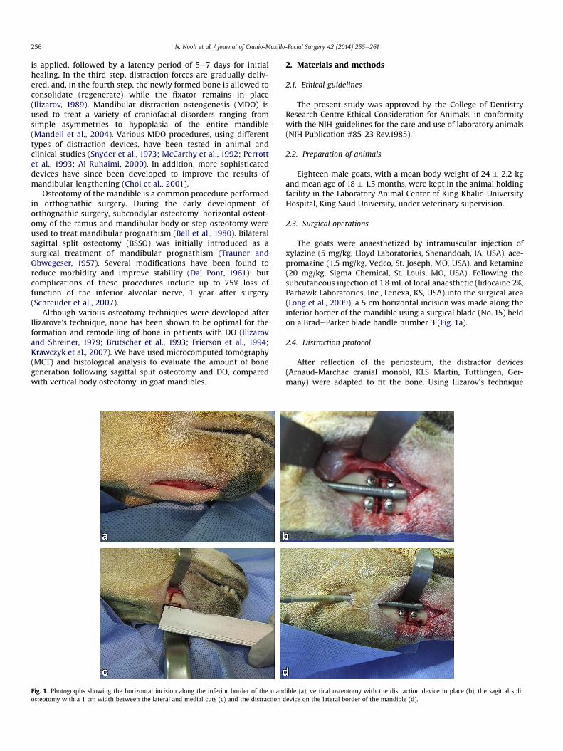
Fig. 1. Photographs showing the horizontal incision along the inferior border of the mandosteotomy with a 1 cm width between the lateral and medial cuts (c) and the distraction d
2. Materials and methods
2.1. Ethical guidelines
The present study was approved by the College of DentistryResearch Centre Ethical Consideration for Animals, in conformitywith the NIH-guidelines for the care and use of laboratory animals(NIH Publication #85-23 Rev.1985).
2.2. Preparation of animals
Eighteen male goats, with a mean body weight of 24 � 2.2 kgand mean age of 18 � 1.5 months, were kept in the animal holdingfacility in the Laboratory Animal Center of King Khalid UniversityHospital, King Saud University, under veterinary supervision.
2.3. Surgical operations
The goats were anaesthetized by intramuscular injection ofxylazine (5 mg/kg, Lloyd Laboratories, Shenandoah, IA, USA), ace-promazine (1.5 mg/kg, Vedco, St. Joseph, MO, USA), and ketamine(20 mg/kg, Sigma Chemical, St. Louis, MO, USA). Following thesubcutaneous injection of 1.8 mL of local anaesthetic (lidocaine 2%,Parhawk Laboratories, Inc., Lenexa, KS, USA) into the surgical area(Long et al., 2009), a 5 cm horizontal incision was made along theinferior border of the mandible using a surgical blade (No. 15) heldon a BradeParker blade handle number 3 (Fig. 1a).
2.4. Distraction protocol
After reflection of the periosteum, the distractor devices(Arnaud-Marchac cranial monobl, KLS Martin, Tuttlingen, Ger-many) were adapted to fit the bone. Using Ilizarov’s technique
ible (a), vertical osteotomy with the distraction device in place (b), the sagittal splitevice on the lateral border of the mandible (d).

Fig. 2. MCT images reconstructed for distraction gaps of goat mandibles 35 days afterthe end of distraction, showing the amount of newly formed bone trabeculae (green)in the gap between the proximal and distal fractured ends. The old and remodelledbones are coloured yellow, while the medullary cavities and soft structures are col-oured brown. The upper images represent vertical osteotomy while the lower imagesrepresent sagittal splitting. The views in (a) and (c) were reconstructed in sagittalplane while the views in (b) and (d) were reconstructed in the axial plane.
N. Nooh et al. / Journal of Cranio-Maxillo-Facial Surgery 42 (2014) 255e261 257
(Ilizarov and Shreiner,1979), the cortical bonewas cut on the lateralside of the mandibular edentulous area, with the distractorinstalled prior to the completion of bone cutting to mark the po-sition of the screws. Following completion of the cuts, the distractorwas secured on the edentulous area of the left side of the mandible(Fig. 1b). On the right side, sagittal splitting of the bone was per-formed, with a distance of 1 cm between themedial and lateral cuts(Fig. 1c and d). The distractor was inserted prior to the completionof bone cutting to mark the position of screws. Following comple-tion of the cuts, the distractor was secured.
The initial gap between the two bone segments was standard-ized at a width of 2 mm. The wounds were closed in layers, using 3/0 vicryl (Ethicon; Johnson & Johnson Intel. Belgium) for the deeplayers and 3/0 silk (Ethicon; Johnson & Johnson Intel. Scotland) forthe skin. In all animals, distraction was started bilaterally after a 5-day latency period. The distraction period lasted for 10 days, at1 mm/day. Six animals each were sacrificed at the end of thedistraction period and 10 and 35 days later.
2.5. MCT analysis
Following euthanasia, the mandibles were dissected, sectionedinto halves and fixed in 10% formaldehyde. Subsequently, thespecimens were wrapped in parafilm (Brand GMBH, Germany) toprevent drying during scanning. All samples were scanned at anenergy of 100 kV and an intensity of 343 mA with a resolution of12.39 mm pixels using an aluminium filter (1 mm) (SkyScan 1176high resolution in vivo X-ray microtomograph, Kontich, Belgium).The rotation step was 0.6� and the exposure time was 15e20 min.The frame averaging was 4 while the tomographic rotation was1800 and the camera was adapted as standard (1000 pixel fieldwidth). Projection images of cone-beam acquisition and recon-struction were saved as 16 bit TIF files. All scanning and recon-struction parameters were identical for all specimens andcalibrations. After the scan, a control computer withWindows 7, 64bit version, launched NRecon reconstruction software, data Viewerscan viewing software and the CT-Analyser analysis program.
The parameters acquired via MCT were bone volume (BV, mm3)and bone mineral density (BMD, g/cm3) of mineralized tissuewithin the region of interest (ROI). BV was defined as the volume ofmineralized tissue formed at the defect site during healing (Parfittet al., 1987; Anderson et al., 1999; Ott, 2008); and BMDwas definedas the density of mineralized tissue within the volume of interest,and is an indicator of the quality of mineralized tissue (Parfitt et al.,1987; Anderson et al., 1999; Ott, 2008).
Following MCT analysis, the ROIs for BV (sites of new bone for-mation between the osteotomy edges) were identified using thecomputer programme. The area of new bone formed was measuredin each section of theMCTscan and the product of themeasured areaand the slice thickness of the scan section was the BV in thatparticular scan section. The sum of the BVs of each section wascalculated to determine the total BV of new bone formed. To calcu-late the BMD of the newly formed bone between the osteotomyedges, eight representative sites were selected from the specimen asthe ROI, four selected sites on the buccal side and four on the lingualside of the specimen. Each site was selected to represent either thesuperior or the inferior aspect of the proximal and distal portions ofthe distracted bone, 2 mm away from the osteotomy edges. Themean of the individual BMD obtained from each site was calculatedto obtain the total BMD of new bone formed.
2.6. Specimen processing and histological evaluation
The specimens were fixed in 10% neutral buffered formalin anddecalcified by immersion in formic acid sodium citrate solution
(1 M/L sodium citrate in 45% formic acid) for 5 weeks. The decal-cified bone was trimmed with a sharp scalpel blade, such that thespecimen included the proximal and distal ends of the distractiongap with safety borders of about 1 mm. The specimens wereembedded in paraffin and 5-mm thick serial sections were stainedwith haematoxylin and eosin.
2.7. Statistical analysis
BV and BMD were compared by two-way analysis of variance(ANOVA) followed by the least significant difference (LSD) post-hoctest. The factors for ANOVAwere examination periods (i.e. 0,10, and35 days after the end of treatment) and type of osteotomy (i.e.sagittal split osteotomy and vertical body osteotomy), with LSDpost-hoc tests performed only for the former. A P-value <0.05 was
N. Nooh et al. / Journal of Cranio-Maxillo-Facial Surgery 42 (2014) 255e261258
considered statistically significant. All statistical analyses wereperformed using SPSS for Windows, version 16 (SPSS; Chicago, IL,USA).
3. Results
3.1. MCT results
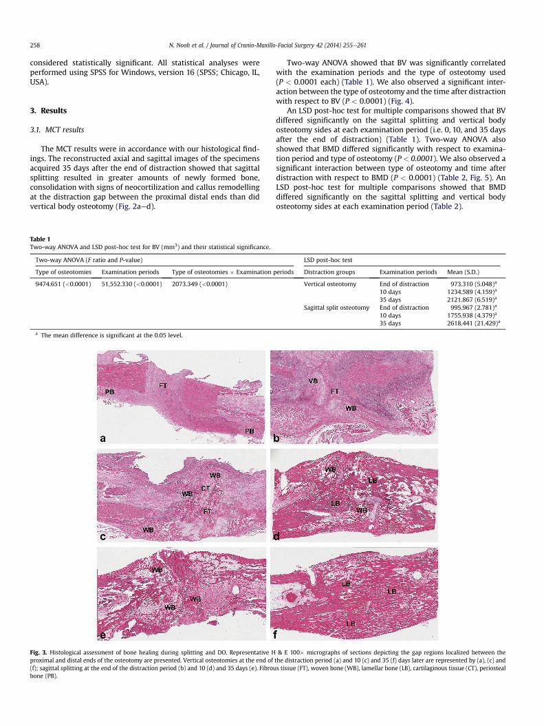
The MCT results were in accordance with our histological find-ings. The reconstructed axial and sagittal images of the specimensacquired 35 days after the end of distraction showed that sagittalsplitting resulted in greater amounts of newly formed bone,consolidation with signs of neocortilization and callus remodellingat the distraction gap between the proximal distal ends than didvertical body osteotomy (Fig. 2aed).
Table 1Two-way ANOVA and LSD post-hoc test for BV (mm3) and their statistical significance.
Two-way ANOVA (F ratio and P-value)
Type of osteotomies Examination periods Type of osteotomies � Examination p
9474.651 (<0.0001) 51,552.330 (<0.0001) 2073.349 (<0.0001)
a The mean difference is significant at the 0.05 level.
Fig. 3. Histological assessment of bone healing during splitting and DO. Representative Hproximal and distal ends of the osteotomy are presented. Vertical osteotomies at the end of(f); sagittal splitting at the end of the distraction period (b) and 10 (d) and 35 days (e). Fibroubone (PB).
Two-way ANOVA showed that BV was significantly correlatedwith the examination periods and the type of osteotomy used(P < 0.0001 each) (Table 1). We also observed a significant inter-action between the type of osteotomy and the time after distractionwith respect to BV (P < 0.0001) (Fig. 4).
An LSD post-hoc test for multiple comparisons showed that BVdiffered significantly on the sagittal splitting and vertical bodyosteotomy sides at each examination period (i.e. 0, 10, and 35 daysafter the end of distraction) (Table 1). Two-way ANOVA alsoshowed that BMD differed significantly with respect to examina-tion period and type of osteotomy (P < 0.0001). We also observed asignificant interaction between type of osteotomy and time afterdistraction with respect to BMD (P < 0.0001) (Table 2, Fig. 5). AnLSD post-hoc test for multiple comparisons showed that BMDdiffered significantly on the sagittal splitting and vertical bodyosteotomy sides at each examination period (Table 2).
LSD post-hoc test
eriods Distraction groups Examination periods Mean (S.D.)
Vertical osteotomy End of distraction 973.310 (5.048)a
10 days 1234.589 (4.159)a
35 days 2121.867 (6.519)a
Sagittal split osteotomy End of distraction 995.967 (2.781)a
10 days 1755.938 (4.379)a
35 days 2618.441 (21.429)a
& E 100� micrographs of sections depicting the gap regions localized between thethe distraction period (a) and 10 (c) and 35 (f) days later are represented by (a), (c) ands tissue (FT), woven bone (WB), lamellar bone (LB), cartilaginous tissue (CT), periosteal

Fig. 4. Line chart histogram showing the interaction between the groups at thedifferent examination periods relative to BV (mm3).
Table 2Two-way ANOVA for BMD and LSD post-hoc test for BMD (g/cm3) and their statistical significance.
Two-way ANOVA (F ratio and P-value) LSD post-hoc test
Type of osteotomies Examination periods Type of osteotomies � Examination periods Distraction groups Examination periods Mean (S.D.)
90.316 (<0.0001) 5553.605 (<0.0001) 20.014 (<0.0001) Vertical osteotomy End of distraction 0.358 (0.012)a
10 days 0.410 (0.012)a
35 days 1.070 (0.019)a
Sagittal split osteotomy End of distraction 0.420 (0.013)a
10 days 0.421 (0.009)a
35 days 1.182 (0.030)a
a The mean difference is significant at the 0.05 level.
Fig. 5. Line chart histogram showing the interaction between the groups at thedifferent examination periods relative to BMD (g/cm3).
N. Nooh et al. / Journal of Cranio-Maxillo-Facial Surgery 42 (2014) 255e261 259
3.2. Histological findings
None of the goats showed any evidence of morbidity or infectionfollowing surgery. At the end of the distraction period, the verticalbody osteotomy fracture gaps showed a central zone of fibroustissue proliferation, along with periosteal bone calluses around theends of the fractured bone segments (Fig. 3a), whereas the sagittalsplit osteotomy fracture gaps showed a central zone of fibroustissue rich in chondrocyte-like cells, fibroblasts, and oval cells thatwere morphologically intermediate between fibroblasts andchondrocytes (Fig. 3b). The proximal and distal zones showed thedevelopment of newlywoven bone rimmed by osteoblasts adjacentto the bone edges and around the fractured ends (Fig. 3b). Ten dayslater, the vertical body osteotomy fracture gaps showed fibroustissue from the central zone growing towards the proximal anddistal ends and subsequently replaced by woven bone with areas ofcartilaginous tissue (Fig. 3c). The ends of the sagittal splittingosteotomy fracture gaps showed the remodelling of woven bone aswell as cartilage into lamellar bone (Fig. 3d). The volume of newlyformed callus in the regenerated bone parallel to the vector ofdistractionwas higher in the sides that underwent sagittal splittingthan in the sides that underwent vertical body osteotomy. After 35days, the sagittally split sides showed greater evidence of robustlamellar bone formation bridging the distraction gap than thevertical body sides. The regenerating bone on the sagittally splitsides showed completion of consolidation with signs of
neocortilization and callus remodelling (Fig. 3e), whereas the sidesthat underwent vertical body osteotomy did not fully consolidate(Fig. 3f).
4. Discussion
Determining the appropriate animal model for experimentalcraniofacial DO is essential for the success of the experimentaldesign. Since the rate and potential for bone repair have beenshown to be inversely related to the evolutionary scale and age ofthe animal, the choice of animal model may have a pronouncedeffect on the results (Bardach and Kelly, 1988). Although goatbones differ physiologically from human bones, as they do notundergo normal haversian remodelling, they have been widelyused in orthopaedic research (An and Friedman, 1998). The goathas been found to be a suitable animal model for testing humanimplants and materials, as goats and humans have similar meta-bolic and bone remodelling rates (Anderson et al., 1999). For DO,the goats were used as animal models to compare the transportdistraction versus costochondral graft for reconstruction oftemporomandibular joint ankylosis (Cheung et al., 2009), in thereconstruction of condylar osteochondral defect using DO com-bined with tissue-engineered cartilage (Yu et al., 2011), to inves-tigate the feasibility of using an internal bidirectional mandibulardistractor to elongate the mandibular bodies and rami (Lin et al.,2012).
N. Nooh et al. / Journal of Cranio-Maxillo-Facial Surgery 42 (2014) 255e261260
The optimal osteotomy technique in DO for the formation andremodelling of bone regeneration is unclear, as is the postoperativestability of the mandible after bilateral lengthening by BSSO(Schendel and Linck, 2004; Krawczyk et al., 2007; Schreuder et al.,2007). Assessment of the postoperative stability of the mandibleafter bilateral lengthening showed no difference between BSSO orDO after 4 years (Baas et al., 2012). In contrast, sagittal split ramusosteotomy was reported to result in greater callus stability (Choiet al., 2001). The amount of callus tissue has been considered thekey factor for successful distraction, with the area of osteotomyinterface being much larger in BSSO than in vertical osteotomy,resulting in the formation of a larger amount of callus (Sahoo andRangarajan, 2011). Our histologic and MCT findings support thelatter results.
The surgical results of sagittal splitting were mainly evaluatedusing X-rays, such as orthopantomograms. Since this method doesnot allow a precise analysis of the split pattern (Plooij et al., 2009),the morphology of the forming and remodelled regenerating tissuecan best be assessed histologically. Our histological results indicatethat, following sagittal splitting and DO, new bone was formedthrough a cascade of cellular events. This bone was of intra-membranous or endochondral type and formed parallel to the di-rection of pull, maturing into lamellar bone following the end ofdistraction and during the consolidation period. These resultssupport clinical findings in 20 patients, showing that, after24 months, mandibular body distraction with BSSO was a betteroption than distraction with vertical osteotomy (Sahoo andRangarajan, 2011). Sagittal splitting was found to allow the im-mediate rotation and lengthening of the proximal segment whilestill providing a large surface interface for bone regeneration(Schendel and Linck, 2004).
Although we observed a significant association between type ofosteotomy and examination periods with respect to BV, this inter-actionmay have been due to the small difference in BV between thetwo types of osteotomy at the end of the distraction period. We alsoobserved a significant association between type of osteotomy andexamination period with respect to BMD, but this may also be dueto the small difference in BMD between the two types of osteotomy10 days after the end of the distraction period.
At each examination period, we found that the mean BV at thedistraction gap and its mean BMD were significantly greater on theside that underwent sagittal splitting and DO than on the side thatunderwent vertical osteotomy and DO. Thus, as expected, we foundthat sagittal splitting significantly improved the regenerative ca-pacity of the gap between the distraction ends. This is in agreementwith a previous study, which demonstrated long-term osseoushealing in the cleavage space between bone fragments 2 years aftersagittal splitting ramus osteotomy (SSRO) surgery, indicating thatremodelling between bone fragments is a major mechanism ofosseous healing after SSRO (Hasegawa et al., 2011).
5. Conclusion
To our knowledge, this study is the first to evaluate new boneformation after DO in animal models. Our results indicate thatsagittal split osteotomy was associated with a better regenerativecapacity than vertical body osteotomy, possibly due to the largeoverlapping surfaces of the split segments. The significant increasein the volumes of newly formed bone and in its mineral densityresulted in increased consolidation following sagittal splitosteotomy.
Sources of support in the form of grantsNot funded.
Conflict of interestThe authors have no conflicts of interest to declare.
Acknowledgement
Special acknowledgement for the College of Dentistry ResearchCenter (CDRC), King Saud University, Saudi Arabia for the approvalof this research as a non-funded research no. FR 0002.
References
Al Ruhaimi KA: A submerged osteodistraction device: an innovative technique forexperimental animal studies. J Craniofac Surg 11: 59e61, 2000
An YH, Friedman RJ: Animal models of orthopedic implant infection. J Invest Surg11: 139e146, 1998
Anderson ML, Dhert WJ, de Bruijn JD, Dalmeijer RA, Leenders H, van Blitterswijk CA,et al: Critical size defect in the goat’s os ilium. A model to evaluate bone graftsand substitutes. Clin Orthop Relat Res 364: 231e239, 1999
Baas EM, Pijpe J, de Lange J: Long term stability of mandibular advancement pro-cedures: bilateral sagittal split osteotomy versus distraction osteogenesis. Int JOral Maxillofac Surg 41: 137e141, 2012
Bardach J, Kelly KM: Role of animal models in experimental studies of craniofacialgrowth following cleft lip and palate repair. Cleft Palate J 25: 103e113, 1988
Bell WH, White RP, Hall HD: Mandibular excess. In: Bell WH, Proffit WR, White RP(eds), Surgical correction of dentofacial deformities, vol. 2. Philadelphia: WBSaunders Co., 844e1013, 1980
Brutscher R, Rahn BA, Ruter A, Perren SM: The role of corticotomy and osteotomy inthe treatment of bone defects using the Ilizarov technique. J Orthop Trauma 7:261e269, 1993
Cheung LK, Zheng LW, Ma L, Shi XJ: Transport distraction versus costochondral graftfor reconstruction of temporomandibular joint ankylosis: which is better? OralSurg Oral Med Oral Pathol Oral Radiol Endod 108: 32e40, 2009
Cho-Lee GY, Naval-Gías L, González-García R, Martos-Díaz PL, Muñoz-Guerra MF,Sastre-Pérez J, et al: Bifocal transport osteogenesis for the reconstruction ofadult calvarial defects: a new surgical technique. J Craniomaxillofac Surg 38:368e373, 2010
Choi JY, Hwang KG, Baek SH, Lee JH, Kim TW, Kim MJ, et al: Original sagittal splitosteotomy revisited for mandibular distraction. J Craniomaxillofac Surg 29:165e173, 2001
Dal Pont G: Retromolar osteotomy for the correction of prognathism. J Oral SurgAnesth Hosp Dent Serv 19: 42e47, 1961
de Gijt JP, Vervoorn K, Wolvius EB, Van der Wal KG, Koudstaal MJ: Mandibularmidline distraction: a systematic review. J Craniomaxillofac Surg 40: 248e260,2012
Figueroa AA, Polley JW: Mandibular distraction osteogenesis. Op Tech OtolaryngolHead Neck Surg 13: 17e28, 2002
Frierson M, Ibrahim K, Boles M, Bote H, Ganey T: Distraction osteogenesis. Acomparison of corticotomy techniques. Clin Orthop Relat Res 301: 19e24, 1994
Hasegawa T, Tateishi C, Uchida R, Nishi C, Furudoi S, Shibuya Y, et al: Osseoushealing after a sagittal splitting ramus osteotomy. Int J Oral Maxillofac Surg 40:475e482, 2011
Ilizarov GA, Shreiner AA: A new method of closed flexion osteoclasia (experimentalstudy). Ortop Travmatol Protez 1: 9e14, 1979
Ilizarov GA: The tension-stress effect on the genesis and growth of tissues: part II.The influence of the rate and frequency of distraction. Clin Orthop Relat Res239: 263e285, 1989
Krawczyk A, Kuropka P, Kuryszko J, Wall A, Dragan S, Kulej M: Experimental studieson the effect of osteotomy technique on the bone regeneration in distractionosteogenesis. Bone 40: 781e791, 2007
Lin X, Zhou L, Shang H, Liu Y, Bo B: Use of an internal bidirectional distractor toelongate goats’ mandibles. Br J Oral Maxillofac Surg 50: e49ee52, 2012
Long J, Tang W, Fan YB, Tian WD, Feng F, Liu L, et al: Effects of rapid distraction rateon new bone formation during mandibular distraction osteogenesis in goats.Injury 40: 831e834, 2009
Mandell DL, Yellon RF, Bradley JP, Izadi K, Gordon CB: Mandibular distraction formicrognathia and severe upper airway obstruction. Arch Otolaryngol HeadNeck Surg 130: 344e348, 2004
McCarthy JG, Schreiber J, Karp N, Thorne CH, Grayson BH: Lengthening themandibular body by gradual distraction. Plast Reconstr Surg 89: 1e10, 1992
Nakase T, Yasui N, Kawabata H, Shimizu N, Ohzono K, Hiroshima K, et al: Correctionof deformity and shortening due to post traumatic epiphyseal arrest bydistraction osteogenesis. Arch Orthop Trauma Surg 127: 659e663, 2007
Ott SM: Histomorphometric measurements of bone turnover, mineralization, andvolume. Clin J Am Soc Nephrol 3(Suppl. 3): S151eS156, 2008
Parfitt AM, Drezner MK, Glorieux FH, Kanis JA, Malluche H, Meunier PJ, et al: Bonehistomorphometry: standardization of nomenclature, symbols, and units.Report of the ASBMR Histomorphometry Nomenclature Committee. J BoneMiner Res 2: 595e610, 1987
Park JT, Lee JG, Kim SY, Kim GH, Hu KS, Cha JY, et al: A piezoelectric motor-basedmicroactuator-generated distractor for continuous jaw bone distraction.J Craniofac Surg 22: 1486e1488, 2011

N. Nooh et al. / Journal of Cranio-Maxillo-Facial Surgery 42 (2014) 255e261 261
Perrott DH, Berger R, Vargervik K, Kaban LB: Use of a skeletal distractiondevice to widen the mandible: a case report. J Oral Maxillofac Surg 51:435e439, 1993
Plooij JM, Naphausen MT, Maal TJ, Xi T, Rangel FA, Swennnen G, et al: 3D evaluationof the lingual fracture line after a bilateral sagittal split osteotomy of themandible. Int J Oral Maxillofac Surg 38: 1244e1249, 2009
Rachmiel A, Emodi O, Gutmacher Z, Blumenfeld I, Aizenbud D: Oral and dentalrestoration of wide alveolar cleft using distraction osteogenesis and temporaryanchorage devices. J Craniomaxillofac Surg 41: 728e734, 2013
Sahoo NK, Rangarajan H: Comparative evaluation of vertical body osteotomy andsagittal split osteotomy for mandibular corpus distraction. J Oral MaxillofacSurg 69: 381e389, 2011
Schendel SA, Linck 3rd DW: Mandibular distraction osteogenesis by sagittal splitosteotomy and intraoral curvilinear distraction. J Craniofac Surg 15: 631e635,2004
Schreuder WH, Jansma J, Bierman MW, Vissink A: Distraction osteogenesis versusbilateral sagittal split osteotomy for advancement of the retrognathic mandible:a review of the literature. Int J Oral Maxillofac Surg 36: 103e110, 2007
Shang H, Xue Y, Liu Y, Zhao J, He L: Modified internal mandibular distraction osteo-genesis in the treatment of micrognathia secondary to temporomandibular jointankylosis: 4-year follow-up of a case. J Craniomaxillofac Surg 40: 373e378, 2012
Snyder CC, Levine GA, Swanson HM, Browne Jr EZ: Mandibular lengthening bygradual distraction. Preliminary report. Plast Reconstr Surg 51: 506e508, 1973
Trauner R, Obwegeser H: The surgical correction of mandibular prognathism andretrognathia with consideration of genioplasty. I. Surgical procedures to correctmandibular prognathism and reshaping of the chin. Oral Surg Oral Med OralPathol 10: 677e689, 1957
Yu H, Yang X, Cheng J, Wang X, Shen SG: Distraction osteogenesis combined withtissue-engineered cartilage in the reconstruction of condylar osteochondraldefect. J Oral Maxillofac Surg 69: e558ee564, 2011

![Advanced Taxation (CFAP5) by Fawad Hassan [Lecture1]](https://static.fdocuments.in/doc/165x107/58ece99f1a28abdb6f8b46e1/advanced-taxation-cfap5-by-fawad-hassan-lecture1.jpg)