Joseph M. Reinhardt, Ph.D....Joseph M. Reinhardt, Ph.D. SERVICE ACTIVITIES Department: Member,...
27
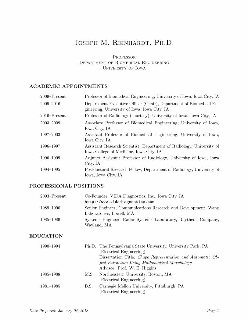
Joseph M. Reinhardt, Ph.D. Professor Department of Biomedical Engineering University of Iowa ACADEMIC APPOINTMENTS 2009–Present Professor of Biomedical Engineering, University of Iowa, Iowa City, IA 2009–2016 Department Executive Officer (Chair), Department of Biomedical En- gineering, University of Iowa, Iowa City, IA 2016–Present Professor of Radiology (courtesy), University of Iowa, Iowa City, IA 2003–2009 Associate Professor of Biomedical Engineering, University of Iowa, Iowa City, IA 1997–2003 Assistant Professor of Biomedical Engineering, University of Iowa, Iowa City, IA 1996–1997 Assistant Research Scientist, Department of Radiology, University of Iowa College of Medicine, Iowa City, IA 1996–1999 Adjunct Assistant Professor of Radiology, University of Iowa, Iowa City, IA 1994–1995 Postdoctoral Research Fellow, Department of Radiology, University of Iowa, Iowa City, IA PROFESSIONAL POSITIONS 2003–Present Co-Founder, VIDA Diagnostics, Inc., Iowa City, IA http://www.vidadiagnostics.com 1989–1990 Senior Engineer, Communications Research and Development, Wang Laboratories, Lowell, MA 1985–1989 Systems Engineer, Radar Systems Laboratory, Raytheon Company, Wayland, MA EDUCATION 1990–1994 Ph.D. The Pennsylvania State University, University Park, PA (Electrical Engineering) Dissertation Title: Shape Representation and Automatic Ob- ject Extraction Using Mathematical Morphology Advisor: Prof. W. E. Higgins 1985–1988 M.S. Northeastern University, Boston, MA (Electrical Engineering) 1981–1985 B.S. Carnegie Mellon University, Pittsburgh, PA (Electrical Engineering) Date Prepared: January 04, 2018 Page 1
Transcript of Joseph M. Reinhardt, Ph.D....Joseph M. Reinhardt, Ph.D. SERVICE ACTIVITIES Department: Member,...
Joseph M. Reinhardt, Ph.D.
ProfessorDepartment of Biomedical Engineering
University of Iowa
ACADEMIC APPOINTMENTS
2009–Present Professor of Biomedical Engineering, University of Iowa, Iowa City, IA
2009–2016 Department Executive Officer (Chair), Department of Biomedical En-gineering, University of Iowa, Iowa City, IA
2016–Present Professor of Radiology (courtesy), University of Iowa, Iowa City, IA
2003–2009 Associate Professor of Biomedical Engineering, University of Iowa,Iowa City, IA
1997–2003 Assistant Professor of Biomedical Engineering, University of Iowa,Iowa City, IA
1996–1997 Assistant Research Scientist, Department of Radiology, University ofIowa College of Medicine, Iowa City, IA
1996–1999 Adjunct Assistant Professor of Radiology, University of Iowa, IowaCity, IA
1994–1995 Postdoctoral Research Fellow, Department of Radiology, University ofIowa, Iowa City, IA
PROFESSIONAL POSITIONS
2003–Present Co-Founder, VIDA Diagnostics, Inc., Iowa City, IAhttp://www.vidadiagnostics.com
1989–1990 Senior Engineer, Communications Research and Development, WangLaboratories, Lowell, MA
1985–1989 Systems Engineer, Radar Systems Laboratory, Raytheon Company,Wayland, MA
EDUCATION
1990–1994 Ph.D. The Pennsylvania State University, University Park, PA(Electrical Engineering)Dissertation Title: Shape Representation and Automatic Ob-ject Extraction Using Mathematical MorphologyAdvisor: Prof. W. E. Higgins
1985–1988 M.S. Northeastern University, Boston, MA(Electrical Engineering)
1981–1985 B.S. Carnegie Mellon University, Pittsburgh, PA(Electrical Engineering)
Date Prepared: January 04, 2018 Page 1

Joseph M. Reinhardt, Ph.D.
HONORS AND AWARDS
2008 Honorable Mention Poster Award, SPIE Symposium on Medical Imag-ing, 2008, “Tracking the motion of the hyoid bone in videofluoroscopicswallowing studies”, Patrick Kellen, Darci Becker, Joseph M. Rein-hardt, and Douglas van Daele
2008 Nominated for Graduate College “Outstanding Graduate Mentor”award for 2008.
2007 Honorable Mention Poster Award, SPIE Symposium on Medical Imag-ing, 2007, “Feature-based pairwise retinal image registration by radialdistortion correction”, Sangyeol Lee, Michael Abramoff, and JosephM. Reinhardt
2007 1st place Siemens Preclinical CT Image of the Year for “Imaging andanalysis for the assessment of the normal mouse lung,” acquired byEric A. Hoffman, Geoffrey McLennan, Joseph M. Reinhardt, Gary E.Christensen, Deokiee Chon, Eman Namati, Jacqueline Namati, LijunShi, Joo Hyun Song, Kunlin Cao, and Jered Sieren from The Univer-sity of Iowa, 2007.
2006 Elected Fellow, AIMBE
2002 Elected Senior Member, IEEE
1998 Tau Beta Pi Excellence in Teaching Award, University of Iowa, IowaCity, IA.
1994 Graduate Research Award, The Pennsylvania State University, Uni-versity Park, PA. Awarded top prize for research in Science and En-gineering category for entry titled “Toward Efficient MorphologicalShape Representation.”
1990–1991 Palmer Graduate Fellowship, The Pennsylvania State University, Uni-versity Park, PA. Awarded for academic excellence.
1984 Tau Beta Pi Honor Society
PROFESSIONAL MEMBERSHIPS
Fellow, AIMBE; Senior Member, IEEE; Member, AAPM; Member, ASEE; Member, SPIE;Member, Tau Beta Pi; Member, IEEE Biomedical Engineering Society; Member, IEEE SignalProcessing Society.
TEACHING ACTIVITIES
I. Graduate Student Supervision
Student Name Degree Grad. Date Current Position
Sarah Gerard Ph.D. May 2018Natalie Ross M.S. May 2019Sandeep Bodduluri M.S. May 2012
Ph.D. Dec 2016 Asst. Professor, Univ. of AlabamaYang Wook Kim M.S. Aug. 2013 Graduate school
Date Prepared: January 04, 2018 Page 2
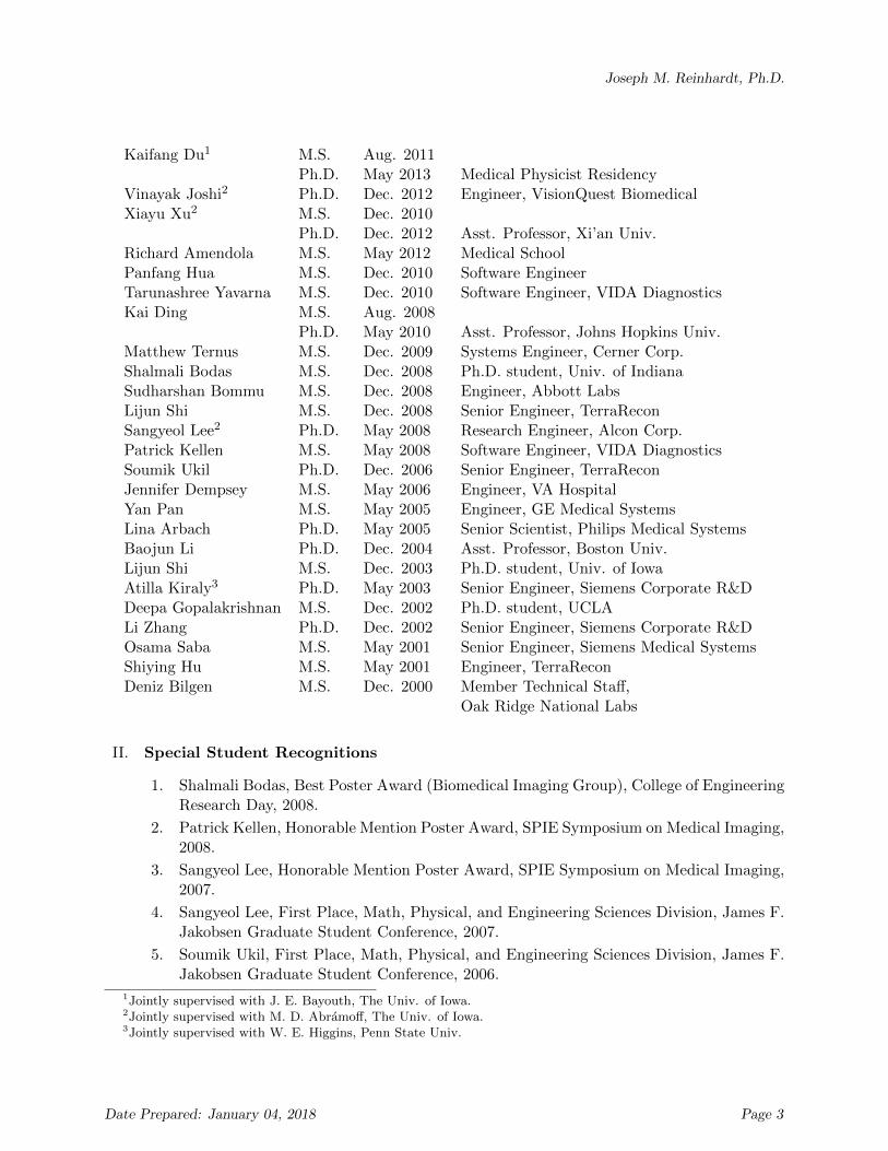
Joseph M. Reinhardt, Ph.D.
Kaifang Du1 M.S. Aug. 2011Ph.D. May 2013 Medical Physicist Residency
Vinayak Joshi2 Ph.D. Dec. 2012 Engineer, VisionQuest BiomedicalXiayu Xu2 M.S. Dec. 2010
Ph.D. Dec. 2012 Asst. Professor, Xi’an Univ.Richard Amendola M.S. May 2012 Medical SchoolPanfang Hua M.S. Dec. 2010 Software EngineerTarunashree Yavarna M.S. Dec. 2010 Software Engineer, VIDA DiagnosticsKai Ding M.S. Aug. 2008
Ph.D. May 2010 Asst. Professor, Johns Hopkins Univ.Matthew Ternus M.S. Dec. 2009 Systems Engineer, Cerner Corp.Shalmali Bodas M.S. Dec. 2008 Ph.D. student, Univ. of IndianaSudharshan Bommu M.S. Dec. 2008 Engineer, Abbott LabsLijun Shi M.S. Dec. 2008 Senior Engineer, TerraReconSangyeol Lee2 Ph.D. May 2008 Research Engineer, Alcon Corp.Patrick Kellen M.S. May 2008 Software Engineer, VIDA DiagnosticsSoumik Ukil Ph.D. Dec. 2006 Senior Engineer, TerraReconJennifer Dempsey M.S. May 2006 Engineer, VA HospitalYan Pan M.S. May 2005 Engineer, GE Medical SystemsLina Arbach Ph.D. May 2005 Senior Scientist, Philips Medical SystemsBaojun Li Ph.D. Dec. 2004 Asst. Professor, Boston Univ.Lijun Shi M.S. Dec. 2003 Ph.D. student, Univ. of IowaAtilla Kiraly3 Ph.D. May 2003 Senior Engineer, Siemens Corporate R&DDeepa Gopalakrishnan M.S. Dec. 2002 Ph.D. student, UCLALi Zhang Ph.D. Dec. 2002 Senior Engineer, Siemens Corporate R&DOsama Saba M.S. May 2001 Senior Engineer, Siemens Medical SystemsShiying Hu M.S. May 2001 Engineer, TerraReconDeniz Bilgen M.S. Dec. 2000 Member Technical Staff,
Oak Ridge National Labs
II. Special Student Recognitions
1. Shalmali Bodas, Best Poster Award (Biomedical Imaging Group), College of EngineeringResearch Day, 2008.
2. Patrick Kellen, Honorable Mention Poster Award, SPIE Symposium on Medical Imaging,2008.
3. Sangyeol Lee, Honorable Mention Poster Award, SPIE Symposium on Medical Imaging,2007.
4. Sangyeol Lee, First Place, Math, Physical, and Engineering Sciences Division, James F.Jakobsen Graduate Student Conference, 2007.
5. Soumik Ukil, First Place, Math, Physical, and Engineering Sciences Division, James F.Jakobsen Graduate Student Conference, 2006.
1Jointly supervised with J. E. Bayouth, The Univ. of Iowa.2Jointly supervised with M. D. Abramoff, The Univ. of Iowa.3Jointly supervised with W. E. Higgins, Penn State Univ.
Date Prepared: January 04, 2018 Page 3

Joseph M. Reinhardt, Ph.D.
SERVICE ACTIVITIES
Department: Member, Medical Imaging Faculty Search Committee, 2017–2018; Depart-ment Executive Officer, March 1, 2009–June 30, 2016; Chair, Undergraduate Program Com-mittee, 2008–2009; Chair, Undergraduate Program Committee, 2007–2008; Chair, Depart-mental Consulting Group on Promotions and Tenure, 2007–2008; Member, DEO SearchCommittee, 2007–2008; Chair, Undergraduate Program Committee, 2006–2007; Member, De-partmental Consulting Group on Promotions and Tenure, 2006–2007; Chair, DepartmentalConsulting Group on Promotions and Tenure, 2005–2006; Undergraduate Program Com-mittee, 2005–2006; Member, Departmental Consulting Group on Promotions and Tenure,2004–2005; Undergraduate Program Committee, 2004–2005; Research Thrust Committee,2004–2005; Member, Departmental Consulting Group on Promotions and Tenure, 2003–2004; Chairman, Undergraduate Program Committee, 2003–2004; Member, Strategic Plan-ning Committee, 2002–2003; Undergraduate Internship Coordinator, 2001-2004; Member,Undergraduate Curriculum Committee, 2002–2003; Member, Graduate Program Enhance-ment Committee, 2001–2002; Assistant Program ABET Coordinator, 2001–2002; Chairman,Teaching Equipment Purchasing Committee, 2000–2001; Chairman, Digital Signal Process-ing Faculty Search Committee, 2001; Chairman, Strategic Planning Committee, 1999–2000;Chairman, Academic Affairs Committee Task force on new BME Fundamentals Course, 2000;Member, Department Enhancement Committee, 1999–2000; Faculty Secretary, 1997–1998;Faculty Secretary, 1998–1999; Member, Graduate Committee, 1997–1998.
College: Member, Dean’s Advisory Instructional Faculty Promotion Committee, 2017-2018;Member, Teaching Committee, 2007–2008; IEEE Student Chapter Faculty Mentor, 2005–2007; Chair, Engineering Faculty Council, 2004–2005; Member, Engineering Faculty Council,2003–2006; Member, EFC Computer Services Committee, 2003–2004; Member, College ofEngineering Curriculum Committee, 2001–2002; Member, Faculty Outreach Planning Com-mittee, Fall 2000; Member, College of Engineering Internship Director search committee, Fall2000; Faculty Secretary, 1999–2000.
University: Faculty Senator, 2017–2018; Member, College of Engineering Decanel ReviewCommittee, Spring 2017; Chair, Search Committee for Director of National Advanced Driv-ing Simulator, University of Iowa, Fall 2015–Spring 2016; Member, Review Committee forDepartment of Radiology, University of Iowa, Spring 2016; Member, TIER Presidential Advi-sory Board, Fall 2015/Spring 2016; Member, Interdisciplinary Program in Health InformaticsExecutive Committee, 2016; Member, Interdisciplinary Program in Health Informatics Exec-utive Committee, Fall 2015; Member, Orthopaedics and Rehabilitation Associate Chair forResearch Search Committee, Fall 2014/Spring 2015; Member, Director of Neurosciences Re-search Institute, Fall 2014/Spring 2015; Member, Head of Orthopaedics and RehabilitationSearch Committee, Spring 2013; Member, Director of Sponsored Programs Search Commit-tee, Spring 2012; Member, Vice President for Research Conflict of Interest In Research Com-mittee, AY 2008-2011; Member, Vice President for Research Internal Selection Committeefor Biological Science Funding Programs, 2008; Judge, Jakobsen Conference, 2008; Mem-ber, Vice President for Research Internal Selection Committee for Biological Science FundingPrograms, 2007; Judge, Jakobsen Conference, 2006; Member, Vice President for Research In-ternal Selection Committee for Major Research Equipment Grant Proposals, 2004; Member,Vice President for Research Advisory Committee on Biological Sciences, 2000-2003; Member,Grant Accounting Internal Review Committee, Fall 2001.
Date Prepared: January 04, 2018 Page 4
Joseph M. Reinhardt, Ph.D.
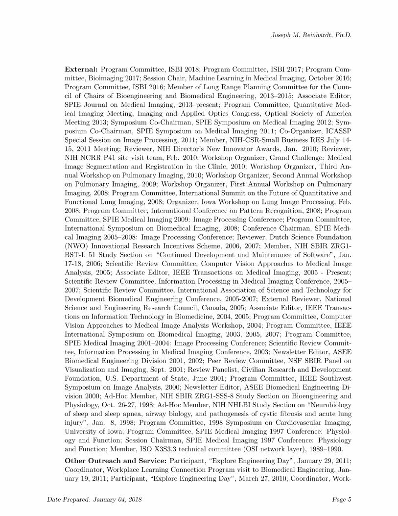
External: Program Committee, ISBI 2018; Program Committee, ISBI 2017; Program Com-mittee, Bioimaging 2017; Session Chair, Machine Learning in Medical Imaging, October 2016;Program Committee, ISBI 2016; Member of Long Range Planning Committee for the Coun-cil of Chairs of Bioengineering and Biomedical Engineering, 2013–2015; Associate Editor,SPIE Journal on Medical Imaging, 2013–present; Program Committee, Quantitative Med-ical Imaging Meeting, Imaging and Applied Optics Congress, Optical Society of AmericaMeeting 2013; Symposium Co-Chairman, SPIE Symposium on Medical Imaging 2012; Sym-posium Co-Chairman, SPIE Symposium on Medical Imaging 2011; Co-Organizer, ICASSPSpecial Session on Image Processing, 2011; Member, NIH-CSR-Small Business RES July 14-15, 2011 Meeting; Reviewer, NIH Director’s New Innovator Awards, Jan. 2010; Reviewer,NIH NCRR P41 site visit team, Feb. 2010; Workshop Organizer, Grand Challenge: MedicalImage Segmentation and Registration in the Clinic, 2010; Workshop Organizer, Third An-nual Workshop on Pulmonary Imaging, 2010; Workshop Organizer, Second Annual Workshopon Pulmonary Imaging, 2009; Workshop Organizer, First Annual Workshop on PulmonaryImaging, 2008; Program Committee, International Summit on the Future of Quantitative andFunctional Lung Imaging, 2008; Organizer, Iowa Workshop on Lung Image Processing, Feb.2008; Program Committee, International Conference on Pattern Recognition, 2008; ProgramCommittee, SPIE Medical Imaging 2009: Image Processing Conference; Program Committee,International Symposium on Biomedical Imaging, 2008; Conference Chairman, SPIE Medi-cal Imaging 2005–2008: Image Processing Conference; Reviewer, Dutch Science Foundation(NWO) Innovational Research Incentives Scheme, 2006, 2007; Member, NIH SBIR ZRG1-BST-L 51 Study Section on “Continued Development and Maintenance of Software”, Jan.17-18, 2006; Scientific Review Committee, Computer Vision Approaches to Medical ImageAnalysis, 2005; Associate Editor, IEEE Transactions on Medical Imaging, 2005 - Present;Scientific Review Committee, Information Processing in Medical Imaging Conference, 2005–2007; Scientific Review Committee, International Association of Science and Technology forDevelopment Biomedical Engineering Conference, 2005-2007; External Reviewer, NationalScience and Engineering Research Council, Canada, 2005; Associate Editor, IEEE Transac-tions on Information Technology in Biomedicine, 2004, 2005; Program Committee, ComputerVision Approaches to Medical Image Analysis Workshop, 2004; Program Committee, IEEEInternational Symposium on Biomedical Imaging, 2003, 2005, 2007; Program Committee,SPIE Medical Imaging 2001–2004: Image Processing Conference; Scientific Review Commit-tee, Information Processing in Medical Imaging Conference, 2003; Newsletter Editor, ASEEBiomedical Engineering Division 2001, 2002; Peer Review Committee, NSF SBIR Panel onVisualization and Imaging, Sept. 2001; Review Panelist, Civilian Research and DevelopmentFoundation, U.S. Department of State, June 2001; Program Committee, IEEE SouthwestSymposium on Image Analysis, 2000; Newsletter Editor, ASEE Biomedical Engineering Di-vision 2000; Ad-Hoc Member, NIH SBIR ZRG1-SSS-8 Study Section on Bioengineering andPhysiology, Oct. 26-27, 1998; Ad-Hoc Member, NIH NHLBI Study Section on “Neurobiologyof sleep and sleep apnea, airway biology, and pathogenesis of cystic fibrosis and acute lunginjury”, Jan. 8, 1998; Program Committee, 1998 Symposium on Cardiovascular Imaging,University of Iowa; Program Committee, SPIE Medical Imaging 1997 Conference: Physiol-ogy and Function; Session Chairman, SPIE Medical Imaging 1997 Conference: Physiologyand Function; Member, ISO X3S3.3 technical committee (OSI network layer), 1989–1990.
Other Outreach and Service: Participant, “Explore Engineering Day”, January 29, 2011;Coordinator, Workplace Learning Connection Program visit to Biomedical Engineering, Jan-uary 19, 2011; Participant, “Explore Engineering Day”, March 27, 2010; Coordinator, Work-
Date Prepared: January 04, 2018 Page 5
Joseph M. Reinhardt, Ph.D.
place Learning Connection Program visit to Cellular Engineering Technologies and VIDADiagnostics, April 14, 2010; Middle school STEM outreach program, April 13, 2010; “In-troduction to Biomedical Engineering” panelist for area high school students, November10, 2009; Participant, Freshman/Parent Summer Orientation Program, 2009; Participant,“Explore Engineering Day”, April 18, 2009; Participant, Freshman/Parent Summer Orien-tation Program, 2007; Participant, Freshman/Parent Summer Orientation Program, 2006;Speaker, Gladbook-Reinbeck High School Career Day, 2006; Speaker, College of Engineer-ing Workplace Learning Connection Program, 2006; Participant, Freshman/Parent SummerOrientation Program, 2005; Participant, Freshman/Parent Summer Orientation Program,2004; Participant, Freshman/Parent Summer Orientation Program, 2003; “Introduction toX-ray Imaging” program for Horn Elementary School science curriculum, Sept. 23, 2003;Participant, Freshman/Parent Summer Orientation Program, 2002; NSPE Review SessionLeader (Circuits), Spring 2002; Participant, Freshman/Parent Summer Orientation Program,2001; Participant, Engineering Day, 2001; NSPE Review Session Leader (Circuits), Spring2001; Participant, Engineering Day, 2000; Participant, Engineering Day, 1999; Participant,Freshman/Parent Summer Orientation Program, 1999; Participant, Engineering Day, 1999;Judge, 1999 State Invention Convention, Ames, Iowa; Participant, Freshman/Parent SummerOrientation Program, 1998; Participant, Engineering Day, 1998.
Journal Reviewing: Computer Vision and Image Understanding ; IEEE Transactions onBiomedical Engineering ; IEEE Transactions on Image Processing ; IEEE Transactions onSignal Processing ; IEEE Transactions on Medical Imaging ; IEEE Transactions on PatternAnalysis and Machine Intelligence; IEEE Computer Graphics and Image Processing ; Au-tomedica; Annals of Biomedical Engineering ; Journal of Visual Communication and ImageRepresentation; Multidimensional Systems and Signal Processing ; Medical Image Analysis;Pattern Recognition
RESEARCH FUNDING
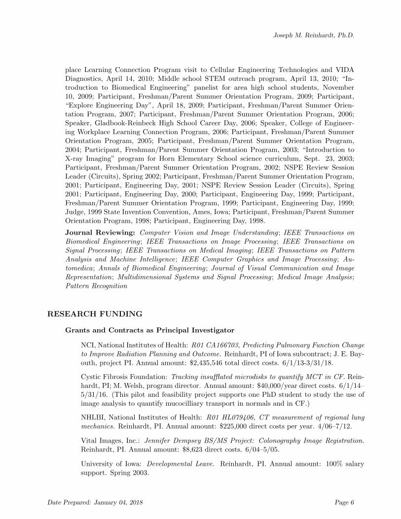
Grants and Contracts as Principal Investigator
NCI, National Institutes of Health: R01 CA166703, Predicting Pulmonary Function Changeto Improve Radiation Planning and Outcome. Reinhardt, PI of Iowa subcontract; J. E. Bay-outh, project PI. Annual amount: $2,435,546 total direct costs. 6/1/13-3/31/18.
Cystic Fibrosis Foundation: Tracking insufflated microdisks to quantify MCT in CF. Rein-hardt, PI; M. Welsh, program director. Annual amount: $40,000/year direct costs. 6/1/14–5/31/16. (This pilot and feasibility project supports one PhD student to study the use ofimage analysis to quantify mucocilliary transport in normals and in CF.)
NHLBI, National Institutes of Health: R01 HL079406, CT measurement of regional lungmechanics. Reinhardt, PI. Annual amount: $225,000 direct costs per year. 4/06–7/12.
Vital Images, Inc.: Jennifer Dempsey BS/MS Project: Colonography Image Registration.Reinhardt, PI. Annual amount: $8,623 direct costs. 6/04–5/05.
University of Iowa: Developmental Leave. Reinhardt, PI. Annual amount: 100% salarysupport. Spring 2003.
Date Prepared: January 04, 2018 Page 6

Joseph M. Reinhardt, Ph.D.
Whitaker Foundation: Transition Year: Measurement of Lung Parenchymal Strainvia X-ray CT. Reinhardt, PI. Annual amount: $76,250 direct costs per year. 9/01–8/02.
National Science Foundation: CAREER: Program in Pulmonary Imaging. Reinhardt, PI.Annual amount: $75,000 direct costs per year. 3/01–2/06.
Whitaker Foundation: Biomedical Engineering Internship Program at the University ofIowa. Reinhardt, PI. Annual amount: $60,000 direct costs per year. 7/00–6/03.
University of Iowa: Old Gold Summer Fellowship. Reinhardt, PI. Annual amount: 1 monthsalary. Summer 1999.
Whitaker Foundation: Measurement of Lung Parenchymal Strain via X-ray CT. Reinhardt,PI. Annual amount: $70,000 direct costs per year. 4/97–2/01.
Environmental Health Sciences Research Center (University of Iowa): Accurate Measure-ment of Intrathoracic Airways. Reinhardt, PI. Annual amount: $15,000 direct costs. 4/97–3/98.
Grants and Contracts as Co-Investigator
NHLBI, National Institutes of Health: U01 HL137880, SPIROMICS II: Biological Un-derpinnings of COPD Heterogeneity and Progression. Reinhardt, co-Investigator, (P.Woodruff, PI). Annual amount: $6,092,935. 09/15/17-05/31/22.
NHLBI, National Institutes of Health: R01 HL111453, Expanding objective CT-based phe-notyping to lungs with enhanced radiodensities. Reinhardt, Co-Investigator (R. Beichel, PI)5% effort. Annual amount: $355,570. 1/1/2012–12/31/2014.
Carver College of Medicine Collaborative Pilot Grant: Quantifying radiation inducedchanges in pulmonary function in irradiated and un-irradiated lung tissue. Reinhardt, Co-investigator (J. Bayouth, PI). Annual amount: $50,000. 7/1/2009–6/30/2010.
NHLBI, National Institutes of Health: R01 HL092056, Lung alterations in acute exacerba-tions of COPD. Reinhardt, Co-investigator10% salary support plus grad RA(K. Doerschug, PI). Annual amount: $250,000 per year. 7/15/2009–6/30/2011.
NHLBI, National Institutes of Health: S10RR024738, Vertical MR System Shared Instru-mentation Grant. Reinhardt, Co-Investigator, 0% salary support, (Eric A. Hoffman, PI).Annual amount: $500,000. 7/09–6/10.
NSF: S10RR22421, Large-Scale Computing and Visualization for Cardiopulmonary Imag-ing. Reinhardt, Co-Investigator, 0% salary support, (Ching-Long Lin, PI). Annual amount:$473,636. 1/08–1/11.
NCI, National Institutes of Health: R01 CA129022, Precise correspondence of 3D Pathol-ogy with radiological features in lung nodules. Reinhardt, Co-Investigator, 3% effort, (G.McLennan, PI). Annual amount: $306,354 per year. 9/07-8/12.
NHLBI, National Institutes of Health: R01 HL080285, Quantitative CT-based lung atlas ofthe mouse. Reinhardt, Co-Investigator, 10% effort plus grad RA, (Eric A. Hoffman, PI).Annual amount: $400,000 per year. 4/06–3/11.
Date Prepared: January 04, 2018 Page 7

Joseph M. Reinhardt, Ph.D.
NEI, National Institutes of Health: R01 EY017066, Low cost, patient friendly, portableimaging of diabetic retinopathy. Reinhardt, Co-Investigator, 5% effort plus grad RA, (M.Abramoff, PI). Annual amount: $237,903 per year. 9/05-7/09.
The University of Iowa: Academic Technologies Instructional Computing Award. Reinhardt,Co-Investigator, 0% salary support, (G. Thomas, PI). Annual amount: $50,000. 12/06–8/07.
NHLBI, National Institutes of Health: Image and Model Based Analysis of Lung Disease.Reinhardt, Co-Investigator, 15% effort plus grad RA, (E. A. Hoffman, PI). Annual amount:$1,993,762 per year. 7/05–6/10.
Pacific Northwest National Labs: Lung atlas development, subcontract #14624. Reinhardt,Co-Investigator, RA salary support, (E. A. Hoffman, PI). Annual amount: $30,000 per year.8/05-7/10.
Confirma, Inc.: Breast MRI lesion classification. Reinhardt, Co-Investigator, Grad RAsalary support, (A. Stolpen, PI). Annual amount: $5,000. 1/04–12/04.
NCI, National Institutes of Health: Virtual true-color bronchoscope to detect lung cancer.Reinhardt, Co-Investigator, 5% effort plus grad RA, (G. McLennan, PI). Annual amount:$300,000 per year. 9/02–9/06.
NHLBI, National Institutes of Health: Lung image database with pathologic correlates.Reinhardt, Co-Investigator, 3% effort plus grad RA, (G. McLennan, PI). Annual amount:$267,170 per year. 9/01–8/06.
NCCAM, National Institutes of Health: Does spinal manipulation speed determine neuralresponse?. Reinhardt, Co-Investigator, 5% effort, (J. Pickar, PI). Annual amount: $125,000per year. 8/01–7/03.
NCI, National Institutes of Health: Quantitative Medical Imaging Training Grant. Rein-hardt, Co-Investigator, RA salary support, (R. Hichwa, PI). Annual amount: $100,000 peryear. 7/00-6/05.
Whitaker Foundation: ASEE Annual Meeting Travel Grant. Reinhardt, Participant, (J.Winters, PI). Annual amount: $750. 6/00.
NHLBI, National Institutes of Health: Image and Model Based Analysis of Lung Disease.Reinhardt, Co-Investigator, 15% salary support plus grad RA, (E. A. Hoffman, PI). Annualamount: $1,846,154 per year. 12/99–11/04.
NHLBI, National Institutes of Health: Inflammatory Parenchymal Lung Disease. Rein-hardt, Co-Investigator, 10% salary support plus grad RA, (E. A. Hoffman, PI). Annualamount: $298,674 per year. 7/99–6/04.
Environmental Protection Agency: Assessment of Variations in Human Airway Geometryand the Implications for Evaluation of Particle Deposition and Dose to Different Popu-lations. Reinhardt, Co-Investigator, Grad RA salary support, (B.S. Cohen, PI). Annualamount: $25,000 per year. 6/99–5/01.
National Science Foundation: 3-D Cardiac Ultrasound Image Analysis. Reinhardt, Co-Investigator, 5% effort, (E. L. Dove, PI). Annual amount: $75,000 per year. 1/99–12/00.
Date Prepared: January 04, 2018 Page 8

Joseph M. Reinhardt, Ph.D.
Cystic Fibrosis Center University of Iowa: Laser fluorescent bronchoscopy. Reinhardt, Co-Investigator, Grad RA salary support, (G. McLennan, PI). Annual amount: $45,000 peryear. 9/98–8/00.
Whitaker Foundation: Collaborative Educational Environment for Functional Cardiovascu-lar Image Analysis. Reinhardt, Co-Investigator, equipment and staff salary support, (K. B.Chandran, PI). Annual amount: $308,517 per year. 2/98–6/02.
Cervical Spine Research Society: Biomechanics of Ligamentous Spine and Spinal Cordduring Whiplash and Post-Whiplash. Reinhardt, Co-Investigator, RA salary support, (V.K. Goel, PI). Annual amount: $30,000 per year. 11/98–10/99.
PATENTS
Geoffrey McLennan, Martin Donnelley, Deepa Gopalakrishnan, Eric Hoffman, Joseph Rein-hardt and Melissa Suter, “Methods and devices useful for analyzing color medical images,”U.S. Patent No. 7,613,335. Issued November 3, 2009.
Joseph M. Reinhardt, Soumik Ukil, Milan Sonka, Geoffrey McLennan, and Eric A. Hoffman,“Methods of smoothing segmented regions,” U.S. Patent No. 8,073,210. Issued December 6,2011.
Michael Abramoff, Sangyeol Lee, Joseph M. Reinhardt, and Meindert Niemeijer, “OptimalRegistration of Multiple Deformed Images using a Physical Distortion Model of the ImagingDistortion,” U.S. Patent No. 8,194,936. Issued June 5, 2012.
Juerg Tschirren, Milan Sonka, Geoffrey McLennan, Eric Hoffman, and Joseph Reinhardt,“Methods and devices for airway tree labeling and/or matching,” U.S. Patent No. 8,155,403.Issued April 10, 2012.
Juerg Tschirren, Milan Sonka, Joseph Reinhardt, Geoffrey McLennan, and Eric Hoffman,“Methods and devices for labeling and/or matching,” U.S. Patent No. 9,820,651. IssuedNovember 21, 2017.
PATENT APPLICATIONS
Joseph M. Reinhardt, John T. Garber, Juerg Tschirren, Milan Sonka, Geoffrey McLennan,and Eric A. Hoffman, “Treatment planning methods, devices, and systems,” U.S. ProvisionalPatent Application No. 60/772,176. Filed September 30, 2005.
PUBLICATIONS
I. Books
[1] K. B. Chandran, H. S. Udaykumar, and J. M. Reinhardt, Eds., Image-Based Compu-tational Modeling of the Human Circulatory and Pulmonary Systems. Springer, 2011.
Date Prepared: January 04, 2018 Page 9

Joseph M. Reinhardt, Ph.D.
II. Conference and Workshop Proceedings
[1] J. M. Fitzpatrick and J. M. Reinhardt, Eds., Medical Imaging 2005: Image Processing,vol. 5747, ser. The Proceedings of the SPIE, International Society for Optical Engineer-ing, 2005.
[2] J. M. Reinhardt and J. P. Pluim, Eds., Medical Imaging 2006: Image Processing,vol. 6144, ser. The Proceedings of the SPIE, International Society for Optical Engi-neering, 2006.
[3] J. M. Reinhardt, B. van Ginneken, and M. Sonka, Guest editorial: Pulmonary imageprocessing, Special Issue on Pulmonary Imaging, Apr. 2006.
[4] J. P. Pluim and J. M. Reinhardt, Eds., Medical Imaging 2007: Image Processing,vol. 6512, ser. The Proceedings of the SPIE, International Society for Optical Engi-neering, 2007.
[5] J. Z. Liang, H. Lu, D. N. Metaxas, and J. M. Reinhardt, Guest editorial: Medicalimaging informatics — information processing from image formation to visualization,Special Issue on Medical Image Reconstruction, Processing and Visualization, Jan. 2007.
[6] J. M. Reinhardt and J. P. Pluim, Eds., Medical Imaging 2008: Image Processing,vol. 6914, ser. The Proceedings of the SPIE, International Society for Optical Engi-neering, 2008.
[7] M. Brown, M. de Bruijne, B. van Ginneken, A. Kiraly, J.-M. Kuhnigk, C. Lorenz, K.Mori, and J. M. Reinhardt, Eds., Proceedings of the First International Workshop onPulmonary Image Analysis, http://www.lulu.com/content/3507981., 2008.
[8] M. Brown, M. de Bruijne, B. van Ginneken, A. Kiraly, J.-M. Kuhnigk, C. Lorenz, J. R.McClelland, K. Mori, A. Reeves, and J. M. Reinhardt, Eds., Proceedings of the SecondInternational Workshop on Pulmonary Image Analysis, http://www.amazon.com/Second-International-Workshop-Pulmonary-Analysis/dp/1448680891., 2009.
[9] M. Brown, M. de Bruijne, B. van Ginneken, K. Ding, A. Kiraly, J.-M. Kuhnigk, J. R.McClelland, K. Mori, and J. M. Reinhardt, Eds., Proceedings of the Third InternationalWorkshop on Pulmonary Image Analysis, http://www.amazon.com/Third-International-Workshop-Pulmonary-Analysis/dp/1453776001, 2010.
III. Rigorously Reviewed Journal Articles
[1] J. M. Reinhardt and W. E. Higgins, “Comparison between the morphological skeletonand morphological shape decomposition,” IEEE Trans. Patt. Anal. Machine Intell., vol.18, no. 9, pp. 951–957, Sep. 1996.
[2] J. M. Reinhardt and W. E. Higgins, “Efficient morphological shape representation,”IEEE Trans. Image Proc., vol. 5, no. 1, pp. 89–101, Jan. 1996.
[3] J. M. Reinhardt, N. D. D’Souza, and E. A. Hoffman, “Accurate measurement of intra-thoracic airways,” IEEE Trans. Medical Imaging, vol. 16, no. 6, pp. 820–827, Dec. 1997.
[4] J. M. Reinhardt and W. E. Higgins, “Paradigm for shape-based image analysis,” OpticalEngineering, vol. 37, no. 2, pp. 570–581, Feb. 1998.
[5] J. M. Reinhardt and E. A. Hoffman, “Quantitative pulmonary imaging: Spatial andtemporal considerations in HRCT,” Acad. Radiol., vol. 5, no. 8, pp. 539–546, Aug.1998.
Date Prepared: January 04, 2018 Page 10
Joseph M. Reinhardt, Ph.D.
[6] J. M. Reinhardt, A. J. Wang, T. P. Weldon, and W. E. Higgins, “Cue-based segmenta-tion of 4D cardiac image sequences,” Comp. Vision and Image Understanding, vol. 77,no. 2, pp. 251–262, Feb. 2000.
[7] S. Hu, E. A. Hoffman, and J. M. Reinhardt, “Automatic lung segmentation for accuratequantitation of volumetric X-ray CT images,” IEEE Trans. Medical Imaging, vol. 20,no. 6, pp. 490–498, Jun. 2001.
[8] The National Emphysema Treatment Trial Research Group, “Patients at high risk ofdeath after lung-volume-reduction surgery,” New England J. Medicine, vol. 345, no. 15,pp. 1–9, Oct. 2001.
[9] L. Arbach, G. Fallouh, J. M. Reinhardt, and L. Bennett, “Distinguishing between ma-lignant and nonmalignant breast masses from mammograms using artificial intelligencetechniques,” Bassel Al-Assad J. for Engineering Science, vol. 16, pp. 103–121, Jul. 2002.
[10] A. Kiraly, W. E. Higgins, G. McLennan, E. A. Hoffman, and J. M. Reinhardt, “3Dhuman airway segmentation for clinical virtual bronchoscopy,” Acad. Radiol., vol. 9,no. 10, pp. 1153–1168, Oct. 2002.
[11] C. P. Rooney, M. Suter, G. McLennan, M. Donnelley, J. M. Reinhardt, A. Delsing, E. A.Hoffman, and J. Zabner, “Laser fluorescence bronchoscopy for detection of fluorescentreporter genes in airway epithelia,” Gene Therapy, vol. 9, no. 23, pp. 1639–1644, Dec.2002.
[12] A. Fishman, F. Martinez, K. Naunheim, S. Piantadosi, R. Wise, A. Ries, G. Weinmann,D. E. Wood, A. P. Fishman, B. A. Bozzarello, et al., “A randomized trial comparinglung-volume-reduction surgery with medical therapy for severe emphysema,” New Eng-land J. Medicine, vol. 348, no. 21, pp. 2059–2073, May 2003, PMID: 12759479. Note:Joseph M. Reinhardt is author 428 of 448 on this manuscript.
[13] D. Aykac, E. A. Hoffman, G. McLennan, and J. M. Reinhardt, “Segmentation andanalysis of the human airway tree from three-dimensional X-ray CT images,” IEEETrans. Medical Imaging, vol. 22, no. 8, pp. 940–950, Aug. 2003.
[14] O. Saba, E. A. Hoffman, and J. M. Reinhardt, “Maximizing quantitative accuracy oflung airway lumen and wall measures obtained from X-ray CT imaging,” J. AppliedPhysiology, vol. 95, pp. 1063–1095, 2003.
[15] E. A. Hoffman, J. M. Reinhardt, M. Sonka, B. A. Simon, J. Guo, O. Saba, D. Chon,S. Samrah, H. Shikata, J. Tschirren, et al., “Characterization of the interstitial lungdiseases via density-based and texture-based analysis of computed tomography imagesof lung structure and function,” Acad. Radiol., vol. 10, no. 10, pp. 1104–1118, Oct. 2003.
[16] B. Li, G. E. Christensen, G. McLennan, E. A. Hoffman, and J. M. Reinhardt, “Estab-lishing a normative atlas of the human lung: Inter-subject warping and registration ofvolumetric CT,” Acad. Radiol., vol. 10, no. 3, pp. 255–265, Mar. 2003.
[17] The National Emphysema Treatment Trial Research Group, “Safety and efficacy ofmedian stenotomy versus video-assisted thoracic surgery for lung volume reductionsurgery,” J. Thorac. Cardiovasc. Surg., vol. 127, pp. 1350–1360, May 2004.
Date Prepared: January 04, 2018 Page 11

Joseph M. Reinhardt, Ph.D.
[18] R. M. Kaplan, A. L. Ries, J. Reilly, Z. Mohsenifar, A. P. Fishman, B. A. Bozzarello, A.Al-Amin, M. Katz, C. Wheeler, E. Baker, et al., “Measurement of health-related qualityof life in the national emphysema treatment trial,” Chest, vol. 126, no. 3, pp. 781–789,Sep. 2004, PMID: 15364757. Note: Joseph M. Reinhardt is author 426 of 446 on thismanuscript.
[19] M. Suter, J. Tschirren, J. Reinhardt, M. Sonka, E. Hoffman, W. Higgins, and G. McLen-nan, “Evaluation of the human airway with multi-detector X-ray computed tomographyand optical imaging,” Physiological Measurement, vol. 25, no. 4, pp. 837–847, Aug. 2004.
[20] M. H. Tawhai, P. Hunter, J. Tschirren, J. M. Reinhardt, G. McLennan, and E. A.Hoffman, “CT-based geometry analysis and finite element models of the human andovine bronchial tree,” J. Applied Physiology, vol. 97, no. 6, pp. 2310–2321, 2004.
[21] E. A. Hoffman, A. V. Clough, G. E. Christensen, C.-L. Lin, G. McLennan, J. M. Rein-hardt, B. A. Simon, M. Sonka, M. Tawhai, J. R. van Beek Edwin, et al., “The compre-hensive imaging-based analysis of the lung: A forum for team science,” Acad. Radiol.,vol. 11, no. 12, pp. 1370–1380, Dec. 2004.
[22] O. I. Saba, D. Chon, K. Beck, G. McLennan, J. Sieren, J. M. Reinhardt, and E. A.Hoffman, “Static versus prospective gated non-breath hold volumetric MDCT imagingof the lungs,” Acad. Radiol., vol. 12, no. 11, pp. 1371–1384, Nov. 2005.
[23] B. A. Simon, G. E. Christensen, D. A. Low, and J. M. Reinhardt, “Computed tomog-raphy studies of lung mechanics,” Proc Am Thorac Soc, vol. 2, no. 6, pp. 517–521,2005.
[24] M. Suter, J. M. Reinhardt, P. Montague, P. Taft, J. Lee, J. Zabner, and G. McLennan,“Bronchoscopic imaging of the pulmonary mucosal vasculature responses to inflamma-tory mediators,” J. Biomed. Optics, vol. 10, no. 3, p. 034 013, May 2005.
[25] M. Suter, G. McLennan, J. M. Reinhardt, D. Riker, and E. A. Hoffman, “Macro-optical color assessment of the pulmonary airways with subsequent three-dimensionalmultidetector-X-ray-computed-tomography assisted display,” J. Biomed. Optics, vol.10, no. 5, p. 051 703, Sep. 2005.
[26] S. Ukil and J. M. Reinhardt, “Smoothing lung segmentation surfaces in 3D X-ray CTimages using anatomic guidance,” Acad. Radiol., vol. 12, no. 12, pp. 1502–1511, Dec.2005.
[27] W. M. Chatila, E. A. Hoffman, J. Gaughan, G. B. Robinswood, G. J. Criner, A. P.Fishman, B. A. Bozzarello, A. Al-Amin, M. Katz, C. Wheeler, et al., “Advanced em-physema in African-American and white patients: Do differences exist?” Chest, vol. 130,pp. 108–118, Jul. 2006, PMID: 16840390. Note: Joseph M. Reinhardt is author 434 of456 on this manuscript.
[28] F. J. Martinez, G. Foster, J. L. Curtis, G. Criner, G. Weinmann, A. Fishman, M. M.DeCamp, J. Benditt, F. Sciurba, B. Make, et al., “Predictors of mortality in patientswith emphysema and severe airflow obstruction,” Amer. J. Respiratory and CriticalCare Medicine, vol. 173, pp. 1326–1334, 2006. Note: Joseph M. Reinhardt is author 444of 465 on this manuscript.
[29] L. Zhang, E. Hoffman, and J. M. Reinhardt, “Lung lobe segmentation in volumetricX-ray CT images,” IEEE Trans. Medical Imaging, vol. 25, no. 1, pp. 1–16, 2006.
Date Prepared: January 04, 2018 Page 12

Joseph M. Reinhardt, Ph.D.
[30] L. A. Meinel, A. H. Stolpen, K. S. Berbaum, L. L. Fajardo, and J. M. Reinhardt, “BreastMRI lesion classification: Improved performance of human readers with a backpropaga-tion neural network computer-aided diagnosis (CAD) system,” J. Magn. Res. Imaging,vol. 25, no. 1, pp. 85–95, Jan. 2007.
[31] B. D. Owen, G. E. Christensen, J. M. Reinhardt, and T. C. Ryken, “Rapid prototypepatient-specific drill template for cervical pedicle screw placement,” Computer AidedSurgery, vol. 12, no. 5, pp. 303–308, Sep. 2007.
[32] M. Suter, J. M. Reinhardt, and G. McLennan, “Integrated CT/bronchoscopy in thecentral airways: Preliminary results,” Acad. Radiol., vol. 15, no. 6, pp. 786–798, Jun.2008.
[33] M. K. Fuld, R. B. Easley, O. I. Saba, D. Chon, J. M. Reinhardt, E. A. Hoffman, andB. A. Simon, “CT measured regional specific volume change reflects regional specificventilation in supine sheep,” J. Applied Physiology, vol. 104, no. 4, pp. 1177–1184, Apr.2008.
[34] J. M. Reinhardt, K. Ding, K. Cao, G. E. Christensen, E. A. Hoffman, and S. V. Bodas,“Registration-based estimates of local lung tissue expansion compared to xenon-CTmeasures of specific ventilation,” Med. Imag. Analysis, vol. 12, no. 6, pp. 752–763, Dec.2008. doi: 10.1016/j.media.2008.03.007.
[35] B. Li, G. E. Christensen, G. McLennan, E. A. Hoffman, and J. M. Reinhardt, “Pul-monary CT image registration and warping for tracking tissue deformation during therespiratory cycle through 3-D consistent image registration,” Medical Physics, vol. 35,no. 12, pp. 5575–5583, 2008, http://dx.doi.org/10.1118/1.3005633.
[36] S. Ukil and J. M. Reinhardt, “Anatomy-guided lung lobar surface detection in X-rayCT images,” IEEE Trans. Medical Imaging, vol. 28, no. 2, pp. 202–214, 2009, PMID:19188109. doi: 10.1109/TMI.2008.929101.
[37] T. C. Ryken, B. D. Owen, G. E. Christensen, and J. M. Reinhardt, “Image baseddrill-templates for cervical pedicle screw placement,” J Neurosurg Spine, vol. 10, no. 1,pp. 21–26, Jan. 2009. doi: 10.3171/2008.9.SPI08229.
[38] T. C. Ryken, B. D. Owen, G. E. Christensen, and J. M. Reinhardt, “Engineering patient-specific drill templates and bioabsorbable posterior cervical plates: a feasibility study,”J Neurosurg Spine, vol. 10, no. 2, pp. 129–132, Feb. 2009.
[39] K. Ding, Y. Yin, K. Cao, G. E. Christensen, C.-L. Lin, E. A. Hoffman, and J. M. Rein-hardt, “Evaluation of lobar biomechanics during respiration using image registration,”in Medical Imaging Computing and Computer Assisted Intervention, G.-Z. Yang, D. J.Hawkes, D. Rueckert, A. Noble, and C. Taylor, Eds., ser. Lecture Notes in ComputerScience, vol. 5761, London: Springer-Verlag, Sep. 2009, pp. 739–746. doi: 10.1007/978-3-642-04268-3_91.
[40] K. Cao, G. E. Christensen, K. Ding, and J. M. Reinhardt, “Intensity-and-landmark-driven, inverse consistent, B-spline registration and analysis for lung imagery,” in SecondInternational Workshop on Pulmonary Image Analysis, M. Brown, M. de Bruijne, B.van Ginneken, A. Kiraly, J. M. Kuhnigk, C. Lorenz, J. R. McClelland, K. Mori, A.Reeves, and J. M. Reinhardt, Eds., 2009, pp. 137–148.
Date Prepared: January 04, 2018 Page 13

Joseph M. Reinhardt, Ph.D.
[41] P. Lo, B. van Ginneken, J. M. Reinhardt, and M. de Bruijne, “Extraction of airways fromCT (EXACT’09),” in Second International Workshop on Pulmonary Image Analysis,M. Brown, M. de Bruijne, B. van Ginneken, A. Kiraly, J. M. Kuhnigk, C. Lorenz, J. R.McClelland, K. Mori, A. Reeves, and J. M. Reinhardt, Eds., 2009, pp. 175–189.
[42] T. E. Robinson, F. R. Long, P. Raman, P. Saha, M. J. Edmond, J. M. Reinhardt,R. Raman, and A. S. Brody, “An airway phantom to standardize CT acquisition inmulti-center clinical trials,” Acad. Radiol., vol. 16, no. 9, pp. 1134–1141, 2009. doi:10.1016/j.acra.2009.02.018.
[43] J. Tschirren, T. Yavarna, and J. M. Reinhardt, “Airway segmentation framework forclinical environments,” in Second International Workshop on Pulmonary Image Analy-sis, M. Brown, M. de Bruijne, B. van Ginneken, A. Kiraly, J. M. Kuhnigk, C. Lorenz,J. R. McClelland, K. Mori, A. Reeves, and J. M. Reinhardt, Eds., 2009, pp. 227–238.
[44] E. A. Hoffman, R. Jiang, H. Baumhauer, M. A. Brooks, J. J. Carr, R. Detrano, J. M.Reinhardt, J. Rodriguez, K. Stukovsky, N. Wong, et al., “Reproducibility and validity oflung density measures from cardiac CT scans–The multi-ethnic study of atherosclerosis(MESA) lung study,” Acad. Radiol., vol. 16, no. 6, pp. 689–699, Jun. 2009.
[45] P. M. Kellen, D. L. Becker, J. M. Reinhardt, and D. J. Van Daele, “Computer-AssistedAssessment of Hyoid Bone Motion from Videofluoroscopic Swallow Studies,” Dysphagia,Oct. 2009, PMID: 19856024.
[46] R. Benzo, M. H. Farrell, C. C. Chang, F. J. Martinez, R. Kaplan, J. Reilly, G. Criner,R. Wise, B. Make, J. Luketich, et al., “Integrating health status and survival data: thepalliative effect of lung volume reduction surgery,” Amer. J. Respiratory and CriticalCare Medicine, vol. 180, pp. 239–246, Aug. 2009, PMID 19483114. Note: Joseph M.Reinhardt is author 442 of 463 on this manuscript.
[47] G. J. Criner, P. Belt, A. L. Sternberg, Z. Mosenifar, B. J. Make, J. P. Utz, F. Sciurba,M. Katz, C. Wheeler, E. Baker, et al., “Effects of lung volume reduction surgery on gasexchange and breathing pattern during maximum exercise,” Chest, vol. 135, pp. 1268–1279, May 2009, PMID: 19420196. Note: Joseph M. Reinhardt is author 423 of 443 onthis manuscript.
[48] W. J. Kim, E. K. Silverman, E. Hoffman, G. J. Criner, Z. Mosenifar, F. C. Sciurba,B. J. Make, V. Carey, R. S. Estepar, A. Diaz, et al., “CT metrics of airway diseaseand emphysema in severe COPD,” Chest, vol. 136, pp. 396–404, Aug. 2009, PMID:19411295. Note: Joseph M. Reinhardt is author 463 of 484 on this manuscript.
[49] J. C. Sieren, J. Weydert, E. Namati, J. Thiesse, J. P. Sieren, J. M. Reinhardt, E. A.Hoffman, and G. McLennan, “A process model for direct correlation between computedtomography and histopathology application in lung cancer,” Acad. Radiol., vol. 17,pp. 169–180, Feb. 2010. doi: 10.1016/j.acra.2009.09.006.
[50] K. Ding, J. E. Bayouth, J. M. Buatti, G. E. Christensen, and J. M. Reinhardt, “4DCT-based measurement of changes in pulmonary function following a course of radiationtherapy,” Medical Physics, vol. 37, no. 3, pp. 1261–1272, Mar. 2010. doi: 10.1118/1.3312210.
[51] M. D. Abramoff, J. M. Reinhardt, S. R. Russell, J. C. Folk, V. B. Mahajan, M. Niemeijer,and G. Quellec, “Automated early detection of diabetic retinopathy,” Ophthalmology,vol. 117, pp. 1147–1154, Jun. 2010. doi: 10.1016/j.ophtha.2010.03.046.
Date Prepared: January 04, 2018 Page 14
Joseph M. Reinhardt, Ph.D.
[52] S. Lee, J. M. Reinhardt, P. C. Cattin, and M. D. Abramoff, “Objective and expert-independent validation of retinal image registration algorithms by a projective imagingdistortion model,” Med. Imag. Analysis, vol. 14, no. 4, pp. 539–549, Aug. 2010. doi:10.1016/j.media.2010.04.001.
[53] L. Tang, T. E. Scheetz, D. A. Mackey, A. W. Hewitt, J. H. Fingert, Y. H. Kwon, G.Quellec, J. M. Reinhardt, and M. D. Abramoff, “Automated quantification of inheritedphenotypes from color images: A twin study of the variability of optic nerve head shape,”Invest Ophthalmol Vis Sci., vol. 51, pp. 5870–5877, Nov. 2010.
[54] J. Thiesse, E. Namati, J. C. Sieren, A. R. Smith, J. M. Reinhardt, E. A. Hoffman, andG. McLennan, “Lung structure phenotype variation in inbred mouse strains revealedthrough in vivo micro-CT imaging,” J. Applied Physiology, vol. 109, pp. 1960–1968,Dec. 2010. doi: 10.1152/japplphysiol.01322.2009.
[55] G. R. Washko, F. J. Martinez, E. A. Hoffman, S. H. Loring, R. S. Estepar, A. A. Diaz,F. C. Sciurba, E. K. Silverman, M. K. Han, M. Decamp, et al., “Physiological andcomputed tomographic predictors of outcome from lung volume reduction surgery,”Amer. J. Respiratory and Critical Care Medicine, vol. 181, pp. 494–500, Mar. 2010.Note: Joseph M. Reinhardt is author 440 of 462 on this manuscript.
[56] M. K. Han, R. Wise, J. Mumford, F. Sciurba, G. J. Criner, J. L. Curtis, S. Murray,A. Sternberg, G. Weinman, E. Kazerooni, et al., “Prevalence and clinical correlates ofbronchoreversibility in severe emphysema,” European Respiratory J., vol. 35, pp. 1048–1056, May 2010. Note: Joseph M. Reinhardt is author 444 of 466 on this manuscript.
[57] Y. Yin, E. A. Hoffman, K. Ding, J. M. Reinhardt, and C.-L. Lin, “A cubic B-spline-basedhybrid registration of lung CT images for a dynamic airway geometric model with largedeformation,” Phys. Med. Biol., vol. 56, pp. 203–218, Jan. 2011. doi: 10.1088/0031-9155/56/1/013.
[58] X. Xu, M. Niemeijer, Q. Song, M. Sonka, M. K. Garvin, J. M. Reinhardt, and M. D.Abramoff, “Vessel Boundary Delineation on Fundus Images using Graph-Based Ap-proach,” IEEE Trans. Medical Imaging, vol. 30, no. 6, pp. 1184–1191, Jan. 2011.
[59] R. E. Amelon, K. Cao, K. Ding, G. E. Christensen, J. M. Reinhardt, and M. L. Ragha-van, “Three-dimensional characterization of regional lung deformation,” J Biomechan-ics, vol. 44, pp. 2489–2495, Sep. 2011.
[60] L. M. Gabe, K. M. Baker, E. J. R. van Beek, G. W. Hunninghake, J. M. Reinhardt, andE. A. Hoffman, “Effect of segmental bronchoalveolar lavage on quantitative computedtomography of the lung,” Acad. Radiol., vol. 18, pp. 876–884, Jul. 2011.
[61] K. Murphy, B. van Ginneken, J. M. Reinhardt, S. Kabus, K. Ding, X. Deng, K. Cao, K.Du, G. E. Christensen, V. Garcia, et al., “Evaluation of registration methods on thoracicCT: the EMPIRE10 challenge,” IEEE Trans. Medical Imaging, vol. 30, pp. 1901–1920,Nov. 2011.
[62] M. A. Puhan, D. Chandra, Z. Mosenifar, A. Ries, B. Make, N. N. Hansel, R. A. Wise,F. Sciurba, A. P. Fishman, B. A. Bozzarello, et al., “The minimal important differenceof exercise tests in severe COPD,” European Respiratory J., vol. 37, pp. 784–790, Apr.2011.
Date Prepared: January 04, 2018 Page 15

Joseph M. Reinhardt, Ph.D.
[63] K. Ding, K. Cao, M. K. Fuld, K. Du, G. E. Christensen, E. A. Hoffman, and J. M. Rein-hardt, “Comparison of image registration based measures of regional lung ventilationfrom dynamic spiral CT with Xe-CT,” Medical Physics, vol. 39, no. 8, pp. 5084–5098,2012. doi: 10.1118/1.4736808.
[64] K. Du, J. E. Bayouth, K. Cao, G. E. Christensen, K. Ding, and J. M. Reinhardt, “Re-producibility of registration-based measures of lung tissue expansion,” Medical Physics,vol. 39, no. 3, pp. 1595–1608, Mar. 2012. doi: http://link.aip.org/link/doi/10.1118/1.3685589.
[65] S. Sommerfeld Ross, J. M. Reinhardt, and J. Fiegel, “Enhanced analysis of bacteriasusceptibility in connected biofilms,” J. Microbiol. Methods, vol. 90, no. 1, pp. 9–14,Jul. 2012.
[66] V. S. Joshi, R. J. Maude, J. M. Reinhardt, L. Tang, M. K. Garvin, A. A. Sayeed,A. Ghose, M. U. Hassan, and M. A. Abramoff, “Automated detection of malarialretinopathy associated retinal hemorrhages,” Invest Ophthalmol Vis Sci., vol. 53, no.10, pp. 6582–6588, Sep. 2012, PMID: 22915035.
[67] P. Lo, B. van Ginneken, J. M. Reinhardt, T. Yavarna, P. de Jong, B. Irving, C. Fetita,M. Ortner, R. Pinho, J. Sijbers, et al., “Extraction of airways from CT (EXACT’09),”IEEE Trans. Medical Imaging, vol. 31, no. 11, pp. 2093–2107, Nov. 2012. doi: http://dx.doi.org/10.1109/TMI.2012.2209674.
[68] B. Li, G. E. Christensen, E. A. Hoffman, G. McLennan, and J. M. Reinhardt, “Estab-lishing a normative atlas of the human lung: Computing the average transformation andatlas construction,” Acad. Radiol., vol. 19, no. 11, pp. 1368–1381, Nov. 2012, PMID:22951110. doi: 10.1016/j.acra.2012.04.025.
[69] K. Cao, G. E. Christensen, K. Ding, K. Du, M. L. Raghavan, R. E. Amelon, K. M.Baker, E. A. Hoffman, and J. M. Reinhardt, “Tracking regional tissue volume andfunction change in lung using image registration,” Int. J. Biomed Imaging, vol. 2012,p. 956 248, 2012, PMID: 23118740. doi: 10.1155/2012/956248.
[70] K. Cao, K. Ding, J. M. Reinhardt, and G. E. Christensen, “Improving Intensity-BasedLung CT Registration Accuracy Utilizing Vascular Information,” Int. J. Biomed Imag-ing, vol. 2012, p. 285 136, 2012, PMID: 23251141. [Online]. Available: http://www.hindawi.com/journals/ijbi/2012/285136/.
[71] X. Xu, J. M. Reinhardt, Q. Hu, B. Bakall, P. S. Tlucek, G. Bertelsen, and M. D.Abramoff, “Retinal vessel width measurement at branchings using an improved electricfield theory-based graph approach,” PLoS ONE, vol. 7, no. 11, e49668, 2012, PMID:23209588.
[72] D. S. Kacmarynski, R. Amendola, J. M. Reinhardt, and R. J. Smith, “Flexible modelsfor planning repair of complex tracheal anomalies,” Laryngoscope, vol. 122 Suppl 4, S77,Dec. 2012, PMID: 23254611.
[73] S. Bodduluri, J. D. Newell, E. A. Hoffman, and J. M. Reinhardt, “Registration-basedlung mechanical analysis of chronic obstructive pulmonary disease (COPD) using asupervised machine learning framework,” Acad. Radiol., vol. 20, no. 5, pp. 527–536,May 2013, PMID: 23570934.
Date Prepared: January 04, 2018 Page 16

Joseph M. Reinhardt, Ph.D.
[74] L. Tang, M. Niemeijer, J. M. Reinhardt, M. K. Garvin, and M. D. Abramoff, “Splat fea-ture classification with application to retinal hemorrhage detection in fundus images,”IEEE Trans. Medical Imaging, vol. 32, no. 2, pp. 364–375, Feb. 2013, PMID: 23193310.
[75] K. Du, J. E. Bayouth, K. Ding, G. E. Christensen, K. Cao, and J. M. Reinhardt,“Reproducibility of intensity-based estimates of lung ventilation,” Medical Physics, vol.40, no. 6, p. 063 504, May 2013, PMID: 23718615. doi: http://dx.doi.org/10.1118/1.4805106.
[76] K. Du, J. M. Reinhardt, G. E. Christensen, K. Ding, and J. E. Bayouth, “Respiratoryeffort correction strategies to improve the reproducibility of lung expansion measure-ments,” Medical Physics, vol. 40, no. 12, p. 123 504, 2013. doi: http://dx.doi.org/10.1118/1.4829519. [Online]. Available: http://scitation.aip.org/content/aapm/journal/medphys/40/12/10.1118/1.4829519.
[77] R. J. Adam, A. S. Michalski, C. Bauer, M. H. Abou Alaiwa, T. J. Gross, M. S. Awadalla,D. C. Bouzek, N. D. Gansemer, P. J. Taft, M. J. Hoegger, et al., “Air trapping andairflow obstruction in newborn cystic fibrosis piglets,” Amer. J. Respiratory and CriticalCare Medicine, vol. 188, no. 12, pp. 1434–1441, 2013, PMID: 24168209.
[78] S. Sommerfeld Ross, M. H. Tu, M. L. Falsetta, M. R. Ketterer, M. R. Kiedrowski, A. R.Horswill, M. A. Apicella, J. M. Reinhardt, and J. Fiegel, “Quantification of confocalimages of biofilms grown on irregular surfaces,” J. Microbiol. Methods, vol. 100, pp. 111–120, May 2014, PMID: 24632515.
[79] V. S. Joshi, J. M. Reinhardt, M. K. Garvin, and M. D. Abramoff, “Automated methodfor identification and artery-venous classification of vessel trees in retinal vessel net-works,” PLoS ONE, vol. 9, no. 2, e88061, 2014, PMID: 24533066.
[80] R. E. Amelon, K. Cao, J. M. Reinhardt, G. E. Christensen, and M. L. Raghavan, “Ameasure for characterizing sliding on lung boundaries,” Ann Biomed Eng, vol. 42, no.3, pp. 642–650, Mar. 2014, PMID: 24114112.
[81] R. L. Amendola, J. M. Reinhardt, M. B. Zimmerman, Y. Sato, H. R. Diggelmann, andD. S. Kacmarynski, “Development of a preliminary pediatric tracheal growth modelfrom magnetic resonance images,” Laryngoscope, vol. 124, no. 8, pp. 1947–1951, Aug.2014, PMID: 24307560.
[82] S. P. Bhatt, S. Bodduluri, J. D. Newell, E. A. Hoffman, J. C. Sieren, M. K. Han, M. T.Dransfield, and J. M. Reinhardt, “CT-derived Biomechanical Metrics Improve Agree-ment Between Spirometry and Emphysema,” Acad. Radiol., vol. 23, no. 10, pp. 1255–1263, Apr. 2016, PMID: 27055745.
[83] G. G. Zhang, K. Latifi, K. Du, J. M. Reinhardt, G. E. Christensen, K. Ding, V. Feygel-man, and E. G. Moros, “Evaluation of the ∆V 4D CT ventilation calculation methodusing in vivo xenon CT ventilation data and comparison to other methods,” J ApplClin Med Phys, vol. 17, no. 2, p. 5985, 2016, PMID: 27074479.
[84] S. Bodduluri, S. P. Bhatt, E. A. Hoffman, J. D. Newell, C. H. Martinez, M. T. Dransfield,M. K. Han, and J. M. Reinhardt, “Biomechanical CT metrics are associated with patientoutcomes in COPD,” Thorax, vol. 72, no. 5, pp. 409–414, May 2017, PMID: 28044005.[Online]. Available: http://thoraxbeta.bmj.com/content/early/2017/01/02/thoraxjnl-2016-209544.
Date Prepared: January 04, 2018 Page 17
Joseph M. Reinhardt, Ph.D.
[85] S. Bodduluri, S. P. Bhatt, and J. M. Reinhardt, “Computed tomography image match-ing in chronic obstructive pulmonary disease,” Critical Reviews in Biomedical Engi-neering, vol. 44, no. 6, pp. 411–425, 2016, issn: 0278-940X.
[86] S. P. Bhatt, S. Bodduluri, E. A. Hoffman, J. D. Newell, J. C. Sieren, M. T. Dransfield,and J. M. Reinhardt, “Computed tomography measure of lung at-risk and lung functiondecline in chronic obstructive pulmonary disease,” Amer. J. Respiratory and CriticalCare Medicine, May 2017, PMID: 28481639.
[87] S. Bodduluri, J. M. Reinhardt, E. A. Hoffman, J. D. Newell, H. Nath, M. T. Dransfield,and S. P. Bhatt, “Signs of gas trapping in normal lung density regions in smokers,” Jul.2017, PMID: 28707983. doi: https://doi.org/10.1164/rccm.201705-0855OC.
[88] S. Bodduluri, J. M. Reinhardt, E. A. Hoffman, J. D. Newell, and S. P. Bhatt, “Recentadvances in CT imaging in chronic obstructive pulmonary disease,” Ann Am ThoracSoc, Aug. 2017. doi: https://dx.doi.org/10.1513/AnnalsATS.201705-377FR.
IV. Rigorously Reviewed Conference Articles
[1] J. M. Reinhardt, J. Guo, L. Zhang, D. Bilgen, S. Hu, R. Uppaluri, R. M. Long, O. I.Saba, G. McLennan, M. Sonka, et al., “Integrated system for objective assessmentof global and regional lung structure,” in Medical Imaging Computing and ComputerAssisted Intervention, W. J. Niessen and M. A. Viergever, Eds., ser. Lecture Notes inComputer Science, vol. 2208, Utrecht: Springer-Verlag, Oct. 2001, pp. 1384–1385.
[2] H. Kitaoka, Y. Park, J. Tschirren, J. M. Reinhardt, M. Sonka, G. McLennan, andE. A. Hoffman, “Automated nomenclature labeling of the bronchial tree in 3D-CTlung images,” in Medical Imaging Computing and Computer Assisted Intervention, T.Dohi and R. Kikinis, Eds., ser. Lecture Notes in Computer Science, vol. 2489, Utrecht:Springer-Verlag, Oct. 2002, pp. 1–11.
[3] J. Tschirren, K. Palagyi, J. M. Reinhardt, E. A. Hoffman, and M. Sonka, “Segmentation,skeletonization, and branchpoint matching—A fully automated quantitative evaluationof human intrathoracic airway trees,” in Medical Imaging Computing and ComputerAssisted Intervention, T. Dohi and R. Kikinis, Eds., ser. Lecture Notes in ComputerScience, vol. 2489, Utrecht: Springer-Verlag, Oct. 2002, pp. 12–19.
[4] J. M. Reinhardt, G. E. Christensen, E. A. Hoffman, K. Ding, and K. Cao, “Registration-derived estimates of local lung expansion as surrogates for regional ventilation,” inInformation Processing in Medical Imaging, N. Karssemeijer and B. Lelieveldt, Eds.,ser. Lecture Notes in Computer Science, vol. 4584, Utrecht: Springer-Verlag, Jul. 2007,pp. 763–774.
[5] K. Ding, K. Cao, G. E. Christensen, M. L. Raghavan, E. A. Hoffman, and J. M. Rein-hardt, “Registration-based lung tissue mechanics assessment during tidal breathing,” inFirst International Workshop on Pulmonary Image Analysis, M. Brown, M. de Bruijne,B. van Ginneken, A. Kiraly, J. M. Kuhnigk, C. Lorenz, K. Mori, and J. Reinhardt, Eds.,2008, pp. 63–72.
[6] J. Guo, M. K. Fuld, S. K. Alford, J. M. Reinhardt, and E. A. Hoffman, “PulmonaryAnalysis Software Suite 9.0: Integrating quantitative measures of function with struc-tural analyses,” in First International Workshop on Pulmonary Image Analysis, M.Brown, M. de Bruijne, B. van Ginneken, A. Kiraly, J. M. Kuhnigk, C. Lorenz, K. Mori,and J. Reinhardt, Eds., 2008, pp. 283–292.
Date Prepared: January 04, 2018 Page 18

Joseph M. Reinhardt, Ph.D.
[7] K. Cao, G. E. Christensen, K. Ding, and J. M. Reinhardt, “Intensity-and-landmark-driven, inverse consistent, B-spline registration and analysis for lung imagery,” in SecondInternational Workshop on Pulmonary Image Analysis, M. Brown, M. de Bruijne, B.van Ginneken, A. Kiraly, J. M. Kuhnigk, C. Lorenz, J. R. McClelland, K. Mori, A.Reeves, and J. M. Reinhardt, Eds., 2009, pp. 137–148.
[8] P. Lo, B. van Ginneken, J. M. Reinhardt, and M. de Bruijne, “Extraction of airways fromCT (EXACT’09),” in Second International Workshop on Pulmonary Image Analysis,M. Brown, M. de Bruijne, B. van Ginneken, A. Kiraly, J. M. Kuhnigk, C. Lorenz, J. R.McClelland, K. Mori, A. Reeves, and J. M. Reinhardt, Eds., 2009, pp. 175–189.
[9] J. Tschirren, T. Yavarna, and J. M. Reinhardt, “Airway segmentation framework forclinical environments,” in Second International Workshop on Pulmonary Image Analy-sis, M. Brown, M. de Bruijne, B. van Ginneken, A. Kiraly, J. M. Kuhnigk, C. Lorenz,J. R. McClelland, K. Mori, A. Reeves, and J. M. Reinhardt, Eds., 2009, pp. 227–238.
[10] K. Ding, Y. Yin, K. Cao, G. E. Christensen, C.-L. Lin, E. A. Hoffman, and J. M. Rein-hardt, “Evaluation of lobar biomechanics during respiration using image registration,”in Medical Imaging Computing and Computer Assisted Intervention, G.-Z. Yang, D. J.Hawkes, D. Rueckert, A. Noble, and C. Taylor, Eds., ser. Lecture Notes in ComputerScience, vol. 5761, London: Springer-Verlag, Sep. 2009, pp. 739–746. doi: 10.1007/978-3-642-04268-3_91.
[11] X. Artaechevarria, D. Perez-Martin, J. M. Reinhardt, A. Munoz-Barrutia, and C. Ortiz-de-Solarzano, “Automated quantitative analysis of a mouse model of chronic pulmonaryinflammation using micro X-ray computed tomography,” in Second International Work-shop on Pulmonary Image Analysis, M. Brown, M. de Bruijne, B. van Ginneken, A.Kiraly, J. M. Kuhnigk, C. Lorenz, J. R. McClelland, K. Mori, A. Reeves, and J. M.Reinhardt, Eds., 2009, pp. 115–124.
[12] K. Cao, K. Ding, G. E. Christensen, M. L. Raghavan, R. E. Amelon, and J. M. Rein-hardt, “Unifying vascular information in intensity-based nonrigid lung CT registra-tion,” in 4th International Workshop on Biomedical Image Registration, ser. LCNS 6204,Springer, Jul. 2010, pp. 1–12.
[13] K. Ding, K. Cao, R. E. Amelon, M. L. Raghavan, G. E. Christensen, and J. M. Rein-hardt, “Comparison of intensity- and Jacobian-based estimates of lung regional ven-tilation,” in Third International Workshop on Pulmonary Image Analysis, M. Brown,M. de Bruijne, B. van Ginneken, K. Ding, A. Kiraly, J.-M. Kuhnigk, J. McClelland,K. Mori, and J. M. Reinhardt, Eds., Beijing, 2010, pp. 49–60.
[14] K. Cao, K. Du, K. Ding, J. M. Reinhardt, and G. E. Christensen, “Regularized nonrigidregistration of lung CT images by preserving tissue volume and vesselness measure,”in Medical Image Analysis for the Clinic — A Grand Challenge, B. van Ginneken, K.Murphy, T. Heimann, V. Pekar, and X. Deng, Eds., Beijing, 2010, pp. 43–54.
[15] K. Murphy, B. van Ginneken, J. M. Reinhardt, S. Kabus, K. Ding, X. Deng, andJ. Pluim, “Evaluation of methods for pulmonary image registration: The EMPIRE10study,” in Medical Image Analysis for the Clinic — A Grand Challenge, B. van Gin-neken, K. Murphy, T. Heimann, V. Pekar, and X. Deng, Eds., Beijing, 2010, pp. 11–22.
Date Prepared: January 04, 2018 Page 19

Joseph M. Reinhardt, Ph.D.
[16] A. Feragen, P. Lo, V. Gorbunova, M. Nielsen, A. Dirksen, J. M. Reinhardt, F. Lauze,and M. de Bruijne, “An airway tree-shape model for geodesic airway branch labeling,”in Third MICCAI Workshop on Mathematical Foundations of Computational Anatomy,X. Pennec, S. Joshi, and M. Nielsen, Eds., 2011.
[17] S. G. Yeary, G. E. Christensen, J. E. Bayouth, S. Bodduluri, Y. Pan, J. Guo, K. Du,J. H. Song, B. Zhao, I. Oguz, et al., “4D lung CT segmentation for radiation therapyapplications,” in ICART: Imaging and Computer Assistance in Radiation Therapy,2015, pp. 50–57.
[18] Y. Pan, G. E. Christensen, O. C. Durumeric, S. E. Gerard, J. M. Reinhardt, and G. D.Hugo, “Current- and varifold-based registration of lung vessel and airway trees,” in 7th
International Workshop on Biomedical Image Registration, Jun. 2016.
[19] B. Zhao, G. E. Christensen, J. Hyun Song, Y. Pan, S. E. Gerard, J. M. Reinhardt, K.Du, T. Patton, J. E. Bayouth, and G. D. Hugo, “Tissue-volume preserving deformableimage registration for 4DCT pulmonary images,” in 7th International Workshop onBiomedical Image Registration, Jun. 2016.
[20] S. E. Gerard, H. J. Johnson, J. E. Bayouth, G. E. Christensen, K. Du, J. Guo, andJ. M. Reinhardt, “Alpha shapes for lung segmentation in the presence of large tumors,”in 6th International Workshop on Pulmonary Image Analysis, 2016, pp. 9–17.
V. Conference Papers and Less Rigorously Reviewed Articles
[1] J. M. Reinhardt and W. E. Higgins, “Toward efficient morphological shape representa-tion,” in Proc. IEEE Int. Conf. Acoust., Speech, Signal Processing, vol. V, Minneapolis,MN, 27-30 April 1993, pp. 125–128.
[2] J. M. Reinhardt and W. E. Higgins, “Flexible search-based approach for morphologicalshape decomposition,” in Proc. SPIE Conf. Visual Comm., vol. 2094, Boston, MA, Aug.1993, pp. 1424–1435.
[3] W. E. Higgins, W. L. Sharp, M. W. Hansen, and J. M. Reinhardt, “A graphical userinterface system for 3D medical image analysis,” in Proc. SPIE Conf. Medical Imaging,vol. 2164, Newport Beach, CA, 13-18 Feb. 1994, pp. 95–106.
[4] W. E. Higgins, J. M. Reinhardt, and W. L. Sharp, “Semi-automatic construction of3D medical image-segmentation processes,” in Proc. SPIE Conf. Visual. in Biomed.Comp., vol. 2359, Rochester, MN, Apr. 1994, pp. 59–71.
[5] J. M. Reinhardt and W. E. Higgins, “Shape representation: Comparison between themorphological skeleton and morphological shape decomposition,” in Proc. IEEE Int.Conf. Image Processing, vol. I, Austin, TX, 13-16 Nov. 1994, pp. 91–95.
[6] J. M. Reinhardt and W. E. Higgins, “Automatic generation of image-segmentationprocesses,” in Proc. IEEE Int. Conf. Image Processing, vol. III, Austin, TX, 13-16 Nov.1994, pp. 791–795.
[7] J. M. Reinhardt and W. E. Higgins, “Strategy for shape-based image analysis,” in Proc.IEEE Int. Conf. Image Processing, vol. I, Washington, DC, 22-25 Oct. 1995, pp. 502–505.
[8] N. D. D’Souza, J. M. Reinhardt, and E. A. Hoffman, “Asap: Interactive quantificationof 2D airway geometry,” in Proc. SPIE Conf. Medical Imaging, vol. 2709, NewportBeach, CA, Oct. 1996, pp. 180–196.
Date Prepared: January 04, 2018 Page 20

Joseph M. Reinhardt, Ph.D.
[9] W. E. Higgins, A. J. Wang, and J. M. Reinhardt, “Semi-automatic 4D analysis ofcardiac image sequences,” in Proc. SPIE Conf. Medical Imaging, vol. 2709, NewportBeach, CA, Oct. 1996, pp. 359–372.
[10] R. A. Chiplunkar, J. M. Reinhardt, and E. A. Hoffman, “Segmentation and quantifi-cation of the primary human airway tree from 3-D X-ray CT,” in Proc. SPIE Conf.Medical Imaging, vol. 3033, Newport Beach, CA, 23-28 Feb. 1997, pp. 403–414.
[11] E. A. Hoffman, J. M. Reinhardt, J. K. Tajik, and B. Q. Tran, “Physiologic assessmentof the lung via X-ray CT,” in Proc. SPIE Conf. Medical Imaging, vol. 3033, NewportBeach, CA, 23-28 Feb. 1997.
[12] J. M. Reinhardt, W. Park, E. A. Hoffman, and M. Sonka, “Intrathoracic airway walldetection using graph search with CT scanner PSF information,” in Proc. SPIE Conf.Medical Imaging, vol. 3033, Newport Beach, CA, 23-28 Feb. 1997, pp. 93–101.
[13] J. M. Reinhardt, S. A. Raab, N. D. D’Souza, and E. A. Hoffman, “Intra-thoracic airwaymeasurement: Ex vivo validation,” in Proc. SPIE Conf. Medical Imaging, vol. 3033,Newport Beach, CA, 23-28 Feb. 1997, pp. 69–80.
[14] R. D. Swift, W. E. Higgins, E. A. Hoffman, G. McLennan, and J. M. Reinhardt, “Au-tomatic axis generation for 3D virtual-bronchoscopic image assessment,” in Proc. SPIEConf. Medical Imaging, vol. 3337, San Diego, CA, 22-23 Feb. 1998, pp. 73–84.
[15] L. Zhang and J. M. Reinhardt, “Detection of lung lobar fissures using fuzzy logic,” inProc. SPIE Conf. Medical Imaging, vol. 3660, San Diego, CA, 20-26 Feb. 1999, pp. 188–199.
[16] J. M. Reinhardt, R. Uppaluri, W. E. Higgins, and E. A. Hoffman, “Pulmonary imaging,”in Medical Image Processing and Analysis, J. M. Fitzpatrick and M. Sonka, Eds., SPIEPress, 2000.
[17] O. I. Saba, E. A. Hoffman, and J. M. Reinhardt, “Computed tomographic-based esti-mation of airway size with correction for scanned plane tilt angle,” in Proc. SPIE Conf.Medical Imaging, vol. 3978, San Diego, CA, Dec. 2000, pp. 58–66.
[18] L. Zhang and J. M. Reinhardt, “3D pulmonary CT image registration with a standardlung atlas,” in Proc. SPIE Conf. Medical Imaging, vol. 3978, San Diego, CA, Dec. 2000,pp. 67–77.
[19] B. Li and J. M. Reinhardt, “Automatic generation of 3-D shape models and theirapplication to tomographic image segmentation,” in Proc. SPIE Conf. Medical Imaging,vol. 4322, San Diego, CA, 17-22 Feb. 2001, pp. 311–322.
[20] F. Li, C.-W. Chen, E. A. Hoffman, and J. M. Reinhardt, “Evaluation and applicationof 3D lung warping and registration model using HRCT images,” in Proc. SPIE Conf.Medical Imaging, vol. 4321, San Diego, CA, 17-22 Feb. 2001, pp. 234–243.
[21] L. Zhang, E. A. Hoffman, and J. M. Reinhardt, “Lung lobar segmentation by graphsearch with 3D shape constraints,” in Proc. SPIE Conf. Medical Imaging, vol. 4321,San Diego, CA, 17-22 Feb. 2001, pp. 204–215.
[22] B. Li, G. E. Christensen, J. Dill, E. A. Hoffman, and J. M. Reinhardt, “3-D inter-subjectwarping and registration of pulmonary CT images for a human lung model,” in Proc.SPIE Conf. Medical Imaging, vol. 4683, San Diego, CA, 23-28 Feb. 2002, pp. 324–335.
Date Prepared: January 04, 2018 Page 21
Joseph M. Reinhardt, Ph.D.
[23] D. Gopalakrishnan, G. McLennan, M. Donnelley, A. Delsing, M. Suter, D. Flaherty,J. Zabner, E. A. Hoffman, and J. M. Reinhardt, “Color bronchoscopic analysis of thehuman airway tree,” in Proc. SPIE Conf. Medical Imaging, vol. 4683, San Diego, CA,23-28 Feb. 2002, pp. 341–351.
[24] J. Guo, J. M. Reinhardt, H. Kitaoka, L. Zhang, G. McLennan, and E. A. Hoffman,“Integrated system for CT-based assessment of parenchymal lung disease,” in 2002International Symposium on Biomedical Imaging, Washington, DC, Jul. 2002, pp. 871–874.
[25] A. Kiraly, W. E. Higgins, E. A. Hoffman, G. McLennan, and J. M. Reinhardt, “3Dhuman airway segmentation for virtual bronchoscopy,” in Proc. SPIE Conf. MedicalImaging, vol. 4683, San Diego, CA, 23-28 Feb. 2002, pp. 16–29.
[26] L. Arbach, L. Bennett, J. M. Reinhardt, and G. Fallouh, “Breast mass classification:Comparison between human readers and a back-propagation neural network,” in Proc.SPIE Conf. Medical Imaging, vol. 5032, San Diego, CA, 2003, pp. 810–818.
[27] L. Arbach, L. Bennett, J. M. Reinhardt, and G. Fallouh, “Mammogram breast massclassification with backpropagation neural network,” in IEEE Canadian Conference onElectrical and Computer Engineering, vol. 3, Montreal, May 2003, pp. 1441–1444.
[28] L. Zhang, E. A. Hoffman, and J. M. Reinhardt, “Atlas-driven lung lobe segmentationin volumetric X-ray CT images,” in Proc. SPIE Conf. Medical Imaging, vol. 5032, SanDiego, CA, 2003, pp. 309–319.
[29] L. Arbach, A. Stolpen, and J. M. Reinhardt, “Classification of breast MRI lesionsusing a backpropagation neural network (BNN),” in 2004 International Symposium onBiomedical Imaging, Washington, DC, 2004, pp. 253–256.
[30] J. de Ryk, E. Namati, J. M. Reinhardt, C. Piker, Y. Xu, L. Liu, E. A. Hoffman, andG. McLennan, “A whole organ serial sectioning and imaging system for correlation ofpathology to computer tomography,” in Proc. SPIE Conf. Photonics West, vol. 5324,San Jose, CA, 2004, pp. 224–234.
[31] S. Krishnan, K. C. Beck, J. M. Reinhardt, K. A. Carlson, B. A. Simon, R. K. Albert, andE. A. Hoffman, “Regional lung ventilation from volumetric CT scans using image warp-ing functions,” in 2004 International Symposium on Biomedical Imaging, Washington,DC, 2004, pp. 792–795.
[32] L. Shi, E. A. Hoffman, and J. M. Reinhardt, “Segmentation of the ovine lung in 3DCT images,” in Proc. SPIE Conf. Medical Imaging, vol. 5369, San Diego, CA, 2004,pp. 455–463.
[33] M. Suter, J. M. Reinhardt, M. Sonka, W. E. Higgins, E. A. Hoffman, and G. McLennan,“Three-dimensional true color topographical analysis of the pulmonary airways,” inProc. SPIE Conf. Medical Imaging, vol. 5369, San Diego, CA, 2004, pp. 189–198.
[34] S. Ukil and J. M. Reinhardt, “Smoothing lung segmentation surfaces in 3D X-ray CTimages using anatomic guidance,” in Proc. SPIE Conf. Medical Imaging, vol. 5370, SanDiego, CA, 2004, pp. 1066–1075.
[35] A. Kiraly, J. M. Reinhardt, E. A. Hoffman, G. McLennan, and W. E. Higgins, “Virtualbronchoscopy for quantitative airway analysis,” in Proc. SPIE Conf. Medical Imaging,vol. 5746, San Diego, CA, 2005, pp. 369–383.
Date Prepared: January 04, 2018 Page 22

Joseph M. Reinhardt, Ph.D.
[36] Z. Markowitz, M. Loew, and J. M. Reinhardt, “The use and benefit of stereology inchoosing a CT scanning protocol for the lung,” in Proc. SPIE Conf. Medical Imaging,vol. 5747, San Diego, CA, 2005, pp. 667–674.
[37] Y. Pan, D. Kumar, E. A. Hoffman, G. E. Christensen, G. McLennan, J. H. Song,A. Ross, B. A. Simon, and J. M. Reinhardt, “Regional lung expansion via 3D imageregistration,” in Proc. SPIE Conf. Medical Imaging, vol. 5746, San Diego, CA, 2005,pp. 453–464.
[38] M. Suter, J. M. Reinhardt, D. Easker, D. Riker, E. A. Hoffman, and G. McLennan,“Classification of pulmonary airway disease based on mucosal color analysis,” in Proc.SPIE Conf. Medical Imaging, vol. 5746, San Diego, CA, 2005, pp. 465–473.
[39] M. Suter, J. M. Reinhardt, E. A. Hoffman, and G. McLennan, “3D pulmonary airwaycolor image reconstruction via shape from shading and virtual bronchoscopy imagingtechniques,” in Proc. SPIE Conf. Medical Imaging, vol. 5747, San Diego, CA, 2005,pp. 775–763.
[40] J. Thiesse, J. M. Reinhardt, J. de Ryk, J. Leinen, W. Recheis, E. A. Hoffman, andG. McLennan, “Three-dimensional visual truth of the normal airway tree for use as aquantitative comparison to micro-CT reconstructions,” in Proc. SPIE Conf. MedicalImaging, vol. 5746, San Diego, CA, 2005, pp. 369–383.
[41] S. Ukil, E. A. Hoffman, and J. M. Reinhardt, “Automatic lung lobe segmentation inX-ray CT images by 3D watershed transform using anatomic information from thesegmented airway tree,” in Proc. SPIE Conf. Medical Imaging, vol. 5747, San Diego,CA, 2005, pp. 556–567.
[42] J. de Ryk, J. Thiesse, J. M. Reinhardt, E. A. Hoffman, and G. McLennan, “Estab-lishing multi-modality datasets with the incorporation of 3D histopathology for softtissue imaging,” in Proc. SPIE Conf. Medical Imaging, vol. 6144, San Diego, CA, 2006,pp. 1028–1035.
[43] S. Ukil, M. Sonka, and J. M. Reinhardt, “Automatic segmentation of pulmonary fissuresin X-ray CT images using anatomic guidance,” in Proc. SPIE Conf. Medical Imaging,vol. 6144, San Diego, CA, 2006, pp. 213–223.
[44] M. Sonka, J. Tschirren, S. Ukil, X. Zhang, Y. Xu, J. M. Reinhardt, E. J. R. van Beek,G. McLennan, and E. A. Hoffman, “Pulmonary CT image analysis and computer aideddetection,” in Biomedical Imaging: From Nano to Macro, ISBI 2007. 4th IEEE Inter-national Symposium on Biomedical Imaging, 2007, pp. 500–503.
[45] J. de Ryk, J. Weydert, G. Christensen, J. Thiesse, E. Namati, J. Reinhardt, E. Hoffman,and G. McLennan, “Three dimensional histopathology of lung cancer with multimodal-ity image registration,” in Proc. SPIE Conf. Medical Imaging, vol. 6512, San Diego,CA, 2007. doi: 10.1117/12.710597.
[46] S. Lee, M. Abramoff, and J. M. Reinhardt, “Feature-based pairwise retinal image regis-tration by radial distortion correction,” in Proc. SPIE Conf. Medical Imaging, vol. 6512,San Diego, CA, 2007. doi: 10.1117/12.710676.
[47] L. Shi, J. Thiesse, G. McLennan, E. A. Hoffman, and J. M. Reinhardt, “Three-dimensionalmurine airway segmentation in micro-CT images,” in Proc. SPIE Conf. Medical Imag-ing, vol. 6511, San Diego, CA, 2007. doi: 10.1117/12.711213.
Date Prepared: January 04, 2018 Page 23

Joseph M. Reinhardt, Ph.D.
[48] S. Lee, J. M. Reinhardt, and M. D. Abramoff, “Validation of retinal image registrationalgorithms by a projective imaging distortion model,” in Proc IEEE Eng Med Biol Soc,vol. 1, 2007, pp. 6471–6474.
[49] P. Kellen, D. Becker, J. M. Reinhardt, and D. van Daele, “Tracking the motion ofthe hyoid bone in videofluoroscopic swallowing studies,” in Proc. SPIE Conf. MedicalImaging, vol. 6914, San Diego, CA, 2008. doi: 10.1117/12.771198.
[50] S. Lee, M. D. Abramoff, and J. M. Reinhardt, “Retinal image mosaicing using theradial distortion correction model,” in Proc. SPIE Conf. Medical Imaging, vol. 6914,San Diego, CA, 2008. doi: 10.1117/12.773161.
[51] S. Miyawaki, G. Constantinescu, T. Nakato, and J. M. Reinhardt, “A numerical studyof flow around freshwater mussels,” in River flow 2008: International conference onfluvial hydraulics, 2008.
[52] K. Ding, K. Cao, G. E. Christensen, E. A. Hoffman, and J. M. Reinhardt, “Registration-based regional lung mechanical analysis: Retrospectively reconstructed dynamic imag-ing versus static breath-hold image acquisition,” in Proc. SPIE Conf. Medical Imaging,vol. 7262, Feb. 2009. doi: 10.1117/12.813694.
[53] K. Cao, K. Ding, G. E. Christensen, and J. M. Reinhardt, “Tissue volume and vesselnessmeasure preserving nonrigid registration of lung CT images,” in Proc. SPIE Conf.Medical Imaging, B. M. Dawant and D. R. Haynor, Eds., vol. 7623, 2010, p. 762 309.doi: 10.1117/12.844541.
[54] G. E. Christensen, N. Burnette, W. Gao, M. Shaker, J. M. Reinhardt, J. E. Cook-Granroth, G. McLennan, and E. A. Hoffman, “Human airway tree structure queryatlas,” in Proc. SPIE Conf. Medical Imaging, R. C. Molthen and J. B. Weaver, Eds.,vol. 7626, 2010, p. 762 611. doi: 10.1117/12.844596.
[55] V. Joshi, J. M. Reinhardt, and M. D. Abramoff, “Automated measurement of retinalblood vessel tortuosity,” in Proc. SPIE Conf. Medical Imaging, N. Karssemeijer andR. M. Summers, Eds., vol. 7624, 2010, 76243A. doi: 10.1117/12.844641.
[56] S. Lee, M. D. Abramoff, and J. M. Reinhardt, “Retinal atlas statistics from color fundusimages,” in Proc. SPIE Conf. Medical Imaging, B. M. Dawant and D. R. Haynor, Eds.,vol. 7623, 2010, p. 762 310. doi: 10.1117/12.843714.
[57] V. Joshi, M. K. Garvin, J. M. Reinhardt, and M. D. Abramoff, “Automated methodfor the identification and analysis of vascular tree structures in retinal vessel network,”in Proc. SPIE Conf. Medical Imaging, R. M. Summers and B. van Ginneken, Eds.,vol. 7963, 2011, pp. 7963–17. doi: 10.1117/12.878712.
[58] X. Xu, M. K. Garvin, M. D. Abramoff, and J. M. Reinhardt, “Simultaneous automaticdetection of the optic disc and fovea on fundus photographs,” in Proc. SPIE Conf.Medical Imaging, B. M. Dawant and D. R. Haynor, Eds., vol. 7962, Orlando, FL, Dec.2011. doi: 10.1117/12.877801.
[59] K. Du, K. Ding, K. Cao, J. E. Bayouth, G. E. Christensen, and J. M. Reinhardt,“Registration-based measurement of regional expiration volume ratio using dynamic4DCT imaging,” in Biomedical Imaging: From Nano to Macro, ISBI 2011. 8th IEEEInternational Symposium on Biomedical Imaging, vol. 1658, 2011.
Date Prepared: January 04, 2018 Page 24

Joseph M. Reinhardt, Ph.D.
[60] V. Joshi, M. K. Garvin, J. M. Reinhardt, and M. D. Abramoff, “Identification and re-connection of interrupted vessels in retinal vessel segmentation,” in Biomedical Imaging:From Nano to Macro, ISBI 2011. 8th IEEE International Symposium on BiomedicalImaging, vol. 1658, 2011.
[61] X. Xu, M. Niemeijer, Q. Song, M. K. Gavin, J. M. Reinhardt, and M. D. Abramoff,“Retinal vessel width measurement based on a graph-theoretic method,” in Biomedi-cal Imaging: From Nano to Macro, ISBI 2011. 8th IEEE International Symposium onBiomedical Imaging, vol. 1658, 2011.
[62] K. Ding, W. Miller, K. Cao, G. E. Christensen, J. M. Reinhardt, S. Benedict, B. Libby,and K. Sheng, “Quantification of regional lung ventilation from tagged hyperpolarizedhelium-3 MRI,” in Biomedical Imaging: From Nano to Macro, ISBI 2011. 8th IEEEInternational Symposium on Biomedical Imaging, vol. 1658, 2011, pp. 1074–1077.
[63] K. Ding, K. Du, K. Cao, G. E. Christensen, and J. M. Reinhardt, “Time-varying lungventilation analysis of 4DCT using image registration,” in Proc. of 2011 IEEE Inter-national Conference on Acoustics, Speech and Signal Processing, 2011, pp. 5772–5775.doi: 10.1109/ICASSP.2011.5947672.
[64] K. Ding, K. Cao, W. Miller, G. Christensen, J. Reinhardt, S. Benedict, B. Libby, andK. Sheng, “Correlation of measures of regional lung ventilation from 4DCT vs. hyper-polarized helium-3 MR,” in Proc. SPIE Conf. Medical Imaging, R. C. Molthen andJ. B. Weaver, Eds., vol. 8317, San Diego, California, USA: SPIE, 2012, 83171E. doi:10.1117/12.912793.
[65] X. Xu, M. D. Abramoff, G. Bertelsen, and J. M. Reinhardt, “Retinal vessel widthmeasurement at branching points using an improved electric field theory-based graphapproach,” in Proc. SPIE Conf. Medical Imaging, D. R. Haynor and S. Ourselin, Eds.,vol. 8314, San Diego, California, USA: SPIE, 2012, 83144K. doi: 10.1117/12.911831.
[66] R. Amelon, K. Cao, J. M. Reinhardt, G. E. Christensen, and M. Raghavan, “Estimationof lung lobar sliding using image registration,” in Proc. SPIE Conf. Medical Imaging,R. C. Molthen and J. B. Weaver, Eds., vol. 8317, San Diego, CA: SPIE, 2012, 83171H.doi: 10.1117/12.911614.
[67] V. S. Joshi, M. K. Garvin, J. M. Reinhardt, and M. D. Abramoff, “Automated artery-venous classification of retinal blood vessels based on structural mapping method,” inProc. SPIE Conf. Medical Imaging, B. van Ginneken and C. L. Novak, Eds., vol. 8315,San Diego, California, USA: SPIE, 2012, p. 83151C. doi: 10.1117/12.911490.
[68] R. L. Amendola, J. M. Reinhardt, Y. Sato, M. B. Zimmerman, H. R. Diggelmann, andD. Kacmarynski, “Graph-based segmentation of the pediatric trachea in MR images tomodel growth,” in Proc. SPIE Conf. Medical Imaging, J. B. Weaver and R. C. Molthen,Eds., vol. 8672, Lake Buena Vista, FL, 2013, p. 867 210. doi: 10.1117/12.2006290.
[69] K. Du, J. M. Reinhardt, G. Christensen, K. Ding, B. Zhao, and J. Bayouth, “Evaluationof composed lung ventilation with 4DCT and image registration,” Medical Physics, vol.41, no. 6, pp. 409–409, 2014, Abstract.
[70] T. Patton, K. Du, G. Christensen, J. M. Reinhardt, and J. Bayouth, “Consistency oflung expansion and contraction during respiration: Implications for quantitative imag-ing,” Medical Physics, vol. 41, no. 6, pp. 449–450, 2014, Abstract.
Date Prepared: January 04, 2018 Page 25

Joseph M. Reinhardt, Ph.D.
[71] T. Patton, K. Du, G. E. Christensen, J. M. Reinhardt, and J. E. Bayouth, “Consistencyof 4DCT-based lung expansion and validation of radiation-induced ventilation changes,”in AAPM North Central Chapter Autumn Meeting, Abstract, 2014.
[72] G. G. Zhang, K. Latifi, K. Du, J. M. Reinhardt, G. E. Christensen, K. Ding, V. Feygel-man, and E. G. Moros, “Evaluation of the ∆V ventilation calculation method usingin vivo Xe CT ventilation data,” in Proceedings of 2015 World Congress on MedicalPhysics and Biomedical Engineering, 2015.
[73] S. Bodduluri, S. Bhatt, M. Han, J. Newell, J. Sieren, M. Dransfield, E. Hoffman, andJ. M. Reinhardt, “Discordance between CT measures of COPD and spirometry: Assess-ment of mechanistic differences,” European Respiratory Journal, vol. 44, no. Suppl 58,P645, 2014, Abstract.
[74] K. Du, T. Patton, J. M. Reinhardt, G. E. Christensen, B. Zhao, S. E. Gerard, Y. Pan,and J. E. Bayouth, “Lobar-level lung ventilation analysis using 4DCT and deformableimage registration,” Medical Physics, vol. 42, no. 6, pp. 3284–3285, 2015, Abstract.
[75] T. Patton, K. Du, G. E. Christensen, J. M. Reinhardt, and J. E. Bayouth, “Predict-ing radiation therapy induced ventilation changes using 4DCT Jacobian calculations,”Medical Physics, vol. 42, no. 6, pp. 3630–3630, 2015, Abstract.
[76] T. Patton, K. Du, G. E. Christensen, J. M. Reinhardt, and J. E. Bayouth, “Predictingventilation change due to radiation therapy: Dependency on pre-RT ventilation andeffort correction,” Medical Physics, vol. 43, no. 6, pp. 3459–3459, 2016, (abstract).
[77] T. Patton, K. Du, G. E. Christensen, J. M. Reinhardt, and J. E. Bayouth, “Quantifyingventilation change due to radiation therapy using 4DCT Jacobian calculations,” MedicalPhysics, vol. 43, no. 6, pp. 3794–3795, 2016, (abstract).
[78] K. Du, T. Patton, J. M. Reinhardt, G. E. Christensen, and J. E. Bayouth, “Correlat-ing lung CT HU with transformation-based and Xe-CT derived ventilation,” MedicalPhysics, vol. 43, no. 6, pp. 3795–3795, 2016, (abstract).
[79] S. Bodduluri, S. G. Yeary, and J. M. Reinhardt, “Image registration-based lung me-chanics for COPD phenotyping,” in Proceedings of World Congress on ComputationalBiomechanics, WCCM XII, (abstract), 2016.
[80] J. Kipritidis, G. Cazoulat, B. Tahir, M. Hofman, S. Siva, J. Callahan, T. Yamamoto,G. Christensen, J. Reinhardt, N. Kadoya, et al., “The VAMPIRE challenge: Results ofan international multi-institutional validation study to evaluate CT ventilation imagingalgorithms,” Medical Physics, 2017, (abstract).
[81] T. Patton, S. E. Gerard, G. E. Christensen, J. M. Reinhardt, and J. E. Bayouth, “Re-moving interpolation artifacts in respiratory-gated 4DCT for improved ventilation esti-mation,” Medical Physics, 2017, (abstract).
[82] J. Bayouth, T. Patton, S. Gerard, G. Christensen, A. Baschnagel, and J. Reinhardt,“Clinical application of functional planning technique designed to improve lung toxic-ity,” Medical Physics, 2017, (abstract).
[83] J. Bayouth, T. Patton, S. Gerard, G. Christensen, A. Baschnagel, and J. Reinhardt,“Reducing patient respiration induced 4DCT image artifacts,” Medical Physics, 2017,(abstract).
Date Prepared: January 04, 2018 Page 26

Joseph M. Reinhardt, Ph.D.
[84] S. Bodduluri, J. M. Reinhardt, E. A. Hoffman, J. D. Newell, J. C. Sieren, M. Dransfield,and S. P. Bhatt, “Weighted density difference between paired computed tomography(CT) scans as a metric of lung function to predict FEV1 decline in chronic obstruc-tive pulmonary disease (COPD),” in A103. COPD: DISEASE PROGRESSION ANDPROGNOSIS, (abstract), American Thoracic Society, 2017, A2712–A2712.
[85] S. Bodduluri, J. M. Reinhardt, E. A. Hoffman, J. D. Newell, H. P. Nath, J. C. Sieren, M.Dransfield, and S. P. Bhatt, “A simple global measure of functional small airway disease(fSAD) in chronic obstructive pulmonary disease (COPD): A “density-mask” basedapproach,” in C22. COPD PHENOTYPES, (abstract), American Thoracic Society,2017, A4991–A4991.
[86] W. Shao, S. E. Gerard, Y. Pan, T. J. Patton, J. M. Reinhardt, O. C. Durumeric, J. E.Bayouth, and G. E. Christensen, “Sensitivity analysis of jacobian determinant used intreatment planning for lung cancer,” in Proc. SPIE Conf. Medical Imaging, SPIE, 2018.
VI. Book Chapters
[1] J. M. Reinhardt and W. E. Higgins, “Cardiac imaging,” in Handbook of Image andVideo Processing, A. C. Bovik, Ed., Academic Press, 2000.
[2] J. M. Reinhardt, R. Uppaluri, W. E. Higgins, and E. A. Hoffman, “Pulmonary imaging,”in Medical Image Processing and Analysis, J. M. Fitzpatrick and M. Sonka, Eds., SPIEPress, 2000.
[3] J. M. Reinhardt and W. E. Higgins, “Cardiac imaging,” in Handbook of Image andVideo Processing, A. C. Bovik, Ed., Academic Press, 2005.
[4] L. A. Meinel and J. M. Reinhardt, “Breast MRI computer-aided diagnosis systems,”in Recent advances in breast imaging, mammography, and computer-aided diagnosis ofbreast cancer, J. S. Suri and R. M. Rangayyan, Eds., SPIE Press, 2006, pp. 791–832.
[5] K. Cao, K. Ding, R. E. Amelon, K. Du, J. M. Reinhardt, M. L. Raghavan, and G. E.Christensen, “Intensity-based registration for lung motion estimation,” in 4D Modelingand Estimation of Respiratory Motion for Radiation Therapy, ser. Biological and Med-ical Physics, Biomedical Engineering, J. Ehrhardt and C. Lorenz, Eds., Springer, 2013,pp. 125–158. doi: 10.1007/978-3-642-36441-9.
[6] K. Ding, K. Cao, K. Du, R. Amelon, G. E. Christensen, M. Raghavan, and J. M.Reinhardt, “Estimation of lung ventilation,” in 4D Modeling and Estimation of Res-piratory Motion for Radiation Therapy, ser. Biological and Medical Physics, Biomed-ical Engineering, J. Ehrhardt and C. Lorenz, Eds., Springer, 2013, pp. 297–317. doi:10.1007/978-3-642-36441-9.
Date Prepared: January 04, 2018 Page 27