Johanna Delladetsimademo.webmicroscope.net/Handouts/HO_161_DelladetsimaHandout.pdf · •The case...
19
Slide seminar: Liver and Pancreatobiliary pathology Johanna Delladetsima Pathology Department Medical School, University of Athens
Transcript of Johanna Delladetsimademo.webmicroscope.net/Handouts/HO_161_DelladetsimaHandout.pdf · •The case...
Slide seminar:
Liver and Pancreatobiliary pathology
Johanna DelladetsimaPathology Department
Medical School, University of Athens

Clinical presentation
• 37-year old man with acute cholestatic syndrome complaining of jaundice and pruritus the last five days before admission
• Medical and family history non significant
• No use of medications or exposure to toxic chemicals
• Alcohol consumption: ~ 320g twice/week, last 8 years
• Occasional cannabis user, cocaine inhaler for a 5-month period until 2 months before the icteric episode
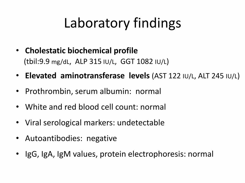
Laboratory findings
• Cholestatic biochemical profile (tbil:9.9 mg/dL, ALP 315 IU/L, GGT 1082 IU/L)
• Elevated aminotransferase levels (AST 122 IU/L, ALT 245 IU/L)
• Prothrombin, serum albumin: normal
• White and red blood cell count: normal
• Viral serological markers: undetectable
• Autoantibodies: negative
• IgG, IgA, IgM values, protein electrophoresis: normal

• Ultrasound and MRCP
No parenchymal abnormalities, no dilation or stenosis of extra-or intrahepatic bile ducts
• Gastroduodenoscopy,colonoscopy
No signs of inflammatory bowel disease

Predominant histological findings
• Majority of interlobular bile ducts:pleomorphic cholangitis rarely with epithelial destruction (infiltration of the epithelium by neutrophils and eosinophils, degenerative epithelial changes with nuclear pleomorphism, cell crowding and flattening)
• Three bile ducts:
sclerosing cholangitis (pericholangitis, concentric periductal fibrosis, focally severely damaged epithelium)
• No bile duct loss
Secondary findings
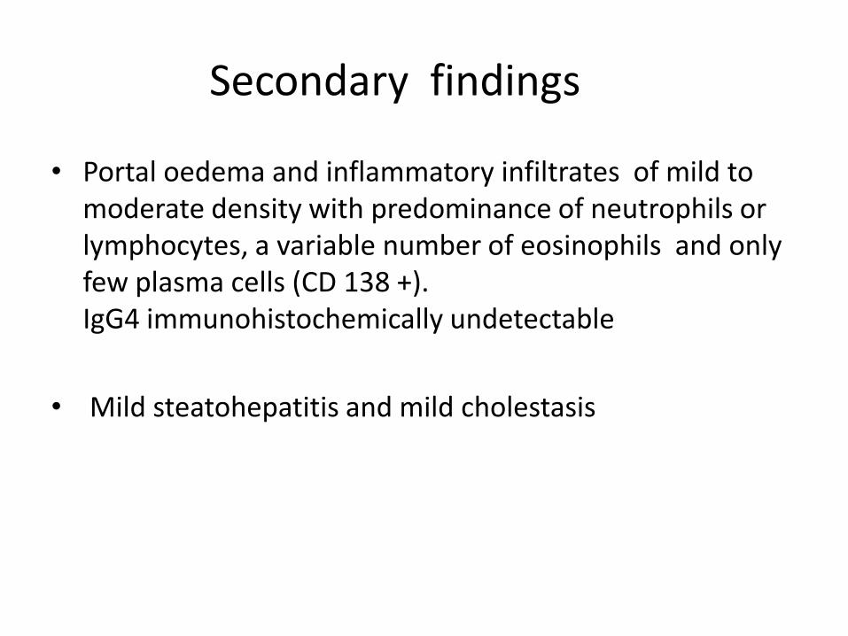
• Portal oedema and inflammatory infiltrates of mild to moderate density with predominance of neutrophils or lymphocytes, a variable number of eosinophils and only few plasma cells (CD 138 +). IgG4 immunohistochemically undetectable
• Mild steatohepatitis and mild cholestasis
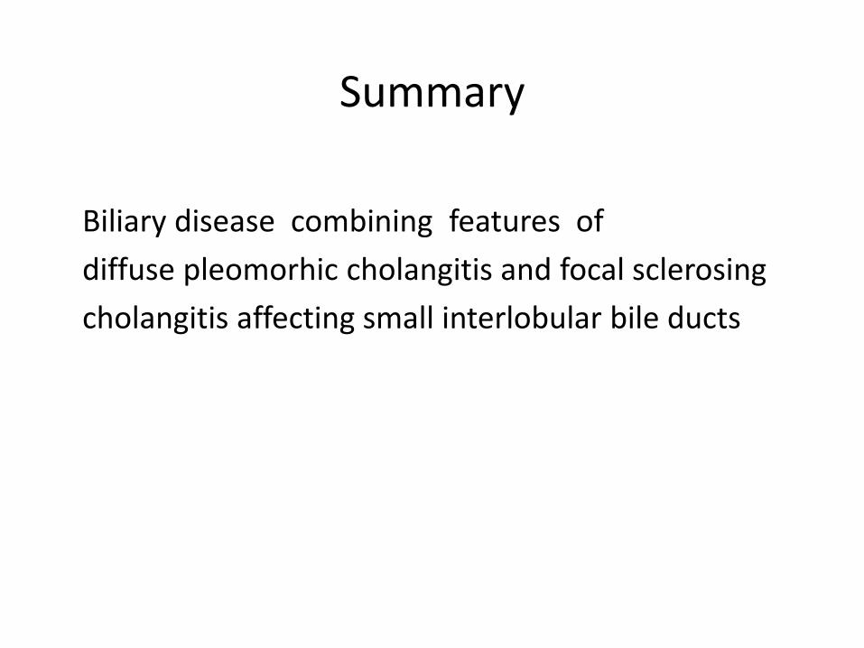
Biliary disease combining features of
diffuse pleomorhic cholangitis and focal sclerosing
cholangitis affecting small interlobular bile ducts
Summary
Clinical outcome
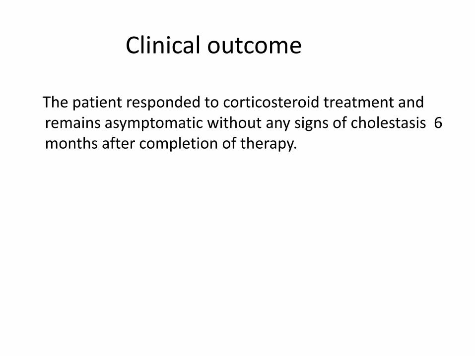
The patient responded to corticosteroid treatment and remains asymptomatic without any signs of cholestasis 6 months after completion of therapy.

Differential diagnosis of small-duct biliary diseases in immunocompetent patients
• Idiopathic adulthood ductopenia (IAD)
• Drug induced bile duct damage
• Hepatitis C virus-associated bile duct loss
• Primary biliary cirrhosis/Autoimmune cholangitis
• Steroid responsive IgG4 -associated cholangitis
• Secondary sclerosing cholangitis complicated
by recurrent or longstanding episodes of suppurative cholangitis
• Primary small- duct sclerosing cholangitis
Excluded by clinical, laboratory findings and histology

Exclusion criteria of primary small-duct PSC:
• acute clinical onset
• no IBD
• response to steroids
• coexistence of diffuse pleomorphic cholangitis

Exclusion criteria of secondary cholangitis complicated by acute ascending cholangitis:
• absence of clinical and laboratory findings of bacterial infection
• absence of suppurative cholangitis histologically

Diagnosis proposed :
Idiopathic adulthood ductopenia (IAD)

IDIOPATHIC ADULTHOOD DUCTOPENIA
• Heterogeneous entity
• Diagnosis one of exclusion
• Young or middle aged adults (median 30 years)
• Distinct male predominance
• Chronic cholestatic disease
• Episodic jaundice and pruritus in 1/3 of the patients

Diagnostic criteria
• Negative AMA
• No large bile duct involvement
• No history of drug use
• No evidence of IBD
• Occasional response to steroids
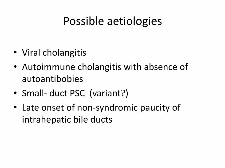
Possible aetiologies
• Viral cholangitis
• Autoimmune cholangitis with absence of autoantibobies
• Small- duct PSC (variant?)
• Late onset of non-syndromic paucity of intrahepatic bile ducts

Ludwig J. Mayo Clinic Proceedings 1998; 73 (3): 285
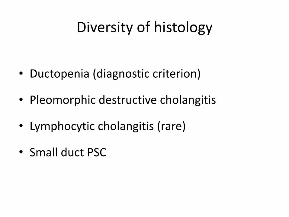
Diversity of histology
• Ductopenia (diagnostic criterion)
• Pleomorphic destructive cholangitis
• Lymphocytic cholangitis (rare)
• Small duct PSC

Conclusion
• The case meets the exclusion and inclusion criteria of IADregarding age, sex, clinical history, clinical presentation and laboratory findings
• The combination of two different bile duct lesions probably reflects different stages of a rapid evolving disease starting as pleomorphic cholangitis and progressing to small-duct PSC
• The effectiveness of corticosteroids points towards an immune mediated process
References
1. Galambos GT, Scott Brooks W. Atypical biliary cirrhosis – or sclerosing cholangitis. J Clinic Gastroenterol 1980; 2: 43-52.
2. Ludwig J, Wiesner RH, LaRusso N. Idiopathic adulthood ductopenia. A cause of chronic cholestatic liver disease and biliary cirrhosis. J Hepatol 1988; 7: 193-199
3. Ludwig J . Idiopathic adulthood ductopenia: an update. Mayo Clin Proc 1998; 73: 285-291
4. Zafrani ES, Metreau JM, Douvin C, Larrey D, Massari R, Reynes M, Doffoel M, Benhamou JP, Dhumeaux D. Idiopathic biliary ductopenia in adults: a report of five cases. Gastroenterology 1990; 99:1823-8.
5. Faa G, van Eyken P, Demelia L, Vallebone E, Costa V, Desmet V. Idiopathic adulthood ductopenia presenting with chronic recurrent cholestasis. A case report. J Hepatol 1991;12: 14-20
6. Khanlou H, Sass D, Rothstein K, Manzarbeitia C, Reich D, Jacobson L, Fleischer D, Muñoz SJ. Idiopathic adulthood ductopenia: case report and review of the literature. Arch Intern Med. 2000, 10;160:1033-6.
7. Kim WR, Ludwig J, Lindor KD. Variant forms of cholestatic diseases involving small bile ducts in adults. Am J Gastroenterol 2000; 95: 1139-1138
8. Burak K, Pearson D, Swain M, Kelly J, Urbanski S, Bridges D. Familial idiopathic adulthood ductopenia: a report of five cases in three generations. Journal of Hepatology 2000; 32: 159-163
9. Park BC, Park SM, Choi EY, Chae HB, Yoon SJ, Sung R, Lee SK. A case of idiopathic adulthood ductopenia. Korean J Intern Med. 2009,24:270-3. Epub 2009 Aug 26.