
Jean Luc Cornille - Horse Trainer,Lameness ,Dressage Jumpers and 3
47
Jean Luc Cornille Jean Luc Cornille 2008 @copyright 1 SOM
Transcript of Jean Luc Cornille - Horse Trainer,Lameness ,Dressage Jumpers and 3

Jean Luc Cornille
Jean Luc Cornille 2008 @copyright 1
SOM

Jean Luc Cornille 2008 @copyright 2
In 1825, the Prussian cavalry’s regulations emphasized the total elevation of the horse’s head and neck. The experiment lasted several decades then was abandoned completely. The theory behind the technique was that total elevation of the neck was enhancing the horse’s balance, developing the back muscles, and engaging the hind legs. By contrast, the Prussian Emperor’s Riding Master, lowered and over-flexed his horses’ necks completely. Paul Splinzner (1853-1920), in fact created the “rollkur”. His theory was that such over-flexion of the neck was enhancing the horse’s balance, developing the back muscles, and engaging the hind legs. Almost contemporarily and on the other side of the Rhine River, Francois Baucher(1776-1873) promoted in his “dernier enseignements” the systematic elevation of the neck. His view was that such elevation of the neck was enhancing the horse’s balance, developing the back muscles, and engaging the hind legs. Half a century later, Jack Licart (1951) reinvented the lowering of the neck. His theory was that the lowering of the neck was enhancing the horse’s balance, developing the back muscles, and engaging the hind legs. Not long ago, Harry Bolt (1973) warned against the practice of over-flexing the neck. Today no one can win without over-flexing the horse’s neck completely.

Obviously, the relationship between neck posture
and vertebral column’s mechanism is more a mater
of opinion than scientific documentation.
Twenty five thousand, seven hundred and thirty six
opinions later, the question: “what does the Rollkur
do to horses,” remains unanswered. Olympic riders
are winning practicing this technique and therefore
anyone anxious to win feels that
over-flexing the horse’s neck is the price of
winning.
Jean Luc Cornille 2008 @copyright 3
Jean Luc Cornille 2008 @copyright 4
In terms of “drowning the fish”, the “Rollkur” largely
surpasses any political lobbying. “Is the Rollkur
damaging for horses?” is a subjective question.
Technology has not been developed to measure
the horse’s level of pain. Furthermore,
paraphrasing the words of Roy R. Pool DVM PhD,
scientific studies often reflect the particular bias or
specific angle of the proponents’ investigative
technique.
Rollkur opponents care for horses. Rollkur
proponents care more for success. Everyone would
profit to know if there is a physical advantage to work
a horse with the neck lowered and
over-flexed.

Jean Luc Cornille 2008 @copyright 5
Scientific references have often been used to
support a specific point, but never has actual
scientific knowledge been compiled to look at the
overall picture. One must begin with the neck
muscles and ligaments and follow the effects on the
horse’s vertebral column and hind and front legs.
Such analysis is the topic of this discussion.

Jean Luc Cornille 2008 @copyright 6
Respectively in 1999, 2002, and 2005, three
pertinent studies have been published. They
investigate neck, base of the neck, and back
structures that are placed under strain in the
rollkur situation. Interestingly, the three studies
have been carefully ignored during this whole
controversy. Probably the publications were too
pertinent and too explicit.
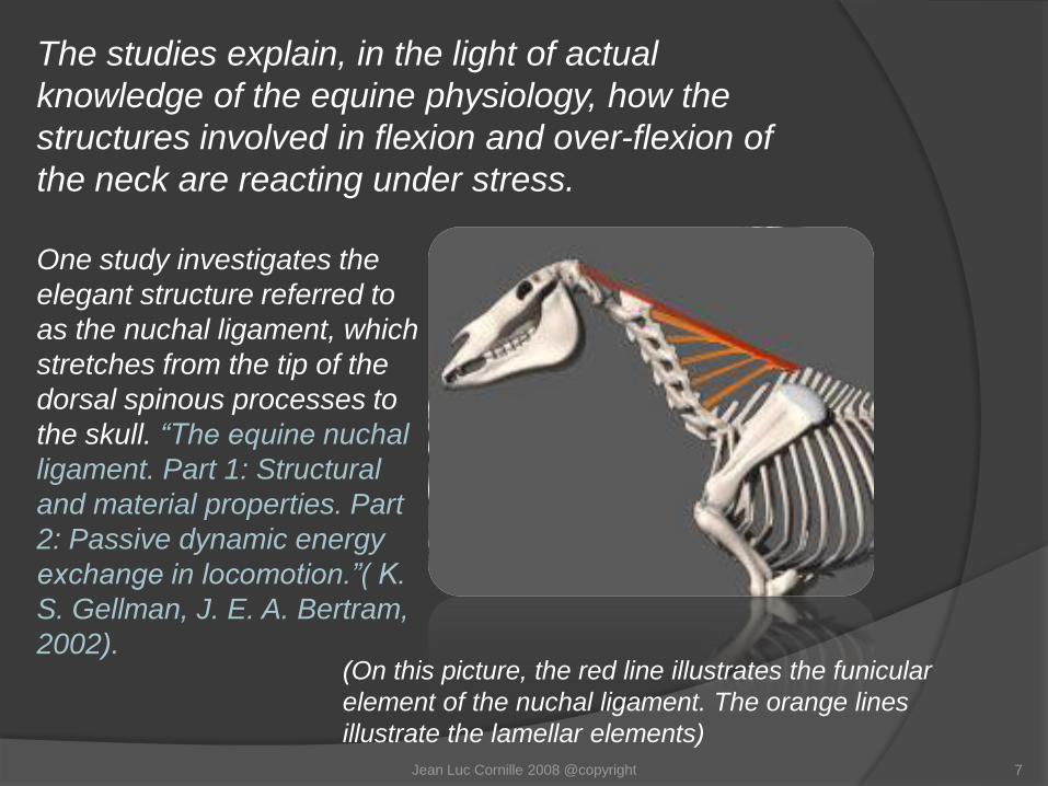
One study investigates the
elegant structure referred to
as the nuchal ligament, which
stretches from the tip of the
dorsal spinous processes to
the skull. “The equine nuchal
ligament. Part 1: Structural
and material properties. Part
2: Passive dynamic energy
exchange in locomotion.”( K.
S. Gellman, J. E. A. Bertram,
2002). (On this picture, the red line illustrates the funicular
element of the nuchal ligament. The orange lines
illustrate the lamellar elements)
Jean Luc Cornille 2008 @copyright 7
The studies explain, in the light of actual
knowledge of the equine physiology, how the
structures involved in flexion and over-flexion of
the neck are reacting under stress.
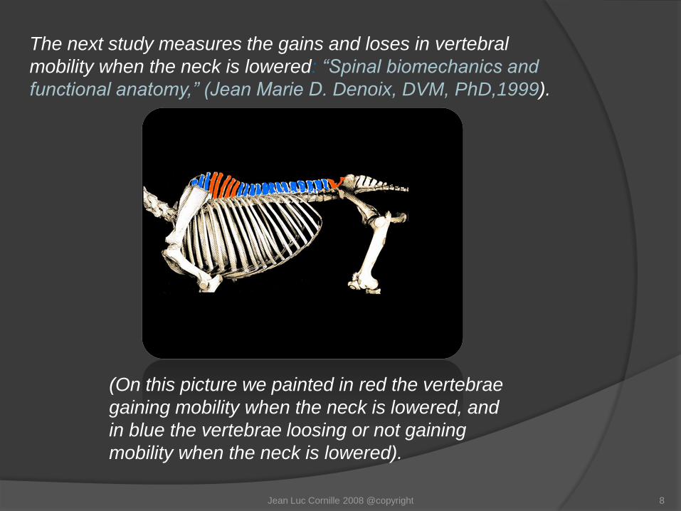
The next study measures the gains and loses in vertebral
mobility when the neck is lowered: “Spinal biomechanics and
functional anatomy,” (Jean Marie D. Denoix, DVM, PhD,1999).
(On this picture we painted in red the vertebrae
gaining mobility when the neck is lowered, and
in blue the vertebrae loosing or not gaining
mobility when the neck is lowered).
Jean Luc Cornille 2008 @copyright 8
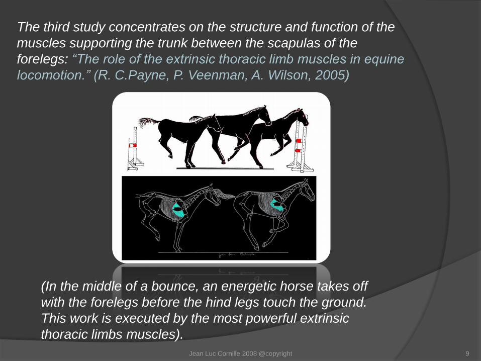
The third study concentrates on the structure and function of the
muscles supporting the trunk between the scapulas of the
forelegs: “The role of the extrinsic thoracic limb muscles in equine
locomotion.” (R. C.Payne, P. Veenman, A. Wilson, 2005)
(In the middle of a bounce, an energetic horse takes off
with the forelegs before the hind legs touch the ground.
This work is executed by the most powerful extrinsic
thoracic limbs muscles).
Jean Luc Cornille 2008 @copyright 9
Jean Luc Cornille
Jean Luc Cornille 2008 @copyright 10
SOM

In the mid fifties, Jack Licart referred to the lowering of the neck
as an “extension”. The words were in agreement with the
author’s philosophy; the thought of a neck extending out of the
shoulders and elongating eventually the back muscles. This
terminology was incorrect and in fact in contradiction with the
horse’s actual physiology. The lowering of the neck is not an
extension but rather a flexion created by concentric contraction
of the lower neck muscles. The total elevation of the neck
promoted by the Prussian cavalry was indeed an extension.
Jean Luc Cornille 2008 @copyright 11

Licart’s philosophy remains the idea behind the lowering of
the neck, an elongation that stretches the upper neck
muscles and sub-consequently the long back muscles. If we
give credit to this theory, we must first verify if the muscles
situated below the cervical vertebrae (ventral muscles) are
capable to elongate the muscles situated above the cervical
vertebrae (dorsal muscles). Then, we should ensure that the
lowering of the cervical vertebrae effectively stretches the
long back muscles connected to the cervical linkage.
Jean Luc Cornille 2008 @copyright 12
A distinction needs to be made between “elongation” and
“stretching”. The words are synonyms but their nuances are
important. One elevating the arms vertically above the head
does elongate the back muscles but does not really stretch
these muscles. Suspended by the hands to a horizontal pool,
one will have the same posture but one’s body weight will add a
force that will elongate further the back muscles. This effort is
more precisely described as stretching.
Jean Luc Cornille 2008 @copyright 13
The renowned Pathologist James R. Rooney does not think
that the ventral neck muscles are capable of stretching the
muscles situated above the cervical vertebrae. “The ventral
cervical muscles do not appear adequate to pull the head and
neck down against the powerful dorsal muscles”. (James R.
Rooney, Biomechanics of Lameness in Horses, 1969)
Jean Luc Cornille 2008 @copyright 14
SOM
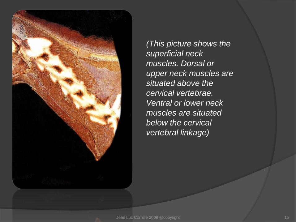
(This picture shows the
superficial neck
muscles. Dorsal or
upper neck muscles are
situated above the
cervical vertebrae.
Ventral or lower neck
muscles are situated
below the cervical
vertebral linkage)
Jean Luc Cornille 2008 @copyright 15

The lowering of the neck is created by concentric
contraction of the lower neck muscles. The lower muscles
are aided in their task by the attraction of gravity. Such
lowering is permitted by the elongation of the dorsal neck
muscles. However, the dorsal neck muscles are also the
muscles carrying the head.
The horse’s head and neck weighs approximately 10% of
the horse’s body mass. The Prussian cavalry’s basic idea
was precisely to reduce the burden of the head and neck
placing the head as close as possible from the vertical of
the shoulders.
Jean Luc Cornille 2008 @copyright 16
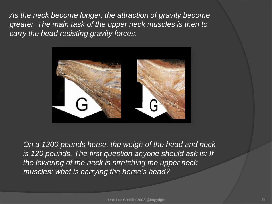
As the neck become longer, the attraction of gravity become
greater. The main task of the upper neck muscles is then to
carry the head resisting gravity forces.
On a 1200 pounds horse, the weigh of the head and neck
is 120 pounds. The first question anyone should ask is: If
the lowering of the neck is stretching the upper neck
muscles: what is carrying the horse’s head?
Jean Luc Cornille 2008 @copyright 17
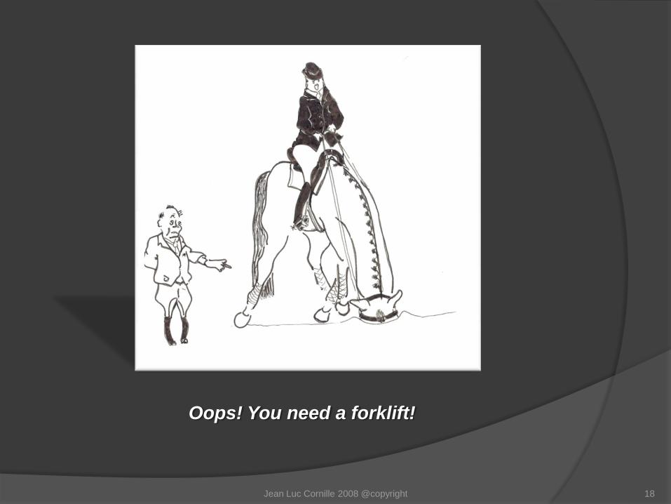
Oops! You need a forklift!
Jean Luc Cornille 2008 @copyright 18
“Active Stretching”
19Jean Luc Cornille 2008 @copyright
As the neck lowers, the dorsal neck muscles elongate
resisting the burden of the head and neck.
SOM
Even if sometimes referred to as “active stretching”, this type of
muscular work is not a yielding at all. “Active stretching” is a term
describing a muscle that elongates while contracting. This type of
muscle work is named “eccentric contraction”. It is in fact the
most powerful type of muscle contraction. “Muscles working
eccentrically can absorb up to 15 times more energy than during
concentric contractions.” (Payne et al.)
Jean Luc Cornille 2008 @copyright 20
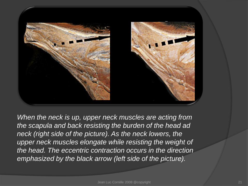
When the neck is up, upper neck muscles are acting from
the scapula and back resisting the burden of the head ad
neck (right side of the picture). As the neck lowers, the
upper neck muscles elongate while resisting the weight of
the head. The eccentric contraction occurs in the direction
emphasized by the black arrow (left side of the picture).
Jean Luc Cornille 2008 @copyright 21

Stretching theories believe that while the neck is lowering
dorsal neck muscles are working in the direction illustrated
on the left side of this picture, pulling on the back muscles. If
it was the case, one would need a forklift.
Jean Luc Cornille 2008 @copyright 22
“So why Does the horse lower
the neck after collected work”?
23Jean Luc Cornille 2008 @copyright
SOM

The nuchal ligament extends from the dorsal spinous process of
the fourth thoracic vertebrae to the back of the skull. Contrary to
traditional beliefs, the nuchal ligament is not under tension when
the neck is held in an alert posture, which is approximately an
angle of 60º.
Jean Luc Cornille 2008 @copyright 24
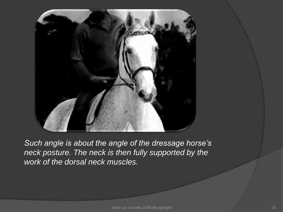
Such angle is about the angle of the dressage horse’s
neck posture. The neck is then fully supported by the
work of the dorsal neck muscles.
Jean Luc Cornille 2008 @copyright 25

As the neck lowers, the nuchal ligament comes under
tension. The nuchal ligament is then assisting the upper neck
muscles.
Jean Luc Cornille 2008 @copyright 26

The backing provided by the nuchal ligament is considerable. At
the walk for instance, measurements have demonstrated that the
nuchal ligament was replacing 55% of the work of the dorsal
neck muscles. At the trot and canter, where the neck is submitted
to lesser movements, the work of the nuchal ligament reduced
the work of the upper neck muscles by a percentage of 32 to
36%.
Jean Luc Cornille 2008 @copyright 27

For simplification, diagrams always represent the whole
structure of the nuchal ligament as clean bungee cords.
On this diagram that was presented earlier in this
discussion, the funicular element is represented in red
and the lamellar elements are represented in orange.
Jean Luc Cornille 2008 @copyright 28
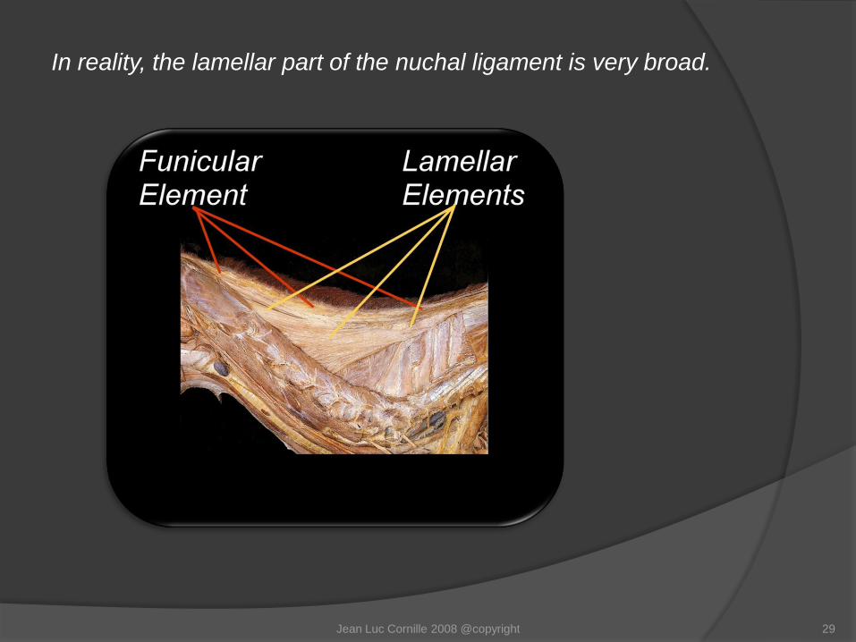
In reality, the lamellar part of the nuchal ligament is very broad.
Jean Luc Cornille 2008 @copyright 29
The broad lamellar part helps to understand how the nuchal
ligament can provide such efficient assistance to the work of
the upper neck muscles.
After collection, the horse lowers the neck increasing the
tension of the nuchal ligament and consequently reducing the
work of the upper neck muscles. However, ease does not
mean relaxation. Even if the nuchal ligament does a large
percentage of the work, the dorsal neck muscles are working
to carry the head against the attraction of gravity.
Jean Luc Cornille 2008 @copyright 30

The nuchal ligament comes under tension when the
neck lowers below and angle of 60º.
On this angle, On this angle and lower,
the nuchal ligament the nuchal ligament is under tension.
is not under tension.
Jean Luc Cornille 2008 @copyright 31
The tension increases as the neck becomes longer and
lower. Each horse’s morphology is different and the
comfort zone varies from one horse to the other. The
comfort zone is the neck posture where the tension
exerted on the nuchal ligament reduces the most
efficiently the work of the dorsal neck muscles.
Jean Luc Cornille 2008 @copyright 32
“But it seems logical to me that
the back muscles inserted on the
cervical vertebrae stretch when
the neck is lowered”.
Jean Luc Cornille 2008 @copyright 33
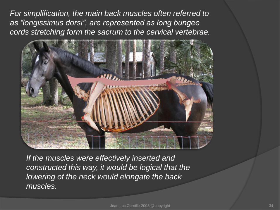
For simplification, the main back muscles often referred to
as “longissimus dorsi”, are represented as long bungee
cords stretching form the sacrum to the cervical vertebrae.
If the muscles were effectively inserted and
constructed this way, it would be logical that the
lowering of the neck would elongate the back
muscles.
Jean Luc Cornille 2008 @copyright 34
In reality, longissimus dorsi is a generic term which includes
the longissimus cervicis, thoracis, lumborum, etc. The
muscles group is composed of two divisions each side of the
spine. The dorsal division is illustrated on the following
diagram with the upper red line. The ventral division is
illustrated with the lower red line .
Jean Luc Cornille 2008 @copyright 35

The logissimus system is composed of fascicles that
bridge three to five vertebrae. The fascicles are
oriented oblique, down and forward. In the light of
such construction, even if the lowering of the neck
was stretching the cervical segments of the
longissimus thoracis, the effect on the back muscles
would not extend further back than the vertebrae of
the wither.
Jean Luc Cornille 2008 @copyright 36
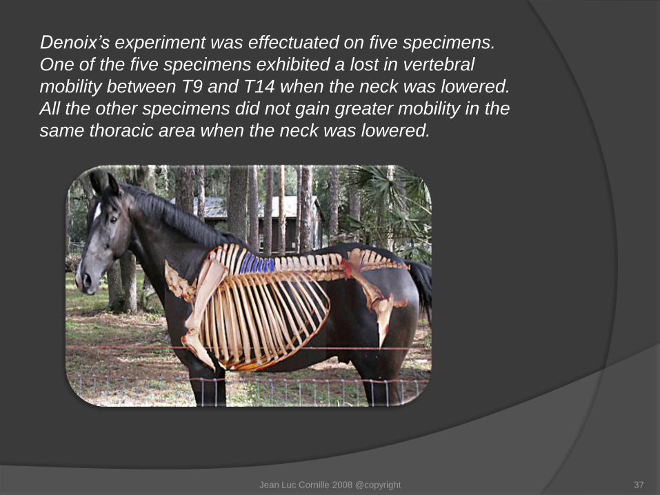
Denoix’s experiment was effectuated on five specimens.
One of the five specimens exhibited a lost in vertebral
mobility between T9 and T14 when the neck was lowered.
All the other specimens did not gain greater mobility in the
same thoracic area when the neck was lowered.
Jean Luc Cornille 2008 @copyright 37
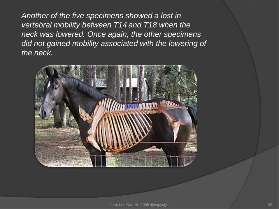
Another of the five specimens showed a lost in
vertebral mobility between T14 and T18 when the
neck was lowered. Once again, the other specimens
did not gained mobility associated with the lowering of
the neck.
Jean Luc Cornille 2008 @copyright 38
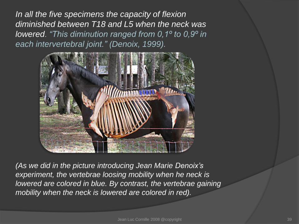
In all the five specimens the capacity of flexion
diminished between T18 and L5 when the neck was
lowered. “This diminution ranged from 0,1º to 0,9º in
each intervertebral joint.” (Denoix, 1999).
(As we did in the picture introducing Jean Marie Denoix’s
experiment, the vertebrae loosing mobility when he neck is
lowered are colored in blue. By contrast, the vertebrae gaining
mobility when the neck is lowered are colored in red).
Jean Luc Cornille 2008 @copyright 39

The average amount of flexion in the lumbo-sacral
area was greater when the neck was lowered.
Such gain in lumbo-sacral rotation is mostly
compensating for the lost of mobility occurring
through the thoracolumbar spine when the
neck is lowered.
Jean Luc Cornille 2008 @copyright 40

In the cranial thoracic area, intervertebral
movements are greater between T6 and T10 when
the neck is lowered
The gain of mobility is primarily due to the action
of the nuchal ligament which continues along the
spine over the top of the dorsal spinous
processes taking the name of supraspinous
ligament.
Jean Luc Cornille 2008 @copyright 41
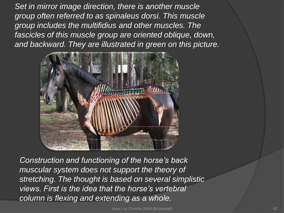
Set in mirror image direction, there is another muscle
group often referred to as spinaleus dorsi. This muscle
group includes the multifidius and other muscles. The
fascicles of this muscle group are oriented oblique, down,
and backward. They are illustrated in green on this picture.
Construction and functioning of the horse’s back
muscular system does not support the theory of
stretching. The thought is based on several simplistic
views. First is the idea that the horse’s vertebral
column is flexing and extending as a whole. Jean Luc Cornille 2008 @copyright 42
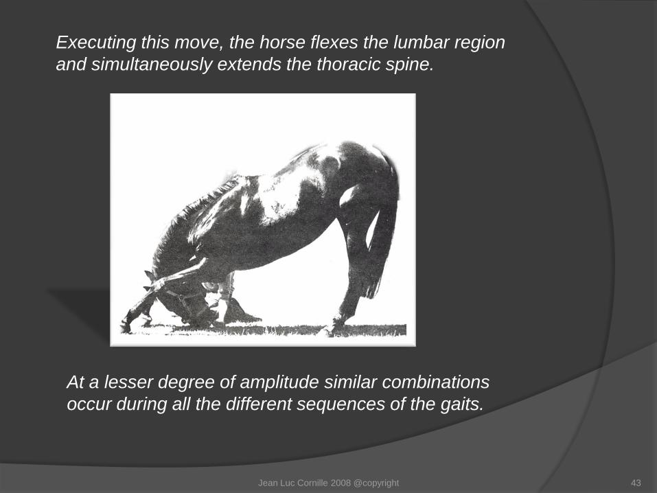
Executing this move, the horse flexes the lumbar region
and simultaneously extends the thoracic spine.
At a lesser degree of amplitude similar combinations
occur during all the different sequences of the gaits.
Jean Luc Cornille 2008 @copyright 43
The second thought is that greater ease, harmony, and
amplitude of the horse’s movements can be achieved
increasing the horse’s vertebral column range of
motion. This idea is in drastic contradiction with actual
knowledge of the equine physiology. Our ancestors
interpreted as movements, the large amount of forces
they perceived on the saddle. Misinterpreting forces as
movements, our predecessors elaborated the thought
that the amplitude of the stride would result from
greater amplitude of the horse’s vertebral column
range of motion.
Jean Luc Cornille 2008 @copyright 44
The misinterpretation gravely compromises the horse’s ability
to perform since at the contrary, the main function of the back
muscles is precisely to prevent the vertebral column linkage
from amplitude of movement exceeding its possible range of
motion. Electromyographic studies “strongly suggest that the
primary function of the back muscles during walking is to
control the stiffening of the back rather that to create
movement.” (Hans Carlson, 1979)
Jean Luc Cornille 2008 @copyright 45
The equestrian language will see the word
“stiffening” as a defect. In the context the meaning is
that during locomotion, the vertebral column muscles
are stabilizing the spine and preventing excessive
movements. The more intensively the stretching
theories will try to increase the vertebral column’s
range of motion, the more intensively the back
muscles will resist such increase.
Jean Luc Cornille 2008 @copyright 46

Jean Luc Cornille 2008 @copyright 47
“But I feel the horse relaxing
when he is lowering the neck”!
The nuchal ligament is another neck structure placed under
strain when the neck is lowered and over-flexed. Since from
the perspective of the upper neck muscles, the concept of
stretching is a theory that does not really translate into reality,
perhaps the “relaxation” that anyone can perceive when the
neck is lowered results from the action of the nuchal ligament
on the vertebrae of the thoracolumbar spine.
This working hypothesis will be the topic of our next newsletter.
Jean Luc Cornille
www.scienceofmotion.com


