JANICE LIOTTA - cpb-us-e1.wpmucdn.com · Foreyt WJ 1997 Veterinary Parasitology Reference Manual...
70
J ANICE L IOTTA
Transcript of JANICE LIOTTA - cpb-us-e1.wpmucdn.com · Foreyt WJ 1997 Veterinary Parasitology Reference Manual...

JANICE LIOTTA

• References:• Foreyt WJ 1997 Veterinary Parasitology Reference
Manual 4th ed SCAVMA • Georgis’ Parasitology for Veterinarians 10e, Bowman ed,
Saunders 2014 • Merck Veterinary Manual
https://www.merckvetmanual.com• http://www.goatbiology.com/• http://blogs.cornell.edu/smallruminantparasites/• https://www.wormx.info/ American Consortium for
small ruminant parasite control• https://web.uri.edu/sheepngoat/ NE Small Ruminant
Parasite control• Janice Liotta: [email protected]• tatiana Stanton: [email protected]

Know the problem!
WHY????

• Understand parasites lifecycles • Infection method• Identify infected animals• Prevent others from getting infected• Interventions/Disrupt cycles• Efficacy - What may or may not work
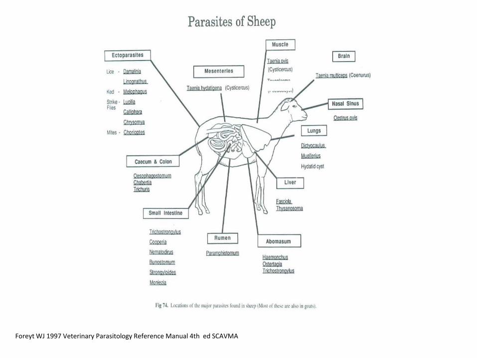
Foreyt WJ 1997 Veterinary Parasitology Reference Manual 4th ed SCAVMA

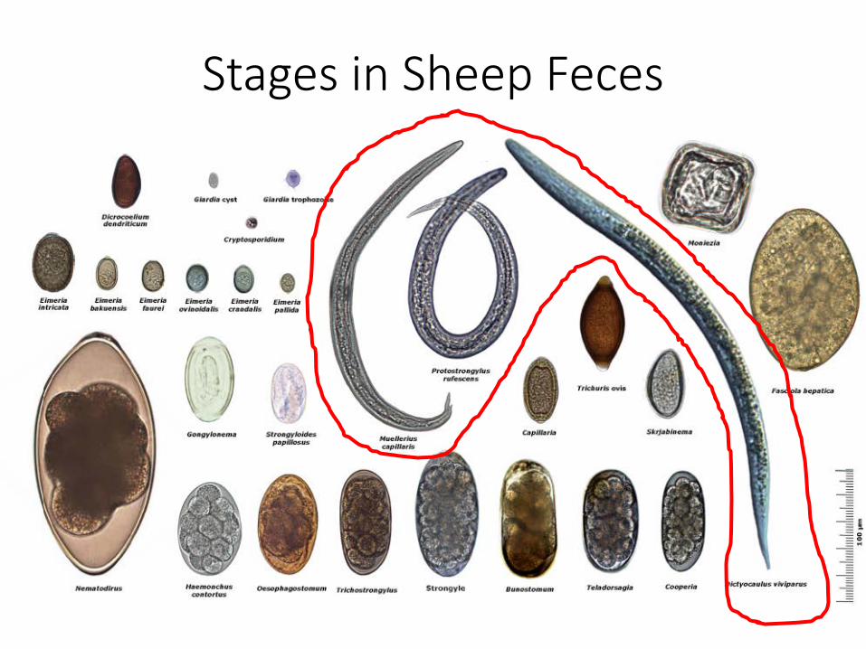
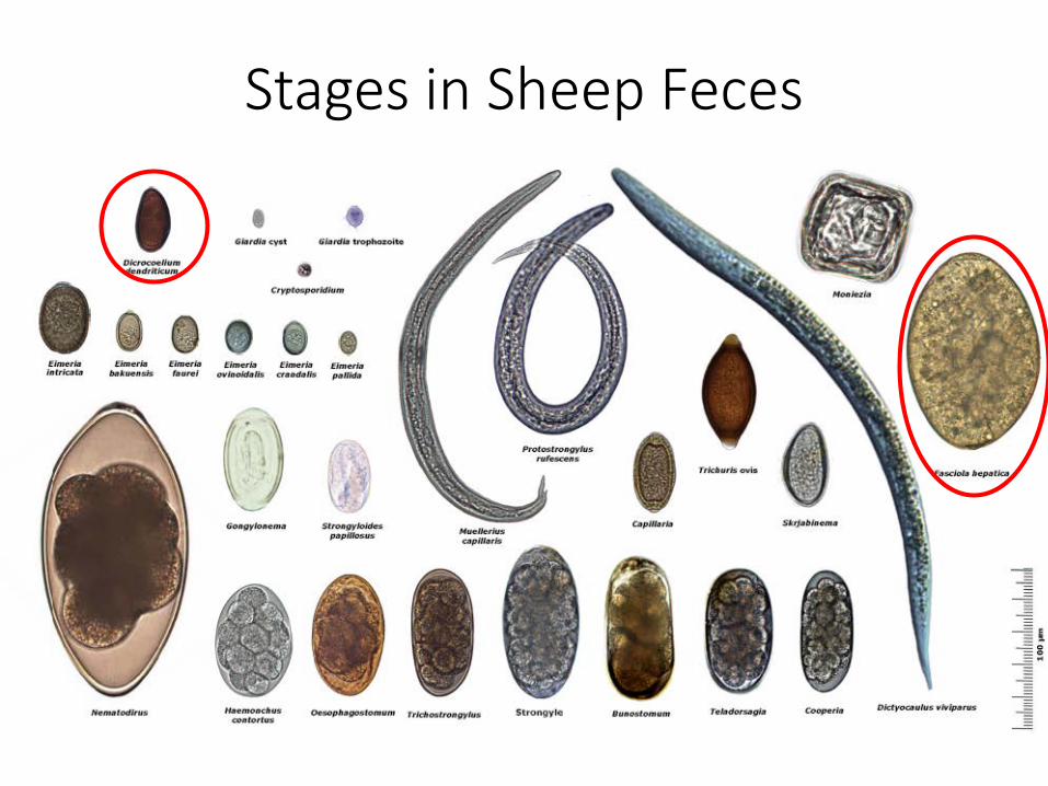
Stages in Sheep Feces

A few NOT found in sheep/goat feces
• Toxoplasma gondii• Fascioloides magna• Parelaphostrongylus tenuis• Echinococcus granulosis

Foreyt WJ 1997 Veterinary Parasitology Reference Manual 4th ed SCAVMA
Fascioloides magna
Dicrocoelium
CNS - Parelaphostrongylus tenuis
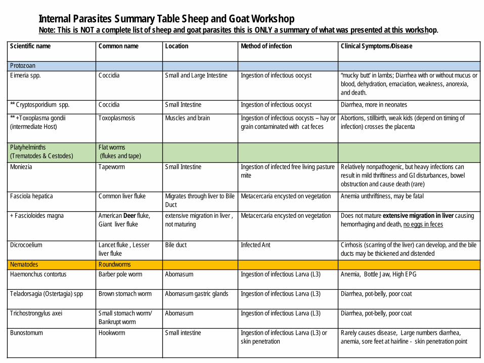
Scientific name Common name Location Method of infection Clinical Symptoms/Disease
ProtozoanEimeria spp. Coccidia Small and Large Intestine Ingestion of infectious oocyst “mucky butt’ in lambs; Diarrhea with or without mucus or
blood, dehydration, emaciation, weakness, anorexia, and death.
** Cryptosporidium spp. Coccidia Small Intestine Ingestion of infectious oocyst Diarrhea, more in neonates
** +Toxoplasma gondii (intermediate Host)
Toxoplasmosis Muscles and brain Ingestion of infectious oocysts – hay or grain contaminated with cat feces
Abortions, stillbirth, weak kids (depend on timing of infection) crosses the placenta
Platyhelminths (Trematodes & Cestodes)
Flat worms(flukes and tape)
Moniezia Tapeworm Small Intestine Ingestion of infected free living pasture mite
Relatively nonpathogenic, but heavy infections can result in mild thriftiness and GI disturbances, bowel obstruction and cause death (rare)
Fasciola hepatica Common liver fluke Migrates through liver to Bile Duct
Metacercaria encysted on vegetation Anemia unthriftiness, may be fatal
+ Fascioloides magna American Deer fluke, Giant liver fluke
extensive migration in liver , not maturing
Metacercaria encysted on vegetation Does not mature extensive migration in liver causing hemorrhaging and death, no eggs in feces
Dicrocoelium Lancet fluke , Lesser liver fluke
Bile duct Infected Ant Cirrhosis (scarring of the liver) can develop, and the bile ducts may be thickened and distended
Nematodes RoundwormsHaemonchus contortus Barber pole worm Abomasum Ingestion of infectious Larva (L3) Anemia, Bottle Jaw, High EPG
Teladorsagia (Ostertagia) spp Brown stomach worm Abomasum gastric glands Ingestion of infectious Larva (L3) Diarrhea, pot-belly, poor coat
Trichostrongylus axei Small stomach worm/Bankrupt worm
Abomasum Ingestion of infectious Larva (L3) Diarrhea, pot-belly, poor coat
Bunostomum Hookworm Small intestine Ingestion of infectious Larva (L3) or skin penetration
Rarely causes disease, Large numbers diarrhea, anemia, sore feet at hairline - skin penetration point
Internal Parasites Summary Table Sheep and Goat WorkshopNote: This is NOT a complete list of sheep and goat parasites this is ONLY a summary of what was presented at this workshop.

Scientific name Common name Location Scientific name Common nameCooperia spp. Intestinal worm Small intestine Ingestion of infectious Larva (L3) Diarrhea, pot-belly, poor coat
Strongyloides papillosus Intestinal threadworm Small intestine Ingestion of infectious Larva (L3) Diarrhea, pot-belly, poor coat
Trichostrongylus colubriformis black scour worm/ hairworm
Small intestine Ingestion of infectious Larva (L3) Diarrhea, pot-belly, poor coat
Nematodirus spp. Thread-necked worm Small intestine Ingestion of infectious Larva (L3) Diarrhea, pot-belly, poor coat
Oesophagostomum Nodular worm Large intestine Ingestion of infectious Larva (L3) Diarrhea, excess mucus with blood, weak, weight loss intermittent diarrhea and constipation, stilted gate often humped back
Chabertia ovina Largemouth bowel worm
Large intestine/colon mucosa
Ingestion of infectious Larva (L3) Infected sheep are unthrifty; the feces are soft, contain much mucus, and may be streaked with blood
Dictyocaulus Lung worm Lumen of the bronchial tree
Infective L3 larva bronchitis or pneumonia, coughing
Muellerius Lung worm Embedded in lung tissues Snail intermediate host bronchitis or pneumonia, coughing
Protostrongylus Small red lung worm of sheep
Snail intermediate host bronchitis or pneumonia, coughing
+ Parelaphostrongylus tenuis Deer Brain WormCNS
Ingestion of infectious Larva (L3) (snails)
Neurological symptoms such as Paralysis & incoordination, larva cause severe inflammation to CNS,
** Zoonotic + Not shed in feces
References:Foreyt WJ 1997 Veterinary Parasitology Reference Manual 4th ed SCAVMA Georgis’ Parasitology for Veterinarians 10e, Bowman ed, Saunders 2014 Merck Veterinary Manual https://www.merckvetmanual.com
Resources: http://www.goatbiology.com/animations/parasites.html

Stages in Sheep Feces

Basic Life Cycle StrongylidNematodes
Feces is a nice place moisture, warmth, and food
Larva develops and hatches 1-6 days depending on temperature/humidity
L1 free living eats bacteria in feces grows & molts (sheds skin like a snake)
L-2 free living eats bacteria, grows but does not shed its skin/cuticle – keeps it = L3
Cuticle protects L3 drying out L3 does not eat; EATEN BY SHEEP
Eggs passed in feces Morulastage
Direct life cycle

Haemonchus contortusBarber pole worm
• Blood sucking – lancet pierces abomasum mucosa• 50 ul of blood lost/worm/day beginning 6 days
after infection• lambs may lose 1/10th to 1/5th of blood volume
per day• Blood Plasma and protein Loss• Short generation time < 3 weeks• Heavy egg producer 5000-10,000egg/worm/day
Kaplan RM. 2010. Small ruminant recommendations for control of parasites. Large animal. Proceedings of the North American Veterinary Conference, Orlando, Florida, USA, 16-20 January 2010 Pages: 348-356

Haemonchus Lancet
From Whitlock JH: Diagnosis of veterinary parasitisms, Philadelphia, 1960, Lea & Febiger.)
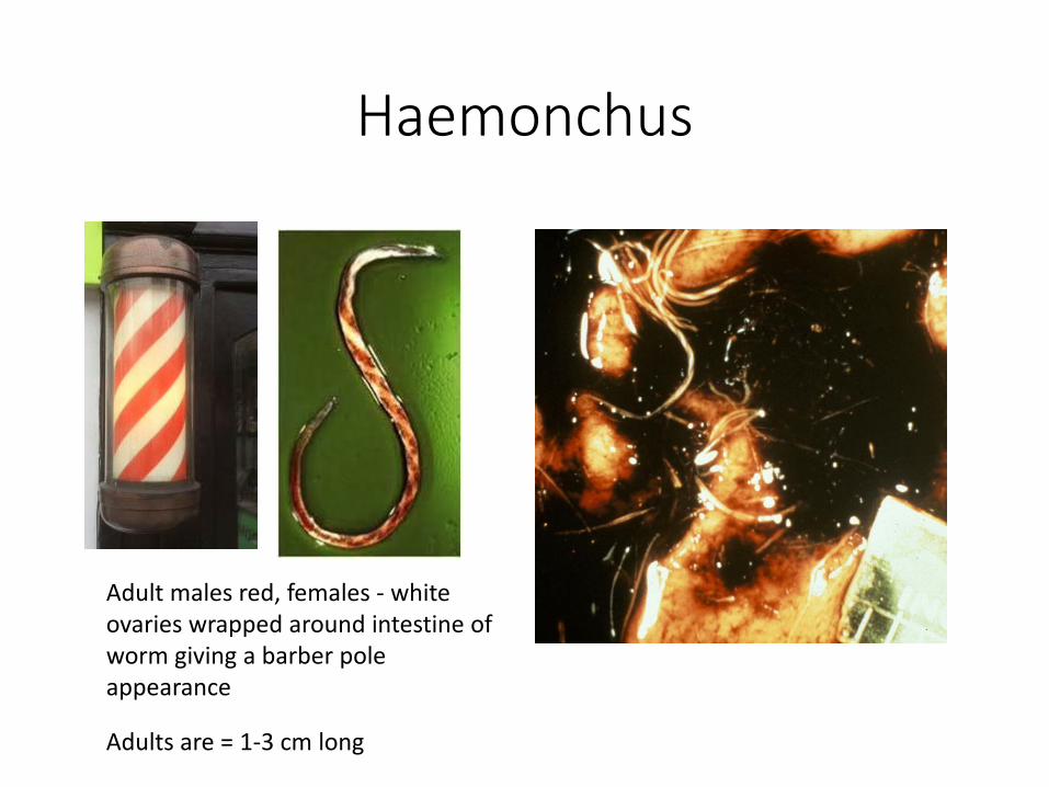
Haemonchus
Adults are = 1-3 cm long
Adult males red, females - white ovaries wrapped around intestine of worm giving a barber pole appearance

Clinical Symptoms• Anemia• Bottle jaw• High fecal egg counts with
anemia• Feces often well formed
may even have constipation (of course, with mixed infections with Trichostrongylus & Teladorsagia may have diarrhea)

FAMACHA• LIVING WITH WORMS
• FAffa MAlan CHArt
Normal
ANEMIA

Other Strongyles• Nematodirus – very large egg (develops to L3 in egg then hatches)
• Trichostrongylus spp. • Teladorsagia (Ostertagia)• Cooperia spp. • Oesophagostomum spp• Bunostomum
• Direct life cycle• Eggs indistinguishable from one another except Nematodirus• Eggs/Larvae tolerate cold temperatures and some even
overwinter on pasture• Clinical Symptoms:
scouring, weight loss, rough hair coat, ill thrift
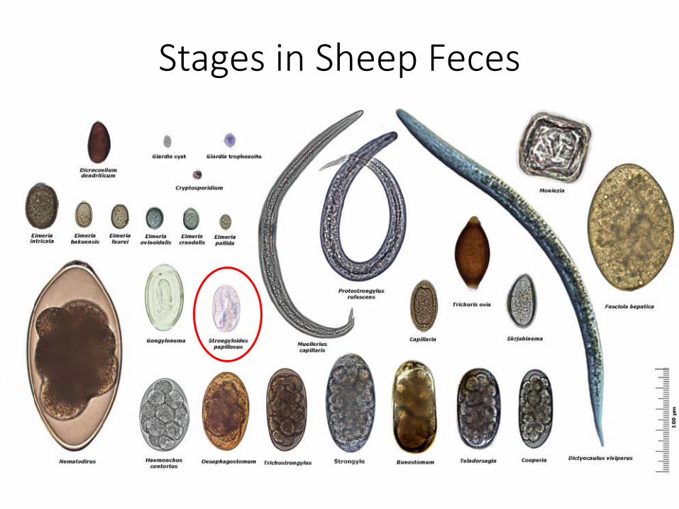
Stages in Sheep Feces

• Complicated life cycle Asexual and sexual
***Larvated egg passed in feces – collect fresh feces
• Infection methods:ingestion –
L3 pasture, Dam’s milk if larvae migrate to her udder
Pentration of skin (i.e. hairline above the hoof in muddy, infected pasture, shed or barn)
Transmammary transmission (larvae migrate through the placenta)
• Not infectious to people
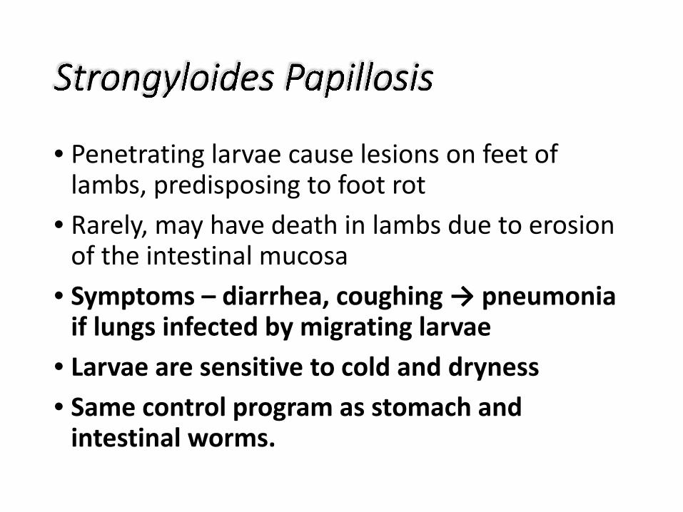
• Penetrating larvae cause lesions on feet of lambs, predisposing to foot rot
• Rarely, may have death in lambs due to erosion of the intestinal mucosa
• Symptoms – diarrhea, coughing → pneumonia if lungs infected by migrating larvae
• Larvae are sensitive to cold and dryness• Same control program as stomach and
intestinal worms.
Stages in Sheep Feces
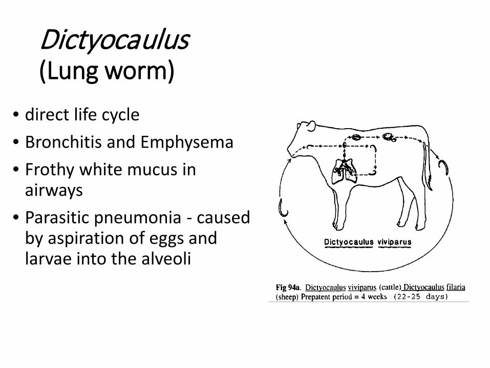
Dictyocaulus(Lung worm)
• direct life cycle• Bronchitis and Emphysema• Frothy white mucus in
airways• Parasitic pneumonia - caused
by aspiration of eggs and larvae into the alveoli

Dictyocaulus
Larvae in feces Adults in Lumen of the bronchial tree
Take fecal sample direct from animal (otherwise can confuse with soil nematodes)

Muellerius & Protostrongylusmore lung worms
• Indirect life cycle• eggs laid in lung, larva hatches,
coughed up and swallowed, passed as larva in feces
• penetrate snail and develop to infective third stage
• host eats snail while grazing• larvae enter lymphatics - right
heart to lung• adults in small bronchi• PPP = 5 to 6 weeks

Lungworms• Indirect or direct life
cycle• Severe infestations
cause coughing, fluid in lungs, pneumonia
• Transmitted in feces • Take fecal sample
direct from animal (otherwise can confuse with soil nematodes)
• Same control program as stomach and intestinal worms.

• Meningeal worm of the white-tailed deer• adults 6 to 10 cm long• Indirect life cycle typical of
metastrongyles• snail - ingested• worm migrates through or along
spinal cord to brain to the brain pan.
• non-pathogenic in deer, however may cause neurological damage in goats, sheep and camelids (alpacas, llamas, etc).
• WE will talk about this parasite in more detail in part 3 of our presentation
Stages in Sheep Feces

Flukes• Indirect life cycles snail/ant intermediate
host• Fasciola hepatica – common liver fluke• Fascioloides magna - Deer Fluke
• nonpatent in sheep/goats One fluke can kill a sheep
• Extensive Migration in liver • nonpathogenic in deer and elk
• Dicrocoelium dendriticum - Lancet fluke

Life cycle Fasciola and Fascioloides

Dicrocoelium dendriticum

Destroyed Liver : BLACK or FLUKE pigment deposited by migrating stages

Liver flukes
• Some farms in NE US have acute or chronic liver fluke populations
• Requires open water, snails (wet conditions)
• Can kill adult liver flukes with Albendazole (Valbazen®) or Ivomec® Plus)

Fasciola hepatica• common liver fluke – used to assume not east of
Mississippi but some veterinarians have observed affected animals in NY
• cycle includes fresh water snails• acute peritonitis (during migration)• Often causes chronic problems afterwards• hypoproteinemia, anemia
(blood leaks into bile)

Fascioloides magna• American deer fluke – found in Adirondacks• natural parasite of deer and elk• sheep and goats abnormal hosts• larval stages continue to migrate through liver
- sheep and goats don’t excrete eggs• ACUTE disease - usually fatal within 6
months

liver of goat killed by fluke

Treatment of liver flukes
Black Liver Disease (deadly) - To try to prevent: 1)try to kill flukes and 2) administer a vaccine for Clostridium novyi B such as Covexin® 8 as soon as possible

Treatment for liver flukes• Prevention – stay off wet areas• Kill flukes in animal
• albendazole – can cause abortion in early pregnancy, 15 to 20 mg/kg to kill adult flukes
• clorsulon orally - adult Fasciola . 3.5 mg/kg sheep, 7 mg/kg goats
• clorsulon orally – 8 wk. old Fasciola, 7 mg/kg sheep, 15 mg/kg goats
• clorsulon for Fascioloides – 21 mg/kg• Prevent Black Liver disease - Clostridium novyi B
vaccination such as Covexin® 8

Stages in Sheep Feces

Moniezia (tapeworm)
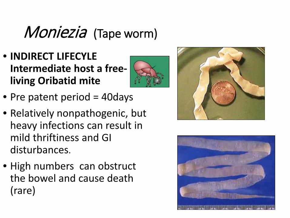
Moniezia (Tape worm)
• INDIRECT LIFECYLE Intermediate host a free-living Oribatid mite
• Pre patent period = 40days• Relatively nonpathogenic, but
heavy infections can result in mild thriftiness and GI disturbances.
• High numbers can obstruct the bowel and cause death (rare)

Moniezia egg
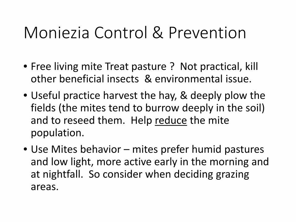
Moniezia Control & Prevention
• Free living mite Treat pasture ? Not practical, kill other beneficial insects & environmental issue.
• Useful practice harvest the hay, & deeply plow the fields (the mites tend to burrow deeply in the soil) and to reseed them. Help reduce the mite population.
• Use Mites behavior – mites prefer humid pastures and low light, more active early in the morning and at nightfall. So consider when deciding grazing areas.

Moniezia Chemical Treatments
• Dewormers will be discussed in the next presentation
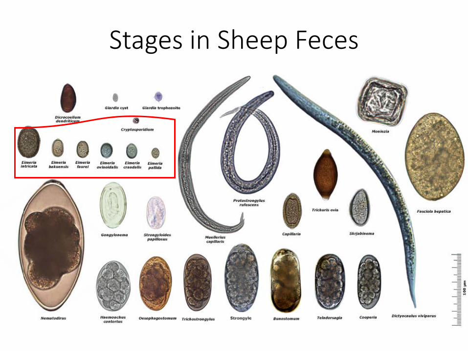
Stages in Sheep Feces

Protozoans
• Eimeria spp –Coccidia• Cryptosporidium –Coccidia
• Toxoplasma gondii - no eggs in feces

Life cycle Eimeria sp
Georgis’ Parasitology for Veterinarians 10e, Bowman ed, Saunders 2014 Foreyt WJ 1997 Veterinary Parasitology Reference Manual 4th ed SCAVMA

Eimeria spp. - Coccidiosis
• Many Eimeria species!• Host specific – direct lifecycle
• No transmission between host species• Site specific
• Develop within certain portions of the intestine• Symptom Severity varies between species• Immunity to each coccidia species develops with
exposure – mild exposure best for first exposure• Intensity of disease dependent upon number of
oocysts ingested• Avoid sudden exposure to large amounts of
infected feces

Eimeria spp. - Coccidiosis Symptoms• Damages small and large intestinal
lining causing malabsorption
• Suspect when calves, lambs or kids get diarrhea after 3 weeks of age (before that, usually bacterial or overindulging on milk)
• “mucky butt” in lambs• Spread through infected feces (contain
oocyst), decomposing feces in soil and bedding

• Disease of crowding• Sporulation happens 1-2 days!• warmth and moisture permit sporulation• Warmth from fecal decomposition can
cause sporulation Even when ambient temperatures below zero
• Can build up in soil, easily survives 2-3 mo. to even 1 year in optimum conditions
• Killed by direct sunlight and low humidity (<25%)
Eimeria spp. - Coccidiosis
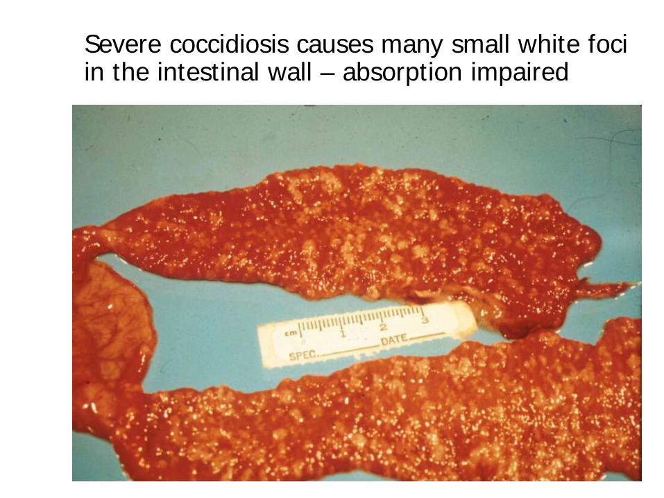
Severe coccidiosis causes many small white foci in the intestinal wall – absorption impaired
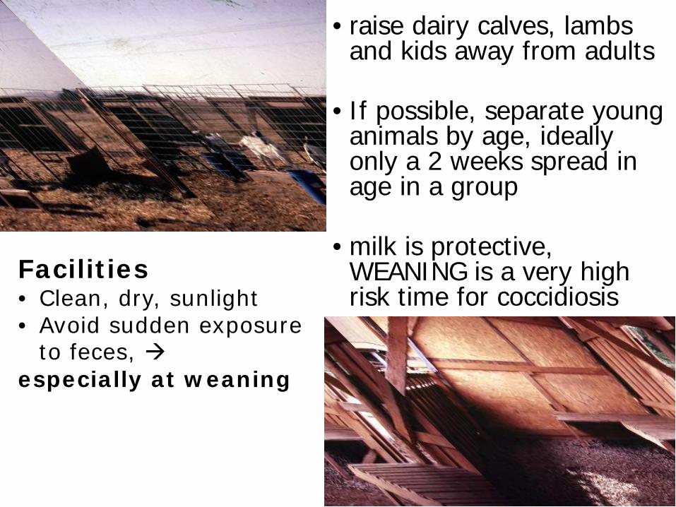
• raise dairy calves, lambs and kids away from adults
• If possible, separate young animals by age, ideally only a 2 weeks spread in age in a group
• milk is protective, WEANING is a very high risk time for coccidiosis
Facilities• Clean, dry, sunlight• Avoid sudden exposure
to feces, especially at weaning

Conventional flocks or herds may use coccidiostats as additives in the feed, salt or water to help prevent:
• Especially in pregnant females starting 1 month before parturition until weaning of their young. Continue in young animals after weaning.
• Lasalocid (Bovatec®)1,3 – non-lactating only
• Monensin (Rumensin®)2,3
• Dequinate (Deccox®)1,2 – non-lactating only,
Deccox M in milk?• Amprolium (Corid®) in water or milk?
1 - FDA-approved for sheep2 - FDA-approved for goats3 - TOXIC to EQUINES!!!!!!

Chemical treatment of coccidiosis• oral sulfonamides –Sulmet, Albon, etc.• Veterinary Feed Directive- new legislation requiring
vet prescription to use and may not be able to be prescribed use in milk.
• Amprolium 25-50 mg/kg per day for 5 days = 1 ml Corid 9.6% per 8 pounds
• can add to water (milk?) or directly drench• Adequate selenium for immunity• Electrolytes, supplemental nutrition• Alleviate stress
Coccidia Eimera sp. (species-specific)
• many Eimeria species, host specific
• immunity to each species of coccidia develops with exposure – mild exposure best at first
• Avoid sudden exposure to large amounts of infected feces
• Vulnerability – stress and age related! Young animals and geriatric animals most susceptible, also orphans, weaning, moving to new home, young mothers
• STOCKING RATE related – low density of animals


Eimeria macusaniensisE. mac
Considered highly pathogenic in naïve animals - Llamas
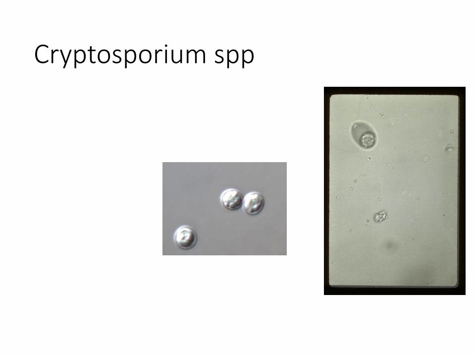
Cryptosporium spp
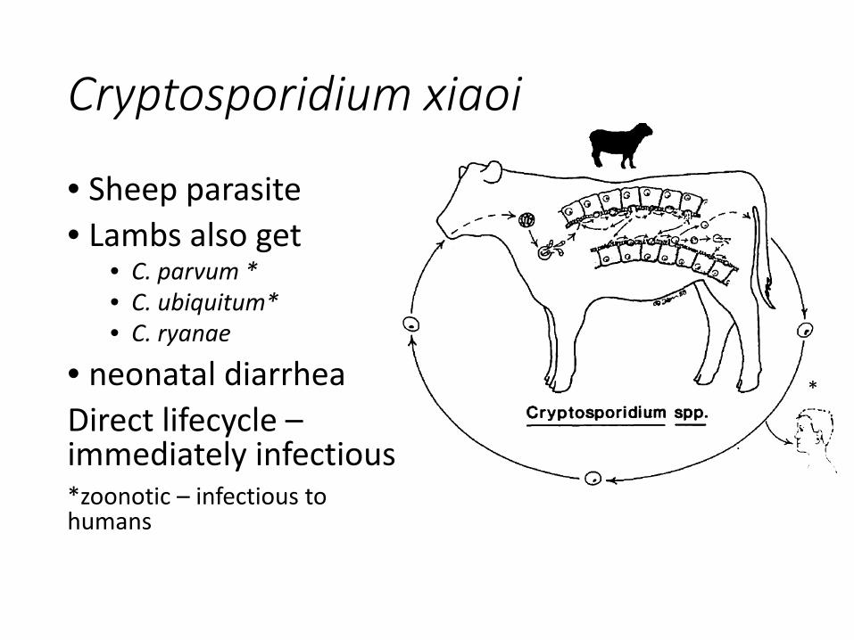
Cryptosporidium xiaoi
• Sheep parasite• Lambs also get
• C. parvum *• C. ubiquitum*• C. ryanae
• neonatal diarrheaDirect lifecycle –immediately infectious*zoonotic – infectious to humans
*

Cryptosporidium• Oocysts survive for several months in cool and moist
conditions• Oocysts are resistant to most disinfectants • Oocyst infectivity can be destroyed by ammonia, formalin,
freeze-drying, and exposure to temperatures <32°F (0°C) or >149°F (65°C). Ammonium hydroxide, hydrogen peroxide, chlorine dioxide and 5% ammonia are effective in destroying oocyst infectivity.
• Infectivity in calf feces is reduced after 1–4 days of drying.• Inactivated with UV exposure (dose 4000 -8000 mJ/cm2)• NO TREATMENT just fluids / electrolytes

Toxoplasma gondii
Intermediate hosts = (do not shed eggs in feces, pass disease through tissue exposure)

Toxoplasma gondii
• Intermediate (required for development) hosts:• Sheep• Humans• Mice • Horses• Pigs• All mammals!
• Vertical transmission- crosses the placenta!• Eggs resistant remain viable for several months• Tissue cyst susceptible high heat cooking

Toxoplasma gondii• Infection acquired during pregnancy by naïve animal
• Effect on pregnancy depends on gestation stage when the infection occurs
<45-55 days, unobserved abortion and expulsion55-90 days, abortion90-120 days, fetal infections; no abortion
• Typical clinical findings• Infection in early or mid-gestation results in fetal death with
reabsorption or mummification. • Occasionally lambs infected in mid-pregnancy survive to term
but are stillborn or are weak and die shortly after birth. • A fetus infected in late pregnancy develops an immune
response and is born live, infected, and immune. • Infected ewes & does rarely show clinical signs and do not
abort in subsequent pregnancies.

Toxoplasma and Humans• Healthy people (non-pregnant) who become infected
with Toxoplasma gondii often do not have symptoms because their immune system usually keeps the parasite from causing illness.
• More than 40 million men, women, and children in the U.S. (~20%) carry the Toxoplasma parasite, but very few have symptoms because the immune system usually keeps the parasite from causing illness. However, women newly infected with Toxoplasma during or shortly before pregnancy and anyone with a compromised immune system should be aware that toxoplasmosis can have severe consequences.
• Mother-to-child (congenital)• Generally if infected longtime prior to pregnancy unborn child is
protected by mothers immunity• If infected just prior to pregnancy or during pregnancy the mother can
pass the infection to the child with deleterious effects ranging from ocular disease, encephalitis, mental disabilities, seizures, miscarriage or still born.
• Toxoplasma antibody titer

Not picking on Cats Here’s Dog Parasite of concern:
Echinococcus granulosus• Cysts can become large and displace
huge amounts of liver tissue.• Source of infection is taeniid (E.
granulosus) eggs in dog or wild canid feces
• Once extremely rare to non-existent in USA except in wolves and elk, Echinococcus canadensis, however, concern that may be seeing the introduction of the domestic species E. granulosus
• Disease can be severe, and treatment requires surgery

1. The adult Echinococcus granulosus worm resides in the small intestine of the definitive hosts (dogs, other canines).
2. Proglottids release eggs, which are passed in the feces.
3. After ingestion by an intermediate host (usually, sheep, goats, swine, cattle, horses, camels, or humans), the egg hatches in the small intestine and releases an oncosphere, which penetrates the intestinal wall and migrates through the circulatory system into various organs, especially the liver and lungs.
4. In these organs, the oncosphere develops into a cyst, which enlarges gradually; protoscolices and daughter cysts form within the cyst. The definitive host becomes infected by ingesting the cyst-containing organs of the infected intermediate host.
5. After ingestion, protoscolices evaginate and attach to the intestinal mucosa.
6. They develop into adult stages in 32 to 80 days.
Image from the Centers for Disease Control and Prevention Image Library.
No Eggs in feces of intermediate hosts: sheep, goats, swine, cattle, horses, camels, humans

Hydatid in a sheep
Reports in Deer in NY• Recently the Wildlife Pathology Unit identified the
hydatid cysts of Echinococcus granulosus in the lungs of a hunter-killed deer from Franklin County.
• This is the third case that we have identified since 2005.
• The first was in a deer from Sullivan County in 2005, and the second in a moose from Jefferson County in 2006.
• It appears that E. granulosus is an emerging parasite in New York.

• The meat of E. granulosus infected cervids (deer and moose) is safe to eat.
• The hydatid cysts found in cervid intermediate hosts are not infective to humans.
• Sheep or goats tend to suffer no ill effects and after initial exposure their resistance generally prevents new cysts from forming.
• Treatment of sheep or goats for Moniezia tapeworm will have no effect on the hydatid cysts in the liver or lungs.
• Dogs should not be allowed to consume uncooked cervid viscera as they are also suitable definitive hosts for E. granulosus.
• Infected dogs are the principle source of eggs for human infections worldwide.

Thanks!