j. Perinat. Med. Perinatal aspects of pregnancy ...
8
KJrkinen et al., Fetal ovarian cysts 245 j. Perinat. Med. Perinatal aspects of pregnancy complicated by fetal ovarian cyst 13(1985)245 P. Kirkinen, R Jouppila Department of Obstetrics and Gynecology, University of Oulu, Finland 1 Introduction . «,,,.«, - /- .- ovarian cyst. The following findings were nee- Fetal ovarian organs are not inactive. In fact, ded for the diagnosis: a female fetus with uni- they undergo great functional development. Pa- or bilateral cystic pelvic tumor, normal fetal thological changes are possible during this pro- kidneys and urinary bladder, and normal ultra- cess. The occurrence of a neonatal ovarian cyst sonic appearance of the fetal gastrointestinal is a recognized but rare pediatric phenomenon, tract and abdominal wall. This suspected diag- Autopsy material of stillbirths and neonatal nosis was established in all cases by postnatal deaths reveal that small follicular cysts are not examinations or during exploratory surgery, unusual in fullterm infants [6]. Only large or The patients were selected from a population complicated cysts are diagnosed and recorded of 21 000 parturients from 1978 to 1983 in the by clinical means, and the true incidence of Department of Obstetrics and Gynecology, these neonatal findings has remained unknown. University of Oulu. Until the middle of the 1980's fewer than 100 cases have been reported in the literature [2, 3, 12]. 3 Results A few cases of antenatal ultrasonic diagnosis of a fetal ovarian cyst have been reported [5, 7], but there is minimal experience with the therapeutic or perinatal aspects in these cases. A recent increase in the use of obstetrical ultra- sound enables the diagnosis of clinically latent fetal abnormalities. We wish to present our experiences with the diagnostic and therapeutic aspects of fetal ovarian cysts. The indications of the antenatal ultrasonic exa- minations, ante- and postnatal findings and pre- and postnatal courses are presented in Tab. I. The typical ultrasonic appearance of the cyst is presented in Figs. 1 —6. In one patient (case number 6) the ovarian cyst underwent twisting and became necrotic. Antenatal ultrasonogra- phy revealed slight echodense material and sep- ta (Fig. 7). 2 Patients and methods Pregnancy was clinically uncomplicated in 7 patients. The above-mentioned pregnancy with the twisted cyst resulted in a spontaneous pre- mature birth during the 34th gestational week. The material consists of eight pregnancies in Elective cesarean section was performed due to which antenatal real-time ultrasonic examina- the large size of the cyst in the 40th gestational tion (Toshiba SAL 20 A, 3,5 MHz linear probe week in one instance (case 8). In another patient and Aloka SSD 280, 3,5 MHz linear/sector pro- (case 2) cesarean section was performed becau- be) demonstrated reliable suggestion of fetal se of a breech presentation. © 1985 by Walter de Gruyter Co. Berlin · New York
Transcript of j. Perinat. Med. Perinatal aspects of pregnancy ...
KJrkinen et al., Fetal ovarian cysts 245
j. Perinat. Med. Perinatal aspects of pregnancy complicated by fetal ovarian cyst13(1985)245
P. Kirkinen, R Jouppila
Department of Obstetrics and Gynecology, University of Oulu, Finland
1 Introduction . « , , , . « , - /- .-ovarian cyst. The following findings were nee-Fetal ovarian organs are not inactive. In fact, ded for the diagnosis: a female fetus with uni-they undergo great functional development. Pa- or bilateral cystic pelvic tumor, normal fetalthological changes are possible during this pro- kidneys and urinary bladder, and normal ultra-cess. The occurrence of a neonatal ovarian cyst sonic appearance of the fetal gastrointestinalis a recognized but rare pediatric phenomenon, tract and abdominal wall. This suspected diag-Autopsy material of stillbirths and neonatal nosis was established in all cases by postnataldeaths reveal that small follicular cysts are not examinations or during exploratory surgery,unusual in fullterm infants [6]. Only large or The patients were selected from a populationcomplicated cysts are diagnosed and recorded of 21 000 parturients from 1978 to 1983 in theby clinical means, and the true incidence of Department of Obstetrics and Gynecology,these neonatal findings has remained unknown. University of Oulu.Until the middle of the 1980's fewer than 100cases have been reported in the literature [2, 3,12]. 3 ResultsA few cases of antenatal ultrasonic diagnosisof a fetal ovarian cyst have been reported [5,7], but there is minimal experience with thetherapeutic or perinatal aspects in these cases.A recent increase in the use of obstetrical ultra-sound enables the diagnosis of clinically latentfetal abnormalities. We wish to present ourexperiences with the diagnostic and therapeuticaspects of fetal ovarian cysts.
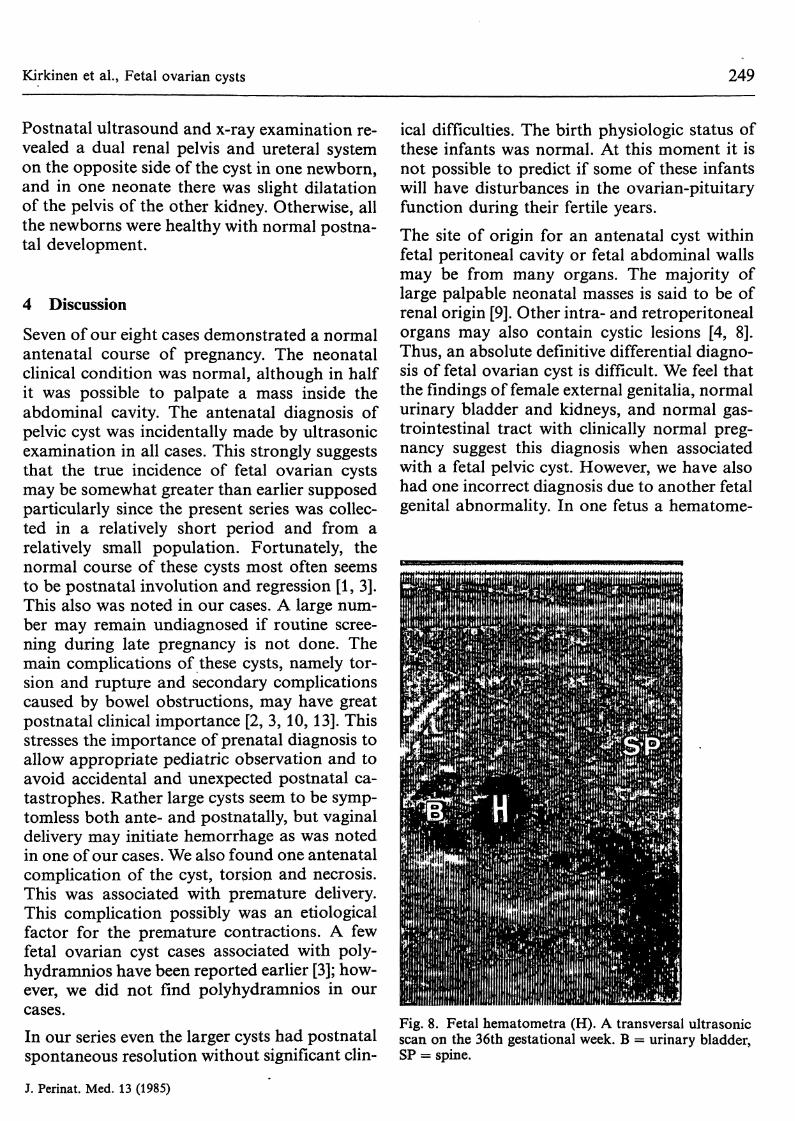
The indications of the antenatal ultrasonic exa-minations, ante- and postnatal findings andpre- and postnatal courses are presented in Tab.I. The typical ultrasonic appearance of the cystis presented in Figs. 1 — 6. In one patient (casenumber 6) the ovarian cyst underwent twistingand became necrotic. Antenatal ultrasonogra-phy revealed slight echodense material and sep-ta (Fig. 7).
2 Patients and methods
Pregnancy was clinically uncomplicated in 7patients. The above-mentioned pregnancy withthe twisted cyst resulted in a spontaneous pre-mature birth during the 34th gestational week.
The material consists of eight pregnancies in Elective cesarean section was performed due towhich antenatal real-time ultrasonic examina- the large size of the cyst in the 40th gestationaltion (Toshiba SAL 20 A, 3,5 MHz linear probe week in one instance (case 8). In another patientand Aloka SSD 280, 3,5 MHz linear/sector pro- (case 2) cesarean section was performed becau-be) demonstrated reliable suggestion of fetal se of a breech presentation.
© 1985 by Walter de Gruyter Co. Berlin · New York
246 Kirkinen et al., Fetal ovarian cysts
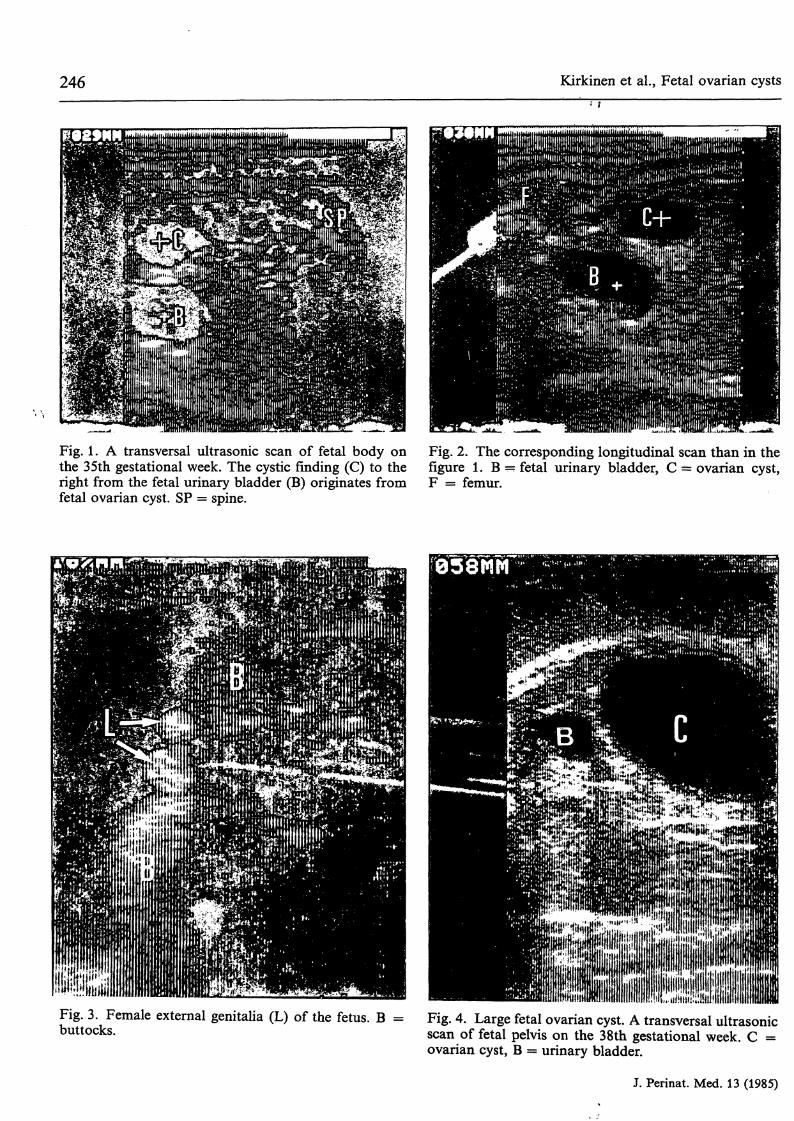
Fig. 1. A transversal ultrasonic scan of fetal body on Fig. 2. The corresponding longitudinal scan than in thethe 35th gestational week. The cystic finding (C) to the figure 1. B — fetal urinary bladder, C = ovarian cyst,right from the fetal urinary bladder (B) originates from F = femur,fetal ovarian cyst. SP = spine.
•WÄWI
Fig. 3. Female external genitalia (L) of the fetus. B =buttocks.
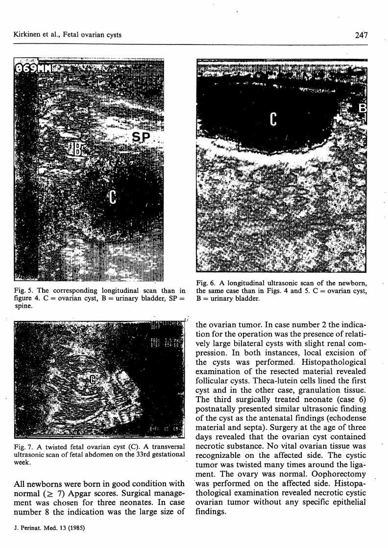
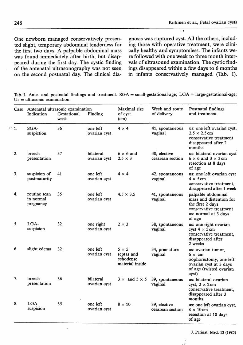
Fig. 4. Large fetal ovarian cyst. A transversal ultrasonicscan of fetal pelvis on the 38th gestational week. C =ovarian cyst, B = urinary bladder.
J. Perinat. Med. 13 (1985)
Kirkinen et al., Fetal ovarian cysts 247
Fig. 5. The corresponding longitudinal scan than infigure 4. C = ovarian cyst, B = urinary bladder, SP =spine.
Fig. 7. A twisted fetal ovarian cyst (C). A transversalultrasonic scan of fetal abdomen on the 33rd gestationalweek.
All newborns were born in good condition withnormal (> 7) Apgar scores. Surgical manage-ment was chosen for three neonates. In casenumber 8 the indication was the large size of
Fig. 6. A longitudinal ultrasonic scan of the newborn,the same case than in Figs. 4 and 5. C = ovarian cyst,B = urinary bladder.
the ovarian tumor. In case number 2 the indica-tion for the operation was the presence of relati-vely large bilateral cysts with slight renal com-pression. In both instances, local excision ofthe cysts was performed. Histopathologicalexamination of the resected material revealedfollicular cysts. Theca-lutein cells lined the firstcyst and in the other case, granulation tissue.The third surgically treated neonate (case 6)postnatally presented similar ultrasonic findingof the cyst as the antenatal findings (echodensematerial and septa). Surgery at the age of threedays revealed that the ovarian cyst containednecrotic substance. No vital ovarian tissue wasrecognizable on the affected side. The cystictumor was twisted many times around the liga-ment. The ovary was normal. Oophorectomywas performed on the affected side. Histopa-thological examination revealed necrotic cysticovarian tumor without any specific epithelialfindings.
J. Perinat. Med. 13 (1985)
248 Kirkinen et al., Fetal ovarian cysts
One newborn managed conservatively presen-ted slight, temporary abdominal tenderness forthe first two days. A palpable abdominal masswas found immediately after birth, but disap-peared during the first day. The cystic findingof the antenatal ultrasonography was not seenon the second postnatal day. The clinical dia-
gnosis was ruptured cyst. All the others, includ-ing those with operative treatment, were clini-cally healthy and symptomless. The infants we-re followed with one week to three month inter-vals of ultrasound examination. The cystic find-ings disappeared within a few days to 6 monthsin infants conservatively managed (Tab. I).
Tab. I. Ante- and postnatal findings and treatment. SGA = small-gestational-age; LGA = large-gestational-age;Us = ultrasonic examination.
Case
'·Antenatal ultrasonic examinationIndication Gestational Finding
week
SGA- 36suspiction
one leftovarian cyst
Maximal sizeof cyst(cm)
4 x 4
Week and routeof delivery
41, spontaneousvaginal
Postnatal findingsand treatment
us: one left ovarian cyst,2.5 χ 2.5cm
2.
3.
4.
breech 37presentation
suspicion of 41postmaturity
routine scan 35in normalpregnancy
LGA-suspicion
breechpresentation
LGA-suspicion
32
slight edema 32
36
35
bilateralovarian cyst
one leftovarian cyst
one leftovarian cyst
one rightovarian cyst
one leftovarian cyst
bilateralovarian cyst
one leftovarian cyst
6 x 6 and2.5 χ 3
4 x 4
4.5 χ 3.5
2 x 3
40, electivecesarean section
42, spontaneousvaginal
41, spontaneousvaginal
38, spontaneousvaginal
5 x 5 34, prematureseptas and vaginalechodensematerial inside
3 χ and 5 x 5 39, spontaneousvaginal
8 χ 10 39, electivecesarean section
conservative treatmentdisappeared after 2monthsus: bilateral ovarian cyst6 x 6 and 3 χ 3 cmresection at 8 daysof ageus: one left ovarian cyst4 χ 5cmconservative treatment,disappeared after 1 weekpalpable abdominalmass and distention forthe first 2 daysconservative treatmentus: normal at 3 daysof ageus: one right ovariancyst 4 χ 5 cmconservative treatment,disappeared after2 weeksus: ovarian tumor,6 χ cmoophorectomy; one leftovarian cyst at 3 daysof age (twisted ovariancyst)us: bilateral ovariancyst, 2 χ 2cmconservative treatment,disappeared after 3monthsus: one left ovarian cyst,8 χ 10cmresection at 10 daysof age
J. Perinat. Med. 13 (1985)
Kirkinen et al., Fetal ovarian cysts 249
Postnatal ultrasound and x-ray examination re-vealed a dual renal pelvis and ureteral systemon the opposite side of the cyst in one newborn,and in one neonate there was slight dilatationof the pelvis of the other kidney. Otherwise, allthe newborns were healthy with normal postna-tal development.
4 DiscussionSeven of our eight cases demonstrated a normalantenatal course of pregnancy. The neonatalclinical condition was normal, although in halfit was possible to palpate a mass inside theabdominal cavity. The antenatal diagnosis ofpelvic cyst was incidentally made by ultrasonicexamination in all cases. This strongly suggeststhat the true incidence of fetal ovarian cystsmay be somewhat greater than earlier supposedparticularly since the present series was collec-ted in a relatively short period and from arelatively small population. Fortunately, thenormal course of these cysts most often seemsto be postnatal involution and regression [1, 3].This also was noted in our cases. A large num-ber may remain undiagnosed if routine scree-ning during late pregnancy is not done. Themain complications of these cysts, namely tor-sion and rupture and secondary complicationscaused by bowel obstructions, may have greatpostnatal clinical importance [2, 3, 10, 13]. Thisstresses the importance of prenatal diagnosis toallow appropriate pediatric observation and toavoid accidental and unexpected postnatal ca-tastrophes. Rather large cysts seem to be symp-tomless both ante- and postnatally, but vaginaldelivery may initiate hemorrhage as was notedin one of our cases. We also found one antenatalcomplication of the cyst, torsion and necrosis.This was associated with premature delivery.This complication possibly was an etiologicalfactor for the premature contractions. A fewfetal ovarian cyst cases associated with poly-hydramnios have been reported earlier [3]; how-ever, we did not find polyhydramnios in ourcases.In our series even the larger cysts had postnatalspontaneous resolution without significant clin-
ical difficulties. The birth physiologic status ofthese infants was normal. At this moment it isnot possible to predict if some of these infantswill have disturbances in the ovarian-pituitaryfunction during their fertile years.The site of origin for an antenatal cyst withinfetal peritoneal cavity or fetal abdominal wallsmay be from many organs. The majority oflarge palpable neonatal masses is said to be ofrenal origin [9]. Other intra- and retroperitonealorgans may also contain cystic lesions [4, 8].Thus, an absolute definitive differential diagno-sis of fetal ovarian cyst is difficult. We feel thatthe findings of female external genitalia, normalurinary bladder and kidneys, and normal gas-trointestinal tract with clinically normal preg-nancy suggest this diagnosis when associatedwith a fetal pelvic cyst. However, we have alsohad one incorrect diagnosis due to another fetalgenital abnormality. In one fetus a hematome-
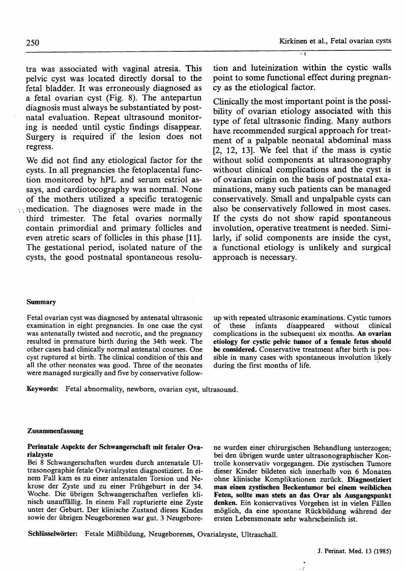
Fig. 8. Fetal hematometra (H). A transversal ultrasonicscan on the 36th gestational week. B = urinary bladder,SP = spine.
J. Perinat. Med. 13 (1985)
250 Kirkinen et al., Fetal ovarian cysts
tra was associated with vaginal atresia. Thispelvic cyst was located directly dorsal to thefetal bladder. It was erroneously diagnosed asa fetal ovarian cyst (Fig. 8). The antepartundiagnosis must always be substantiated by post-natal evaluation. Repeat ultrasound monitor-ing is needed until cystic findings disappear.Surgery is required if the lesion does notregress.We did not find any etiological factor for thecysts. In all pregnancies the fetoplacental func-tion monitored by hPL and serum estriol as-says, and cardiotocography was normal. Noneof the mothers utilized a specific teratogenic
v medication. The diagnoses were made in thethird trimester. The fetal ovaries normallycontain primordial and primary follicles andeven atretic scars of follicles in this phase [11].The gestational period, isolated nature of thecysts, the good postnatal spontaneous resolu-
tion and luteinization within the cystic wallspoint to some functional effect during pregnan-cy as the etiological factor.Clinically the most important point is the possi-bility of ovarian etiology associated with thistype of fetal ultrasonic finding. Many authorshave recommended surgical approach for treat-ment of a palpable neonatal abdominal mass[2, 12, 13]. We feel that if the mass is cysticwithout solid components at ultrasonographywithout clinical complications and the cyst isof ovarian origin on the basis of postnatal exa-minations, many such patients can be managedconservatively. Small and unpalpable cysts canalso be conservatively followed in most cases.If the cysts do not show rapid spontaneousinvolution, operative treatment is needed. Simi-larly, if solid components are inside the cyst,a functional etiology is unlikely and surgicalapproach is necessary.
Summary
Fetal ovarian cyst was diagnosed by antenatal ultrasonicexamination in eight pregnancies. In one case the cystwas antenatally twisted and necrotic, and the pregnancyresulted in premature birth during the 34th week. Theother cases had clinically normal antenatal courses. Onecyst ruptured at birth. The clinical condition of this andall the other neonates was good. Three of the neonateswere managed surgically and five by conservative follow-
up with repeated ultrasonic examinations. Cystic tumorsof these infants disappeared without clinicalcomplications in the subsequent six months. An ovarianetiology for cystic pelvic tumor of a female fetus shouldbe considered. Conservative treatment after birth is pos-sible in many cases with spontaneous involution likelyduring the first months of life.
Keywords: Fetal abnormality, newborn, ovarian cyst, ultrasound.
Zusammenfassung
Perinatale Aspekte der Schwangerschaft mit fetaler Ova-rialzysteBei 8 Schwangerschaften wurden durch antenatale Ul-trasonographie fetale Ovarialzysten diagnostiziert. In ei-nem Fall kam es zu einer antenatalen Torsion und Ne-krose der Zyste und zu einer Frühgeburt in der 34.Woche. Die übrigen Schwangerschaften verliefen kli-nisch unauffällig. In einem Fall rupturieite eine Zysteunter der Geburt. Der klinische Zustand dieses Kindessowie der übrigen Neugeborenen war gut. 3 Neugebore-
ne wurden einer chirurgischen Behandlung unterzogen;bei den übrigen wurde unter ultrasonographischer Kon-trolle konservativ vorgegangen. Die zystischen Tumoredieser Kinder bildeten sich innerhalb von 6 Monatenohne klinische Komplikationen zurück. Diagnostiziertman einen zystischen Beckentumor bei einem weiblichenFeten, sollte man stets an das Ovar als Ausgangspunktdenken. Ein konservatives Vorgehen ist in vielen Fällenmöglich, da eine spontane Rückbildung während derersten Lebensmonate sehr wahrscheinlich ist.
Schlüsselwörter: Fetale Mißbildung, Neugeborenes, Ovarialzyste, Ultraschall.
J. Perinat. Med. 13 (1985)
Kirkinen et al., Fetal ovarian cysts 251
Resume
Aspects perinataux de la grossesse compliquee du kysteovarien foetalOn a diagnostique par echographie antenataie un kysteovarien foetal au cours de 8 grossesses. Dans un cas, lekyste etait tordu et necrotique avant la naissance, et lagrossesse a abouti ä une naissance prematuree au coursde la 34eme semaine. Les autres cas ont eu une evolutionantenatale cliniquement normale. Un kyste s'est rompua la naissance. L'etat clinique dans ce cas et chez tousles autres nouveaux-nes etait bon. Trois nouveaux-nes
ont cte operes et 5 ont eu une surveillance conservatriceavec des echographies repetees. Les kystes de ces enfantsont disparu sans complications ciiniques, au cours des6 mois suivants. On dcvrait se souvenir chez les foetusfeminins de 1'etiologie ovarienne en face d'une tumeurpelvienne kystique. Le traitement conservateur apres lanaissance est possible dans de nombreux cas et l'involu-tion spontanee est probable au cours des premiers moisde la vie.
Mots-cles: Anomalie foetale, kyste ovarien, nouveau-ne, ultrasons.
Bibliography
[1] AHLVIN, R., W. BAUER: Luteinized cysts in ovariesof infants born of diabetic mothers. Am. J. Dis.Child. 93 (1957) 107
[2] AHMED, S.: Neonatal and childhood ovarian cysts.J. Pediatr. Surg. 6 (1971) 702
[3] BOWER, R., L. DEHNER, J. TBRNBERG: Bilateralovarian cysts in the newborn. Am. J. Dis. Child.128 (1974) 731
[4] CAMPBELL, S., J. PEARCE: The prenatal diagnosisof fetal structural anomalies by ultrasound. Clin.Obstet. Gynecol. 10 (1983) 475
[5] GRADE, M., M. GILLOOLY, K. TAYLOR: In uterodemonstration of an ovarian cystic mass byultrasound. J. Clin. Ultrasound 8 (1980) 251
[6] DES A, D. J.: Follicular ovarian cysts in stillbirthsand neonates. Arch. Dis. Child. 50 (1975) 45
[7] JOUPPILA, P., P. KIRKJNEN, S. TuoNONEN: Ultrasonicdetection of bilateral ovarian cysts in the fetus. Eur.J. Obstet. Gynaecol. Reprod. Biol. 13 (1982) 87
[8] KURJAK, A., V. LATIN, V, D'ADDARIO: Abnormal-ities of the fetus, placenta and umbilical cord. Prog.Med. Ultrasound 3 (1982) 87
[9] LONCINO, L., L. MARTIN: Abdominal masses in thenewborn infant. Pediatrics 21 (1958) 596
[10] MAINOLFI, F., W. STANDIFORD, T. HUBBARD:Ruptured ovarian cyst in the newborn. J. Pediatr.Surg. 3 (1968) 612
[11] PELLINIEMI, L., D. MARTIN: The fetal gonadal andsexual differentiation. In: TULCHINSFCY & RYAN(eds.): Maternal — fetal endocrinology. SaundersCompany, Philadelphia 1980
[12] TOWNE, B., H. MAHOUR, M. WooLLEYet al.: Ovari-an cysts and tumors in infancy and childhood. J.Pediatr. Surg. 10(1975)311
[13] WELCH, K.J.: Ovarian cysts and tumors. In:RAVITCH, WELCH, BENSON et al. (eds.): PediatricSurgery. Year Book of Medical Publishers, Inc.,Chicago 1979
Received September 10, 1984. Accepted January 30,1985.
P. Kirkinen, M. D.Department of Obstetrics and GynecologyUniversity of Oulu90220 Oulu 22Finland
J. Perinat. Med. 13 (1985)
• f
Selected Topics inClinical EnztTonologywolume 2Proceedings (selected) of the Fourth InternationalCongress on Clinical Enzymology · Washington, D.C.,U.SA, July 30-August 2,1983Editors M. Werner, D. M. Goldberg1984.17 cm χ 24 cm. XXII, 667 pages. Numerous illustrations. Hardcover. DM 260,-ISBN 311 0102331This book covers practical and theoretical aspects of enzymology. Over 50contributions by an international array of experts bring together currentdevelopments in analytical methodology, diagnostic use of enzymes, the role ofenzymes in pathophysiology, pharmacology and therapeutics as well as veterinarymedicine. A special section is devoted to emerging insights on enzymeheterogeneity. Each contribution contains an abstract for rapid survey of itscontents, and a carefully edited bibliography.
Contents (Main Chapters)Changing Methods · Diagnosis · Pathophysiology, Pharmacology and Thera-peutics · Enzyme Heterogeneity · Veterinary · Subject Index · Index of Contributors.
>4/so available
Selected Topics in Clinical EnzymologyEditors D. M. Goldberg, M. Werner1983. XVIII, 362 pages. DM 160,-
de Gruyter · Berlin · New York