Growth Response and Feed Utilization of Clarias gariepinus Fingerlings Fed with Bambara Groundnut as

~ 97 ~
International Journal of Fisheries and Aquatic Studies 2015; 3(1): 97-110 ISSN: 2347-5129 (ICV-Poland) Impact Value: 5.62 (GIF) Impact Factor: 0.352 IJFAS 2015; 3(1): 97-110 © 2015 IJFAS www.fisheriesjournal.com Received: 24-06-2015 Accepted: 27-07-2015 Ayotunde Ezekiel Olatunji Department of Fisheries and Aquatic Science, Faculty of Agriculture and Forestry, Cross River University of Technology, P.M.B. 102, Obubra Campus, Calabar, Nigeria. Ada Fidelis Bekeh Department of Fisheries and Aquatic Science, Faculty of Agriculture and Forestry, Cross River University of Technology, P.M.B. 102, Obubra Campus, Calabar, Nigeria. Ogbeche Lydia Otelahu Department of Fisheries and Aquatic Science, Faculty of Agriculture and Forestry, Cross River University of Technology, P.M.B. 102, Obubra Campus, Calabar, Nigeria. Inah Cornelius Department of Fisheries and Aquatic Science, Faculty of Agriculture and Forestry, Cross River University of Technology, P.M.B. 102, Obubra Campus, Calabar, Nigeria. Correspondence Ayotunde Ezekiel Olatunji Department of Fisheries and Aquatic Science, Faculty of Agriculture and Forestry, Cross River University of Technology, P.M.B. 102, Obubra Campus, Calabar, Nigeria.
Haematological and Histopathological Responses of Catfish Clarias gariepinus Juvenile to Coconut Water
Cocos nucifera
Ayotunde Ezekiel Olatunji, Ada Fidelis Bekeh, Ogbeche Lydia Otelahu, Inah Cornelius Abstract The toxicity of Coconut water Cocos nucifera L. (Arecaceae) water was determined on the Juvenile Catfish Clarias gariepinus. The 20, 24, 36, 48, 72 and 96LC50 of Coconut water to juvenile catfish were 750mg/l, 650mg/l, 550mg/l, 450mg/l, 350mg/l, and 250mg/l. While the total mortality occurred at a concentration of 750mg/l within 24hours exposure period respectively. Haematological examination showed an increase in blood cell count, and Platelet while there was a decrease in Haematocrit, MCV, MCH, LYMH, HMB, RBC in higher concentration of 750mg/l throughout the test in juvenile catfish Clarias gariepinus. There was a decrease in moisture, ether extract, nitrogen free extract and energy, from 73.78 ±10.73, 3.76 ± 0.34, 0.50 ± 0.08 in the control to 68.13 ±0.38, 3.17 ± 0.09, 0.37 ± 0.06 and 1.39 ±1.41, in higher concentration of 750mg/l, also there was an increase in Ash and Protein, from 0.6 ± 0.54 and 27.01 ±0.42mg/l in the control to 0.84 ± 0.14 and 26.28 ± 0.92mg/l in the concentration of 350mg/l. In the plasma electrolyte of catfish juvenile to coconut water, there was an increase in sodium from 1.06± 4.83 in the control to 74.54 ±26.09 in the concentration of 750mg/l, while in potassium; there was a decrease from 40.54 ± 3.43 in the control to 24.14 ±10.34 in higher concentration of 750mg/l. The result of histopathological responses of skin, gill and liver of the test organisms to coconut water showed different level of degeneration of cells, hypertrophy of gill arch, disarrangement of hepatic cell, necrosis, vacuolation, sign of trauma the gill, detachment of lamella, vessels congested with blood vessels, liver hypoxia, degeneration of the upper dermis and severe thinning of the underlying granular layer. Damages became severs with increase in concentration of aqueous extract and time exposure. The result of the tests provided the baseline information and established safe limits of using coconut water in fresh water fish farm. Keywords: Toxicity, haematological, Cocos nucifera, water, Catfish Clarias gariepinus 1. Introduction Cocos nucifera Linn. Belonging to the family Arecaceae, is commonly known as Coconut in English. The coconut palm is cultivated in more than 90 tropical countries and it represents an important income source. Nigeria, Indonesia, Philippines and India are the major producers and account for about 75% of World production (FOASTAT 2011) [27]. It is a large palm of about 60 to 90 feet height found across the sea levels of the tropical and subtropical world. It is found throughout Nigeria mainly at the Coastal regions. The coconut is known for its great versatility as observed in the many edible, domestic, commercial and industrial uses of its different parts Harries (1978) [23]. Unlike the endosperms of other plants (e.g., Wheat and Corn), the cellularization process in a coconut fruit does not fill up the entire embryo sac cavity, but instead leaves the cavity solution-filled. This solution is commonly known as coconut water and it is of cytoplasmic origin (Janick and Pall 2008) [39]. Nutrients from coconut water are obtained from the seed apoplasm (surrounding cell wall) and are transported symplasmically (through plasmodesmata, which is the connection between cytoplasms of adjacent cells) into the endosperm, (Patrick 2001) [52]. The aqueous part of the coconut endosperm is termed coconut water. Coconut water contains 94% water. Coconut water has been extensively studied in human medicine since its introduction to the scientific community in the 1940s. In its natural form, it is a refreshing and nutritious beverage which is widely consumed due to its beneficial properties to health, some of which are based on cultural/traditional beliefs (Janick and Pall 2008) [39], Sandhya and

~ 98 ~
International Journal of Fisheries and Aquatic Studies
Rajamohan 2008 [61], Asian and Pacific Coconut Community, APCC 1994 [14], Seow and Gwee 1984, George and Sherrington 1984 [32], Campbell-Falck et al. 2000 [19, 20], Pummer et al. 2001 [53], and Anurag and Rajamohan 2003) [5]. It has certain traditional medicinal uses also. The whole coconut tree may be utilized, but the main products are obtained from the fruit: copra and oil, lauric acid, coconut milk, fiber, flour, coconut water (from immature fruit), The consumption of liquid albumen (or coconut water) of the immature coconut is so important to this country, that resulted in deployment of crops aimed mainly to this application, which does not happen in other producing countries. The increasing demand for natural and healthy foods is one factor that has raised the consumption of this drink that reaches around 350 million liters per year in fresh and industrialized form. Besides highly appreciated for its taste and freshness, it is considered an excellent natural isotonic, so it is also consumed for its nutritional qualities. Despite consumption of green coconut water is very beneficial, the demand increasing for this product generates a very large amount of waste in places such beaches where the consumption of this drink is common. Although the solid endosperm or green coconut pulp is edible, generally only water is extracted from fresh fruit. It helps in avoiding the spots left after it and also a good way to avoid dehydration. Besides its nutritional role, coconut water also some active ingredients that have growth regulatory properties, e.g., cytokinin-type activity, (George and Sherrington 1984) [32]. Some of the most significant and useful components in coconut water are Cytokinins, which are a class of phytohormones Kende, and Zeevaart (1997) [40]. Other components found in coconut water include sugars, sugar alcohols, lipids, amino acids, nitrogenous compounds, organic acids and enzymes [Arditti (2008) Tulecke, et al. 1961; Santoso 1996; USDA 2009] [7, 69, 63, 71] and they play different functional roles in plant and human systems due to their distinct chemical properties. The present study aimed to establish the toxicity, haematological and Histological profile of Cocos nucifera water in juvenile Catfish Clarias gariepinus. 2. Materials and Methods Two hundred live and apparently healthy juvenile Catfish Clarias gariepinus measuring 11.5 - 14.6 cm total length and 65.6 - 112.4 g weight were identified using taxonomic key of Reed et al. (1967). The specimen used for the experiment was collected from Cross River University of Technology fish farm at Calabar, Obubra Campus. The specimen was acclimated for 1 week in the laboratory inside transparent, rectangular glass tanks (75 x 45 x 45 cm) of 121.5 L capacity. The tanks were filled with 50 L unchlorinated well water. The fish was fed to apparent satiation twice daily (0900 and 1600 h) with a commercial pelleted fish diet containing 35% crude protein during the acclimation period. Feeding was discontinued 48 h before the commencement of the experiment to minimize the production of wastes in the test container. The maximum admissible toxicant concentration established for adult tilapia was derived by multiplying a constant 0.01 - 0.1 by 96 h LC50 (Koesomadinata, 2000) [46]. 2.1. Water quality analysis Water quality monitoring was done prior to the experiment, during the experiment and after the experiment. pH was determined using a digital pH meter (Mettle Toledo 320). The
electrode was inserted into the bottle containing the water sample after standardization in different buffer, after which the reading was taken. DO2 was measured using a digital, dissolved oxygen meter (Jenway, 9071) once in a day at 8.00 a.m. While, temperature was measured using a mercury in-glass thermometer, which was placed in the medium inside the test container until reading was taken. The reading was taken at 10.00 a.m. on each day of the experiment. 2.2. Preparation of aqueous extract of Coconut water and acclimation of test fish 240 mature fruits of coconut were purchased from the farmers around Obubra village, in Cross River State, Nigeria. The seed powder was prepared by opening the mature fruit of pawpaw and the fresh seeds extracted and were sun dried. The seeds were ground to a fine powder, using the coffle mill attachment of a Moulinex domestic food blender. The powder was kept in desiccators for later use in stock solutions. A preliminary range finding test was conducted to determine the toxicity level of pawpaw seed powder using standard method (American Public Health Association APHA, 1987). 2.3. Acute toxicity test The preliminary range finding test was conducted to determine the actual concentration for the test. One control and five tests in triplicates were set up for the experiment. Pawpaw seed powder was introduced randomly, and tested for 24 h, the behavior and mortality of the test fishes in each tank was monitored and recorded every 15 min for the first hour, once every hour for the next 3 h and every 4 h for the rest 24 h period. Eighteen (75 x 45 x 45 cm) glass tanks of 121.5 L capacity each were filled with 50 L aerated dechlorinated well water. Adult O. niloticus was batch-weighed with a top-loading mettler balance (Mettler Toledo (K), and distributed randomly in triplicate per treatment. The glass tanks were covered with mosquito net to prevent fish from jumping out; there was no aeration, no water change nor feeding throughout the test. This was done prior to the introduction of the toxicant. The toxicant was introduced at 2, 4, 6, 8 and 10 mg/l with a control of 0 mg/l in triplicate. The test lasted for 24 h. The definitive test was conducted using concentration of 4.0, 4.2, 4.4, 4.6, 4.8 and 5.0 mg/l of pawpaw seed powder earlier determined for the range finding test. This test comprised one sublethal toxicity test according to the standard method/procedures (American Public Health Association, 1987). Fish mortality was monitored and recorded hourly for the first 4 h, 4 h for the next 24 h, and subsequently every 24 h, for the next 96 h. The inability of fish to respond to external stimuli was used as an index of death. Apart from monitoring and recording fish mortality the fish behaviour such as erratic swimming, air gulping, loss of reflex, discolouration, and molting was monitored. LC50, which is the concentration of pawpaw seed powder, estimated to be lethal to 50% of test organism after exposure time of 96 h, was determined graphically using probit transformation (Herwig, 1979; USEPA, 2000) [38, 78]. Haematological examination of fish followed the method described by Svobodova et al. (1991) [70]. The moisture, ash and other extracts like crude fibre, crude fat, and protein were then determined using standard methods (AOAC, 1990) [7]. Nitrogen was determined by the micro-kjedahl method (Pearson, 1976) and the crude protein was taken as N% x 6.25 (constant factor) where N is equal to Nitrogen content per 100g sample. Total carbohydrate was determined using the phenol-sulphuric acid method (Adeyeye

~ 99 ~
International Journal of Fisheries and Aquatic Studies
and Faleye, 2004) [4]. The crude fibre was obtained by dry ashing of the sample at 550oC dissolved in 10% HCl(25ml) and 5% Lanthanum Chloride (2ml) boiled, filtered and made up to standard volume with distilled water. Na and K were determined by the Jenway 1997, Flame photometer method. Phosphorus was determined using the spectronic 20 calorimeter by the phosphovando molybdate method (AOAC, 1990) and (Dubois et al., 1956). Ca and Mg were obtained by EDTA method (Ademoroti, 1996) [3], while the heavy metals were determined using atomic absorption spectrophotometer. 2.4. Heamatological Analysis Blood (1-2ml) was sampled from groups of fish by inserting a syringe into the vertebral caudal blood vessel. The blood was empty into 5ml heparinized blood bottles treated with Ethyl Diamine Tetracetic Acid (EDTA). Blood samples were centrifuged (1500g for 20 min) to obtain plasma. Plasma samples were stored -20°C until analysis of mineral content. Plasma mineral contents (Na+, K+, -) were measured in Lloyt Na/K analyser by ISE method. Dietary pH was measured as described by Tarakci. Dry matter (105°C, overnight), ash (550°C, overnight), crude protein (nitrogenx6.25, Gerhard Kjeldatherm, Königswinter, DE), ether extract (Velp Scientifica 148 Solvent Exractor Milan, IT), crude fiber (Ancom 220, Fiber Analyzer. 2.5. Blood Cell Count System KX-2INTM Automated Hematology Analyzer was used in blood cell count, the KX-2IN is an ideal hematology analyzer for a clinical satellite laboratory or research testing. It provide a CBN with 17reportable parameter and 3-part WBC differential, 2.6. Histopathological Analysis of Test Organs At the end of the experiment one fish per each treatment that is one fish per level of concentration was sampled after 96hours of exposure for histopathological analysis. The fish were killed with a blow on the head using a mallet and dissecting kits the gill, liver and skin were successfully removed. The organs were weighed and submersed in 10% formalin for three days after which tissue was dehydrated in periodic acid Schiff's reagent (PAS) following the methods of Hughes and Perry (1976). The organ was then embedded in malted wax. The tissues were sectioned into thin section (5.7um) by means of rotatory microtone and were being dehydrated and stain with Harris haematoxylin - eosin (H- and E) stain. Bancroft and cook (1994), using microtone and each section was cleared by placing warm water (38 0c), Where it was picked with clean slides and oven-dried at 580c for 30minutes to meet the wax. The slide containing sectional materials/tissue was cleared using xylene and graded level of 50%, 70%, 90%, 95%, and 100% alcohol for two minutes each. The section was stained in haematoxylin -eosin for ten minutes. The stained slide was observed under a light microscope at varying X 100 - 500 magnification, section was examined and photographed using an Olympus BH2 microscope filled with photographic attachment (Olympus C35 AD4), a camera (Olympus C40 AB-4) and an automated light experience unit (Olympus pm (35p). 2.7. Statistical analysis All results were collated and analysed using computerized, probit and logit analysis (Lichfielf and Wilcoxon, 1949). The median lethal concentration LC50 at selected period of
exposure, and an associated 95% confidence interval for each replicate toxicity test was subjected to logit and probit analysis (Finney, 1971) using Statistical Package for Social Sciences (SPSS) 17.0 for Windows Vista. 3. Results 3.1. Toxicity The 96 hour LC50 of coconut water to juvenile catfish (Clarias gariepinus) is presented in table 1 is 250mg/l and the graphical method of determination of LC50 is presented in Figure 1-6 and the concentration of the 100% fish is 750mg/l. The percentage cumulative mortality of coconut water to juvenile catfish Clarias gariepinus is presented in table 2 and 3. And fig4 to 9 the values are 0,250,350,450,550,650 and 750mg/l the concentration of the treatment required bringing about 50percent mortality of Clarias gariepinus juvenile within 96 hour period. The admissible toxicant concentration of 2.5 -25mg/l established for juvenile Catfish, was derived by multiplied the 96-hLC50 with an application factor of between 0.1 -0.01 according to Koemsomadinata (1980) [41]
Table 1: The LC50 value for Cocos nucifera to Catfish juveniles
S/n Time LC50 MATC 1 20hr 750mg/l 7.5mg/l - 75mg/l 2 24hr 650mg/l 6.5mg/l -65mg/l 3 36hr 550mg/l 5.5mg/l - 55mg/l 4 48hr 450mg/l 4.5mg/l - 45mg/l 5 72hr 350mg/l 3.5mg/l -35mg/l 6 96hr 250mg/l 2.5mg/l - 25mg/l
The acute toxicity of Cocos nucifera increases with increase in concentration with time. The total mortality 0f catfish juveniles occurred at a concentration of 750mg/l of coconut water. The maximum toxicant concentration of 2.5mg/l-7.5mg/l established for catfish juveniles was derived by multiplying the 96hours LC50 3.2. The method of determination of LC50 using graphical method
Fig 4: Determination of LC50 0f Coconut water to juvenile catfish (Clarias gariepinus) at 20hr

~ 100 ~
International Journal of Fisheries and Aquatic Studies
Fig 5: Determination of LC50 of coconut water to juvenile catfish (Clarias gariepinus) at 24hrs
Fig 6: Determination of LC50 of juvenile Catfish (Clarias gariepinus) to coconut water at 36hrs
Fig 7: Determination of LC50 of Coconut water to Juvenile catfish (Clarias gariepinus) at 48hrs
Fig 8: Determination of LC50 of Coconut water to Juvenile catfish (Clarias gariepinus) at 72hrs
Fig 9: Determination LC50 coconut water to juvenile catfish (Clarias gariepinus) at 96hrs
3.3. Observation of Range finding Test of Catfish Juvenile Clarias gariepinus Exposed to coconut water The change in the behavior of catfish juveniles observed in the range finding test is summarized in Table 2. During the range finding test, juveniles of Clarias gariepinus exposed to coconut water exhibited different behaviors in relation to concentration, such as air gulping, molting erratic swimming, loss of reflex, operculum movement, fin movement, discoloration. The abnormal behavior displayed by the fish exposed to Cocos nucifera increased with increasing concentration. 3.4. Observation of definitive Test of Catfish juvenile exposed to Coconut Erratic swimming was observed in the fish; they also exhibited discoloration, loss of reflex air gulping. The behavioral change observed from the definitive test are summarized in table 3. Discloration was observed to be directly proportional to the increase in the concentration of the coconut water and duration exposure.

~ 101 ~
International Journal of Fisheries and Aquatic Studies
Tables 4: Length-Weight Relationship and Condition Factors of Catfish (Clarias Gariepinus)
Conc Weight (Gm) Standard Lenght Conc Weight (Gm) Standard Lenght
Range Finding Test Definitive Test
0mg/l 61.10 ± 6.4 18.70 ± 0.4 0mg/l 61.110±6.39 18.89±038
150mg/l 66.50 ± 5.2 20.47 ± 1.9 250mg/l 66.50±5.22 20.47±1.93
350mg/l 66.03 ± 6.8 19.51 ± 0.9 350mg/l 66.04±5.6.78 19.52±0.95
450mg/l 64.03 ± 4.7 19.57 ± 0.7 450mg/l 64.03±4.75 19.57±0.67
600mg/l 57.33 ± 6.5 18.72 ± 0.7 550mg/l 57.34±6.55 18.72±070
750mg/l 61.97 ± 7.0 18.68 ± 1.2 650mg/l 60.80±7.19 18.69±1.16
900mg/l 63.67 ± 6.3 19.42 ± 0.8 750mg/l 63.36±1.40 19.14±1.12
Table 5: Summary of Water Quality Parameter of Coconut Water to Catfish Clarias Gariepinus (Mean±Sd)
Conc. Temp.(0c) Do (M/L) Ph
Conc. Temp.(0c) Do (M/L) Ph
Range Finding Definitive
To(0mg/l) 25.67±0.58 7.50±0.27 6.64±0.15 To 24.67±11.15 7.24±0.47 7.17±0.33
T1(150mg/l) 25.34±0.58 7.27±0.38 7.14±0.24 T1(250mg/l) 25.34±0.58 7.27±0.15 7.19±0.21
T2(30mg/l) 25.34±0.58 7.17±0.11 7.10±0.29 T2(350mg/l) 25.10±0.90 7.00±0.27 7.09±0.22
T39450mg/l) 25.57±0.58 6.88±0.37 7.45±0.18 T(450mg/l) 25.34±0.58 6.80±0.10 7.27±0.11
T4(600mg/l) 25.34±0.58 5.77±0.47 6.86±0.23 T4(550mg/l) 24.34±0.58 6.80±0.16 8.35±0.39
T5(750mg/l) 25.00±0.00 5.80±0.27 6.87±0.20 T5(650mg/l) 24.68±0.58 7.07±0.25 7.14±0.23
T6(90mg/l) 25.67±0.58 5.07±0.38 7.40±0.20 T6(750mg/l) 25.00±1.00 7.10±0.31 8.27±0.21
Table 2: Percentage Cumulative Mortality of Clarias garepinus Exposed to Coconut Water (Range Finding Test)
Trt/conc 15mins 30mins 45mins 60mins 2hrs 3hrs 4hrs 6hrs 8hrs 10hrs 16hrs 20hrs 24hrs
0mg/l 0 0 0 0 0 0 0 0 0 0 0 0 0
150mg/l 0 0 0 0 0 0 0 0 0 0 0 0 0
300mg/l 0 0 0 0 0 0 0 0 0 0 0 0 0
450mg/l O 0 0 3.34 6.67 6.67 13.34 13.34 13.34 16.67 20 20 20
600mg/l 0 0 0 3.34 13.34 16.67 16.67 16.67 16.67 23.34 23.34 33.34 33.34
750mg/l 0 0 3.34 6.67 16.67 16.67 26.67 26.67 33.34 36.67 36.67 43.34 46.67
900mg/l 0 0 3.34 13.34 26.67 30.0 36.67 43.34 50 70 76.67 90 93.34
Table 3: Percentage Cumulative Mortality of Clarias garepinus Exposed to Coconut Water (Definitive Test)
Conc. 1hr 2hrs sss3hrs 4hrs 6hrs 8hrs 12hrs 16hrs 20hrs 24hrs 36hrs 48hrs 72hrs 96hrs
Omg/l 0 0 0 0 0 0 0 0 0 0 0 0 0 0
250mg/l 0 0 0 0 0 0 0 0 3.34 6.67 10 10 13.34 13.34
350mg/l 0 0 3.34 3.34 6.67 10 10 10 13.34 13.34 16.67 16.67 23.34 23.34
450mg/l 0 0 0 3.34 10 23.34 23.34 23.34 26.67 26.67 33.34 40 40 60
550mg/l 0 0 0 0 0 6.67 13.34 20 20 33.34 40 46.67 60 66.67
650mg/l. 0 3.34 13.34 16.67 16.67 20 33.34 33.34 43.34 50 66.67 70 76.67 96.67
750mg/l. 3.34 13.34 30 63.34 30 46.67 50 50 53.34 63.34 76.67 83.34 86.67 100

~ 102 ~
International Journal of Fisheries and Aquatic Studies
Table 6: The Summary of effect of coconut water on haematological parameters of juvenile catfish Clarias gariepinus (Mean±SD)
con
c./m
g/l
Wh
ite
blo
od c
ell (
ul)
Red
blo
od c
ell (
ul)
Hae
mog
lob
in (
g/d
l)
Hae
mat
ocri
pt
(%)
Mea
n c
ell v
olu
me
(fl)
Mea
n c
ell h
aem
oglo
bin
(p
g)
Pla
tele
t (u
l)
Lym
ph
ocyt
e (%
)
MC
H
RC
D-W
S
RC
D-W
CV
0 1.69±33.96 6.37±6.87 9.63±2.44 25.27±5.59 1.23±8.24 46.50±1.60 1.98±16709.68 97.700±0.45 37.94±1.50 39.30±2.44 11.10±0.52
250 1.48±25.12 1.61±3.58 8.60±1.21 19.47±3.75 1.18±2.48 52.43±3.57 4.65±12182.09 91.84±4.22 44.60±2.26 59.64±8.44 19.03±0.56
350 1.97±103.87 9.50±9.70 6.60±2.08 10.37±11.55 1.01±11.15 16.44±28.47 5.61±20802.64 31.77±55.02 14.33±24.82 16.70±28.92 51.54±27.78
450 1.60±18.73 1.21±1.05 7.80±1.90 20.06±3.45 1.20±2.53 45.50±4.79 1.80±13568.09 95.70±2.06 37.74±3.75 46.20±15.6 20.50±4.44
550 1.77±13.98 2.09±4.60 11.40±2.36 22.94±4.58 1.10±5.72 56.53±18.67 6.72±41315.98 93.30±5.70 51.60±18.41 24.54±21.39 20.37±9.38
650 1.97±70.35 5.27±7.35 1.97±3.40 6.24±8.87 1.90±69.10 14.37±24.28 6.87±3592.12 31.20±54.04 12.00±20.97 11.00±19.05 3.90±6.75
750 1.14±100.06 1.30±1.12 5.47±4.77 15.03±13.08 1.07±66.78 28.00±22.59 3.60±3377.86 64.84±56.16 24.20±20.97 22.00±19.06 10.2±8.87
Table 7: Summary of effect of coconut water on Biochemical parameters of catfish Clarias gariepinus r (Mean ± SD)
Treatment Moisture (%) Fibre (%) Protein (%) Ether Extract (%) Ash (%) Nitrogen Free Extract (%) Energy (kcal/100g)
0mg/l 73.78±10.73 00.00±00 27.01±0.42 3.76±0.34 0.62±0.54 0.50±0.08 1.44±2.10
250mg/l 64.24±6.37 00.00±00 27.72±1.22 2.82±0.67 0.85±0.47 0.37±0.17 1.41±1.55
350mg/l 69.52±0.74 00.00±00 26.33±1.17 3.13±0.18 0.88±0.09 0.73±0.15 1.36±1.87
450mg/l 69.80±1.39 00.00±00 26.28±0.92 2.1±0.58 0.84±0.14 0.26±0.04 1.35±1.72
550mg/l 68.73±0.65 00.00±00 27.06±0.18 3.22±0.14 0.84±0.44 0.37±0.58 9.33±68.07
650mg/l 68.74±0.58 00.00±00 27.229±1.12 3.09±0.58 0.89±0.09 0.46±0.78 1.34±4.50
750mg/l 68.13±0.38 00.00±00 27.42±0.46 3.17±0.09 0.88±0.12 0.37±0.06 1.39±1.41
Table 8: Summary on plasma electrolyte of Catfish to coconut water
Treatments Sodium Na (ϻmol) Potassium K (ϻmol)
0mg/l 1.06±4.83 40.54±3.43
250mg/l 73.76±5.49 38.77±5.66
350mg/l 67.27±11.32 36.34±12.62
450mg/l 1.10±74.57 56.40±44.23
550mg/l 55.77±17.57 30.17±3.65
650mg/l 70.24±36.69 22.34±4.17
750mg/l 74.54±26.09 24.14±10.34

~ 103 ~
International Journal of Fisheries and Aquatic Studies
Table 9: Biological Monitoring of Catfish Juvenile of Clarias gariepinus Exposed to Differents Concentration of Coconut Water (Range finding Test)
Behavior/Exposure Time 12hrs 16hrs 20hrs 24hrs
Concentration ((Mg/l) O 150 300 450 600 750 900 O 150 300 450 600 750 900 O 150 300 450 600 7509 900 O 150 300 450 600 750 9 900
Air Gulping - - + + + + + - + + + + + + - + + + + + + - - + + + + +
Erratic Swimming - - - - - - - - - - - - - - - + - - - - - - + - + - - -
Molting - - - - - - - - - - - + + - - + - + + + + - - - - - - -
Discoloration - - - - - - - - - - + - + + - + - + + + + - + - - + + +
Barbell Deformation - - - - - - - - - - - - - - - - - - - - - - - - - - - -
Tall beat frequency - - + + - - - - - - - - - - - - - - - - - - + - - - - -
Excessive mucus secretion - - - - - - - - - - - - - - - - - - - - - - - - - - - -
Operculum Movement - + + + + + + - + - + + - + - + + + - + + - + + + + + +
Loss of reflex - - - + - - - - + - + + - + - - + - - + + - - - - + + + -
Fin movement - + + + - - - - + + + + + - - - + - - + + - + + + - + -
Table 10: Behavioral Monitoring of Catfish Juvenile of Clarias gariepinus Exposed to Different Concentration of Coconut Water (Definitive Test)
Behavior/Exposure Time 24hrs 48hrs 72hrs 96hrs
Concentration ((Mg/l) 0 250
350
450
550
650
750 0 250
350
450
550
650
750 0 250
350
450
550
650
750 0 250
350
450
550
650
750
Air Gulping - + + + + + + - + - + + + + - - + + + + + - + + + + + + Erratic Swimming - - - - - + - - - - - - - - - - - - - - - - - - - + + -
Molting - - - - - - - - - - - - - - - - - - - - - - - - - _ - Discoloration - - - - - - - - - - - - - - - - - + - - + - - + - + + -
Barbell Deformation - - - - - - - - - - - - - - - - - - - + - - - - + - - Excessive mucus secretion - - - - - - - - - + - - - - - - - - - - - - - - - - + -
Tall beat frequency - - - - - - - - - - - - - + - - - - - - + - - - - - + - Operculum Movement - - - - - - - - + + - - - - - - - - - - + - - - - + + -
Loss of reflex - - - - - - - - - - - - - - - - - - - + + - - + - - - + Fin movement - + + + + + + - - + + + + + - + + + - + - - - - - - -
Table 11: Histological changes in the liver of Clarias gariepinus exposed Coconuut Water
Concentration (mg/l) Figures 1-7 Histological changes
Omg/l 1 Normal hepatic tissue, showing normal hepatocytes with no architectural defect
250mg/l 2 Section showing liver tissue with slightly degenerated blood vessel with central vein congested with red blood cell.
350mg/l 3 Section showing normal appearance of the architectural pattern of the tissue but a section of the liver showing fatty changed probably due to hypoxia
450mg/l 4 Section showing ballooning of the hepatocytes and blood vessel congested with blood.
550mg/l 5 Section showing degenerated liver, with thickening of the detachment of the lamella.
650mg/l. 6 Section shows liver tissue with thickened reticular fibres, congested blood vessels with several prominent central vein containing red blood cell.
750mg/l. 7 Section shows degenerated blood vessel with moderate fatty layer.

~ 104 ~
International Journal of Fisheries and Aquatic Studies
~ 105 ~
International Journal of Fisheries and Aquatic Studies
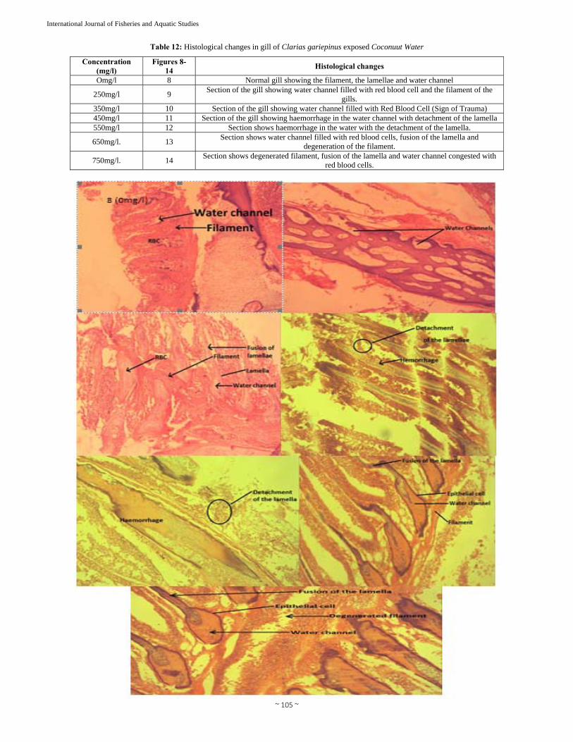
Table 12: Histological changes in gill of Clarias gariepinus exposed Coconuut Water
Concentration (mg/l)
Figures 8- 14
Histological changes
Omg/l 8 Normal gill showing the filament, the lamellae and water channel
250mg/l 9 Section of the gill showing water channel filled with red blood cell and the filament of the
gills. 350mg/l 10 Section of the gill showing water channel filled with Red Blood Cell (Sign of Trauma)450mg/l 11 Section of the gill showing haemorrhage in the water channel with detachment of the lamella 550mg/l 12 Section shows haemorrhage in the water with the detachment of the lamella.
650mg/l. 13 Section shows water channel filled with red blood cells, fusion of the lamella and
degeneration of the filament.
750mg/l. 14 Section shows degenerated filament, fusion of the lamella and water channel congested with
red blood cells.

~ 106 ~
International Journal of Fisheries and Aquatic Studies
Table 13: Histological changes in skin of Clarias gariepinus exposed Coconuut Water
Concentration (mg/l) Figures 15 - 21 Histological changes Omg/l 15 Normal skin tissue, showing the epidermis and the underlying dermis.
250mg/l 16 Skin tissue showing normal keratin of the skin.
350mg/l 17 Tissue showing normal appearance of the skin with normal keratin without
epidermal thickening 450mg/l 18 Normal appearance of the skin with normal keratin appearance.
550mg/l 19 Normal appearance of the skin epidermal layer with no visible thickening of the
epidermis and degeneration of the upper dermis.
650mg/l. 20 Section shows keratin that is histologically abnormal, with severe thinning of the
granular layer.
750mg/l. 21 Section shows noticeable loss of pigmentation of the keratinlayer.
4. Discussion The 96hour LC50 of Cocos nucifera to juvenile African Catfish Clarias gariepinus is presented in table 1, and fig 4-9 is 2.5mg/l with 95% confidence interval 47.1576 -100.3957 and
the maximum safe concentration range between 2.5-25mg/l to 7.5 -75mg/l, Koesoemadinata (1980) [41], he stated that the safe level of a compound is derived by multiplying the 96 -hLC50 with an application factor of 0.1 -0.01 such application factor

~ 107 ~
International Journal of Fisheries and Aquatic Studies
are applied to acute toxicity test data to estimate the concentration that is safe for chronic exposure and is closely related to the work of Ayuba and Ofojekwu (2002) [16] who reported the effect of acute toxicity of the root extract of Datura innoxia to African catfish (Clarias gariepinus) fingerlings and reported that the 96hourLC50 was 204.17mg/l with lower and upper confidence limit of 125.89 and 384.59mg/l. The 96h LC50 in the present result is lower than the result reported by Oti (2002) [50] who reported 96hLC50 of 50.12mg/l on the toxic effect of cassava mill effluent on the African Catfish Heteroclarias hybrid of Heterbranchus bidorsalis male and Clarias gariepinus female The result of haematological parameters of Cocos nucifera to juvenile African Catfish Clarias gariepinus is presented in table 6 which shown increases in red blood count, RCD-WS, RCD-WCV from 39.30 ±2.44, 11.10 ± 0.52, 1.97 ±103.87 and 1.98±16709.68 in control to 59.64 ±8.44, 51.54± 27.78, 1.97 ± 70.35 and 6.87 ±3592.12, in the concentration of 250mg/l, 350mg/l and 650mg/l, agrees with the work of Annune et al. (2002) [11] who reported increase in red blood count RCD-WS, RCD-WCV in lower concentration of ringworm plant, Senna alata used in poisoning water. The result of biochemical parameters of Cocos nucifera to juvenile African Catfish Clarias gariepinus is presented in table 7 shows an increase in Ash and Protein from 0.6± 0.54 and 27.01 ± 0.24 in control to 0.88 ± 0.12 and 27.42 ± 0.46 in the concentration of 750mg/l is close to the work of Orue, and Uner,(1999) [24] who reported increase in liver protein follow in exposure to 2,4 Diamine for 30 days and they was a decrease in moisture, ether extract, nitrogen free extract and energy, from 73.78 ±10. 73, 3.76 ± 0.34, 0.50±0.08 and 1.44 ±2.10 in control to 68.13 ±0.38, 3.17 ± 0.09,0.37 ±0.06 and 1.39 ± 1.41 in higher concentration of 750mg/l and is similar to the work of Bradified and Rees (1978) who reported that toxicants act by disruption of cell membrane permeability replacing the structural or electrochemical important elements in cell which cause functional failure of the organism. The result of plasma electrolytes Cocos nucifera to juvenile African Catfish Clarias gariepinus is presented in table 8, which showed increase in sodium from 1.06±4.83 in the control to 74.54±26.09 and the potassium(K) value exhibited similar trend to sodium from 40.54 ±3.43 in 0mg/l to 24. 14 ±10.34 in 750mg/l, the sodium and potassium were significantly(p <0.05)lower than the exposed groups than the control recorded in this work is in agreement with the work of Kori-siakpere (2000) [42] who reported the electrolytes response to sub-lethel concentrations of potassium permanganate in African Catfish Clarias gariepinus in higher concentration of 750mg/l similar result to the work of Etsler and Edmunds (1906) who observed an increase concentration of sodium, potassium and chlorine in the blood plasma of Clarias gariepinus exposed to various concentration of glyphosate herbicide. The behavioural respons of Clarias gariepinus agreed with the work of Aleem (1988) [10], who observed an increase in erratic swimming and loss of reflex for the test fish as the concentration of tobacco dust were increase and this could probably be due to irritation from increased nicotine content of tobacco 5. Conclusion This study was conducted to determined effect of coconut water to a cultivable fish species, the African catfish (Clarias gariepinus) juveniles. The 20, 24, 36, 48, 72 and 96LC50 of
coconut water to juvenile catfish were 750mg/l, 650mg/l, 550mg/l, 450mg/l, 350mg/l, and 250mg/l. While the total mortality occurred at a concentration of 750mg/l within 24hours exposure period respectively. Haematological examination shown an increase in blood cell count (RCD-WS), (RCD-WCV), and Platelet from, 39.30 ±2.44, 11.10 ± 0.52, 1.97 ±103.87 and 1.98±16709.68 in control to 59.64 ±8.44, 51.54± 27.78, 1.97 ± 70.35 and 6.87 ±3592.12, in the concentration of 250mg/l, 350mg/l and 650mg/l, while there was a decrease in Haematocript, MCV, MCH, LYMH, HMB, RBC from,25.27 ±5.59, 1.23 ± 8.24, 46.50±1.60, 97.700 ± 0.45, 37.94 ± 1.50, 9.63 ± 2.41 and 6.37 ±6.87 in control to 15.03 ± 13.08, 1.07 ± 66.78, 28.00 ± 22.59, 64.84 ±56.16, 24.20 ±20.97, 5.47 ± 4.77 and 1.30 ±1.12 in higher concentration of 750mg/l throughout the test in juvenile catfish Clarias gariepinus. This work is close to the work of Olalade et al., (2010) [46], who reported that Clarias gariepinus fingerlings exposed to sub - lethal concentration of D. innoxia root extract for 12 weeks caused a significance decrease in RBC, HB, and MCV valves. The result of biochemical parameters shows a decrease in moisture, ether extract, nitrogen free extract and energy, from 73.78 ±10.73, 3.76 ± 0.34, 0.50 ± 0.08 in the control to 68.13 ±0.38, 3.17 ± 0.09, 0.37 ± 0.06 and 1.39 ±1.41, in higher concentration of 750mg/l, Also there was an increase in Ash and Protein, from 0.6 ± 0.54 and 27.01 ±0.42 in the control to 0.84 ± 0.14 and 26.28 ± 0.92 I the concentration of 350mg/l. In the plasma electrolyte of catfish juvenile to coconut water, there was an increase in sodium from 1.06± 4.83 in the control to 74.54 ±26.09 in the concentration of 750mg/l, while in potassium; there was a decrease from 40.54 ± 3.43 in the control to 24.14 ±10.34 in higher concentration of 750mg/l. Behavioral responses of African catfish Clarias gariepinus to coconut water include: air gulping, operculum movement, loss of reflex, discoloration, erratic swimming. The result of histopathological responses of skin, gill and liver of the test organisms to aqueous extract of coconut water showed different level of degeneration of cells, hypertrophy of gill arch, disarrangement of hepatic cell, necrosis, vacuolation, sign of trauma the gill, detachment of lamella, vessels congested with blood vessels, liver hypoxia, degeneration of the upper dermis and severe thinning of the underlying granular layer. Damages became severs with increase in concentration of aqueous extract and time exposure. There are no significant changes in the water quality during and after the experiment. The result of the tests provided the baseline information and established safe limits of using coconut water in fresh water fish farm. The fish finally settled at the bottom motionless with slow operculum movement. The results of physio-chemical parameters obtained before the test, during the test and after the test showed that the death of fish is not as a result of poor water quality but coconut because in the control the fish do not undergo any behavioral sign or death. The result of the tests provided the baseline information and established safe limits of using coconut water in fresh water fish farm. 6. References 1. Adakole JA. Changes in some Hematological parameters
of the African catfish (clarias gariepinus) to rubber research in biology. 2012, 3(4).
2. Adewoye SO. Haematological and biochemical changes in Clarias gariepinus exposed to Trephosia vogelii extract. Advances in Applied Science Research. 2010;

~ 108 ~
International Journal of Fisheries and Aquatic Studies
1(1):74-79. 3. Ademoroti, C.M.A Standard Methods for Water and
Effluent Analysis (1st ed), March Print and Consultancy, Benin, Nigeria, 1996.
4. Adeyeye E.I. and I.I. Faleye, Proximate and mineral composition of Bamboy uonopozense, A Bevr Proceeding of the 27th International Conference of the Chemical Society of Nigeria, 2004; 230-232.
5. Adhikari S, Sarkar B. et al. Effect of cypermethrin and carbopuran on certain hematological parameters and prediction of their recovery in a freshwater teleost, labeo rohita (Ham). Ecotoxicol. Environ. saf 2004; 58:220-226.
6. Aiyeloga AA, Bello OA. Ethrobotanical potentials of common herbs in Nigeria. A case study of Enugu State Educational Research and Review. 2006, 1.
7. AOAC Association of Official Analytical Chemists international (AOAC) Official methods of analysis, 17th edition AOAC International, Gaithersburg, MD, USA 2000.
8. Anurag P, Rajamohan T. Cardioprotective effect of tender coconut water in experimental myocardial infarction. Plant Foods. Hum. Nutr 2003; 58:1-12.
9. Alleyne T, Roache S, Thomas C, Shirley A. The control of hypertension by use of coconut water and mauby: Two tropical food drinks. West Indian Med. J. 2005; 54:3-8.
10. Arditti J. Micropropagation of Orchids, 2nd ed.; Blackwell Publishing: Oxford, UK, 2008, II.
11. Ajani F, Awogbade AA. Hematological Changes of the African Catfish Clarias gariepinus (Burchell, 1822) Juveniles Induced by Diuron. British Biotechnology Journal. 2012; 2(4):247-256
12. Akinrotomi OA, Gabriel UU, Ariweriokuma SV. Heamatoxicity of cypermethrin to African catfish clarias gariepious under laboratory conditions. Journal of environmental engineering and technology. 2012, 2.
13. Aleem An assessment of tobacco waste for control gastropod Tympanotonus fucatus (Linnacus) in brackish water fish ponds Aquaculture, View at scopus 1988; 73(1-14):19-25.
14. Annune PA, Ekpendu FOE, Egbonaya nc. Acute toxicity of aqueos extracts of senna alata to juvenile tilapia Oreochromis niloticcus (Trewavas) Book of Abstract, FISON I8'TH-22'nd Nov. Uyo, Nigeria, 2002.
15. Aragão WM, Ribeiro FE, Tupinambá EA, Siqueira ER Variedades e híbridos de coqueiro. In: Aragão WM (Ed.) Coco: Pós-Colheita. Embrapa Informação Tecnológica, Brasília, DF. (Série Frutas do Brasil, 29), 2002, 26-34
16. Ayoola SO. Modern fish farming techniques (Aquaculture). Glamour books Ltd. Dugbe, Ibadan Nigeria, 2010, 180.
17. Asian and Pacific Coconut Community (APCC). International Codes and Standard for Aqueous Coconut Products, 2nd draft. Standards Task Force, Asian and Pacific Coconut Community: Jakarta, Indonesia, 1994.
18. Ayotunde EO. Toxicity of Drumstick Moringa oleifera to Nile Tilapia Oreochromis niloticus (Linn 1757), and African Catfish Clarias gariepinus (BURCHELL 1822). Ph.D. Thesis Federal University of Technology, Akure, Ondo state, Nigeria, 2006.
19. Ayuba VO, Ofojekwu PC. Acute toxicity of the jimson's weed (datura innoxia) to the African Catfish (Clarias gariepinus) Fingerlings AJOL. J Aqua Sci. 2002, 17(2).
20. Bennett BC. Twenty five important plant families B. C. Bennett, Editor. UNESCO Encyclopedia of life support
system, 2007. 21. Campbell D, Obuya S, Spoo M. A simple method for
small scale propagation of Clarias gariepinus in Western Kenya, Field document no. 2, FAO/TCP/KEN/4551, 1995, 27.
22. Campbell-Falck D, Thomas T, Falck TM, Tutuo N, Clem K. The intravenous use of coconut water. Am. J Emerg. Med. 2000; 18:108-111.
23. Campbell-Falck D, Thomas T, Falck TM, Tutuo N, Clem, K. The intravenous use of coconut water. Am J Emerg. Med. 2000; 18:108-111.
24. Coconut Research centre Coconut (cocos nucifera). Retrieved 7, June, 2014 from, 2004. http://www.coconutresearchcentre.org.
25. Dahunsi S, Oranusi U. Hematological responsed of clarias gariepinus, to rubber processing influent. Annual review and research in biology. Science domain international, 2013, 3(4).
26. Harries HC. The evolution, dissemination and classification of Cocos nucifera L. The Botanical Rev 1978; 44:265-319.
27. Orue EO, Uner N. Effect of 2, 4-Diamine on parameters of Protein and Carbohydrate Metabolism in the serum, muscle and liver of Cyprinus carpio. Environ. poll 1999; 105:267-272. Esi.stanford.edu/…/circulations.htm
28. Etsler R, Edmunds PH. Effect of endrim on blood and tissue chemistry of a marine fish. Trans. Soc 1966; 95:153-159.
29. FAO Fishery Information Data and Statistical Services. Aquaculture Production FAO fisheries circular 2003; 815:20-21.
30. FAOSTAT. Production. Crops. Coconut, 2011. http://faostat.fao.org.
31. Federal Department of Fisheries (FDF), Fisheries statistic of Nigeria, published by federal department of fisheries, 2nd edition, 1990, 31.
32. Ferturoti EO. Effect of supplementary feeding and organic manuring on the production of African Catfish, Clarias gariepinas (Beurchell 1822). J West Apri fish. 1989; 4:187-195.
33. Narayana Rao, Srikanth GBK, Ramu G. Haematological Changes In The Fresh Water Fish, Channa Punctatus Due To The Effect of Rayon Industry Effluents. Fisheries Research Laboratory, 1999.
34. Gertjan, Johannes. Handbook On The Artificial Reproduction And Pond Rearing Of The African Catfish Clarias Gariepinus In Sub-Saharan Africa FAO, Fisheries Technical Paper 362 Rome, 1996.
35. George EF, Sherrington PD. Plant Propagation by Tissue Culture-Handbook and Directory of Commercial Laboratories; Exegetics Ltd: Edington, UK, 1984.
36. Goh GYI, Koren G. Folic acid in pregnancy and fetal outcomes J Obstet. Gynaecol. 2008; 28:3-13.
37. Gunni Bee, Luc Baudouin, Kenneth Olsen M. "independent origins of cultivated coconut (cocos nucifera L) in the old world tropics.doi10-1377 T-Journal Pone 0021143. 2011.
38. Herwig N. Handbook of drug and chemical used in the treatment of fish disease: A manual of fish farm and material Media. Charles Thomas Pub. Sprinfield Illinois U.S.A. 1979.
39. Heo HJ, Hong SC, Shin DH. Inhibitory Effect of Zeatin, Isolated from Faitoua Villosa, on acetyicholinesterase

~ 109 ~
International Journal of Fisheries and Aquatic Studies
activity from pciz cells. Mol. Cells 2002; 13:113-117. 40. Ighwela KA, Ahmad AB, Abol-Munaf AB.
Haematological Changes in Nile Tilapia (Oreochromis niloticus) Fed with Varying Dietary Maltose Levels. World Journal of Fish and Marine Sciences. 2012; 4(4):376-381
41. Jackson JC, Gordon A, Wizzard G, McCook K, Rolle R. Change in chemical composition of coconut (cocos nucifera L. ) water during maturation of fruit J Sci. food Agri. 2004; 84:1049-1652.
42. Jean WH, Young, Liya Ge, Yan Fei Ng, Swee Ngin Tan. The chemical Composition and Biological properties of coconut (cocos Nucifera L) water molecules 2009; 14:5144-5164.
43. Janick J, Paull RE. The Encyclopedia of Fruit & Nuts; CAB International: Wallingford, UK, 2008, 112.
44. Kende H, Zeevaart J. The five Classical plant hormones. Plant Cell 1997; 9:1197-1210
45. Koesomadinata S. Pesticides as a major constraint to integrated agricultural integrated agriculture-aquaculture-farming system. ICLRM conference proceedings 4. International Center for Living Aquatic Resources Management, Manila Philippines and Southeast Asian Regional Centre for Graduate study and Research in Agriculture, Los banos, Phillipine, 1980.
46. Koesomadinata, S. acute toxicity of the insecticide formulation of endosulphan, chlorpyrifos, and chlorfluazuron to three freshwater fish species and freshwater giant prawn. Jurnal penelitian perikan Indonesia. 2000; 4(3-4):36-43.
47. Kori-siakpere Ovie. (2000) Electrolytes response to sub-lethel concentration of potassium permanganate in African Catfish; Clarias gariepinus (Burchell, 1822). International Journal of Integrative Biology. 2009, 5(1).
48. Letham DS, Zeatin A. facter Inducing cell division isolated from zea mays life Sci. 1963; 2:569-573.
49. Matieswaran R, Devapaul A, Muralidharan S, Volmurugan B, Ignacimuhu S. Hematological studies of fresh water fish, clarias, batrachus (L) exposed to mercuric chloride international journal of integrative biology. 2008.
50. Miller Skoog CO, Saltza Von F, Strong MH, Kinetin FM. A cell division factions from deoxyribon onucleic acid. J Am Chem Soc. 1955; 77:1392-1393.
51. Olalade IA, Oginni O. Toxic Stress and Haematological effects of Nickel on Clarias gariepinus fingerlings. Journal of Environmental Chemistry and Ecotoxicology. 2010; 2(2):14-19.
52. Nirankush Paul, Rajarshi Roy, Sanjib Bhattacharya, Moulisha Biswas. Acute and sub-chronic toxicity study of Cocos nucifera leaf extracts in Mice. Journal of Advanced Pharmacy Education & Research. 2012; 2(2):74-81
53. Onisiriuka BC. Effect of sub lethal concentration of formalin on weight gain on African catfish Clarias gariepinus (Teugels). Ajol J Aqua Sci. 2002, 1(1).
54. Onusiiuka BC. Effect of sub lethal concentration of formalin on weight gain African Catfish, Clarias gariepinus (L). AJOL. J Aqua Sci. 2002, 15.
55. Oti EE. Acute toxicity of cassava mill effluent to the African Catfish Fingerlings AJOL: J of Aquatic Sci. 2002, 17(1).
56. Parma MJ, Loteste A, Campana M, Bacchetta C. Changes of hematological parameters in Prochilodus lineatus (Pisces, Prochilodontidae) exposed to sublethal
concentration of cypermethrin. Journal of Environmental Biology. 2007; 28(1):147-149
57. Patrick JW, Offler CE. Compartmentation of transport and transfer events in developing seeds. J Exp. Bot. 2001; 52:551-564.
58. Pumer S, Hail P, Maleck W, Petrioanu G. Influence of coconut water on homeostasis. Am. J Emerg. Med. 2001; 19:287-289.
59. Railo E, Nikinmae M et al. Effect of Sampling on blood parameters in the rainbow trout, salmogairderi, J Fish Res. Bd. Can. 1985; 26:725-732.
60. Rattan SIS, Clark BFC. Kinetin delays the onset of ageing characteristics in human fibroblasts. Biochem.Biophys, B. Micronutrients: oxidant/antioxidant status, Br. J Nutr. 2001; 85:567-574.
61. Romesh A, Saravanam M. Hematological and biochemical responses in a freshwater fish cyprinus carpio exposed to chloropyripos. Internal journal of integrative Biology. 2008.
62. Saiangurunathan B. Xavier Innocent and muthulakshimi Immune modulatory effect of Dietary Nelumbo Nucifera (lotus) in Growth and Hematology of Cirrhinus Mrigala challenged with pseudomonas aneruginosa. Journal of applied pharmaceutical science. 2012.
63. Sathaeshkumar P, Ananthan G, Senthil D, Kumar D. "Hematological and Biochemical parameters of different feeding behavior of teleost fishes velar estuary, India Springer Verlag Limited, 2011.
64. Satyabalan K. Coconut Varieties and Cultivars: Their Classification. Asian and Pacific Coconut Community, Jakarta, 1997.
65. Skelton P. A complete guide to the freshwater fishes of Southern Africa. Struik Publishers, Cape Town, 2001.
66. Sandhya VG, Rajamohan T. Comparative evaluation of the hypolipidemic effects of coconut water and lovastatin in rats fed fat-cholesterol enriched diet. Food Chem. Toxicol 2008; 45:3585-3592.
67. Seow CC, Gwee CN. Coconut milk: Chemistry and technology, Int. J Food Sci. Tech. 1997; 32:189-201.
68. Santoso U, Kubo K, Ota T, Tadokoro T, Maekawa A. Nutrient composition of kopyor coconuts (Cocos nucifera L.). Food Chem 1996; 57:299-304.
69. Summarwar S. Comparative Haematological Studies Of Clarius Batrachus In Bisalpur Reservoir And Pushkar Lake. Indian Journal of Fundamental and Applied Life Sciences. 2012, 2(2).
70. Svobodova D. Ravds, J. & Palackova. Unified method of Haematological examination of fish. Research Inst. of Fish Culture and Hydrobiology. Vonnony Czechoslovakia. 1991.
71. Summarwar, Santosh Verma. Study of selected Hematological indices of fresh water fish from Bisalpur Reservoir. Indian Journal of fundamental and applied life sciences. 2012, 2(2).
72. Teugels GG. Preliminary results of a morphological study of five nominal species of the subgenus Clarias (Pisces; Clariidae). J Nat. Hist. 1982a; 16(3):439-464.
73. Teugels GG. Preliminary data of a systematic outline of the African species of the genus Clarias (Pisces; Clariidae). Rev. Zool. afr 1982b; 96(4):731-748.
74. Teugels GG. The nomenclature of African Clarias species used in aquaculture. Aquaculture 1984; 38:373-374.
75. Tulecke W, Weinstein L, Rutner A, Laurencot H. The biochemical composition of coconut water (coconut milk)

~ 110 ~
International Journal of Fisheries and Aquatic Studies
as related to its use in plant tissue culture. Contrib. Boyce Thompson Inst 1961; 21:115-128.
76. Ugwen U, Gabriel Ojo, Akinrotimi A, Funkeye Eseimkumo. "Hematological Responses of wild life Tilapia creochromis niloticus after acclimation to captivity. Jordan journal of biological sciences. 2011, 44.
77. United States Department of Agriculture (USDA). National Nutrient Database for Standard Reference, 2008. Nuts, coconut water [Online]. Available: http://www.nal.usda.gov/fnic/foodcomp/cgi-bin/list_nut_edit.pl/, accessed on 9 December, 2009.
78. USEPA. Methods for measuring the acute toxicity of effluents to freshwater and marine organisms. 4th ed. Environmental Monitouring and support Laboratory, U.S. Environmental Protection Agency, Cincinati, Ohio. EPA 600/4-85/013. 2000.
79. Vazguez Rey G, Guerero GA. Characterization of blood cells and hematological parameters in cichlasom dimerus (Teleostei perciformes). Universidad de Beunos Aires, Crudad Universitarian, Pubellon, U. C1428EHA, Basenos, Aires, Argentina, 2007.
80. Viveen WJAR, Richer CJJ, Van Ocrdt PGWJ, Janssen JAL Husisman EA. Practical Manual for the Culture of the African Catfish; Clarias gariepinus, Joint Pub. Of D.G. I.C. Ministry of foreign affair, D.F.C.F.A.U. Uni of wageningen, and R.G. C.E. Dept. Zoology, Uni of Utrecht Nehterlands. 1986.