Isolation of apolipoproteins from carotenoid- carrying ... · Isolation of apolipoproteins from...
8
Isolation of apolipoproteins from carotenoid- carrying lipoprotein in the serum of chum salmon, Oncorhynchus keta Seiichi Ando' and Mutsuo Hatano Laboratory of Food Chemistry I, Faculty of Fisheries, Hokkaido University, 3-1-1, Minato, Hakodate 041, Japan Abstract Carotenoid-carryinglipoprotein (CCL) was rapidly isolated from the high density lipoprotein (HDL) fraction of the upstream migrating male chum salmon (Oncorhynchw &a) by a single-step density gradient ultracentrifugation. The two apo- lipoproteins (Mr = 24,000 and 12,000; designated apo-I and apo-11, respectively) were readily dissociated and separated in 0.1% SDS by gel liltration chromatography. Prominent features of the amino acid composition in the CCL included the relative high levels of glutamic acid, alanine, leucine, and lysine, and the low cysteine content. Apo-I, as well as the CCL, was rich in glu- tamic acid, alanine, leucine, and lysine. Compared to the amino acid composition of apo-I, apo-I1 included relatively high levels of glycine and tyrosine, and low threonine, serine, and arginine contents. When the intact CCL particle was treated with tryp- sin, apo-I was rapidly proteolyzed, while apo-I1 was resistant. However, both apo-I and apo-I1 isolated from the CCL particle were readily digested with trypsin. This suggested that a differ- ent structural arrangement rather than the amino acid composi- tions of the apolipoproteinswas associated with the limited tryp- sin digestion of the CCL particle. Apo-I1 may be sheltered from the aqueous environment and lie partly within the CCL particle. The properties of both the HDL fraction and apolipoproteins from pink salmon (Oncorhynchus gorbuscha) were similar to those of the CCL from chum salmon. -Ando, S., and M. Hatano. Isolation of apolipoproteins from carotenoid-carrying lipopro- tein in the serum of chum salmon, Oncorhynchus keta. J. Lipid Res. 1988. 29: 1264-1271. Supplementary key words centrifuge * gel filtration * high density lipoprotein apolipoproteins density gradient ultra- Pacific salmon (Oncorhynchus spp.) are typically anadro- mous fish, developing in the ocean and breeding in fresh water. This biological feature governs the pattern of changes in their chemical composition (1). For example, after migrating chum salmon enter a river, the muscle dis- colors and the integument becomes dark yellow or red. These changes in muscle or integument color may be closely associated with the physiological state of the fish which is probably controlled by sex hormones (2-7). Kitahara (8) has reported that carotenoid pigments (mainly astaxanthin) absorbed through the food (zoo- plankton) are carried from the muscle to the integument and gonads via the serum during spawning migration of chum salmon. Little has been reported concerning carrier proteins of carotenoids in the serum of salmon, although several studies on the distribution and characterization of serum lipoproteins in salmon have been made (9-12). Nakamura, Hata, and Hata (13) have recently found that astaxanthin in the serum of upstream migrating chum salmon was exclusively transported by the high density lipoprotein (HDL) fraction. We have previously demon- strated that three types of carotenoid-carrying lipopro- teins (CCLs) such as low density lipoprotein (LDL), HDL, and very high density lipoprotein (VHDL) frac- tions were present in the serum of chum salmon. The CCL from the VHDL fraction was derived from vitel- logenin, a female-specific serum protein (14-17). The CCL from the HDL fraction became a main component during spawning migration (14, 18-20). The CCL from the HDL fraction in both males and females was associ- ated with carotenoid transport from the muscle to the integument, while the carotenoids were transported into the ovaries via vitellogenin. We have recently isolated the CCL from the HDL fraction in the serum of fipstream migrating chum salmon by a sequential ultracentrifugal technique, and DEAE-cellulose and gel filtration column chromatography (18). The CCL gave rise to two apolipo- proteins whose molecular weights were 24,000 (apo-I) and 12,000 (apo-11), and no disulfide bond was detected be- tween the two apolipoproteins. The molar ratio of apo-I to apo-I1 was close to 1:l. The CCL from the HDL frac- Abbreviations: HDL, high density lipoprotein; CCL, carotenoid- carrying lipoprotein; LDL, low density lipoprotein; VHDL, very high density lipoprotein; Tris, tris(hydroxymethy1)amino methane; TLC, thin-layer chromatography; SDS, sodium dodecyl sulfate; PAGE, poly- acrylamide gel electrophoresis. 'Address reprint requests (until May 1989) to: Seiichi Ando, Ph.D., c/o Dr. W. J. Schneider, Department of Biochemistry, Faculty of Medicine, University of Alberta, Edmonton, Alberta, Canada T6G 2H7. 1264 Journal of Lipid Research Volume 29, 1988 by guest, on June 21, 2018 www.jlr.org Downloaded from
Transcript of Isolation of apolipoproteins from carotenoid- carrying ... · Isolation of apolipoproteins from...

Isolation of apolipoproteins from carotenoid- carrying lipoprotein in the serum of chum salmon, Oncorhynchus keta
Seiichi Ando' and Mutsuo Hatano
Laboratory of Food Chemistry I, Faculty of Fisheries, Hokkaido University, 3-1-1, Minato, Hakodate 041, Japan
Abstract Carotenoid-carrying lipoprotein (CCL) was rapidly isolated from the high density lipoprotein (HDL) fraction of the upstream migrating male chum salmon (Oncorhynchw &a) by a single-step density gradient ultracentrifugation. The two apo- lipoproteins (Mr = 24,000 and 12,000; designated apo-I and apo-11, respectively) were readily dissociated and separated in 0.1% SDS by gel liltration chromatography. Prominent features of the amino acid composition in the CCL included the relative high levels of glutamic acid, alanine, leucine, and lysine, and the low cysteine content. Apo-I, as well as the CCL, was rich in glu- tamic acid, alanine, leucine, and lysine. Compared to the amino acid composition of apo-I, apo-I1 included relatively high levels of glycine and tyrosine, and low threonine, serine, and arginine contents. When the intact CCL particle was treated with tryp- sin, apo-I was rapidly proteolyzed, while apo-I1 was resistant. However, both apo-I and apo-I1 isolated from the CCL particle were readily digested with trypsin. This suggested that a differ- ent structural arrangement rather than the amino acid composi- tions of the apolipoproteins was associated with the limited tryp- sin digestion of the CCL particle. Apo-I1 may be sheltered from the aqueous environment and lie partly within the CCL particle. The properties of both the HDL fraction and apolipoproteins from pink salmon (Oncorhynchus gorbuscha) were similar to those of the CCL from chum salmon. -Ando, S., and M. Hatano. Isolation of apolipoproteins from carotenoid-carrying lipopro- tein in the serum of chum salmon, Oncorhynchus keta. J. Lipid Res. 1988. 29: 1264-1271.
Supplementary key words centrifuge * gel filtration * high density lipoprotein
apolipoproteins density gradient ultra-
Pacific salmon (Oncorhynchus spp.) are typically anadro- mous fish, developing in the ocean and breeding in fresh water. This biological feature governs the pattern of changes in their chemical composition (1). For example, after migrating chum salmon enter a river, the muscle dis- colors and the integument becomes dark yellow or red. These changes in muscle or integument color may be closely associated with the physiological state of the fish which is probably controlled by sex hormones (2-7).
Kitahara (8) has reported that carotenoid pigments (mainly astaxanthin) absorbed through the food (zoo-
plankton) are carried from the muscle to the integument and gonads via the serum during spawning migration of chum salmon. Little has been reported concerning carrier proteins of carotenoids in the serum of salmon, although several studies on the distribution and characterization of serum lipoproteins in salmon have been made (9-12). Nakamura, Hata, and Hata (13) have recently found that astaxanthin in the serum of upstream migrating chum salmon was exclusively transported by the high density lipoprotein (HDL) fraction. We have previously demon- strated that three types of carotenoid-carrying lipopro- teins (CCLs) such as low density lipoprotein (LDL), HDL, and very high density lipoprotein (VHDL) frac- tions were present in the serum of chum salmon. The CCL from the VHDL fraction was derived from vitel- logenin, a female-specific serum protein (14-17). The CCL from the HDL fraction became a main component during spawning migration (14, 18-20). The CCL from the HDL fraction in both males and females was associ- ated with carotenoid transport from the muscle to the integument, while the carotenoids were transported into the ovaries via vitellogenin. We have recently isolated the CCL from the HDL fraction in the serum of fipstream migrating chum salmon by a sequential ultracentrifugal technique, and DEAE-cellulose and gel filtration column chromatography (18). The CCL gave rise to two apolipo- proteins whose molecular weights were 24,000 (apo-I) and 12,000 (apo-11), and no disulfide bond was detected be- tween the two apolipoproteins. The molar ratio of apo-I to apo-I1 was close to 1:l. The CCL from the H D L frac-
Abbreviations: HDL, high density lipoprotein; CCL, carotenoid- carrying lipoprotein; LDL, low density lipoprotein; VHDL, very high density lipoprotein; Tris, tris(hydroxymethy1)amino methane; TLC, thin-layer chromatography; SDS, sodium dodecyl sulfate; PAGE, poly- acrylamide gel electrophoresis.
'Address reprint requests (until May 1989) to: Seiichi Ando, Ph.D., c/o Dr. W. J. Schneider, Department of Biochemistry, Faculty of Medicine, University of Alberta, Edmonton, Alberta, Canada T6G 2H7.
1264 Journal of Lipid Research Volume 29, 1988
by guest, on June 21, 2018w
ww
.jlr.orgD
ownloaded from
tion was a unique lipoprotein with high carotenoid levels (20, 21). Thus, the interaction between apolipoproteins and carotenoids in the CCL is of interest.
In order to broaden our basic understanding of struc- ture and function in the CCL, we have isolated apolipo- proteins from the CCL of the upstream migrating male chum salmon, Omorlynchus kctq and we report their amino acid compositions. A difference in exposure of the two apolipoproteins in the aqueous environment is also suggested from the results of amino acid composition and limited trypsinization of the CCL from the HDL fraction.
MATERIALS AND METHODS
Animals
Male adult chum salmon, Oncor&nchus ha, 3 and 4 years old, were caught at the Yurappu River of Hokkaido. The gonadosomatic index (gonad weight x 100/body weight) and hepatosomatic index (liver weight x 100/body weight) were 3.6-4.3 and 1.7-1.9, respectively.
Preparation of lipoproteins
Blood was collected from the caudal vasculature of live salmon and left at room temperature for several hours. The clotted blood was centrifuged at 3,500 rpm for 15 min at 10°C to obtain the serum. The serum was analyzed by density gradient ultracentrifugation (22). An equal amount of 0.75% NaCl was gently layered over 4.74 ml of serum containing 1.9 g of KBr, and centrifuged at 218,000 g for 5 hr at 10°C. The carotenoid-carrying lipoprotein (CCL), which forms a sharp band at density 1.16 gfml, was re- moved by injecting a needle directly into the centrifuge tube beneath the CCL band and withdrawing the solution containing the bright orange-colored portion. The KBr was then removed by dialysis overnight against 0.75% NaCl-20 mM Tris-HC1 buffer (pH 7.5).
Density, 1ipid:protein ratio, and lipid and carotenoid analyses
The density of CCL was measured from the refractive index of KBr solution on an Abbe refractometer (Atago Co., Ltd.) following density gradient ultracentrifugation. Protein level in the CCL was measured by the biuret method (23), using bovine serum albumin as the stan- dard. Lipid extraction from the CCL was done by the method of Bligh and Dyer (24). The extracted lipid was analyzed quantitatively by thin-layer chromatography (TLC). The TLC plates (Kieselgel 60, ready-made plate from Merck) were developed using n-hexane-diethyl- ether-acetic acid 85:15:1 (by volume) for neutral lipids, and chloroform-methanol-acetic acid-water 25:15:4:2 (by volume) for phospholipids. The TLC plate was sprayed with 3% copper acetate-8% phosphoric acid, heated on
a hot plate, and quantitated using a Cosmo F-808 densi- tometer. The amount of phospholipid was calculated from lipid-phosphorus assayed by the method of Fiske and Subbarow (25). Carotenoid content was calculated as- suming the value in acetone at 477 nm to be 2,200. High performance liquid chromatographic (HPLC) anal- ysis of carotenoids was carried out on a Shimadzu LC-6A instrument with a Shimadzu SPD-PA VIS spectropho- tometer set at 470 nm. The column used was a 250 x 4 mm i.d. stainless steel column packed with 5 pm Sumipax OA-2000 (Sumitomo Chemical Co., 'Ltd.). Separation was achieved with a mobile phase of n-hexane-dichloro- methane-ethanol 50:20:0.5 (by volume) at a flow-rate of 0.8 ml/min. Identification of each carotenoid was accom- plished by co-TLC and co-HPLC with authentic speci- mens. The authentic carotenoids used were as follows: astaxanthin diester, astaxanthin monoester, and astaxan- thin were extracted and purified from the Antarctic krill Euphausia superba (26, 27); zeaxanthin was extracted and isolated from Sfiirulinu maxim (28).
Separation of apolipoproteins
Apolipoproteins of the CCL were dissociated in 1% SDS-1% 2-mercaptoethanol at 25OC overnight, and iso- lated by gel filtration chromatography on a 3.2 x 89 cm Sephadex G-100 column equilibrated in 0.1% SDS-20 mM Tris-HC1 buffer (pH 7.5) containing 0.02% NaNs. Column eluent was monitored at 278 nm and protein peaks were combined and dialyzed against distilled water.
Amino acid analysis
The amino acid compositions of the delipidated CCL and the apolipoproteins were determined on a Hitachi model 835 automatic amino acid analyzer after hydrolysis with 4 N methanesulfonic acid at 115OC for 24 hr.
Gel electrophoresis
Electrophoresis in 5 % polyacrylamide gel was carried out at pH 8.3, 2 mA per tube, as described by Davis (29). SDS-slab-15 % polyacrylamide gel electrophoresis (PAGE) was performed in the presence of 0.1% SDS, by the method of Laemmli (30). After electrophoresis, gels were stained with Coomassie Brilliant Blue.
Limited trypsinization
The native CCL and the apolipoproteins from the CCL were subjected to limited trypsinization. One mg of the CCL and the apolipoproteins in 20 mM Tris-HC1 buffer (pH 7.5) was incubated for various times with trypsin (Sigma Chemical Go., Ltd.) at a concentration of 1:lOO (trypsinxample) for times ranging from 0 to 60 min at 25OC. The reaction was initiated by the addition of trypsin and stopped by addition of SDS-PAGE sample treatment buffer (pH 6.8) containing 20 mM Tris-HCI, 2% SDS, 2% 2-mercaptoethanol, and 40% glycerol, and boiling at
Ando and Hatano Apolipoproteins in salmon serum lipoprotein 1265
by guest, on June 21, 2018w
ww
.jlr.orgD
ownloaded from
100°C for 2 min. Samples were subsequently analyzed by SDS-slab-15% PAGE.
1 - 2 =
3 4 L R
RESULTS
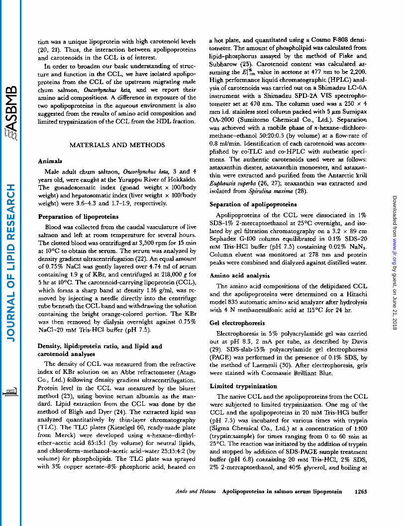
The major lipoprotein in the serum of upstream migrating male chum salmon was isolated from the high density lipoprotein (HDL) fraction by a single-step den- sity gradient ultracentrifugal separation. A bright orange band was present in the middle portion, corresponding to the HDL fraction (d 1.16 g/ml) of the tube. The homogen- eity of the HDL fraction, which corresponded to carote- noid-carrying lipoprotein (CCL), was checked by PAGE. Under native conditions the CCL migrated as a single homogeneous band. The CCL gave two apolipoproteins whose molecular weights were 24,000 (apo-I) and 12,000 (apo-11) as judged by SDS-slab-PAGE (Fig. 1).
Lipid and carotenoid analyses
Table 1 shows the properties of CCL, along with those of the HDL fraction from pink salmon (9). Compared to the HDL fraction from pink salmon, the CCL contained more protein and less lipid, which explains the differences in density. In each of them, cholesteryl ester, cholesterol, and triglyceride accounted for most of the neutral lipid. The major components of phospholipid were phosphatidyl- choline and sphingomyelin. Also, the CCL was bright orange, due to the presence of carotenoids, mainly asta- xanthin.
Separation of apolipoproteins
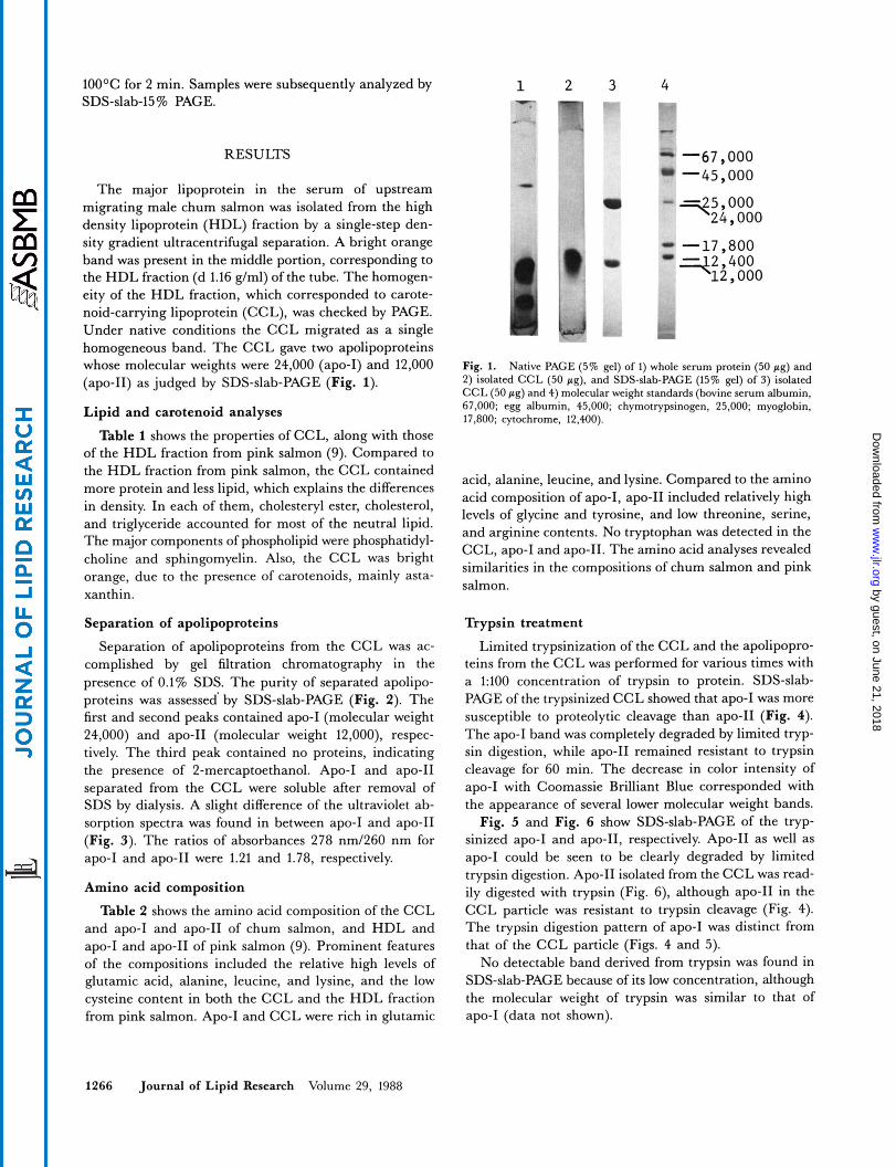
Separation of apolipoproteins from the CCL was ac- complished by gel filtration chromatography in the presence of 0.1% SDS. The purity of separated apolipo- proteins was assessed' by SDS-slab-PAGE (Fig. 2). The first and second peaks contained apo-I (molecular weight 24,000) and apo-I1 (molecular weight 12,000), respec- tively. The third peak contained no proteins, indicating the presence of 2-mercaptoethanol. Apo-I and apo-I1 separated from the CCL were soluble after removal of SDS by dialysis. A slight difference of the ultraviolet ab- sorption spectra was found in between apo-I and apo-I1 (Fig. 3). The ratios of absorbances 278 nm/260 nm for apo-I and apo-I1 were 1.21 and 1.78, respectively.
Amino acid composition
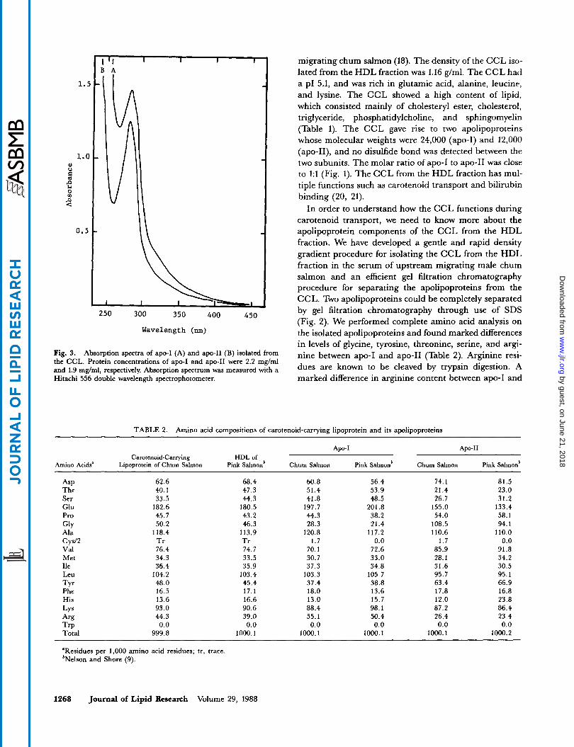
Table 2 shows the amino acid composition of the CCL and apo-I and apo-I1 of chum salmon, and HDL and apo-I and apo-I1 of pink salmon (9). Prominent features of the compositions included the relative high levels of glutamic acid, alanine, leucine, and lysine, and the low cysteine content in both the CCL and the HDL fraction from pink salmon. Apo-I and CCL were rich in glutamic
I
-67,000 -45,000
5,000 -24,000
-17,800
Fig. 1. Native PAGE (5% gel) of 1) whole serum protein (50 pg) and 2) isolated CCL (50 pg), and SDS-slab-PAGE (15% gel) of 3) isolated CCL (50 pg) and 4) molecular weight standards (bovine serum albumin, 67,000; egg albumin, 45,000; chymotrypsinogen, 25,000; myoglobin, 17,800; cytochrome, 12,400).
acid, alanine, leucine, and lysine. Compared to the amino acid composition of apo-I, apo-I1 included relatively high levels of glycine and tyrosine, and low threonine, serine, and arginine contents. No tryptophan was detected in the CCL, apo-I and apo-11. The amino acid analyses revealed similarities in the compositions of chum salmon and pink salmon.
Trypsin treatment
Limited trypsinization of the CCL and the apolipopro- teins from the CCL was performed for various times with a 1:lOO concentration of trypsin to protein. SDS-slab- PAGE of the trypsinized CCL showed that apo-I was more susceptible to proteolytic cleavage than apo-I1 (Fig. 4). The apo-I band was completely degraded by limited tryp- sin digestion, while apo-I1 remained resistant to trypsin cleavage for 60 min. The decrease in color intensity of apo-I with Coomassie Brilliant Blue corresponded with the appearance of several lower molecular weight bands.
Fig. 5 and Fig. 6 show SDS-slab-PAGE of the tryp- sinized apo-I and apo-11, respectively. Apo-I1 as well as apo-I could be seen to be clearly degraded by limited trypsin digestion. Apo-I1 isolated from the CCL was read- ily digested with trypsin (Fig. 6), although apo-I1 in the CCL particle was resistant to trypsin cleavage (Fig. 4). The trypsin digestion pattern of apo-I was distinct from that of the CCL particle (Figs. 4 and 5).
No detectable band derived from trypsin was found in SDS-slab-PAGE because of its low concentration, although the molecular weight of trypsin was similar to that of apo-I (data not shown).
1266 Journal of Lipid Research Volume 29, 1988
by guest, on June 21, 2018w
ww
.jlr.orgD
ownloaded from
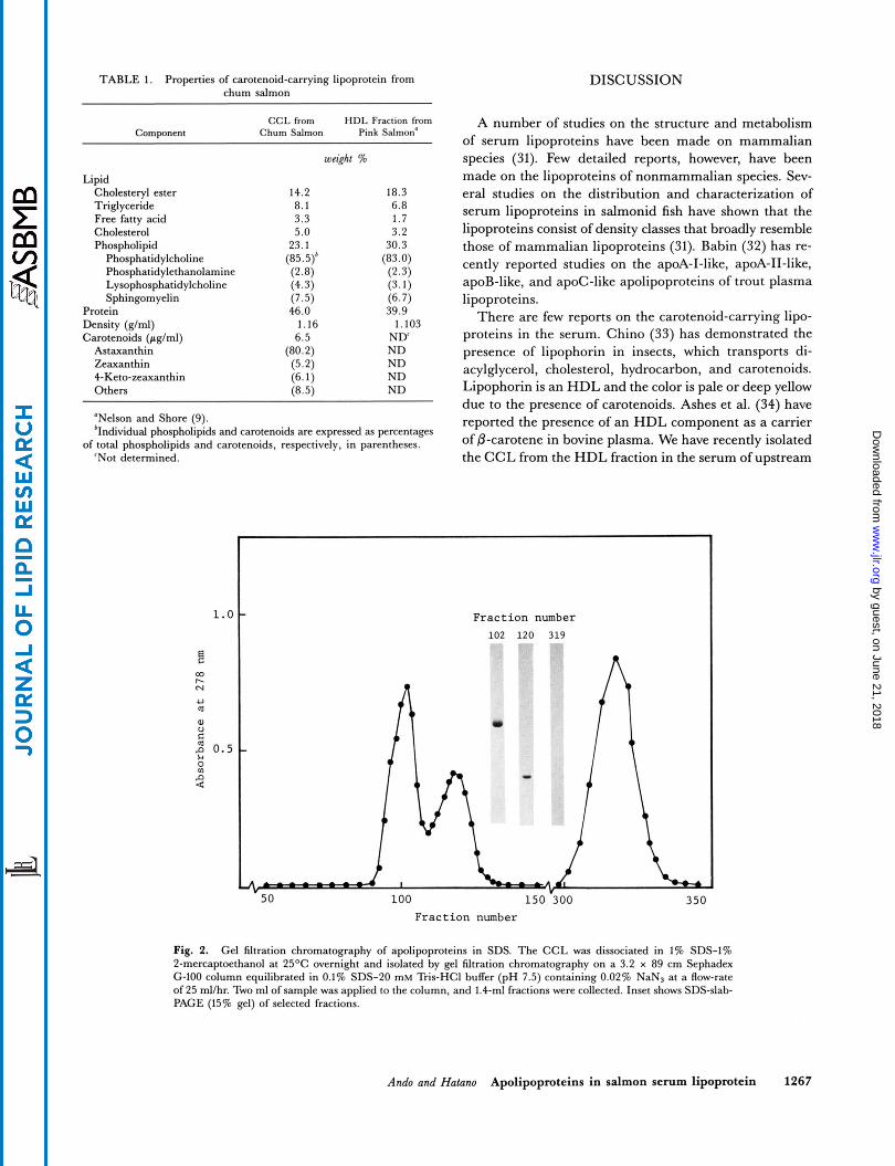
TABLE 1. Properties of carotenoid-carrying lipoprotein from chum salmon
CCL from HDL Fraction from Component Chum Salmon Pink Salmon'
weicht %
Lipid Cholesteryl ester 14.2 18.3 Triglyceride 8.1 6.8 Free fatty acid 3.3 1.7 Cholesterol 5.0 3.2 Phospholipid 23.1 30.3
Phosphatidylcholine (85.5)b (83.0) Phosphatidylethanolamine (2.8) (2.3) Lysophosphatidylcholine (4.3) (3.1) Sphingomyelin (7.5) (6.7)
Protein 46.0 39.9 Density (glml) 1.16 1.103 Carotenoids (pglml) 6.5 ND'
Astaxanthin (80.2) ND Zeaxanthin (5.2) ND 4-Keto-zeaxanthin (6.1 ) ND Others (8.5) ND
"Nelson and Shore (9). 'Individual phospholipids and carotenoids are expressed as percentages
'Not determined. of total phospholipids and carotenoids, respectively, in parentheses.
DISCUSSION
A number of studies on the structure and metabolism of serum lipoproteins have been made on mammalian species (31). Few detailed reports, however, have been made on the lipoproteins of nonmammalian species. Sev- eral studies on the distribution and characterization of serum lipoproteins in salmonid fish have shown that the lipoproteins consist of density classes that broadly resemble those of mammalian lipoproteins (31). Babin (32) has re- cently reported studies on the apoA-I-like, apoA-11-like, apoB-like, and apoC-like apolipoproteins of trout plasma lipoproteins.
There are few reports on the carotenoid-carrying lipo- proteins in the serum. Chino (33) has demonstrated the presence of lipophorin in insects, which transports di- acylglycerol, cholesterol, hydrocarbon, and carotenoids. Lipophorin is an HDL and the color is pale or deep yellow due to the presence of carotenoids. Ashes et al. (34) have reported the presence of an HDL component as a carrier of @-carotene in bovine plasma. We have recently isolated the CCL from the HDL fraction in the serum of upstream
1.0
B Q)
N PI
U a
F r a c t i o n number 102 120 319
h
F r a c t i o n number
Fig. 2. Gel filtration chromatography of apolipoproteins in SDS. The CCL was dissociated in 1% SDS-1% 2-mercapteethanol at 25OC overnight and isolated by gel filtration chromatography on a 3.2 x 89 cm Sephadex '2-100 column equilibrated in 0.1% SDS-20 mM Tris-HCI buffer (pH 7.5) containing 0.02% NaNs at a Row-rate of 25 ml/hr. Two ml of sample was applied to the column, and 1.4-ml fractions were collected. Inset shows SDS-slab- PAGE (15% g e l ) of selected fractions.
Ando and Hatano Apolipopmteins in salmon serum lipoprotein 1267
by guest, on June 21, 2018w
ww
.jlr.orgD
ownloaded from
I I I I I B A
250 300 350 400 450
Wavelength (nm)
Fig. 3. Absorption spectra of apo-I (A) and apo-11 (B) isolated from the CCL. Protein concentrations of apo-I and apo-I1 were 2.2 mglml and 1.9 mg/ml, respectively. Absorption spectrum was measured with a Hitachi 556 double wavelength spectrophotometer.
migrating chum salmon (18). The density of the CCL iso- lated from the HDL fraction was 1.16 g/ml. The CCL had a p1 5.1, and was rich in glutamic acid, alanine, leucine, and lysine. The CCL showed a high content of lipid, which consisted mainly of cholesteryl ester, cholesterol, triglyceride, phosphatidylcholine, and sphingomyelin (Table 1). The CCL gave rise to two apolipoproteins whose molecular weights were 24,000 (apo-I) and 12,000 (apo-11), and no disulfide bond was detected between the two subunits. The molar ratio of apo-I to apo-I1 was close to 1:l (Fig. 1). The CCL from the HDL fraction has mul- tiple functions such as carotenoid transport and bilirubin binding (20, 21).
In order to understand how the CCL functions during carotenoid transport, we need to know more about the apolipoprotein components of the CCL from the HDL fraction. We have developed a gentle and rapid density gradient procedure for isolating the CCL from the HDL fraction in the serum of upstream migrating male chum salmon and an efficient gel filtration chromatography procedure for separating the apolipoproteins from the CCL. Two apolipoproteins could be completely separated by gel filtration chromatography through use of SDS (Fig. 2). We performed complete amino acid analysis on the isolated apolipoproteins and found marked differences in levels of glycine, tyrosine, threonine, serine, and argi- nine between apo-I and apo-I1 (Table 2). Arginine resi- dues are known to be cleaved by trypsin digestion. A marked difference in arginine content between apo-I and
TABLE 2. Amino acid compositions of carotenoid-carrying lipoprotein and its apolipoproteins
Apo-I Apo-I1 Carotenoid-Carrying HDL of
Amino Acids" Lipoprotein of Chum Salmon Pink Salmonb Chum Salmon Pink Salmon* Chum Salmon Pink Salmonb
ASP Thr Ser Glu Pro
Ala
Val Met Ile Leu TY Phe His LY s Arg TrP Total
GlY
c ysl2
62.6 40.1 33.5
182.6 45.7 50.2
118.4 Tr 76.4 34.3 36.4
104.2 48.0 16.5 13.6 93.0 44.3 0.0
999.8
68.4 47.3 44.3
180.5 43.2 46.3
113.9 Tr 74.7 33.5 35.9
103.4 45.4 17.1 16.6 90.6 39.0 0.0
1000.1
60.8 51.4 41.8
197.7 44.3 28.3
120.8 1.7
70.1 30.7 37.3
103.3 37.4 18.0 13.0 88.4 55.1 0.0
1000.1
56.4 53.9 48.5
201.8 38.2 21.4
117.2 0.0
72.6 33.0 34.8
105.7 38.8 13.6 15.7 98.1 50.4 0.0
1000.1
74.1 21.4 26.7
155.0 54.0
108.5 110.6
1.7 85.9 28.1 31.6 95.7 63.4 17.8 12.0 87.2 26.4 0.0
1000.1
81.5 23.0 31.2
133.4 58.1 94.1
110.0 0.0
91.8 34.2 30.5 95.1 66.9 16.8 23.8 86.4 23.4 0.0
1000.2
"Residues per 1,000 amino acid residues; tr, trace bNelson and Shore (9).
1268 Journal of Lipid Research Volume 29, 1988
by guest, on June 21, 2018w
ww
.jlr.orgD
ownloaded from

1 2 3 4 5 6 7 8 9 1 2 3 4 'i 6 7 8 9
24,000- """"
12,000- 12,000-
k -. Fig. 4. SDS-slab-PAGE analysis of CCL particles after incubation wlth trypsin for 0 (l), 2.5 (2), 5 (3), 10 (4), 15 (5), 20 (6), 25 (7), 30 (8), and 60 (9) min. One mg of the CCL in 20 mM Tris-HCI buffer (pH 7.5) was incubated with trypsin at a concentration of 1:lOO (trypsinsample) for times ranging from 0 to 60 min at 25OC. The reaction was initiated by the addition of trypsin and stopped by addition of SDS-PAGE sample treatment buffer (pH 6.8) containing 20 mM Tris-HCI, 2% SDS, 2% 2-mercaptoethanol, and 40% glycerol, and boiling at 100°C for 2 min. Sixty pI (30 pg) of sample was subjected to SDS-slab-15% PAGE.
apo-I1 may affect the limited trypsin digestion of the CCL particle. SDS-slab-PAGE of the trypsinized CCL particle showed that apo-I was much more susceptible to proteo- lysis than apo-I1 (Fig. 4). Apo-I and apo-I1 isolated from
1 2 3 4 5 6 7 8 9 I
24,000-
Fig. 5. SDS-slab-PAGE of apo-I isolated from the CCL following in- cubation with trypsin for 0 (I), 2.5 (2). 5 (3), 10 (4), 15 (5). 20 (6), 25 (7), 30 (8). and 60 (9) min. Apo-I was isolated from the CCL by Sepha- dex G-100 column (3.2 x 89 cm) equilibrated with 0.1% SDS-20 mM Tris-HCI buffer (pH 7.5) containing 0.02% NaN, and dialyzed against distilled water. One mg of apo-I in 20 mM Tris-HCI buffer (pH 7.5) was incubated with trypsin at a concentration of 1:lOO (trypsin:sample) in the same manner as the CCL. Sixty p1 (30 pg) of sample was subjected to SDS-slab-15% PAGE.
Fig. 6. SDS-slab-PAGE of apo-I1 isolated from the CCL following in- cubation with trypsin for 0 (1). 2.5 (2), 5 (3), 10 (4). 15 (5) 20 (6), 25 (7). 30 (8). and 60 (9) min. Apo-I1 was isolated from the CCL in the same manner as apo-I. One mg of apo-I1 in 20 mM Tris-HCI buffer (pH 7.5) was incubated with trypsin at a concentration of 1:lOO (tryp- sin:sample) in the same manner as the CCL. Sixty pI (30 pg) of sample was subjected to SDS-slab-15% PAGE.
the CCL, however, were readily digested with trypsin (Figs. 5 and 6). This suggests that the different structural arrangement rather than the amino acid compositions of the apolipoproteins is associated with the limited trypsin digestion of the CCL particle. Apo-I1 may be sheltered from the aqueous environment and lie partly within the CCL particle.
The accessibility of the protein component of human HDL to trypsin has been compared with apoHDL (35). Human apoHDL was easily attacked by trypsin and de- graded to small-size peptides, while humarr HDL was resistant to trypsin. This agreed with the results of the present study, in which apo-I and apo-I1 isolated from the CCL were readily digested with trypsin (Figs. 5 and 6). However, Camejo (35) did not mention which component of human apoHDL was more digested by trypsin. Taking into account the similarity of amino acid composition be- tween human apoA-I and salmon apo-I (31), apoA-I as well as apo-I may be more exposed to the aqueous en- vironment.
Lipophorin related to the lipid transport in insects has two apolipoproteins, apolipophorin-I (apoLp-I, mol wt 250,000-270,000) and apoLp-I1 (mol wt 85,000-88,000) (33). Pattnaik et al. (36), Shapiro, Keim, and Law (37), and Robbs et al. (38) have carried out structural studies on lipophorins using immunological probes and limited proteolysis. ApoLp-I was more susceptible than apoLp-I1 to reactions with antibodies and with trypsin digestion. The results of these studies indicated that apoLp-I was more exposed to aqueous medium than apoLp-11. A simi- lar behavior of apolipoprotein arrangement was found
Ando and Halano Apolipoproteins in salmon serum lipoprotein 1269
by guest, on June 21, 2018w
ww
.jlr.orgD
ownloaded from
with CCL, although the molecular weights of the apo- lipoproteins were different from each other. The proper- ties of both the HDL fraction and apolipoproteins from pink salmon were similar to those of the CCL (Tables 1 and 2), suggesting that the CCL was associated with carotenoid transport in salmonids including chum salmon and pink salmon. Apo-I1 may play an important role in interaction with carotenoids, because carotenoids are water-insoluble pigments and may be sheltered from the aqueous environment. Immunological approach is re- quired to further reveal the homogeneity of the CCL in the HDL fraction of salmonids. Bll
We greatly appreciate the excellent technical assistance of Kayo Narita. We thank Beverly Bellamy for expert assistance in preparation of this manuscript. Manuscript received 23 October 1987 and in reuised form 19 April 1988.
REFERENCES
1. Ando, S., M. Hatano, and K. Zama. 1985. Deterioration of chum salmon (Oncorhynchus keh) muscle during spawning migration. I. Changes in proximate composition of chum salmon muscle during spawning migration. Comp. Biochem. Physiol. 8OB: 303-307.
2. Idler, D. R., I. I. Bitners, and P. J. Schmidt. 1961. 11-Keto-
3.
4.
5.
6.
7.
a.
9.
10.
11.
testosterone: an androgen for sockeye salmon. Can. J. Bio- chem. Physiol. 39: 1737-1742. Fagerlund, U. H. M., and E. M. Donaldson. 1969. The effect of androgens on the distribution and secretion of cortisol in gonadectomized male sockeye salmon (Oncorhyn- chus ncrka). Gm. Comp. Endocn'nol. 12: 438-448. van Overbeeke, A. P., and J. R. McBride. 1971. Histologi- cal effects of 11-ketotestosterone, 17a-methyltestosterone, estradiol, estradiol cypionate, and cortisol on the interrenal tissue, thyroid gland, and pituitary gland of gonadec- tomized sockeye salmon (Oncorhynchus nerka). J. Fish. Res. Bd. Can. 28: 477-484. Yamazaki, F. 1972. Effects of methyltestosterone on the skin and the gonad of salmonids. Gen. Comp. Endocrinol. Suppl. 3:
Ando, S., E Yamazaki, M. Hatano, and K. Zama. 1986. Deterioration of chum salmon (Oncorhynchus keta) muscle during spawning migration. 111. Changes in protein com- position and protease activity of juvenile chum salmon muscle upon treatment with sex steroids. Comp. Biochem. Physiol. 83B: 325-330. Ando, S., E Yamazaki, and M. Hatano. 1986. Effect of 17a- methyltestosterone on muscle composition of chum salmon. Nippon Suisan Gakkaishi. 52: 565-571. Kitahara, T. 1983. Behavior of carotenoids in the chum salmon (Oncorhynchus ktu) during anadromous migration. Comp. Biochem. Physiol. 76B: 97-101. Nelson, G. J., and V. G. Snore. 1974. Characterization of the serum high density lipoproteins and apolipoproteins of pink salmon. J. Biol. Chem. 249 536-542. Skinner, E. R., and A. Rogie. 1978. The isolation and par- tial characterization of the serum lipoproteins and apolipo- proteins of the rainbow trout. Biochem. J. 173: 507-520. Chapman, M. J,, S. Goldstein, G. L. Mills, and C. Leger. 1978. Distribution and characterization of the serum lipo-
741-750.
12.
13.
14.
15.
16.
17.
18.
19.
20.
21.
22.
23.
24.
25.
26.
27.
28.
29.
proteins and their apoproteins in the rainbow trout (Salmo gaidnerii). Biochemisty 17: 4455-4464. Fremont, L., and D. Marion. 1982. A comparison of the lipoprotein profiles in male trout (Salmo gairdncri) before maturity and during spermiation. Comp. Biochem. Physiol.
Nakamura, K., M. Hata, and M. Hata. 1985. A study on astaxanthin in salmon Oncorhychus keta serum. Nippon Suisan Gakhishi. 51: 979-983. Ando, S., T. Takeyama, M. Hatano, and K. Zama. 1985. Carotenoid-carrying lipoproteins in the serum of chum salmon (Oncorhynchus kh) associated with migration. Agric. Biol. Chem. 49: 2185-2187. Ando, S., T. Takeyama, and M. Hatano. 1986. Transport associated with serum vitellogenin of carotenoid in chum salmon (Oncorhynchus keto). Agric. Biol. Chem. 5 0 557-563. Ando, S., and M. Hatano. 1986. Deterioration of chum salmon Oncorhynchus keta muscle during spawning migra- tion. XIV. Carotenoids in the serum lipoproteins of chum salmon associated with migration. Bull. Fac. Fish. Hokkuido Uniu. 37: 148-156. Hara, A. 1976. Iron-binding activity of female-specific serum proteins of rainbow trout (Salmo gairdneri) and chum salmon (Oncorhynchus ketu). Biochim. Biophys. Acta. 427: 549-557. Ando, S., T. Takeyama, and M. Hatano. 1986. Isolation and characterization of a carotenoid-carrying lipoprotein in the serum of chum salmon (Oncorhynchus hh) during spawn- ing migration. Apt. Biol. C h . 50: 907-914. Ando, S. 1986. Studies on the food biochemical aspects of changes in chum salmon, Oncorhynchus keh, during spawn- ing migration: mechanisms of muscle deterioration and nuptial coloration. Mem. F a . Fish. Hokkaido Uniu. 33: 1-95. Ando, S., and M. Hatano. 1987. Metabolic pathways of carotenoids in chum salmon Oncorhynchus keta during spawning migration. Comp. Biochem. Physiol. 87B: 411-416. Ando, S., and M. Hatano. 1988. Bilirubin-binding protein in the serum of spawning-migrating chum salmon, On- corhynchus keta: its identity with carotenoid-carrying lipo- protein. Fish Physiol. Biochem. In press. Chung, B. H., T. Wilkinson, J. C. Geer, and J. P. Segrest. 1980. Preparative and quantitative isolation of plasma lipo- proteins: rapid, single discontinuous density gradient ultracentrifugation in a vertical rotor. J. Lipid Res. 21:
Gornall, A. G., C. J, Bardawill, and M. M. David. 1949. Determination of serum proteins by means of the biuret reaction. J. Biol. Chem. 177: 751-766. Bligh, E. G., and W. J. Dyer. 1959. A rapid method of total lipid extraction and purification. Can. J. Biochem. Physiol.
Fiske, C. H., and Y. Subbarow 1925. The colorimetric determination of phosphorus. J. Biol. Chem. 66: 375-400. Yamaguchi, K., W. Miki, N. Toriu, Y. Kondo, M. Mura- kami, S. Konosu, M. Satake, and T. Fujita. 1983. The com- position of carotenoid pigments in the Antarctic krill Euphausia superba. Nippon Suisan Gakkaishi. 49: 1411-1415. Maoka, T., M. Katsuyama, N. Kaneko, and T. Matsuno. 1985. Stereo-chemical investigation of carotenoids in the Antarctic krill Euphausia superba. Nippon Suisan Gakkaishi. 51:
Miki, W., K. Yamaguchi, and S. Konosu. 1986. Carotenoid composition of Spirulina maxima. Nippon Suisan Gakkaishi. 52:
Davis, B. J. 1964. Disc electrophoresis. 11. Method and ap-
73~: 849-855.
284-291.
37: 911-917.
1671-1673.
1225-1227.
1270 Journal of Lipid Research Volume 29, 1988
by guest, on June 21, 2018w
ww
.jlr.orgD
ownloaded from
plication to human serum proteins. Ann. N Y had. Sci. 121:
30. Laemmli, U. K. 1970. Cleavage of structural proteins dur- ing the assembly of the head of bacteriophage T4. Natun.
31. Chapman, M. J. 1980. Animal lipoproteins: chemistry, structure, and comparative aspects. J. Lipid &s. 21:
32. Babin, P. J. 1987. Apolipoproteins and the association of egg yolk proteins with plasma high density lipoproteins after ovulation and follicular atresia in the rainbow trout (Salmo gainlnm’). J. Biol. C h . 262: 4290-4296.
33. Chino, H. 1985. Lipid transport: biochemistry of hemo- lymph lipophorin. Comp. Insect Physiol. B i o c h . Phannacol.
34. Ashes, J. R., R. W. Burley, G. S. Sidhu, and R. W. Sleigh. 1984. Effect of particle size and lipid composition of bovine
404-42 7.
227: 680-685.
789-4353.
IO: 115-135.
blood high density lipoprotein on its function as a carrier of 0-carotene. Biochim. Biophy. Acta. 797: 171-177.
35. Camejo, G. 1969. The structure of human density lipo- protein: a study of the effect of phospholipase A and trypsin on its components and of the behavior of the lipid and pro- tein moieties at the air-water interphase. Biochim Biophys.
36. Pattnaik, N. M., E. C. Mundall, B. G. Trambusti, J. H. Law, and E J. Kkzdy. 1979. Isolation and characterization of a larval lipoprotein from the hemolymph of Manduca sexta. Comp, Biochem. Pfcysiol. 63B: 469-476.
37. Shapiro, J. P., P. S. Keim, and J. H. Law. 1984. Structural studies on lipophorin, an insect lipoprotein. J. Biol. C h .
38. Robbs, S. L., R. 0. Ryan, J. 0. Schmidt, P. S. Keim, and J. H. Law. 1985. Lipophorin of the larval honeybee, Apis mellijka L. J. Lipid Res. 26 241-247.
h t a . 175: 290-300.
259: 3680-3685.
Ando and Hatano Apolipoproteins in salmon serum lipoprotein 1271
by guest, on June 21, 2018w
ww
.jlr.orgD
ownloaded from