Isolation and characterization of a novel human gene (HFB30) which encodes a protein with a RING...
5
Short sequence-paper Isolation and characterization of a novel human gene (HFB30) which encodes a protein with a RING ¢nger motif 1 Nobuhide Ueki 2 ; a , Naohiko Seki b , Kazuhiro Yano a , Yasuhiko Masuho a , Toshiyuki Saito b , Masa-aki Muramatsu a;c ; * a Biological Technology Laboratory, Helix Research Institute, 1532-3 Yana, Kisarazu, Chiba 292-0812, Japan b Genome Research Group, National Institute of Radiological Sciences, Anagawa, Inage-ku, Chiba 263-8555, Japan c Department of Biological Cybernetics, Medical Research Institute, Tokyo Medical Dental University, Bunkyo-ku, Tokyo 113-8510, Japan Received 2 February 1999; accepted 18 February 1999 Abstract A human cDNA, HFB30, encoding a novel protein that contains a RING finger (C 3 HC 4 -type zinc finger) motif was isolated. This cDNA clone consists of 3056 nucleotides and encodes an open reading frame of a 474 amino acid protein. From RT-PCR analysis, the messenger RNA was ubiquitously expressed in various human tissues. The gene was located to the chromosome 5q23.3-q31.1 region by PCR-based analyses with both a human/rodent monochromosomal hybrid cell panel and a radiation hybrid mapping panel. Furthermore, the gene consists of nine exons that span about 20 kb of genome DNA. ß 1999 Elsevier Science B.V. All rights reserved. Keywords : RING ¢nger motif; Chromosome 5q23.3-q31.1; RH mapping; Genome structure Zinc ¢nger motifs are composed of several subfa- milies based on their di¡erent types of ¢ngers cate- gorized by the nature and spacing of their zinc-che- lating residues [1,2]. An increasing number of genes encoding zinc ¢nger motifs is being identi¢ed in the course of genome sequencing projects, and to date zinc ¢nger proteins constitute the largest gene super- family [3]. For example, a survey for the complete genome sequences of Saccharomyces cerevisiae and Caenorhabditis elegans have identi¢ed 147 and 535 zinc ¢nger containing genes, respectively [4]. The RING ¢nger is a C 3 HC 4 -type zinc ¢nger motif, and members of RING ¢nger proteins can be divided into several groups according to their sequence ho- mology, domain structure and assumed biological functions [5]. RING ¢nger proteins are mostly nu- clear proteins and the motif is involved in both pro- tein-DNA and protein-protein interactions [6^8]. The partial cDNA clone of HFB30 was initially isolated using the nuclear transportation trap meth- od [9]. To clone the full-length cDNA, we have screened a human fetal brain cDNA library using GeneTrapper (Gibco BRL, Gaithersburg, MD, USA). The isolated cDNA was 3056 bp in length 0167-4781 / 99 / $ ^ see front matter ß 1999 Elsevier Science B.V. All rights reserved. PII:S0167-4781(99)00045-7 * Corresponding author, at address a. Fax: +81 (438) 523952; E-mail : [email protected] 1 The nucleotide sequence data reported in this paper have been deposited to DDBJ, EMBL and GenBank database under accession No. AB022663. 2 Present address: Pharmaceuticals Discovery Laboratory, Mitsubishi Chemical Corporation, Kamoshida-cho 1000, Aoba- ku, Yokohama 227-8502, Japan. Biochimica et Biophysica Acta 1445 (1999) 232^236
-
Upload
nobuhide-ueki -
Category
Documents
-
view
214 -
download
2
Transcript of Isolation and characterization of a novel human gene (HFB30) which encodes a protein with a RING...

Short sequence-paper
Isolation and characterization of a novel human gene (HFB30) whichencodes a protein with a RING ¢nger motif1
Nobuhide Ueki 2;a, Naohiko Seki b, Kazuhiro Yano a, Yasuhiko Masuho a,Toshiyuki Saito b, Masa-aki Muramatsu a;c;*
a Biological Technology Laboratory, Helix Research Institute, 1532-3 Yana, Kisarazu, Chiba 292-0812, Japanb Genome Research Group, National Institute of Radiological Sciences, Anagawa, Inage-ku, Chiba 263-8555, Japan
c Department of Biological Cybernetics, Medical Research Institute, Tokyo Medical Dental University, Bunkyo-ku, Tokyo 113-8510, Japan
Received 2 February 1999; accepted 18 February 1999
Abstract
A human cDNA, HFB30, encoding a novel protein that contains a RING finger (C3HC4-type zinc finger) motif wasisolated. This cDNA clone consists of 3056 nucleotides and encodes an open reading frame of a 474 amino acid protein.From RT-PCR analysis, the messenger RNA was ubiquitously expressed in various human tissues. The gene was located tothe chromosome 5q23.3-q31.1 region by PCR-based analyses with both a human/rodent monochromosomal hybrid cellpanel and a radiation hybrid mapping panel. Furthermore, the gene consists of nine exons that span about 20 kb of genomeDNA. ß 1999 Elsevier Science B.V. All rights reserved.
Keywords: RING ¢nger motif; Chromosome 5q23.3-q31.1; RH mapping; Genome structure
Zinc ¢nger motifs are composed of several subfa-milies based on their di¡erent types of ¢ngers cate-gorized by the nature and spacing of their zinc-che-lating residues [1,2]. An increasing number of genesencoding zinc ¢nger motifs is being identi¢ed in thecourse of genome sequencing projects, and to datezinc ¢nger proteins constitute the largest gene super-
family [3]. For example, a survey for the completegenome sequences of Saccharomyces cerevisiae andCaenorhabditis elegans have identi¢ed 147 and 535zinc ¢nger containing genes, respectively [4]. TheRING ¢nger is a C3HC4-type zinc ¢nger motif,and members of RING ¢nger proteins can be dividedinto several groups according to their sequence ho-mology, domain structure and assumed biologicalfunctions [5]. RING ¢nger proteins are mostly nu-clear proteins and the motif is involved in both pro-tein-DNA and protein-protein interactions [6^8].
The partial cDNA clone of HFB30 was initiallyisolated using the nuclear transportation trap meth-od [9]. To clone the full-length cDNA, we havescreened a human fetal brain cDNA library usingGeneTrapper (Gibco BRL, Gaithersburg, MD,USA). The isolated cDNA was 3056 bp in length
0167-4781 / 99 / $ ^ see front matter ß 1999 Elsevier Science B.V. All rights reserved.PII: S 0 1 6 7 - 4 7 8 1 ( 9 9 ) 0 0 0 4 5 - 7
* Corresponding author, at address a. Fax: +81 (438) 523952;E-mail : [email protected]
1 The nucleotide sequence data reported in this paper havebeen deposited to DDBJ, EMBL and GenBank database underaccession No. AB022663.
2 Present address: Pharmaceuticals Discovery Laboratory,Mitsubishi Chemical Corporation, Kamoshida-cho 1000, Aoba-ku, Yokohama 227-8502, Japan.
BBAEXP 91260 27-4-99
Biochimica et Biophysica Acta 1445 (1999) 232^236
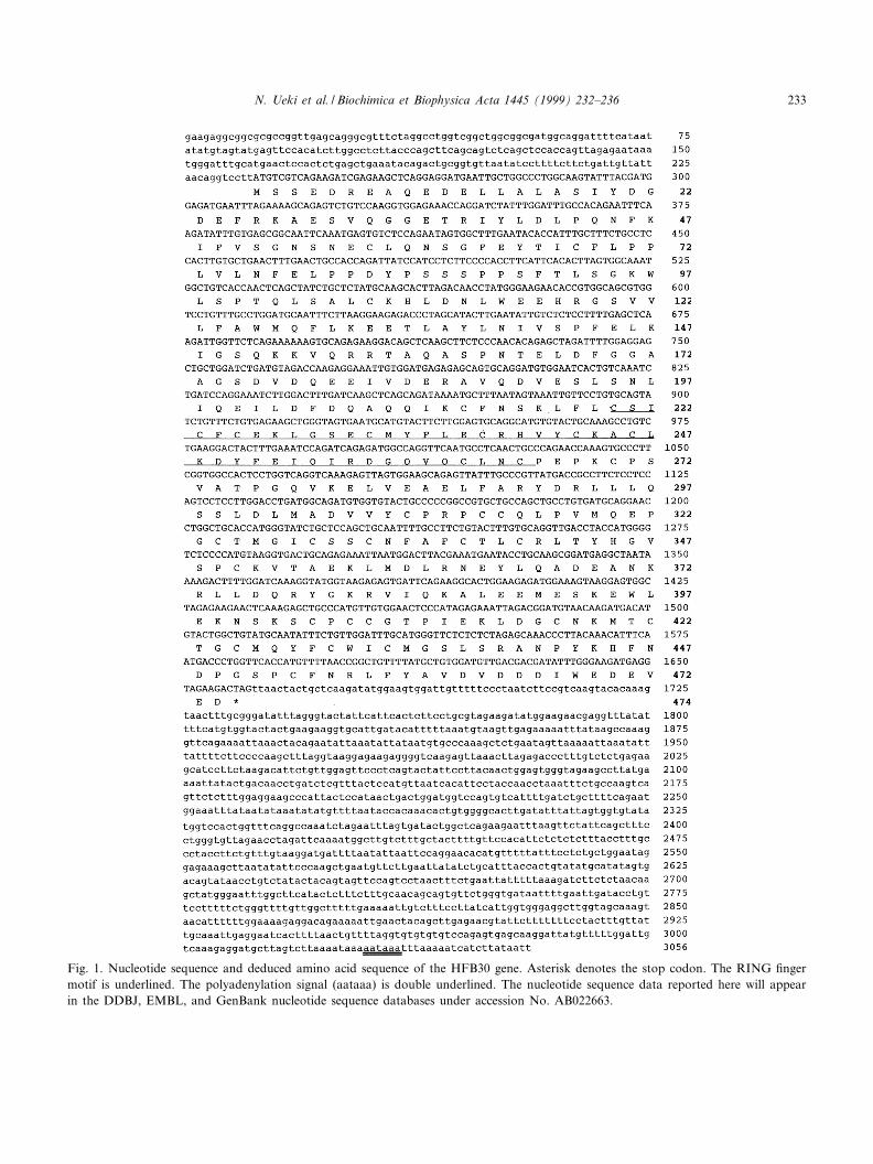
Fig. 1. Nucleotide sequence and deduced amino acid sequence of the HFB30 gene. Asterisk denotes the stop codon. The RING ¢ngermotif is underlined. The polyadenylation signal (aataaa) is double underlined. The nucleotide sequence data reported here will appearin the DDBJ, EMBL, and GenBank nucleotide sequence databases under accession No. AB022663.
BBAEXP 91260 27-4-99
N. Ueki et al. / Biochimica et Biophysica Acta 1445 (1999) 232^236 233

and the presumed initiation methionine was precededin the cDNA by an in-frame stop codon (Fig. 1). Acanonical polyadenylation signal, AATAAA, was lo-cated 22 bp upstream from the poly(A). The pre-dicted protein of 474 amino acids had a calculatedmolecular mass of approx. 53 kDa. The nucleotidesequence data reported here will appear in theDDBJ, EMBL, and GenBank nucleotide sequencedatabases under the accession No. AB022663. Thesequence of the cDNA clone revealed a highly con-served motif encompassing amino acid residues 220^265 (CSICFCEKLGSECMYFLECRHVYCLKAC-LKDYFEIQIRDGQVQCLNC) consistent with aRING ¢nger (C3HC4-type zinc ¢nger) domain. Asearch of the amino acid sequences in the databaserevealed that the C. elegans hypothetical protein,F56D2.6 (accession No. U13644), was highly homol-ogous to the HFB30 gene (28.8% overall identity atamino acid level).
We examined the tissue distribution of HFB30transcript in various tissues by reverse transcrip-tion-coupled polymerase chain reaction (RT-PCR)as described previously [10]. Primers used for RT-PCR correspond to the coding region of the gene(5P-CCATTTGCTTTCTGCCTCCAC-3P and 5P-TT-CCACTAACTCTTTGACCTG-3P). The primer setgave the longer PCR product from genomic DNAwhich was easily distinguished from the 656 bp prod-uct from the mRNA. The PCR product was gener-ated in all tissues examined indicating that the tran-script is ubiquitously expressed in a wide variety ofhuman tissues (Fig. 2). Therefore, HFB30 described
in the present study seems to be involved in a basichousekeeping function of cells.
We determined the chromosomal location of theHFB30 gene using a human/rodent somatic cell hy-brid panels (Mapping Panel #2, Coriell Cell Reposi-tories, Camden, NJ, USA) and a radiation hybridpanel (Genebrigde 4, Research Genetics, Huntsville,AL, USA) in the same manner as in previous reports[10^12]. Primers used for PCR ampli¢cation corre-spond to the 3P UTR region of the gene (5P-TCATT-
Fig. 2. Tissue distribution analysis using RT-PCR. The 12 tis-sues examined are indicated above each lane. The templates ofthe human tissues of poly(A)�RNAs were purchased fromClontech (Palo Alto, CA, USA). The cDNA templates for RT-PCR were synthesized from 2 Wg of poly(A)� using excessamounts of Superscript II reverse transcriptase (Gibco BRL)and random hexamer primers. PCR was carried out in a ¢nalvolume of 10 Wl containing 1ULA-PCR bu¡er (Takara, Kyoto,Japan), 2 WM each primer, 200 WM each dNTP, 1 Wl of tem-plate DNA and 0.01 units of LA-Taq DNA polymerase (Ta-kara). Temperature and time schedule were 30 cycles of 95³Cfor 20 s and 62³C for 1 min. PCR products were separated on2.5% Nusieve GTG agarose gel (FMC, Rockland, ME, USA)with a 1 kb ladder DNA marker (Gibco BRL).
Table 1Genomic structure of the HFB30 gene
Exon No. Exon sizea Splice acceptorb Splice donorb
1 56 GCTGGCGGCGgtgagtgggc
2 174 ttttctgcagATGGCAGGAT... ...TTATTAACAGgtactaactg
3 160 atgttttcagGTCCTTATGT... ...TTTGTGAGCGgttagttaat
4 152 tttggttcagGCAATTCAAA... ...ACCAACTCAGgttagacttg
5 528 tttctcctagTCTGCTCTAT... ...TCCTGGTCAGgtaaatgttt
6 229 ttccctccagGTCAAAGAGT... ...GTGACTGCAGgtatgtttta
7 173 cactttgtagAGAAATTAAT... ...TCCCATAGAGgtaaatgttt
8 131 ttcccaccagAAATTAGACG... ...GTTTTAACCGgtatgtatac
9 1453 atgcttccagGCTGTTTTAT
Intron-exon boundaries of the HFB30 gene. Intron-exon junctions were established by comparison of cDNA and genomic sequence.aSize in base pairs.bSequences at the splice junction. Exonic sequences are shown in capital letters while intronic sequences in lowercase letters. Invariantnucleotides (ag/gt) are in boldface type.
BBAEXP 91260 27-4-99
N. Ueki et al. / Biochimica et Biophysica Acta 1445 (1999) 232^236234

CACTCTTCCTGCGTAG-3P and 5P-ACTATTCA-GAGCTTTGGGCAC-3P) (182 bp PCR product).First, a speci¢c ampli¢ed product for human wasdetected only from the hybrid containing humanchromosome 5 (data not shown). Then, we per-formed further mapping analysis using a PCR-basedradiation hybrid panel with the same primers as usedin the assay for the human/rodent somatic cell hybridpanel. The radiation hybrid mapping data were proc-essed using the RHMAPPER software package(http://carbon.wi.mit.edu:8000/cgi-bin/contig/rhmap-per.pl). The data vector for the HFB30 gene was
0000010100 0000000010 0011110010 00010110000001011000 1000000000 0000000000 01000101000001000010 100 and the consequent statistical reportindicated that the gene was mapped to 3.87 cR distalfrom NIB1948 (lod s 3.0) which maps cytogeneti-cally to the 5q23.3-q31.1 region (Fig. 3).
Sequences of the exon-intron boundaries of thegene were identi¢ed by aligning the cDNA sequencewith a genomic sequence (accession No. AC005740).As shown in Table 1, all the splicing sites conformedto the AG-GT rule, in that there is always an AGand a GT dinucleotide at the splice acceptor and
Fig. 3. Chromosomal placement of the HFB30 gene at a relative distance to framework markers on the WICGR radiation hybridmap of the human genome (http://carbon.wi.mit.edu:8000/cgi-bin/contig/phys_map). The approximate corresponding cytogenetic loca-tion of the gene on the chromosome 5q23.3-q31.1 region is indicated. Distances are in centirays (cR) and in centimorgans (cM) fromthe top of the chromosome 5 linkage group.
BBAEXP 91260 27-4-99
N. Ueki et al. / Biochimica et Biophysica Acta 1445 (1999) 232^236 235

donor sites, respectively. The HFB30 gene is dividedinto nine exons, which range in size from 57 bp (exon1) to 1453 bp (exon 9). The third exon is 160 bp andit contains the putative ATG start codon. The lastexon of 1453 bp contained the TAG translation stopcodon followed by a 1395 bp 3P-untranslated region.Thus, the genome structure of HFB30 gene consist-ing of nine exons and spans approx. 20 kb of thegenome DNA.
Several members of the RING ¢nger protein fam-ily are implicated in human diseases. For example,the RING ¢nger protein BRCA1 is a tumor suppres-sor in an early onset breast cancer [13], anothermember, PML, produces a fusion protein with theretinoic acid receptor A in acute promyelocytic leu-kemia [14^17] and the human 52 kDa SS-A/RoRING ¢nger protein is the nuclear antigen in Sjo«g-ren's autoimmune disease [18,19]. The chromosomalposition, genomic structure, and expression pro¢le ofsuch a gene might contribute toward ongoing posi-tional candidate approaches for disease genes linkedto this genomic locus.
References
[1] J.W. Schwabe, A. Klug, Zinc mining for protein domains,Nat. Struct. Biol. 1 (1994) 345^349.
[2] J.P. Mackay, M. Crossley, Zinc ¢nger interaction, TrendsBiochem. Sci. 23 (1998) 1^4.
[3] R.L. Tatusov, E.V. Koonin, D.J. Lipman, A genomic per-spective on protein families, Science 278 (1997) 631^637.
[4] D. Neil, N.D. Clarke, J.M. Berg, Zinc ¢ngers in Caenorhab-ditis elegans : ¢nding families and probing pathways, Science282 (1998) 2018^2022.
[5] A.J. Saurin, K.L. Borden, M.N. Boddy, P.S. Freemont,Does this have a familiar RING?, Trends Biochem. Sci. 21(1996) 208^214.
[6] P.S. Freemont, I.M. Hanson, J. Trowsdale, A novel cystein-rich sequence motif, Cell 64 (1991) 483^484.
[7] P.N. Barlow, B. Luisi, A. Milner, M. Elliott, R. Everett,Structure of the C3HC4 domain by 1H-nuclear magneticresonance spectroscopy. A new structural class of zinc-¢nger,J. Mol. Biol. 237 (1994) 201^211.
[8] K.L. Borden, M.N. Boddy, J. Lally, N.J. O'Reilly, S. Mar-tin, K. Howe, E. Solomon, P.S. Freemont, The solution
structure of the RING ¢nger domain from the acute pro-myelocytic leukaemia proto-oncoprotein PML, EMBO J.114 (1995) 1532^1541.
[9] N. Ueki, T. Oda, M. Kondo, K. Yano, T. Noguchi, M.Muramatsu, Selection system for genes encoding nuclear-tar-geted proteins, Nat. Biotechnol. 16 (1998) 1338^1342.
[10] N. Seki, A. Hattori, A. Hayashi, S. Kozuma, M. Ohira, T.Hori, T. Saito, Structure, expression pro¢le and chromoso-mal location of an isolog of DNA-Pkcs interacting protein(KIP) gene, Biochim. Biophys. Acta (1999) in press.
[11] N. Seki, Y. Nimura, M. Ohira, T. Saito, S. Ichimiya, N.Nomura, A. Nakagawara, Identi¢cation and chromosomeassignment of a human gene encoding a novel phosphatidyl-inositol-3 kinase, DNA Res. 4 (1997) 355^358.
[12] T. Saito, N. Seki, H. Ishii, M. Ohira, A. Hayashi, S. Kozu-ma, T. Hori, Complementary DNA cloning and chromoso-mal mapping of a novel phosphatidylinositol kinase gene,DNA Res. 4 (1997) 301^305.
[13] Y. Miki, J. Swensen, D. Shattuck-Eidens, P.A. Futreal, K.Harshman, S. Tavtigian, Q. Liu, C. Cochran, L.M. Bennett,W. Ding et al., A strong candidate for the breast and ovar-ian cancer susceptibility gene BRCA1, Science 266 (1994)66^71.
[14] A.D. Goddard, J. Borrow, P.S. Freemont, E. Solomon,Characterization of a zinc ¢nger gene disrupted by thet(15;17) in acute promyelocytic leukemia, Science 254(1991) 1371^1374.
[15] A. Kakizuka, W.H. Miller Jr., K. Umesono, R.P. WarrellJr., S.R. Frankel, V.V. Murty, E. Dmitrovsky, R.M. Evans,Chromosomal translocation t(15;17) in human acute pro-myelocytic leukemia fuses RAR alpha with a novel putativetranscription factor, PML, Cell 66 (1991) 663^674.
[16] H. de The, C. Lavau, A. Marchio, C. Chomienne, L. Degos,A. Dejean, The PML-RAR alpha fusion mRNA generatedby the t(15;17) translocation in acute promyelocytic leuke-mia encodes a functionally altered RAR, Cell 66 (1991) 675^684.
[17] P. Kastner, A. Perez, Y. Lutz, C. Rochette-Egly, M.P.Gaub, B. Durand, M. Lanotte, R. Berger, P. Chambon,Structure, localization and transcriptional properties of twoclasses of retinoic acid receptor alpha fusion proteins inacute promyelocytic leukemia (APL): structural similaritieswith a new family of oncoproteins, EMBO J. 11 (1992) 629^642.
[18] E.K. Chan, J.C. Hamel, J.P. Buyon, E.M. Tan, Molecularde¢nition and sequence motifs of the 52-kD component ofhuman SS-A/Ro autoantigen, J. Clin. Invest. 87 (1991) 68^76.
[19] K. Itoh, Y. Itoh, M.B. Frank, Protein heterogeneity in hu-man Ro/SSA ribonucleoproteins, J. Clin. Invest. 87 (1991)117^186.
BBAEXP 91260 27-4-99
N. Ueki et al. / Biochimica et Biophysica Acta 1445 (1999) 232^236236