IRIDOCYCLITIS Dr. Pranav Bhagwat. Reader, Dept. of Shalakya Tantra. Gomantak Ayurveda College,...
59
IRIDOCYCLITIS Dr. Pranav Bhagwat. Reader, Dept. of Shalakya Tantra. Gomantak Ayurveda College, Shiroda, Goa.
-
Upload
mark-moody -
Category
Documents
-
view
243 -
download
3
Transcript of IRIDOCYCLITIS Dr. Pranav Bhagwat. Reader, Dept. of Shalakya Tantra. Gomantak Ayurveda College,...
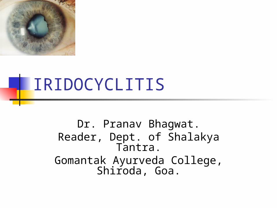
IRIDOCYCLITIS
Dr. Pranav Bhagwat.Reader, Dept. of Shalakya Tantra.
Gomantak Ayurveda College, Shiroda, Goa.
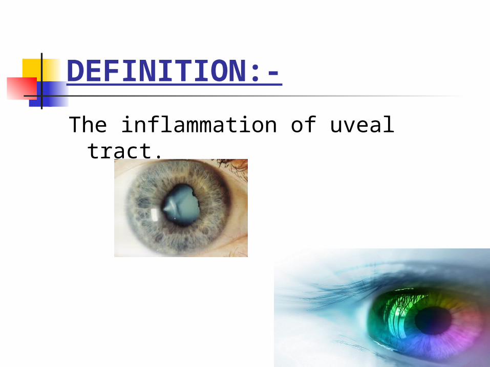
DEFINITION:-
The inflammation of uveal tract.

Classification-
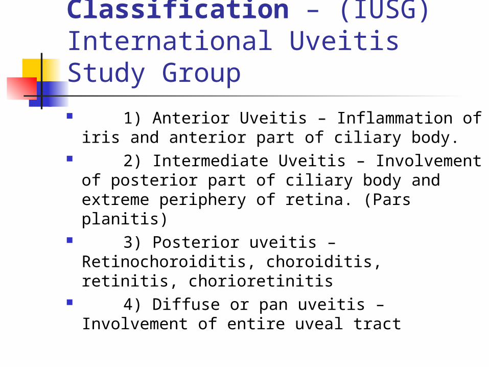
A. Anatomical Classification – (IUSG) International Uveitis Study Group
1) Anterior Uveitis – Inflammation of iris and anterior part of ciliary body.
2) Intermediate Uveitis – Involvement of posterior part of ciliary body and extreme periphery of retina. (Pars planitis)
3) Posterior uveitis – Retinochoroiditis, choroiditis, retinitis, chorioretinitis
4) Diffuse or pan uveitis – Involvement of entire uveal tract
B. Clinical Classification -
1) Acute – sudden symptomatic onset. Persists for 6 weeks or less.
2) Chronic – Frequently insidious and asymptomatic. Persists for months or years.
C. Etiological Classification
One of the most difficult problems in ophthalmology.
In most of the cases, probably, allergy is the cause. 1) Exogenous-
introduction of organism into the eye through a perforating wound or ulcer. acute iridocyclitis of suppurative type, pan-ophthalmitis.
2) Secondary infection- Due to direct spread from adjoining structures-
Cornea Sclera Retina
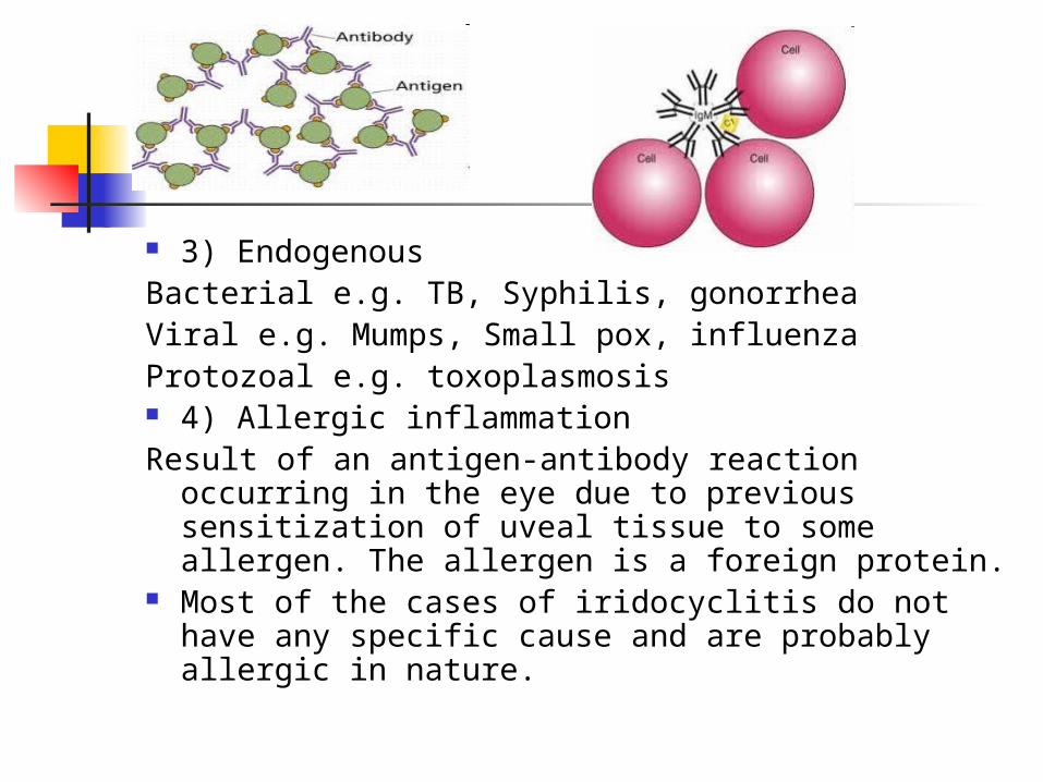
3) EndogenousBacterial e.g. TB, Syphilis, gonorrheaViral e.g. Mumps, Small pox, influenzaProtozoal e.g. toxoplasmosis 4) Allergic inflammationResult of an antigen-antibody reaction occurring in
the eye due to previous sensitization of uveal tissue to some allergen. The allergen is a foreign protein.
Most of the cases of iridocyclitis do not have any specific cause and are probably allergic in nature.

5)Auto-immune/Constitutional-a)Immune disorders affecting the body as a whole
have ocular manifestations in the form of iridocyclitis.
e.g. rheumatoid arthritis, SLE, ankylosing spondylitis, Reiter’s syndrome, Behcet’s Syndrome.
b) response to antigenic stimuli in other part of the eye. Iridocyclitis is a common accompaniment of severe corneal infection and choroiditis of retinal inflammation.
HLA antigenic involvement Disproportionately high percentage of patients of
B-27 antigenic group develop acute anterior uveitis.
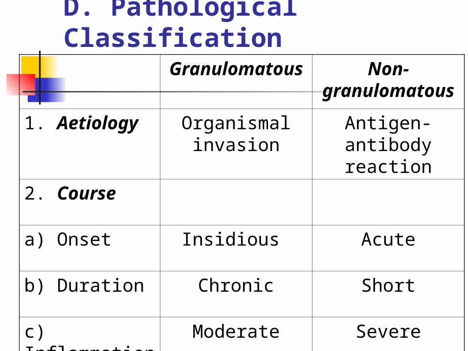
Granulomatous
Non-granulomato
us
1. Aetiology Organismal invasion
Antigen-antibody reaction
2. Course
a) Onset Insidious Acute
b) Duration Chronic Short
c) Inflammation
Moderate Severe
D. Pathological Classification

Granulomatous
Non-granulomato
us
3. Pathology
a) Lesion Circumscribed Diffuse
b) Iris Focal reaction Diffuse reaction
c) Keratic precipitates
Mutton fat Fine plenty
d) Iris adhesions
Coarse, few, thick
Fine, plenty, thin
4. Investigations
May be positive
Negative

PATHOLOGY AND CLINICAL SIGNS-
Inflammation of iris and ciliary body
Dilatation of blood vessels
Iris stromal edema.SIGNS - Iris pattern altered.Iris
colour altered. Iris thickened.Also accompanied by, ciliary congestion, conjunctival hyperaemia and chemosis of conjunctiva.

Exudation of fibrin-rich fluid and inflammatory cells in the tissues
Exudates escape into anterior chamber
Plasmoid aqueous SIGNS - Aqueous flare (like the
beam of projector in smokey theatre)
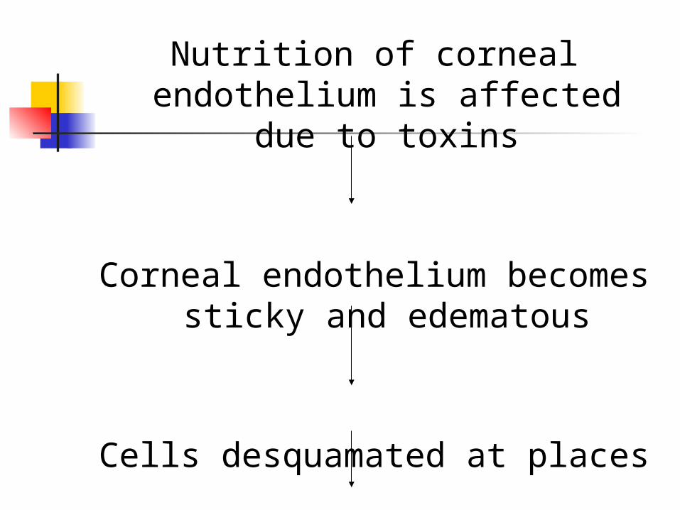
Nutrition of corneal endothelium is affected due to toxins
Corneal endothelium becomes sticky and edematous
Cells desquamated at places
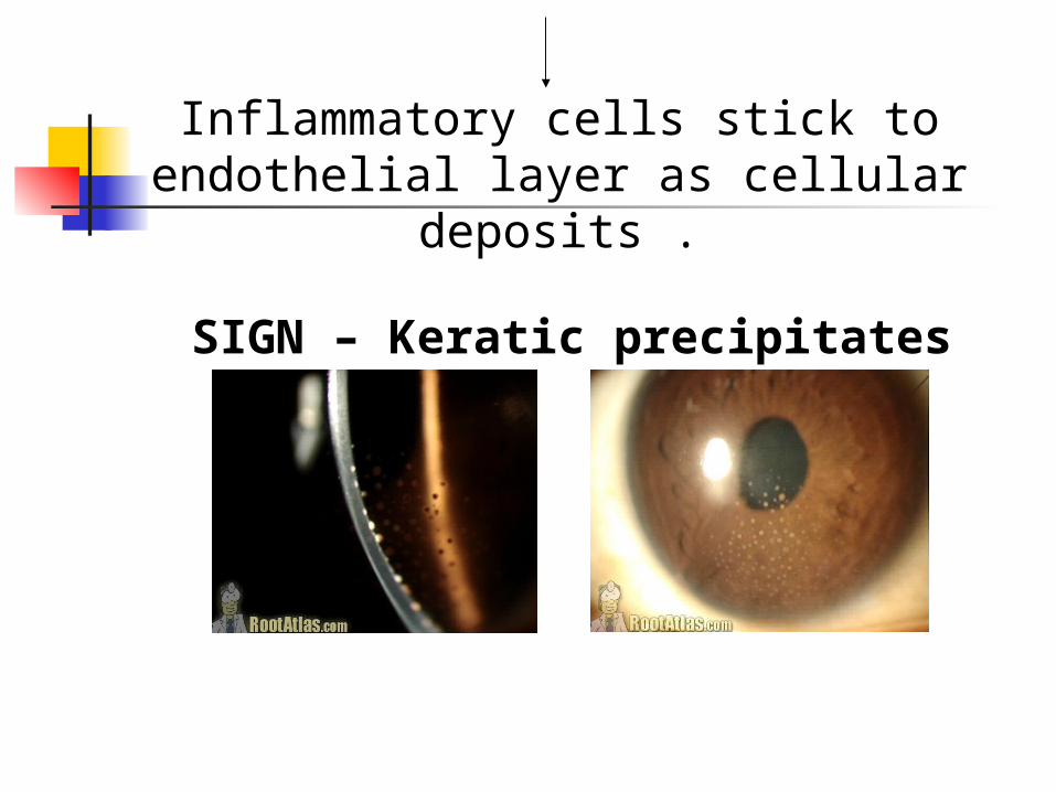
SIGN – Keratic precipitates
Inflammatory cells stick to endothelial layer as cellular deposits .
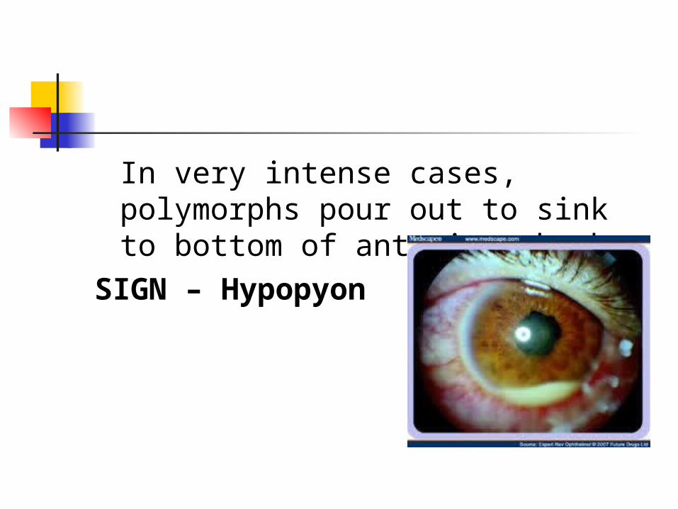
In very intense cases, polymorphs pour out to sink to bottom of anterior chamber
SIGN – Hypopyon
Exudates cover the iris as a thin film and spread over pupillary area
SIGN – Irritation of iris musculature constrictor being more powerful
than dilator, spasm results in miosis.
If exudate is profuse SIGN – Plastic iritis
Blockage of pupilSIGN – impairment of sight.
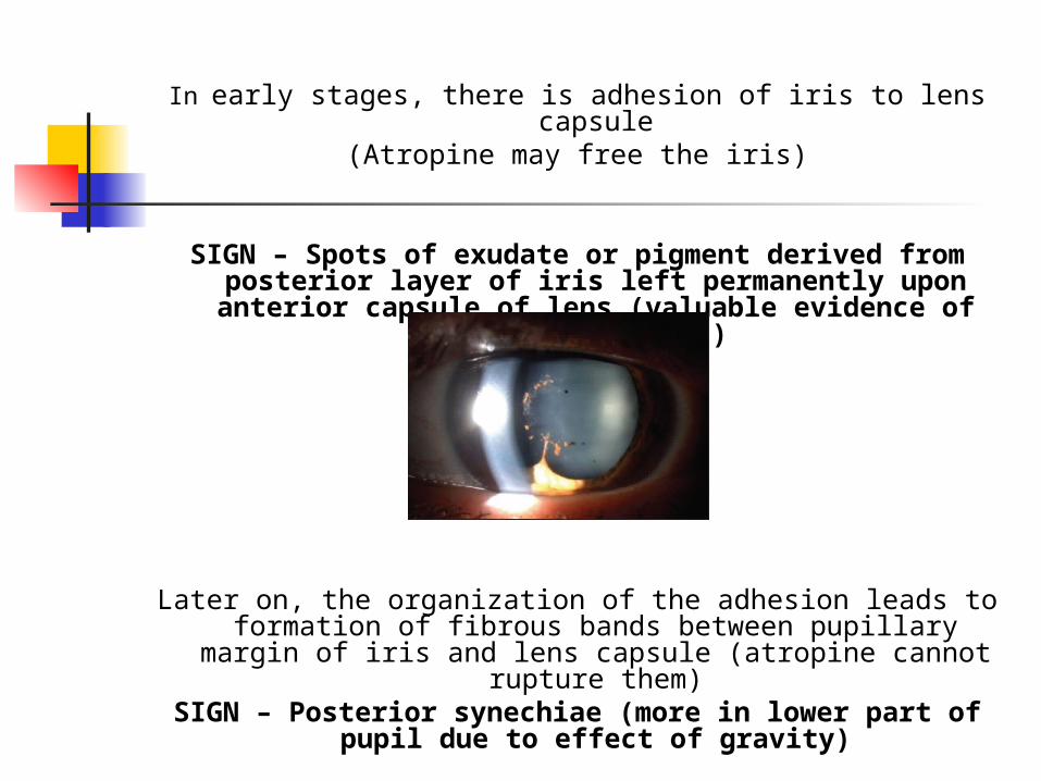
In early stages, there is adhesion of iris to lens capsule(Atropine may free the iris)
SIGN – Spots of exudate or pigment derived from posterior layer of iris left permanently upon
anterior capsule of lens (valuable evidence of previous iritis)
Later on, the organization of the adhesion leads to formation of fibrous bands between pupillary margin of
iris and lens capsule (atropine cannot rupture them)SIGN – Posterior synechiae (more in lower part of
pupil due to effect of gravity)
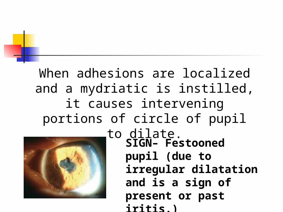
When adhesions are localized and a mydriatic is instilled, it causes intervening portions of circle of
pupil to dilate.
SIGN– Festooned pupil (due to irregular dilatation and is a sign of present or past iritis.)
Pigment epithelium on posterior surface is pulled around pupillary
margin so that patches of pigment on anterior surface of iris are seen.
SIGN – Ectropion of uveal pigment (due to contraction of organizing exudates upon iris)
With recurrent attacks or severe cases, the whole circle of pupillary margin gets tied to lens capsule.
SIGNS – Annular or ring synechiae or Seclusio pupillae
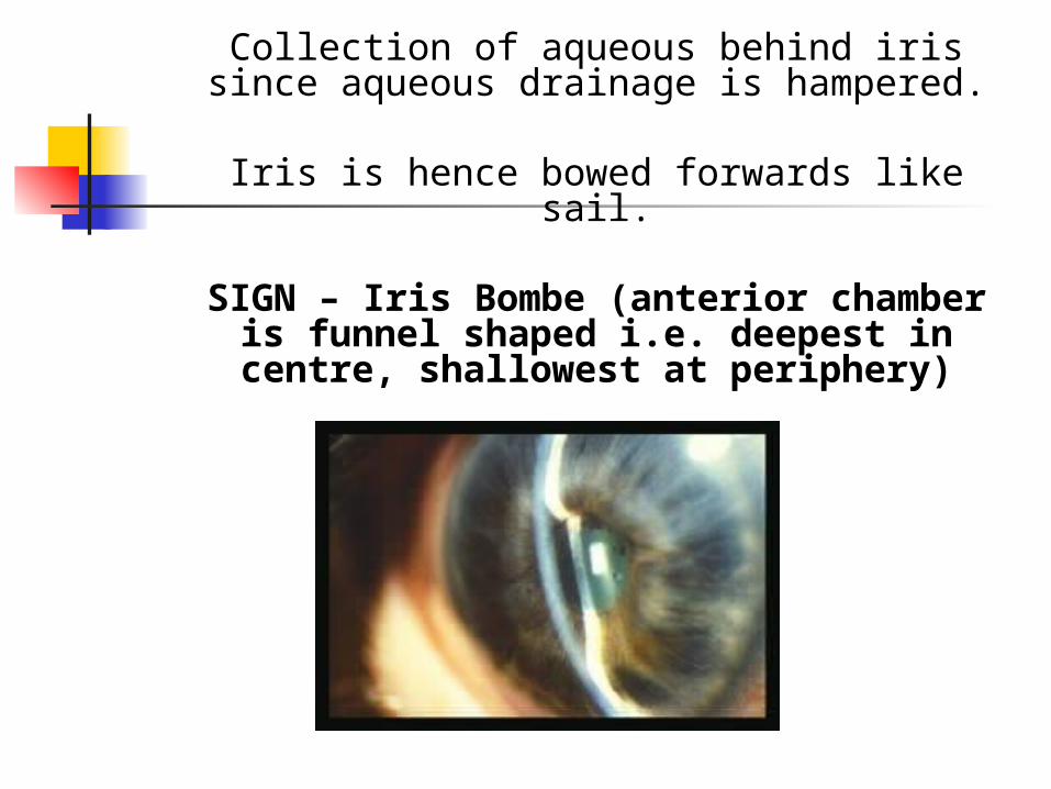
Collection of aqueous behind iris since aqueous drainage is hampered.
Iris is hence bowed forwards like sail.
SIGN – Iris Bombe (anterior chamber is funnel shaped i.e. deepest in centre, shallowest at periphery)
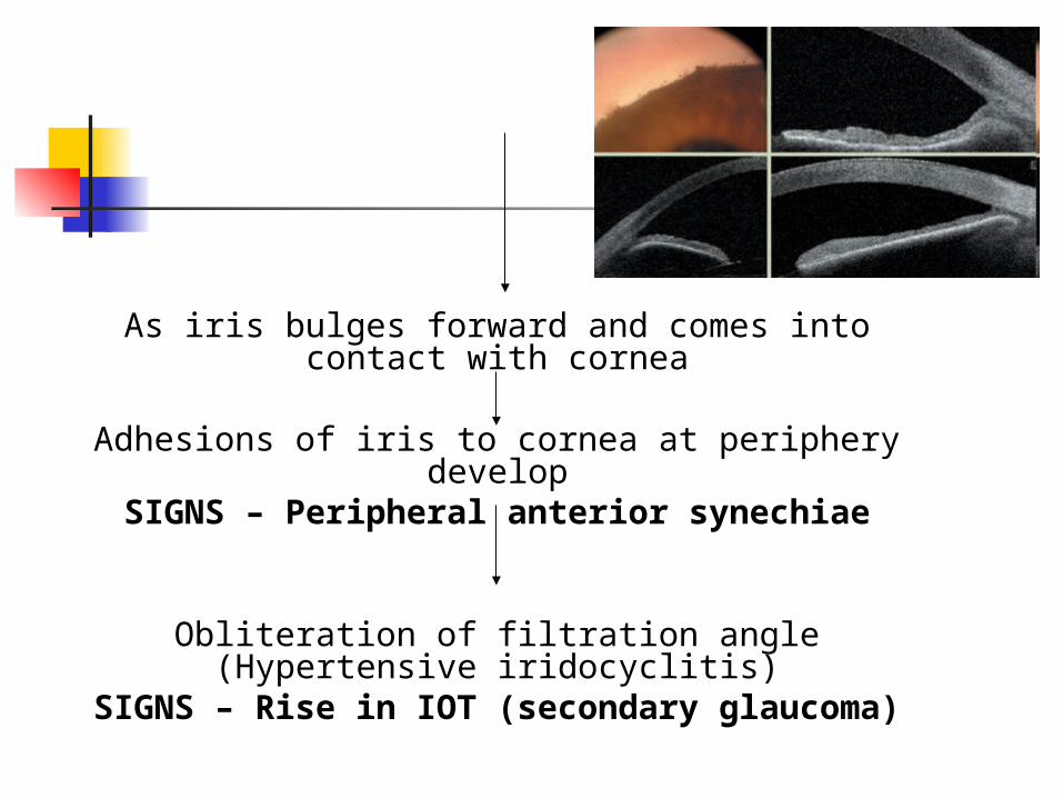
As iris bulges forward and comes into contact with cornea
Adhesions of iris to cornea at periphery develop
SIGNS – Peripheral anterior synechiae
Obliteration of filtration angle (Hypertensive iridocyclitis)
SIGNS – Rise in IOT (secondary glaucoma)
When exudate is more extensive
Organization of exudate across entire pupillary area
Film of opaque fibrous tissue in pupillary area
SIGNS – Occlusio pupillae or Blocked pupil
Exudates fill up posterior chamber if there is much of cyclitis
When these adhesions organize, the iris adheres to lens capsule.
SIGNS – Total posterior synechiae
When these adhesions organize, the iris adheres to lens capsule.
SIGNS – Total posterior synechiae
Retraction of peripheral part of iris
Anterior chamber is abnormally deep at periphery
In worst cases of plastic iridocyclitis
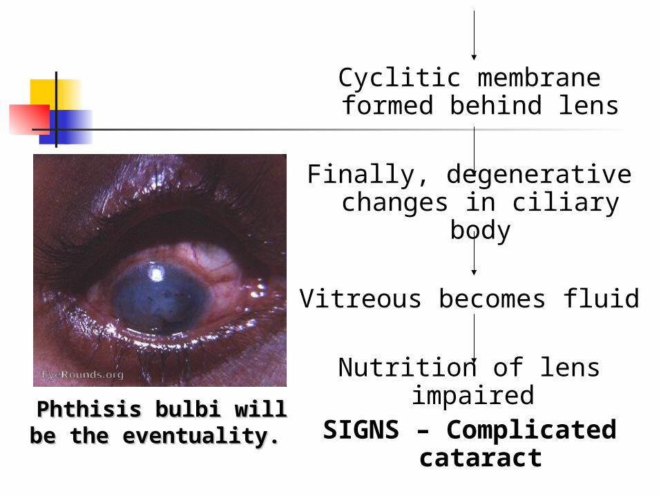
Cyclitic membrane formed behind lens
Finally, degenerative changes in ciliary
body
Vitreous becomes fluid
Nutrition of lens impaired
SIGNS – Complicated cataract
Phthisis bulbi will be the Phthisis bulbi will be the eventuality. eventuality.
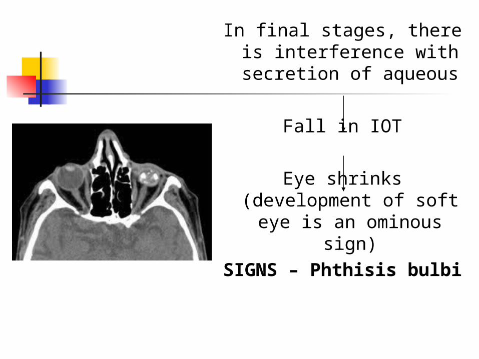
In final stages, there is interference with
secretion of aqueous
Fall in IOT
Eye shrinks (development of soft
eye is an ominous sign)
SIGNS – Phthisis bulbi
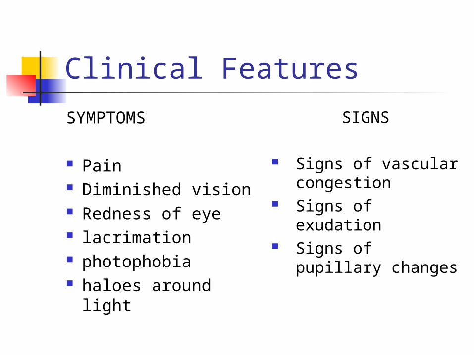
Clinical Features
SYMPTOMS
Pain Diminished vision Redness of eye lacrimation photophobia haloes around light
SIGNS
Signs of vascular congestion
Signs of exudation
Signs of pupillary changes
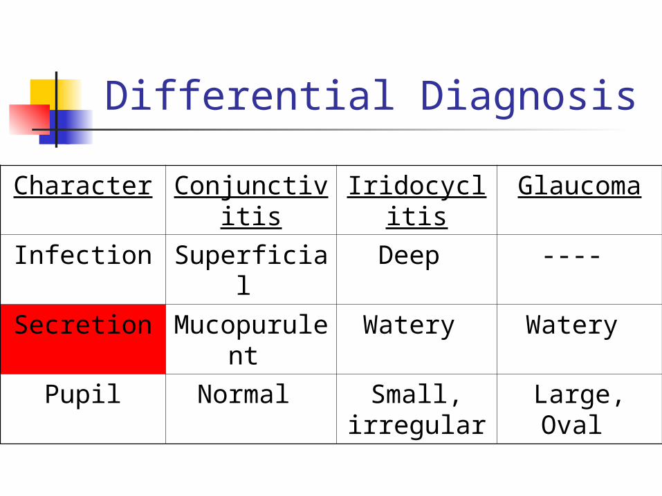
Differential Diagnosis
Character Conjunctivitis
Iridocyclitis Glaucoma
Infection Superficial Deep ----
Secretion Mucopurulent
Watery Watery
Pupil Normal Small, irregular
Large, Oval
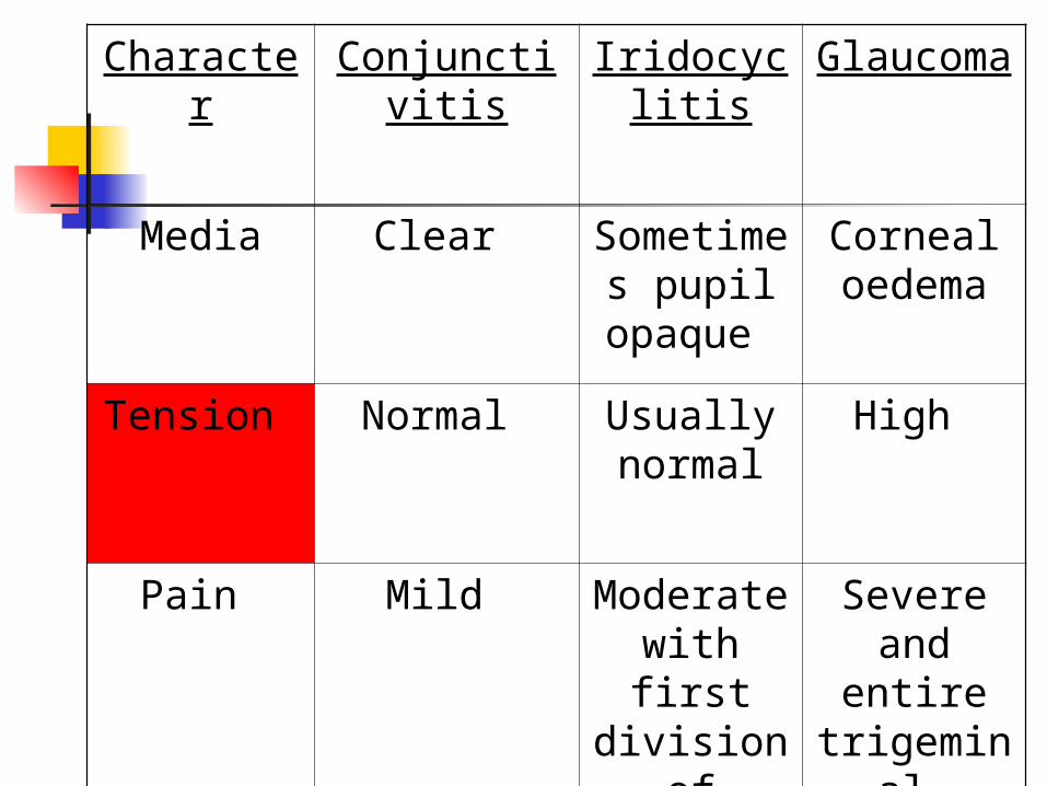
Character Conjunctivitis
Iridocyclitis
Glaucoma
Media Clear Sometimes pupil opaque
Corneal oedema
Tension Normal Usually normal
High
Pain Mild Moderate with first division
of trigemina
l
Severe and
entire trigemina
l
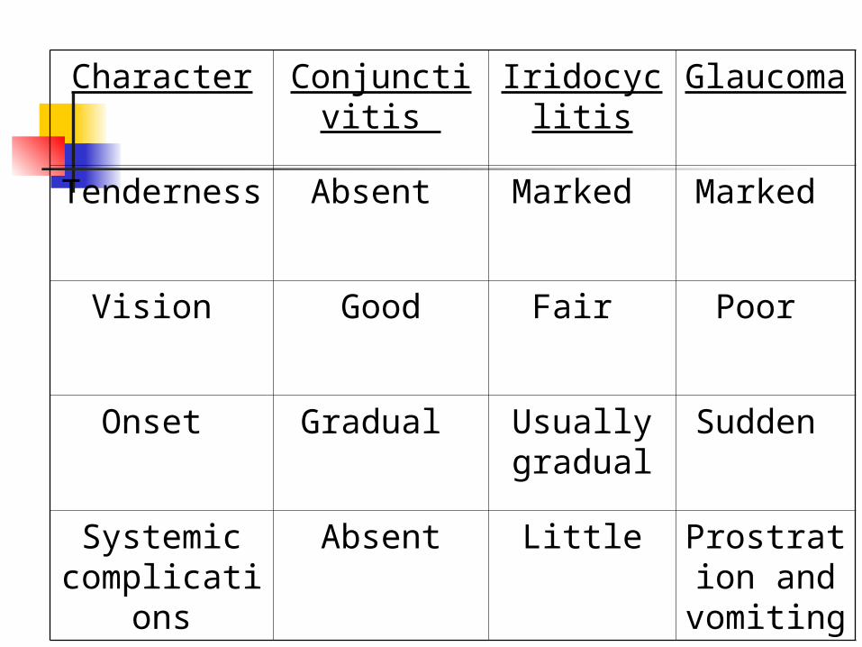
Character Conjunctivitis
Iridocyclitis
Glaucoma
Tenderness Absent Marked Marked
Vision Good Fair Poor
Onset Gradual Usually gradual
Sudden
Systemic complicatio
ns
Absent Little Prostration and
vomiting
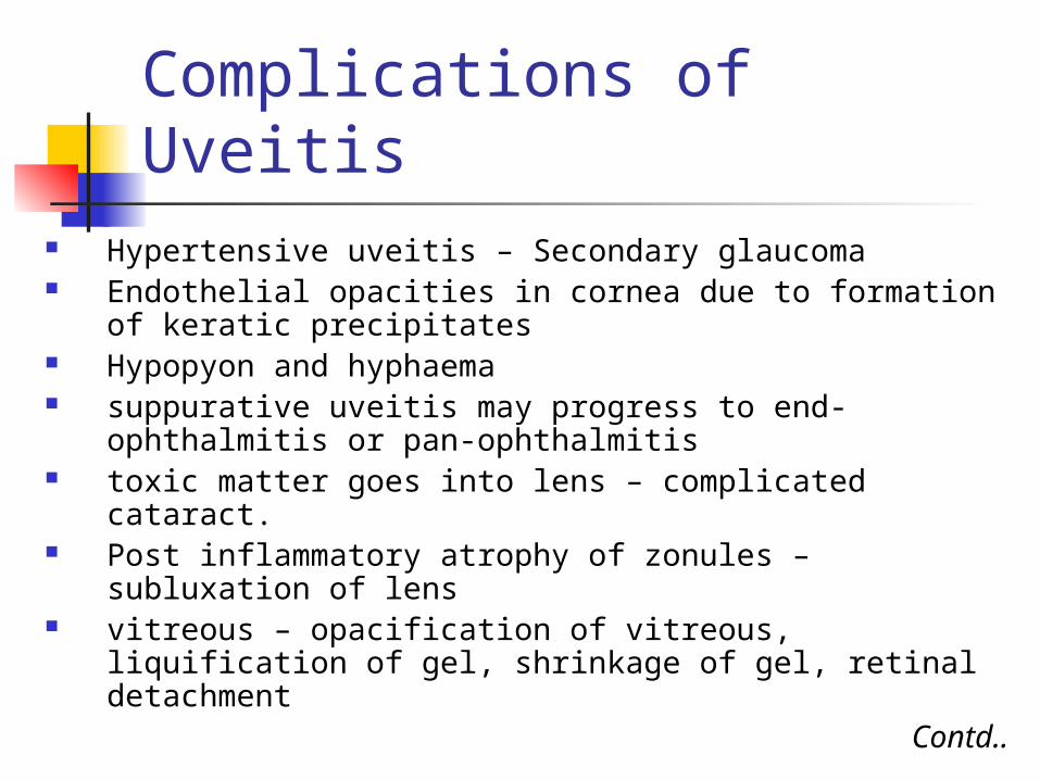
Complications of Uveitis Hypertensive uveitis – Secondary glaucoma Endothelial opacities in cornea due to formation of
keratic precipitates Hypopyon and hyphaema suppurative uveitis may progress to end-
ophthalmitis or pan-ophthalmitis toxic matter goes into lens – complicated cataract. Post inflammatory atrophy of zonules – subluxation
of lens vitreous – opacification of vitreous, liquification of
gel, shrinkage of gel, retinal detachmentContd..
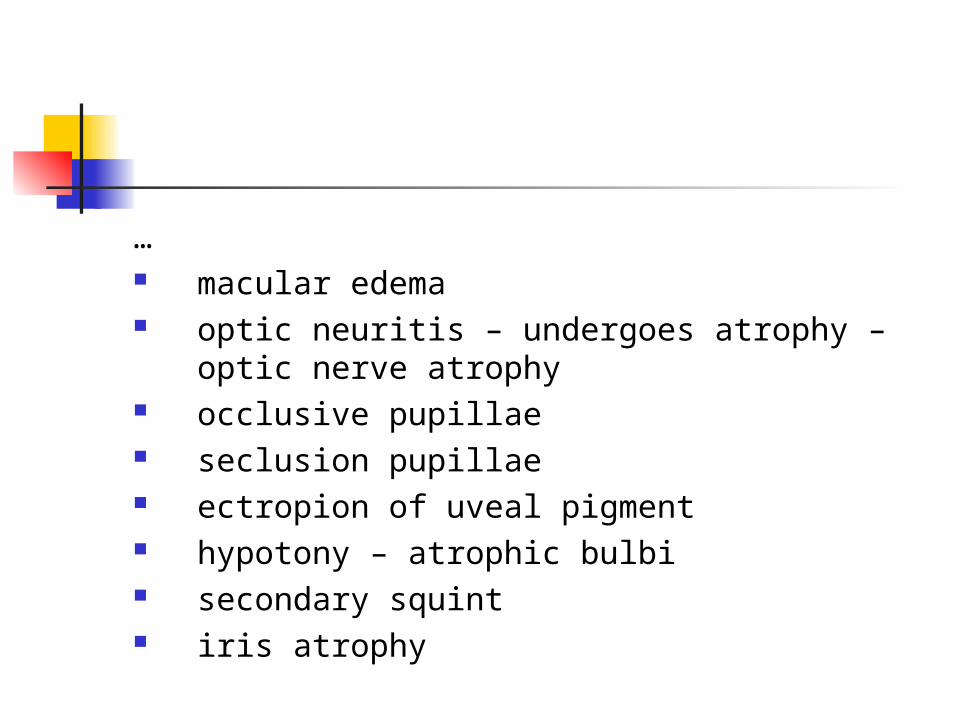
… macular edema optic neuritis – undergoes atrophy – optic
nerve atrophy occlusive pupillae seclusion pupillae ectropion of uveal pigment hypotony – atrophic bulbi secondary squint iris atrophy
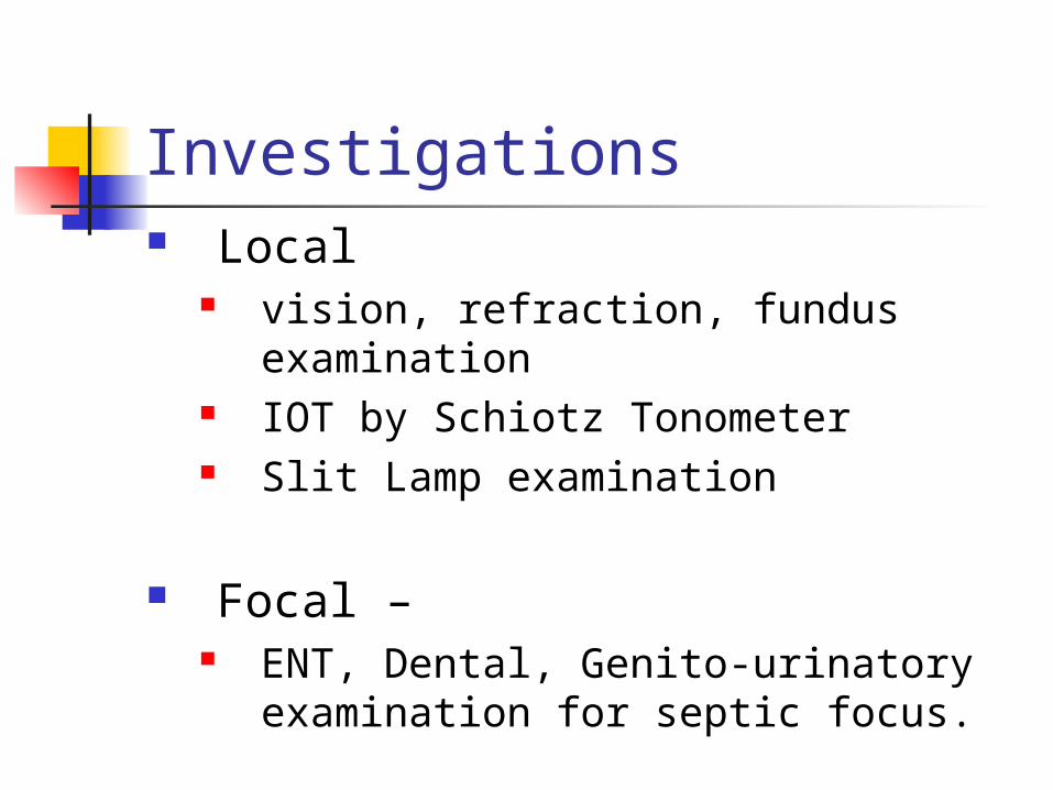
Investigations Local
vision, refraction, fundus examination
IOT by Schiotz Tonometer Slit Lamp examination
Focal – ENT, Dental, Genito-urinatory
examination for septic focus.
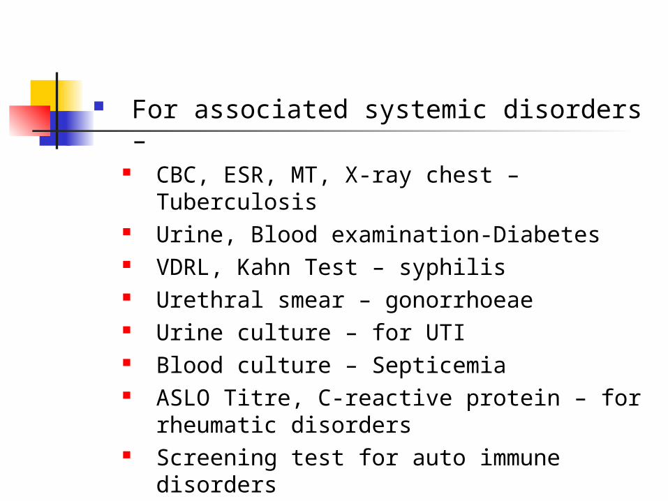
For associated systemic disorders – CBC, ESR, MT, X-ray chest –
Tuberculosis Urine, Blood examination-Diabetes VDRL, Kahn Test – syphilis Urethral smear – gonorrhoeae Urine culture – for UTI Blood culture – Septicemia ASLO Titre, C-reactive protein – for
rheumatic disorders Screening test for auto immune
disorders
Treatment
1. of iridocyclitis2. of complications and sequelae.
Treatment of Iridocyclitis
Drugs used – Mydriatics Steroids Cytotoxic agents Cyclosporin
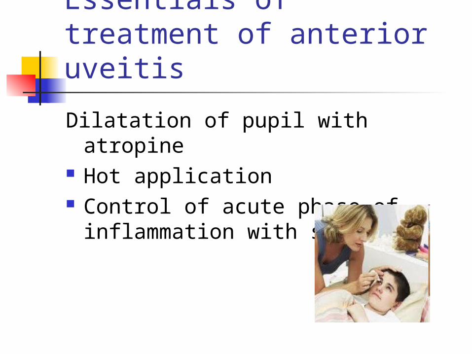
Essentials of treatment of anterior uveitis
Dilatation of pupil with atropine Hot application Control of acute phase of
inflammation with steroids
Atropine
Acts in 3 ways by keeping the iris and ciliary body
at rest by diminishing hyperaemia by preventing formation of posterior
synechiae and breaking down any already formed.

Method of administration and dose: Atropine may be used in form of drops
or ointment (1%) ,every four hours is usually sufficient.
When pupil is well dilated, twice a day suffices.
If atropine irritation ensues, one or the other substitutes for this drug may be used.
e.g. Homatropine, Cyclopentolate.
Mydriasis -the sub-conjunctival injection of 0.3 ml. of mydricaine, a mixture of atropine, procaine and adrenaline.
To avoid relapse-Atropine, or its equivalent -continued for at least 10 days to a fortnight after the eye appears to be quiet.
Hot application extremely soothing to patient by
diminishing the pain. of therapeutic service in increasing
the circulation.

Corticosteroids Administered as drops or ointment, or
more effectively as subconjunctival injections are of great value in controlling the inflammation in the acute phase.
Occasionally, results are dramatic and eye becomes white with great rapidity.
Minimize damages of antigen antibody reaction.
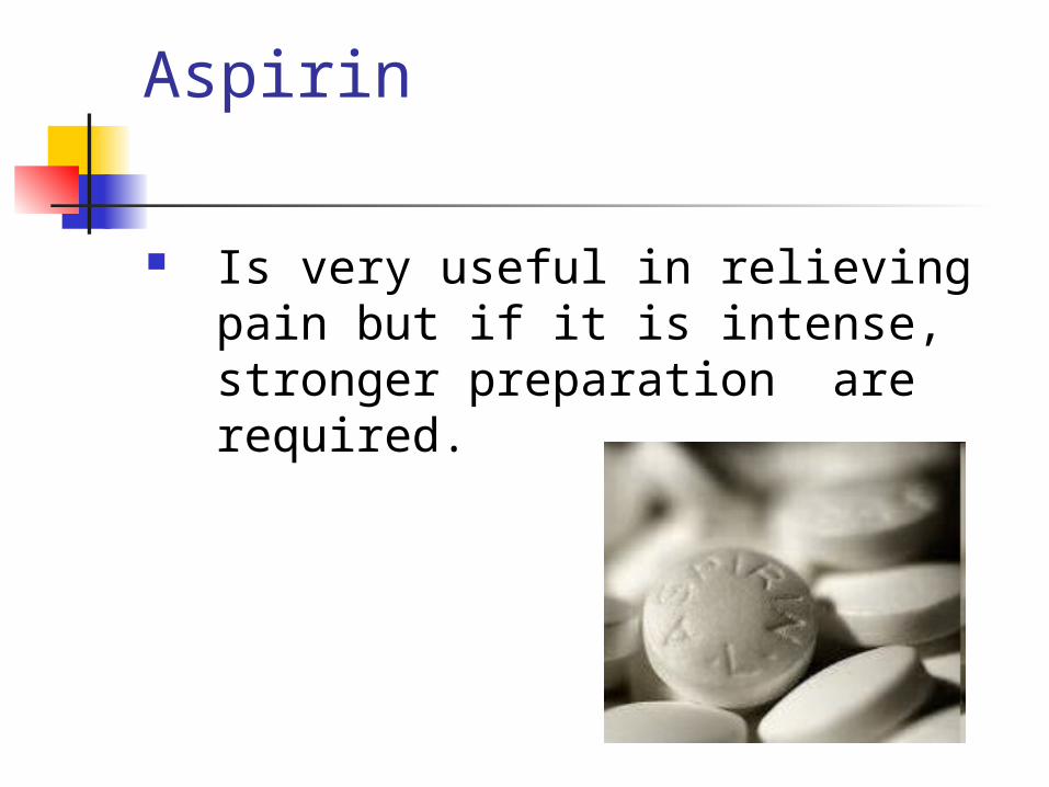
Aspirin
Is very useful in relieving pain but if it is intense, stronger preparation are required.
Cytotoxic drugs in Behcet’s disease Sympathetic uvitis Intermediate uveitis Juvenile chronic arthritis
Cyclosporin -T-cell immunosuppressive drug. Used
in resistant cases. Broad spectrum antibiotic - In case of suppurative uveitis. Specific Chemotherapy for
Tuberculosis, syphilis, gonorrhoea. Increasing body resistance by multi-
vitamins.
Treatment of complications and sequelae-
Secondary glaucoma- Before formation of posterior or
peripheral synechiae,- intensify atropinisation in order to allay the inflammatory congestion.
Corticosteroids - topically and acetazolamide - systematically are very useful in such cases..
Annular synechiae- Iridectomy ‘ ( No operative procedure of this kind must be
undertaken during an acute attack of iritis if it can be avoided. Reason – operation will set up a traumatic iritis which will result in the opening getting filled with exudates.)
preventive iridectomy- Since ring synechiae is the result of recurrent attacks, iridectomy can be performed during quiescent interval.
Difficulty – iris is atrophied, friable. Haemorrhage is common. Synechiae can be broken with YAG Laser.
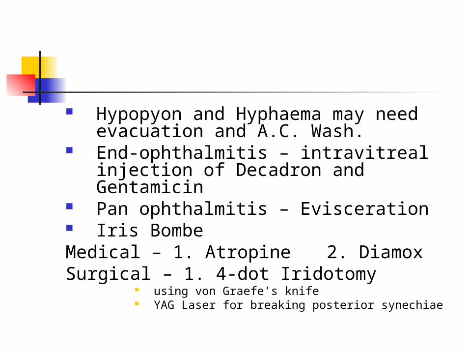
Hypopyon and Hyphaema may need evacuation and A.C. Wash.
End-ophthalmitis – intravitreal injection of Decadron and Gentamicin
Pan ophthalmitis – Evisceration Iris BombeMedical – 1. Atropine 2. DiamoxSurgical – 1. 4-dot Iridotomy
using von Graefe’s knife YAG Laser for breaking posterior synechiae

Ayurvedeeya approach
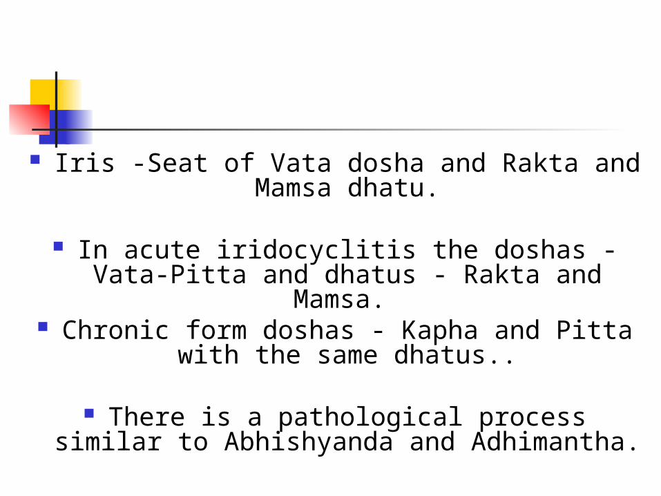
Iris -Seat of Vata dosha and Rakta and Mamsa dhatu.
In acute iridocyclitis the doshas - Vata-Pitta and dhatus - Rakta and Mamsa.
Chronic form doshas - Kapha and Pitta with the same dhatus..
There is a pathological process similar to Abhishyanda and Adhimantha.
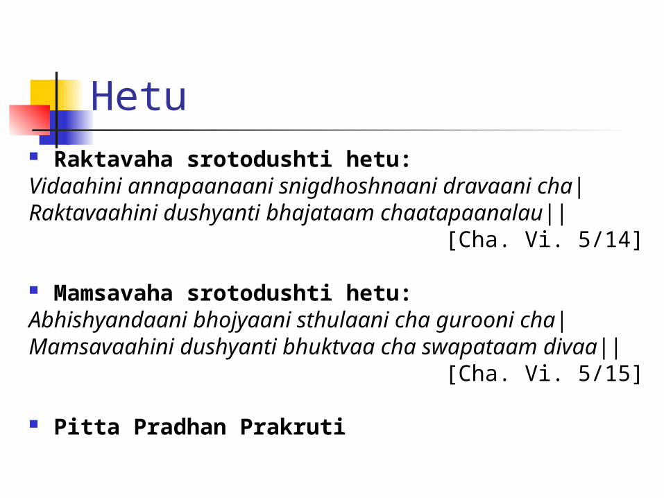
Hetu Raktavaha srotodushti hetu:Vidaahini annapaanaani snigdhoshnaani dravaani cha|Raktavaahini dushyanti bhajataam chaatapaanalau||
[Cha. Vi. 5/14]
Mamsavaha srotodushti hetu: Abhishyandaani bhojyaani sthulaani cha gurooni cha|Mamsavaahini dushyanti bhuktvaa cha swapataam
divaa||[Cha. Vi. 5/15]
Pitta Pradhan Prakruti
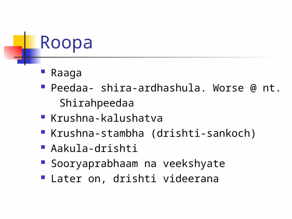
Roopa Raaga Peedaa- shira-ardhashula. Worse @ nt. Shirahpeedaa Krushna-kalushatva Krushna-stambha (drishti-sankoch) Aakula-drishti Sooryaprabhaam na veekshyate Later on, drishti videerana

Samprapti
Hetusevan
Kapha-Pitta, Rakta-Mamsa dushti
Doshaanaam Urdhwagamanam
Netraanusaari Siraasu Praveshaah
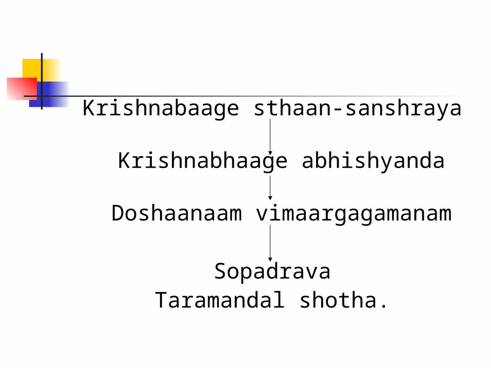
Krishnabaage sthaan-sanshraya
Krishnabhaage abhishyanda
Doshaanaam vimaargagamanam
SopadravaTaramandal shotha.
Treatment
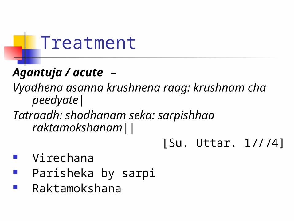
Agantuja / acute –Vyadhena asanna krushnena raag: krushnam
cha peedyate|Tatraadh: shodhanam seka: sarpishhaa
raktamokshanam||[Su. Uttar. 17/74]
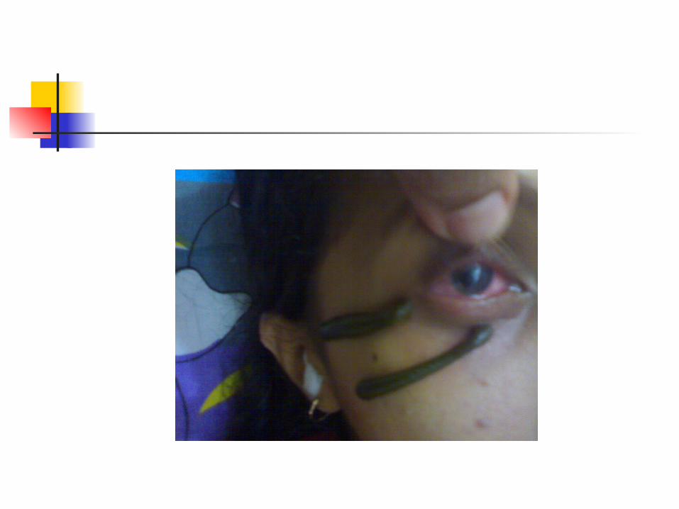
Virechana Parisheka by sarpi Raktamokshana
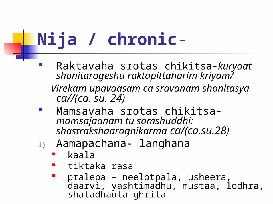
Nija / chronic- Raktavaha srotas chikitsa-kuryaat
shonitarogeshu raktapittaharim kriyam/ Virekam upavaasam ca sravanam
shonitasya ca//(ca. su. 24) Mamsavaha srotas chikitsa-
mamsajaanam tu samshuddhi: shastrakshaaragnikarma ca/(ca.su.28)
1) Aamapachana- langhana kaala tiktaka rasa pralepa – neelotpala, usheera, daarvi,
yashtimadhu, mustaa, lodhra, shatadhauta ghrita
2) Shodhana
adhashodhana raktamokshana anjanam- raktabhishyanda (Patlyadi
anjana) putapaka- same as above nasyam- shodhana followed by
shamana.
3) rakta-mamsa bala vardhaka – sariva, loha, abhraka, ashwagandha. Kapha-pittahara
Nasyam- to reduce abhisyandaa in shiras.
To reduce saamata in Rakta and Mamsa dhatu, required medications like Manjishtha,darvi, musta, Patola should be used.
Treatment of aamavaata- Langhana, tiktaka rasa, deepana drugs, swedanam help to reduce abhishyanda and hence useful in this context.
This might be the appropriate treatment for Taramandal shotha.

THANK YOU!THANK YOU!--Dr. Pranav Bhagwat.Dr. Pranav Bhagwat.